Abstract
Today’s pacemakers and defibrillators include diagnostic tools for detecting and treating cardiac arrhythmias like silent atrial fibrillation as atrial high rate episodes (AHREs). This diagnostic capability is crucial to prevent the potential embolic complications this AHREs are related to. However, sometimes data retrieved from diagnostic counters may be misleading reflecting limitations of detection algorithms, which must follow mathematical rules to classify events on a beat-to-beat basis. The incorporation of stored electrograms has been an important milestone in improving the diagnostic capabilities of these devices confirming the arrhythmia diagnosis.
Keywords: Pacemaker, defibrillator, silent atrial fibrillation (AF), atrial electrograms (EGMs), atrial high rate episodes (AHREs)
Clinical vignette and discussion
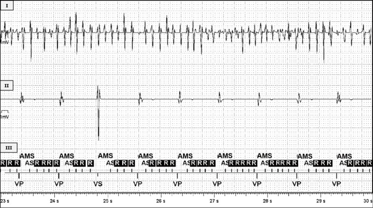
A 76-year-old patient with a dual-chamber pacemaker implanted in 2012 due to complete atrioventricular (AV)-block, attended to routinary pacemaker check-up. The device interrogation showed the presence of multiple atrial high rate episodes (AHRE) with variable duration, from few minutes to several hours, compatible with silent atrial fibrillation (AF). Intracardiac electrograms (EGMs) confirmed the diagnosis of AF (Figure 1). During the episodes the patient was on ventricular pacing at a normal heart rate and completely asymptomatic. On the other hand, the presence of AHRE compatible with silent AF is related with a higher risk of stroke (1). However, the application of anticoagulation therapy in patients with device-detected AHRE is yet unclear and challenging in the absence of randomized studies.
Figure 1.

Stored intracavitary electrograms (EGMs) showing an automatic mode switch (AMS) episode from a St. Jude Medical (Abbott) pacemaker. From top to bottom: (I) atrial EGM channel showing a fast and chaotic activity compatible with AF; (II) ventricular EGM channel showing ventricular stimulated activity in all the beats but the third one showing different morphology and amplitude corresponding to a sensed spontaneous ventricular activity and (III) marker channel showing the registered activity labeling. The device detects correctly the episode as AMS episode when the atrial rate reached the programmed limit. AS, atrial sensing; R, atrial signals in refractory period; VP, ventricular pacing; VS, ventricular sensing; AF, atrial fibrillation.
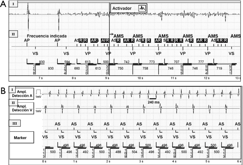
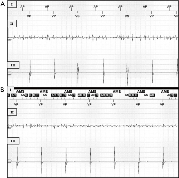
Diagnosis of silent paroxysmal AF represents a challenge, since the arrhythmia may be brief, completely asymptomatic and difficult to detect. Today’s cardiac implantable electronic devices (pacemakers and defibrillators) diagnostics include system diagnostics revealing battery status, energy consumption, lead impedance…but also algorithms for detecting and treating cardiac arrhythmias (2). The device detects the onset of AF episodes as AHRE when the atrial rate exceeds the programmed arrhythmia detection rate during a programmable number of beats. However, when interpreting counter data, we should evaluate for possible inappropriate device behavior or programming. Factors that limit the value of diagnostic counters include over-sensing, under-sensing, far-field sensing, cross-talk, interferences (noise), detection criteria not fulfilled by an arrhythmia, inappropriate programmed detection criteria… (3) (Figure 2). Under-sensing is not uncommon in AF, due to the small amplitude of atrial EGMs (Figure 3). The incorporation of stored EGMs has been an important milestone in improving the diagnostic capabilities of these devices. Multiple studies have clearly shown that data retrieved from diagnostic counters may be misleading (4). This does not imply that the devices are functioning inappropriately. It merely emphasizes the limitations of detection algorithms, which follow mathematical rules to classify events on a beat-to-beat basis. Stored EGMs are an essential tool to evaluate an appropriate or inappropriate detection documenting the intracavitary signals during the episodes and therefore to confirm the diagnosis of the arrhythmia detected by the device (5).
Figure 2.

Stored intracavitary EGMs from St. Jude Medical (Abbott) devices. (A) An automatic mode switch (AMS) episode in a pacemaker. From top to bottom: (I) atrial EGM channel showing signals with a high amplitude corresponding to atrial paced beats and a fast and irregular low-voltage activity compatible with noise; (II) marker channel showing the labeling of the activity registered by the device. The device wrongly classifies the episode as AMS episode in the 9 second (lower strip) when the atrial sensed activity reaches the programmed rate limit, however, the device is sensing noise in the atrial channel as we can confirm with the stored EGM; (B) a fast ventricular rate episode from a CRT pacemaker. From top to bottom: (I) atrial EGM channel showing a fast and regular activity (cycle length 240 ms, 250 bpm) compatible with atrial flutter; (II) ventricular EGM channel showing a regular and slower ventricular sensed activity with a 2:1 relation with the atrial activity; (III) marker channel showing the labeling of the activity registered by the device. Note that the device is not detecting in the marker channel every other atrial activity coincidental with the ventricular activity (post-ventricular atrial blanking period), therefore, the device is classifying the episode based on the detected high ventricular rate neglecting the faster atrial rate. This example illustrates an AHRE corresponding to an atrial flutter, misinterpreted by the device, in which stored EGM are key to make the diagnosis. AS, atrial sensing; AR, atrial signals in refractory period; AP, atrial pacing; VP, ventricular pacing; VS, ventricular sensing; EGM, electrogram; CRT, cardiac resynchronization therapy; AHRE, atrial high rate episode.
Figure 3.
Stored intracavitary EGMs from a St. Jude Medical (Abbott) pacemaker. (A) Real-time EGMs during initial pacemaker interrogation, from top to bottom: (I) marker channel showing the labeling of the activity registered by the device; (II) atrial EGM channel showing a fast and chaotic activity compatible with AF; (III) ventricular EGM channel showing ventricular stimulated activity in all the beats but the third and 5th ones showing different morphology corresponding to sensed spontaneous ventricular beats. The device is not detecting the AF episode, it is not detecting the atrial activity and therefore it is delivering AP at the programmed heart rate; (B) real-time EGM during pacemaker interrogation after modifying atrial sensitivity: (I) marker channel showing the labeling of the activity registered by the device; (II) atrial EGM channel showing AF; (III) ventricular EGM channel showing ventricular stimulated activity. After increasing atrial sensitivity, the device now correctly detects the atrial activity and the AMS is activated. EGM, electrogram; AF, atrial fibrillation; AP, atrial pacing; AS, atrial sensing; VP, ventricular pacing; VS, ventricular sensing; AMS, automatic mode switch.
Acknowledgements
None.
Footnotes
Conflicts of Interest: The authors have no conflicts of interest to declare.
References
- 1.Benezet-Mazuecos J, Rubio JM, Farré J. Atrial high rate episodes in patients with dual-chamber cardiac implantable electronic devices: unmasking silent atrial fibrillation. Pacing Clin Electrophysiol 2014;37:1080-6. 10.1111/pace.12428 [DOI] [PubMed] [Google Scholar]
- 2.Seidl K, Meisel E, VanAgt E, et al. Is the atrial high rate episode diagnostic feature reliable in detecting paroxysmal episodes of atrial tachyarrhythmias? Pacing Clin Electrophysiol 1998;21:694-700. 10.1111/j.1540-8159.1998.tb00125.x [DOI] [PubMed] [Google Scholar]
- 3.Almehairi M, Baranchuk A, Johri A, et al. An unusual cause of automatic mode switching in the absence of an atrial tachyarrhythmia. Pacing Clin Electrophysiol 2014;37:777-80. 10.1111/pace.12296 [DOI] [PubMed] [Google Scholar]
- 4.Nowak B. Pacemaker stored electrograms: teaching us what is really going on in our patients. Pacing Clin Electrophysiol 2002;25:838-49. 10.1046/j.1460-9592.2002.t01-1-00838.x [DOI] [PubMed] [Google Scholar]
- 5.Pollak WM, Simmons JD, Interian A, Jr, et al. Clinical utility of intraatrial pacemaker stored electrograms to diagnose atrial fibrillation and flutter. Pacing Clin Electrophysiol 2001;24:424-9. 10.1046/j.1460-9592.2001.00424.x [DOI] [PubMed] [Google Scholar]


