pharmaceutics
Article
Evaluation of the Hemocompatibility and Anticancer Potential
of Poly(ε-Caprolactone) and Poly(3-Hydroxybutyrate)
Microcarriers with Encapsulated Chrysin
Eleftherios Halevas 1,2, * , Chrysoula Kokotidou 3,4 , Elda Zaimai 2 , Alexandra Moschona 5,6 , Efstratios Lialiaris 7 ,
Anna Mitraki 3,4 , Theodore Lialiaris 7 and Anastasia Pantazaki 2, *
1
2
3
4
5
6
7
*
����������
�������
Citation: Halevas, E.; Kokotidou, C.;
Zaimai, E.; Moschona, A.; Lialiaris, E.;
Mitraki, A.; Lialiaris, T.; Pantazaki, A.
Evaluation of the Hemocompatibility
and Anticancer Potential of
Poly(ε-Caprolactone) and
Poly(3-Hydroxybutyrate)
Microcarriers with Encapsulated
Chrysin. Pharmaceutics 2021, 13, 109.
https://doi.org/10.3390/
pharmaceutics13010109
Received: 30 December 2020
Accepted: 14 January 2021
Published: 16 January 2021
Publisher’s Note: MDPI stays neutral with regard to jurisdictional clai-
Institute of Biosciences & Applications, National Centre for Scientific Research “Demokritos”,
15310 Athens, Greece
Laboratory of Biochemistry, Department of Chemistry, Aristotle University of Thessaloniki,
54124 Thessaloniki, Greece; eldazaimai@gmail.com
Department of Materials Science and Technology, University of Crete, Voutes Campus,
70013 Heraklion, Greece; chkokoti@hotmail.com (C.K.); mitraki@materials.uoc.gr (A.M.)
Institute for Electronic Structure and Laser FORTH, N. Plastira 100, 70013 Heraklion, Greece
Laboratory of Organic Chemistry, Department of Chemical Engineering, Aristotle University of Thessaloniki,
54124 Thessaloniki, Greece; alexmoschona@gmail.com
Laboratory of Natural Resources and Renewable Energies, Chemical Process and Energy Resources Institute,
Centre for Research and Technology-Hellas (CERTH), 6th km Harilaou-Thermis, 57001 Thermi, Greece
Laboratory of Genetics, Medical School, Democritus University of Thrace, 68100 Alexandroupolis, Greece;
stratoslialiaris1998@hotmail.gr (E.L.); lialiari@med.duth.gr (T.L.)
Correspondence: lefterishalevas@gmail.com (E.H.); natasa@chem.auth.gr (A.P.); Tel.: +30-210-650-3558 (E.H.);
+30-2310-99-7838 (A.P.); Fax: +30-210-651-1767 (E.H.); +30-2310-99-7689 (A.P.)
Abstract: In this work, novel chrysin-loaded poly(ε-caprolactone) and poly(3-hydroxybutyrate)
microcarriers were synthesized according to a modified oil-in-water single emulsion/solvent evaporation method, utilizing poly(vinyl alcohol) surfactant as stabilizer and dispersing agent for the
emulsification, and were evaluated for their physico-chemical and morphological properties, loading
capacity and entrapment efficiency and in vitro release of their load. The findings suggest that the
novel micro-formulations possess a spherical and relatively wrinkled structure with sizes ranging
between 2.4 and 24.7 µm and a highly negative surface charge with z-potential values between
(−18.1)–(−14.1) mV. The entrapment efficiency of chrysin in the poly(ε-caprolactone) and poly(3hydroxybutyrate) microcarriers was estimated to be 58.10% and 43.63%, whereas the loading capacity
was found to be 3.79% and 15.85%, respectively. The average release percentage of chrysin was
estimated to be 23.10% and 18.01%, respectively. The novel micromaterials were further biologically
evaluated for their hemolytic activity through hemocompatibility studies over a range of hematological parameters and cytoxicity against the epithelial human breast cancer cell line MDA-MB 231.
The poly(ε-caprolactone) and poly(3-hydroxybutyrate) microcarriers reached an IC50 value with
an encapsulated chrysin content of 149.19 µM and 312.18 µM, respectively, and showed sufficient
blood compatibility displaying significantly low (up to 2%) hemolytic percentages at concentrations
between 5 and 500 µg·mL−1 .
ms in published maps and institutional affiliations.
Keywords: chrysin; poly(ε-caprolactone) and poly(3-hydroxybutyrate) biodegradable polymers;
drug micro-encapsulation and delivery; human blood compatibility; anticancer activity
Copyright: © 2021 by the authors. Licensee MDPI, Basel, Switzerland.
This article is an open access article
distributed under the terms and conditions of the Creative Commons Attribution (CC BY) license (https://
creativecommons.org/licenses/by/
4.0/).
1. Introduction
In recent years, nano- or micro-sized drug delivery systems have become indispensable tools in the pharmaceutical and medical fields and have been extensively investigated
due to the growing demand for the controlled administration of pharmacologically active
materials to specific cells, tissues, and organelles [1]. Micro-encapsulation offers several significant advantages in vitro and in vivo, as it is used to maintain the structural integrity of
Pharmaceutics 2021, 13, 109. https://doi.org/10.3390/pharmaceutics13010109
https://www.mdpi.com/journal/pharmaceutics
�Pharmaceutics 2021, 13, 109
2 of 22
−
compounds that are normally difficult to administer due to insolubility, reactivity, volatility,
and hygroscopicity, and to ensure controlled release and protection from degradation of
the encapsulated contents. In the pharmaceutical industry micro-encapsulation is widely
applied for the specific oral, transdermal, stomach, colon, and small intestine delivery
of drugs. More specifically, it has been reported that the encapsulation of drugs into
micro-formulations suitable for oral administration reduces the exposure to the harsh conditions of the upper gastrointestinal tract (GIT). Moreover, micro-encapsulation provides
immunoisolation and immunoprotection, important factors for the efficient in vivo delivery
−
and implantation of mammalian cells, and –cell and tissue engineering applications [2,3].
, λ For the past few decades, natural or synthetic biodegradable polymers such as
polyesters, poly(ortho esters), polyanhydrides, polyphosphazenes, chitosan, hyaluronic
acid, alginic acid, etc., either as microcapsules, microspheres,biochemical
or nanoparticles generated
cancer [4–8], have been widely applied as carriers for the
under different synthetic techniques
tumor
controlled
delivery of bioactive proteins and peptides, as well as hydrophilic or hydrophobic drugs of varied molecular weights [9–14] to specific locations in vivo, disintegrating
into biocompatible byproducts through enzyme-catalyzed or chemical hydrolysis [15]. The
drug-loaded polymeric formulations maintain the biocompatible, physico-chemical and
morphological properties of the carrier [16–18] and the therapeutic efficacy of the loaded
drug [19,20], presenting controllable biodegradation kinetics [21–23] and sufficient thermodynamic compatibility between the biopolymer and the encapsulated compound [24].
Two of the best-known groupsβof biodegradable polymers are polyhydroxyalkanoates
(PHAs) (Figure 1A) and poly(ε-caprolactones) (PCLs). PHAs are intracellular aliphatic
– monomers accumulating as energy storage materipolyesters of diverse hydroxyalkanoate
als by water-insoluble, discrete nano-sized, and optically dense granular inclusions located
in the cytoplasm of several bacterial and some archaeal cells, usually in the presence of
excess carbon source and under unbalanced growth conditions [1,25]. PHAs are considered exceptional alternatives to various synthetic polymers in a wide range of biomedical
applications as drug delivery vehicles or scaffolds in tissue engineering, potentially due
to their biodegradability, biocompatibility, and ease of insertion into the human body
without having to be removed again and generating significant foreign-body responses
to implantation [26]. One of the most widely used PHAs is poly(3-hydroxybutyric acid)
(PHB) (Figure 1B). PHB has gained particular attention as drug carrier or scaffold biomaterial because, compared to other biodegradable chemically produced polymers such as
poly(lactide-co-glycolide) (PLGA), polylactate (PLA), and polyglycolate (PGA), it displays
significant advantages, which include remarkable biodegradability and biocompatibility,
easier processibility and controllable retarding properties [25].
Figure 1. (A) General structure of PHAs, (B) structure of PHB, (C) structure of PCL, (D) structure of
PVA, (E) structure of chrysin.
PCL (Figure 1C) is a saturated aliphatic polyester composed by hexanoate repeated
units. Based on the range of the weight-average molecular weights, it can be generally
–
–
�Pharmaceutics 2021, 13, 109
3 of 22
described as a semicrystalline material [27]. Due to their ability to be completely degraded
by fungal and bacterial enzymes, including esterases and lipases, PCL-based materials are
of particular interest in biodegradable material applications [28]. Furthermore, PCL-based
formulations, either as blends or as copolymers with synthetic or other biopolymers, due to
their remarkable penetrability, nontoxicity, and exceptional biocompatibility, have attracted
great attention as controlled drug delivery systems, in cell cultivation and in implants for
regenerative medicine as tissue engineering materials [29,30].
However, apart from the significant advantages of both types of biopolymers, their
widespread application as drug delivery systems has been restricted by specific shortcomings, such as the slow degradation rate due to their relatively high crystallinity and
hydrophobicity. The incorporation of highly hydrophilic, biocompatible, and chemically
stable polymers, such as poly(vinyl alcohol) (PVA) (Figure 1D), as stabilizers and dispersing agents with favorable mechanical properties for the emulsification procedure of the
biopolymers, results in the generation of formulations with improved hydrophilicity and
optimized degradation rates [31].
Flavonoid chrysin (5,7-dihydroxyflavone) (Figure 1E) (Empirical formula: C15 H10 O4 ,
Molecular weight: 254.24 g·mol−1 , Melting point: 284–286 ◦ C, Solubility: 0.1 M in NaOH
0.008 g·L−1 , λmax : 348 nm) is present in honey, propolis, and honeycombs and is also a
constituent of the blue passion flowers extract [32]. Chrysin (Chr) displays antioxidant,
antiallergic, anti-inflammatory [33], and important pharmacological and biochemical properties associated with the prevention of cancer [34], functioning as an inhibitor for cell
proliferation and tumor angiogenesis in vivo [35], and tumor cell apoptosis in vitro [36].
However, despite its significant biological properties, the (a) short terminal half-life, (b)
quick metabolism, (c) low absorption rate, and (d) poor bioavailability limit its therapeutic
efficacy [37]. As a result, several types of formulations have been produced for the efficient encapsulation of chrysin in an effort to overcome limitations arisen through its low
aqueous solubility and bioavailability. However, until today, only nanosized formulations
of encapsulated chrysin have been reported in the literature utilizing combinations of
PLGA/PVA, methoxy poly(ethylene glycol)-β-PCL, PLGA-poly(ethylene glycol), PLGApoly(ethylene glycol)-PLGA, magnetic SiO2 /poly(ethylene glycol), or PLGA-poly(ethylene
glycol) chemically produced polymers [38–43].
Aiming at the development of novel multifunctional pharmaceutical micro-formulations
with enhanced bioavailability and therapeutic efficacy involving bioactive flavonoids, we
report herein, for the first time, the synthesis, physico-chemical characterization, and biological evaluation of empty and chrysin-loaded PVA-stabilized PCL and PHB microcarriers
(MCs) (mentioned henceforth as EPCL/PVAMCs, EPHB/PVAMCs, ChrPCL/PVAMCs,
and ChrPHB/PVAMCs, respectively). The newly synthesized micromaterials were compared and evaluated for their suitability as potential MCs with controlled release and
optimized solubility and bioavailability of chrysin, and their structural and textural properties were determined by different and complementary physico-chemical characterization
techniques. The cytotoxic effect of the novel micro-formulations was evaluated against
the epithelial human breast cancer cell line MDA-MB-231, and their hemolytic capacity
was determined through human blood compatibility studies over a range of hematological
parameters.
2. Experimental
2.1. Materials
The initial materials used include: Polycaprolactone (PCL) (average mol wt. 45,000),
poly[(R)-(3-hydroxybutyric acid)] (PHB) (average mol wt. 10,000), poly(vinyl alcohol) (PVA)
(87–90% hydrolyzed, average mol wt 30,000–70,000), chrysin powder (purity 97%), sodium
hydroxide pellets (NaOH), phosphate buffered saline pH 7.4 (PBS). These materials were
purchased from commercial sources (Sigma, Fluka, St. Louis, MO, USA) and were used
without further purification. Ultrapure water, chloroform, dichloromethane, methanol, and
dimethyl sulfoxide (DMSO) were used as solvents.
�Pharmaceutics 2021, 13, 109
4 of 22
The epithelial human breast cancer cell line MDA-MB-231 was from our cell bank
at the Institute of Molecular Biology and Biotechnology (IMBB), FORTH, and was free of
mycoplasma contamination. The media/agents for the cell cultures were purchased from
Thermo Fisher Scientific (Waltham, MA, USA). The MTT reagent (3-[4,5-dimethylthiazol-2yl]-2,5-diphenyl-tetrazolium bromide) was bought from Sigma-Aldrich (Darmstadt, Germany).
2.2. Methods
2.2.1. Fourier-Transform Infrared Spectrometry
Fourier-transform infrared spectra (FT-IR) were recorded on a Perkin Elmer 1760X
FT-infrared spectrometer (Perkin-Elmer, San Francisco, CA, USA). 90 mg of KBr was mixed
with 10 mg of each sample by grinding in agate mortar. A disk was made using the
obtained powdered mixture under a hydraulic pressure of 600 kg. Subsequently, the FT-IR
spectra were recorded between 4000 and 450 cm−1 , with a spectral resolution of 2 cm−1 .
2.2.2. Field Emission Scanning Electron Microscopy
The morphology and detailed structural features of the EPCL/PVAMCs, EPHB/PVAMCs,
ChrPCL/PVAMCs, and ChrPHB/PVAMCs samples were investigated by field emission
scanning electron microscopy (FESEM), using a JEOL JSM-7000F microscope (JEOL, Welwyn Garden City, Hertfordshire, UK). A 10 µL sample (PBS dispersion, diluted 1:10) was
deposited on a circular cover glass (immobilized on a double-sided carbon tape) and was
air dried overnight. Samples were additionally covered with 10 nm Au/Pd sputtering. The
analyses were performed in high vacuum mode in a 15 kV accelerating voltage.
2.2.3. Dynamic Light Scattering
Mean particle size was determined through Dynamic Light Scattering (DLS) using
Photon Correlation Spectroscopy (Malvern S4700 PCS System, Malvern Instruments Ltd.,
Malvern, UK). The analysis was performed at a scattering angle of 90◦ and at a temperature
of 25 ◦ C, using appropriately diluted samples (10 mg of each sample in 50 mL PBS, pH
7.4). Before the measurements, the samples were sonicated for 5 min. For each sample, the
mean diameter ± standard deviation (±SD) of six determinations was calculated applying
multimodal analysis.
2.2.4. Zeta-Potential
Zeta-potential measurements of EPCL/PVAMCs, EPHB/PVAMCs, ChrPCL/PVAMCs,
and ChrPHB/PVAMCs samples were determined by Laser Doppler Anemometry (Malvern
Zetasizer IV, Malvern Instruments Ltd., Malvern, UK). All analyses were performed on
samples appropriately diluted with 1 mM PBS buffer (adjusted to pH 7.4) in order to
maintain constant ionic strength and after sonication for 5 min and subsequent filtration.
For each sample, the mean value ±SD of four determinations was established.
2.2.5. High Performance Liquid Chromatography
The determination of entrapment efficiency and loading capacity, and the in vitro release study of chrysin were performed through High Performance Liquid Chromatography
(HPLC) at a ThermoFinnigan Spectra HPLC system (San Jose, CA, USA) model UV 6000
LP, equipped with EZChromeElite software, Version 3.1.7, four Q-Grad pumps, a diode
array detector (DAD) and a Grace Smart RP C-18 column (250 × 4.6 mm id.; 5 µm particle
size). The injection volume was 20 µL, and the wavelength used was 268 nm. The mobile
phases were 2% (v/v) acetic acid in milli-Q H2 O (eluent A) and 100% acetonitrile (eluent B)
with a flow rate of 1 mL·min−1 . The elution profile was as follows: from 0 min, 100% A;
25 min, 36% A/64% B; 35 min, 25% A/75% B.
�Pharmaceutics 2021, 13, 109
5 of 22
2.3. Synthesis of Chrysin-Loaded MCs
2.3.1. Synthesis of ChrPCL/PVAMCs
The ChrPCL/PVAMCs were formulated via a modification of the oil-in-water (O/W)
single emulsion/solvent evaporation method [39]. More specifically, 500 mg of PCL
(average mol wt. 45,000) was dissolved in 100 mL of dichloromethane under continuous
stirring at room temperature in a 200 mL closed vessel. Subsequently, a solution of 100 mg
of chrysin in 40 mL of a dichloromethane/methanol mixture (3:1, v/v) was added to
the above mixture and left stirred for another 24 h at room temperature. After complete
homogenization of the mixtures (organic phase), the O/W emulsion was prepared by the
dropwise addition of the organic phase to an aqueous solution of PVA (20 mL, 5% w/v PVA,
87–90% hydrolyzed, average mol wt 30,000–70,000) (aqueous phase). The emerged mixture
was homogenized at 15,000 rpm for 30 min, emulsified by sonication for two 5 min periods
interrupted by a 2 min resting period in an ice bath, and left stirred with an overhead
propeller under a flow hood at 600 rpm for 12 h for the complete evaporation of the organic
solvents. ChrPCL/PVAMCs were collected as a yellow precipitate through centrifugation
at 6000 rpm for 30 min. They were washed two times with PBS and centrifuged again
at 6000 rpm for 10 min to ensure the removal of non-encapsulated chrysin. Finally, the
emerged material was immediately freeze-dried at −35 ◦ C and 0.4 mbar for 72 h and stored
at 4 ◦ C until further analysis.
EPCL/PVAMCs were prepared following the same experimental procedure without the addition of chrysin and were used as control samples in the ensuing biological
experiments.
2.3.2. Synthesis of ChrPHB/PVAMCs
The ChrPHB/PVAMCs were formulated based on a similar synthetic procedure, as in
the case of ChrPCL/PVAMCs. More specifically, 500 mg of PHB (granule, 5 mm nominal
granule size) was dissolved in 100 mL of a chloroform/methanol mixture 4:1 (v/v) under
continuous stirring at room temperature in a 200 mL closed vessel. Subsequently, a solution
of 100 mg of chrysin in 40 mL of a chloroform/methanol mixture (3:1, v/v) was added to
the above mixture and left stirred for another 24 h at room temperature. After complete
homogenization of the mixtures (organic phase), the O/W emulsion was prepared by the
dropwise addition of the organic phase to an aqueous solution of PVA (20 mL, 5% w/v
PVA, 87–90% hydrolyzed, average mol wt 30,000–70,000) (aqueous phase). The mixture
was homogenized at 15,000 rpm for 30 min, emulsified by sonication for two 5 min periods
interrupted by a 2 min resting period in an ice bath, and left stirred with an overhead
propeller under a flow hood at 600 rpm for 12 h for the complete evaporation of the organic
solvents. ChrPCL/PVAMCs were collected as a yellow precipitate through centrifugation
at 6000 rpm for 30 min. They were washed two times with PBS and centrifuged again
at 6000 rpm for 10 min to ensure the removal of non-encapsulated chrysin. Finally, the
emerged material was immediately freeze-dried at −35 ◦ C and 0.4 mbar for 72 h and stored
at 4 ◦ C until further analysis.
EPHB/PVAMCs were prepared following the same experimental procedure without the addition of chrysin and were used as control samples in the ensuing biological
experiments.
2.4. Determination of Chrysin Entrapment Efficiency and Loading Capacity
The determination of the entrapment efficiency and loading capacity of chrysin into
the PCL/PVAMCs and PHB/PVAMCs was estimated via HPLC analysis. More specifically,
50 mg of dry ChrPCL/PVAMC or ChrPHB/PVAMC sample was ground and immersed in
a 50 mL PTFE beaker, into 20 mL of a DMSO/methanol mixture (1:1, v/v) and then filtered
through a 0.45 µm PTFE-membrane syringe filter. Appropriate dilutions were applied
�Pharmaceutics 2021, 13, 109
6 of 22
for the HPLC measurements, and the chrysin content was determined according to the
following calibration curve:
CChr = 5 × 10⁸ × peak area − 5 × 10⁶ (R2 = 0.998),
where CChr stands for chrysin concentration in the sample (mg·mL−1 ) and the peak area is
the area of the sample measured at 268 nm.
The entrapment efficiency of chrysin was calculated using the following equation:
Entrapment Efficiency (%) = Chri /Chrt × 100,
where Chri is the amount of chrysin incorporated into each type of MCs and Chrt is the
initially added amount of chrysin.
The loading capacity of chrysin was calculated using the following equation:
Loading Capacity (%) = Chri /WChr-loaded MCs × 100,
where Chri is the amount of chrysin incorporated into each type of MCs and WChr-loaded MCs
is the weight of the synthesized chrysin-loaded MCs after freeze drying.
Experiments were carried out in triplicates, and the results were expressed as mean ±SD.
2.5. In Vitro Chrysin Release Study
The determination of the chrysin release profile from ChrPCL/PVAMCs and ChrPHB/
PVAMCs was performed as follows: 50 mg of each type of chrysin-loaded MCs was
ground and immersed into 20 mL of a PBS (pH 7.4, 1% v/v DMSO) solution [44,45]. The
release medium temperature was set at 37 ± 1 o C under continuous stirring at a rate of ca.
250 rpm [46]. Aliquots of 1.5 mL were withdrawn with a syringe at fixed time intervals for
analysis followed by appropriate dilutions. Following removal of insoluble solid chrysinloaded MCs by centrifugation (13,000 rpm, 1 min) and filtration (0.45 µm PTFE-membrane
syringe filter), the remaining clear solution was analyzed, and the amount of chrysin
released was determined by HPLC with the aid of the aforementioned calibration curve
(vide supra). The cumulative release percentages of chrysin were calculated according to
the following equation:
Cumulative chrysin release (%) = ChrRELEASED /ChrENTRAPPED × 100
The percentages of the insoluble solid chrysin-loaded MCs are presented in the Supporting Information, Table S1.
Experiments were carried out in triplicates, and the results were expressed as mean
±SD.
2.6. Biological Evaluation
2.6.1. Cell Lines and Culture Conditions
Epithelial human breast cancer MDA-MB-231 cells were grown in Dulbecco’s Modified
Eagle′ s—Medium (DMEM) growth medium (pH 7.4) supplemented with 10% Fetal Bovine
Serum (FBS) and 50 µg·mL−1 gentamycin at 37 ◦ C in a 5% humidified CO2 incubator.
2.6.2. Cell Viability of Human Breast Cancer Cell Line (MDA-MB-231)
In the present study, the cell viability of MDA-MB-231 breast cancer line was evaluated
via the MTT assay. The method relies on the conversion of the yellow 3-(4,5-dimethylthiazol2-yl)-2,5-diphenyl tetrazolium bromide (MTT) to purple formazan crystals. The reduction
of MTT is catalyzed by the mitochondrial dehydrogenase enzyme and is therefore a measure for cell viability. MDA-MB-231 cells were exposed to free chrysin, ChrPCL/PVAMCs,
or ChrPHB/PVAMCs in their exponential phase of growth. More specifically, in a 96-well
plate (Corning, NY, USA), a number of 1 × 10⁴ cells/well were seeded, and after 24 h
(80% confluency) they were treated with different concentrations of ChrPCL/PVAMCs or
�Pharmaceutics 2021, 13, 109
7 of 22
ChrPHB/PVAMCs ranging between 0–400 µg·mL−1 . Due to the different loading capacity
of chrysin in each type of MCs, free chrysin (dissolved in DMSO) was added accordingly, in
equal concentrations (µM), with the encapsulated chrysin in the MCs, as estimated in Section 3.5. The cells were incubated with the chrysin-loaded MCs or with free chrysin for 48 h.
After incubation, the medium was replaced with 90 µL of fresh DMEM and 10 µL of MTT
(5 mg·mL−1 ) per well. The plate was then incubated for 4 h at 37 ◦ C in a 5% humidified
CO2 incubator. Subsequently, the content of the wells was carefully removed, and 100 µL
of a solution of DMSO/isopropanol in a 1:1 ratio was added to achieve the dissolution of
the formazan crystals, and then the plate was incubated for 15 min in 37 ◦ C and 15 min in
4 ◦ C. Finally, a Synergy HTX BioTEK plate reader (with a reference wavelength of 630 nm)
was used to determine the absorbance measurement at 545 nm. All the experiments were
performed in triplicate.
2.7. Blood Sample Collection and Handling
Human blood samples were freshly collected from ten healthy volunteers and divided
into tubes containing the anticoagulant agent ethylenediamine tetraacetic acid (EDTA),
according to the protocols approved by the National Institute of Health and the Food and
Drug Administration.
2.7.1. Blood Profile Analysis
The blood profile analysis was performed using an automatic hematological analyzer
Beckman Coulter ACT 5 Diff OV, (Beckman Coulter International S.A., Nyon, Switzerland)
for the determination of different hematological parameters, such as red blood cells (RBCs)
count (1012 /µL), hemoglobin (HGB) g·dL−1 ), hematocrit (HCT) (%), mean corpuscular
volume (MCV) (in femtoliters, fl), mean corpuscular hemoglobin (MCH) (pg), mean corpuscular hemoglobin concentration (MCHC), red cell distribution width (RDW), white
blood cells (WBCs) (109 /L), neutrophils (NE) (%), lymphocytes (LY) (%), monocytes (MO)
(%), eosinophils (EO) (%), basophils (BA) (%), and platelets (PLTs) (109 /L), after exposure to free chrysin, ChrPCL/PVAMCs, or ChrPHB/PVAMCs, and without exposure to
any agent. Briefly, the sample preparation was performed as follows: A 100 µL sample
of whole blood was added to 900 µL of PBS. Then, either chrysin, ChrPCL/PVAMCs,
or ChrPHB/PVAMCs were added to this diluted blood to achieve three concentrations:
low, high, and very high (5, 80, and 200 µg·mL−1 , respectively). The negative control
sample used consisted of 100 µL of whole blood diluted with 900 µL PBS. In parallel,
100 µL samples of whole blood were subjected to the same treatment with the addition of
EPCL/PVAMCs or EPHB/PVAMCs. The suspensions were incubated at 37 ◦ C for 1 h. All
the experiments were performed in triplicate.
2.7.2. Hemocompatibility Studies
The hemocompatibility of free chrysin and the produced micro-formulations was
evaluated via the hemolysis assay performed using a biochemical analyzer Konelab 30,
Thermo Scientific [47]. The experimental procedure was as follows: Whole blood samples
were centrifuged for 10 min at 1500 rpm to remove plasma. The obtained cell pellets
were washed three times with sterile PBS (10 mM, pH 7.2) to separate the red blood cells
(RBCs) from other blood components, such as the white blood cells, plasma proteins, and
excess antibodies, centrifuged and finally re-suspended at 5 mL PBS. The hemolysis assay
was performed by adding 100 µL of the RBC suspension to 900 µL of PBS, containing
several concentrations (5, 20, 40, 60, 80, 100, 200, 300, 400, 500 µg·mL−1 ) of free chrysin,
ChrPCL/PVAMCs, or ChrPHB/PVAMCs. The positive (+) control sample of hemolysis
used (100% hemolysis) consisted of 900 µL of ultrapure water and 100 µL of washed
RBCs. The negative (−) control sample (0% hemolysis) consisted of 900 µL PBS and
100 µL of washed RBCs. PBS and PBS containing either free chrysin, ChrPCL/PVAMCs, or
ChrPHB/PVAMCs were used as blank samples. All samples were incubated for 24 h at
37 ◦ C, under agitation at 120 rpm. After incubation, they were centrifuged at 700 rpm for 5
�Pharmaceutics 2021, 13, 109
8 of 22
min, and the absorbance of the supernatant was measured at 541 nm. The % hemolysis
was calculated after subtracting the blank values, and by setting the control (+) value as
100% of hemolysis. The absorbance was transformed to hemolysis percentage using the
following equation:
Percentage of hemolysis (%) =
ODSample − ODNegative control
ODPositive control − ODNegative control
where OD stands for Optical density.
2.7.3. Statistical Analysis
Data are the mean of at least three independent experiments. The statistical significance of changes in different groups was evaluated by one-way analysis of variance
(ANOVA) followed by Student t-tests, using GraphPad Prism 6.0 software (Science Plus
Group, Groningen, The Netherlands). For each experiment, data are expressed as the mean
±SD, * p ≤ 0.05, ** p ≤ 0.01, *** p ≤ 0.001, ns (not significant) > 0.05.
3. Results and Discussion
3.1. Synthesis of Chrysin-Loaded MCs
The chrysin-loaded PCL/PVA and PHB/PVA MCs were synthesized according to a
modified O/W single emulsion/solvent evaporation method which is usually employed
for the encapsulation of hydrophobic compounds, such as chrysin. The emulsification step
was performed by the addition of the organic phase with the dissolved biopolymer (PCL or
PHB) and flavonoid to an aqueous PVA solution, followed by high-speed homogenization,
sonication, and subsequent evaporation of the organic solvents. The PVA used was 87–90%
hydrolyzed, which is a degree of hydrolysis that ensures the optimum solubility of PVA in
water [48]. In general, the addition of a PVA surfactant as a stabilizing and emulsifying
agent enhances the stability of the dispersed phase droplets formed during the process
of emulsification via the emerging interactions between the hydroxyl groups in its structure with the aqueous phase and the vinyl chain with the organic phase, thus inhibiting
microsphere flocculation and coalescence [49,50]. Furthermore, the addition of the highly
hydrophilic PVA limits the hydrophobic nature of the produced micro-formulations, promoting the formation of more amphiphilic species [31]. Chrysin is encapsulated within
the produced MCs through non-covalent interactions forming molecule-in-molecule assemblies via hydrogen bonds and weak van der Waals forces with the functional groups
of the MC hosts [51]. The effective encapsulation aims at enhancing chrysin aqueous
solubility and therefore its systemic bioavailability, targeted delivery, circulation time, and
therapeutic potential (Figure 2).
3.2. FT-IR Spectroscopy
FT-IR spectra of chrysin, EPCL/PVAMCs, EPHB/PVAMCs, ChrPCL/PVAMCs, and
ChrPHB/PVAMCs are shown in Figure 3A,B. In the FT-IR spectrum of free chrysin the
strong absorption band at 1650 cm−1 is assigned to the stretching vibrations of the carbonyl
group v(C=O) coupled with the double band in the γ-benzopyrone ring [52,53]. Moreover,
the absorption bands observed at 1450 cm−1 , 1580 cm−1 , and 1610 cm−1 are assigned to
the ν(C=C) carbon vibrations in the γ-pyrone and benzene rings [52,53]. The absorption
bands observed at 1360 cm−1 and at 1310 cm−1 are attributed to the coupled ν(C−O) and
δ(O–H) vibrational modes, respectively [52,53]. In addition, the sharp absorption band at
1250 cm−1 is assigned to the v(C–O–C) stretching vibrations, whereas the broad band in the
3090–2640 cm−1 range is attributed to the v(C–H) and v(O–H) stretching vibrations [52,53].
�Pharmaceutics 2021, 13, 109
9 of 22
Figure 2. PBS-dispersed (1% v/v DMSO) (A) chrysin, (B) EPCL/PVAMCs, (C) ChrPCL/PVAMCs, (D) EPHB/PVAMCs,
and (E) ChrPHB/PVAMCs.
Chrysin
EPCL/PVAMCs
ChrPCL/PVAMCs
v(C=O)
(O-H)
v(C=C)
T (%)
v(C-O)
v(O-H)
v(C-O-C)
v(C=O)
v(CH2)
v(C-O) v(C-C)
4000
3500
3000
2500
2000
1500
1000
-1
Wavelengths (cm )
(A)
Figure 3. Cont.
500
�Pharmaceutics 2021, 13, 109
10 of 22
Chrysin
EPHB/PVAMCs
ChrPHB/PVAMCs
v(C=O)
(O-H)
v(C=C)
T (%)
v(C-O)
v(O-H)
v(CH2)
v(C-O)
v(-CH)
v(C=O)
4000
3500
3000
2500
2000
1500
1000
500
-1
Wavenumbers (cm )
(B)
Figure 3. FT-IR spectra of (A) chrysin, EPCL/PVAMCs, ChrPCL/PVAMCs, and (B)
chrysin, EPHB/PVAMCs, ChrPHB/PVAMCs.
In the spectrum of EPCL/PVAMCs the characteristic bands of the symmetric and
asymmetric aliphatic ν(CH2 ) stretching vibrations of PCL can be observed at 2845 cm−1 and
2925 cm−1 , respectively. The strong absorption band at 1730 cm−1 can be attributed to the
carbonyl ν(C=O) stretching vibrations. The absorption bands of the PCL backbone ν(C–O)
and ν(C–C) stretching vibrations are located at 1370 cm−1 and 1300 cm−1 , respectively.
Furthermore, the symmetric and asymmetric ν(C–O–C) vibrations appear at 1150 cm−1
and 1230 cm−1 , respectively [54]. The broad absorption band in the 3590–3118 cm−1 range
can be attributed to the ν(O–H) stretching vibrations of the PCL terminal hydroxyl groups
and the PVA alcoholic moieties [55].
In the spectrum of EPHB/PVAMCs the two strong absorption bands observed at
1720 cm−1 and 1290 cm−1 are attributed to the carbonyl ν(C=O) stretching vibrations of the
ester group and the ν(–CH) group, respectively. The absorption bands located in the range
between 980 cm−1 and 1230 cm−1 can be assigned to the ν(C–O) stretching vibrations of the
ester group. The absorption bands observed at 2980 cm−1 and 2930 cm−1 are indicative of
the alkyl ν(–CH3 ) stretching vibrations, whereas the absorption band located at 1380 cm−1
is attributed to the ν(–CH3 ) symmetric bending vibrations. The band at 1460 cm−1 is
assigned to the ν(–CH2 ) or ν(–CH3 ) asymmetric bending vibrations [56,57]. Moreover, the
broad band at 3440 cm−1 can be attributed to the v(O–H) stretching vibrations of the PHB
terminal hydroxyl groups and the PVA alcoholic moieties [55].
In the spectra of ChrPCL/PVAMCs and ChrPHB/PVAMCs all the important peaks of
the biopolymers and chrysin are present. Variations in the IR peak intensity of both the
host and guest molecules could be related to the intermolecular interactions induced by
the encapsulation process [58].
3.3. FESEM Analyses
The morphological and structural characteristics of EPCL/PVAMCs, EPHB/PVAMCs,
ChrPCL/PVAMCs, and ChrPHB/PVAMCs were examined by FESEM, and the results are
presented in Figure 4. The EPCL/PVAMC sample (Figure 4A) consists of distinct globular
microparticles with a highly wrinkled surface and sizes around 2 µm. FESEM images
�Pharmaceutics 2021, 13, 109
11 of 22
of the ChrPCL/PVAMC sample (Figure 4B) indicate the presence of scattered, spherical
microparticles with a relative smooth surface. Their sizes range between 1.1 and 12.1
µm, showing a relatively wide size distribution. The EPHB/PVAMC sample (Figure 4C)
consists of spherical, relatively wrinkled microparticles with sizes around 10.9 µm and
scattered amorphous agglomerates, whereas FESEM images of the ChrPHB/PVAMC
sample (Figure 4D) indicate the presence of globular microparticles with a highly spongy
and wrinkled structure and sizes around 21.3 µm. The observed increase in the diameter
of the chrysin-loaded species compared to their empty counterparts could be attributed
to the encapsulation of chrysin molecules inside the polymeric structure, which induces
the swelling of the microparticles. Literature reports on PVA-stabilized PCL microspheres
loaded with flavonoid quercetin showed that all tested samples possessed a spherical
morphology and wrinkled surface, but with large diameters, ranging between 61 and
171 µm. Furthermore, the increase of quercetin entrapment efficiency induced the size
enlargement of the quercetin-loaded species compared to their empty counterparts [59].
Spherical morphology has also been observed for curcumin-loaded PHB/PVA microformulations exhibiting a semi smooth surface with pores of different sizes and mean
diameters around 6.98 ± 1.89 µm [60].
(A)
(B)
(C)
(D)
Figure 4. FESEM images of (A) EPCL/PVAMCs, scale bar: 100 nm, (B) ChrPCL/PVAMCs, scale bar: 10 µm, (C)
EPHB/PVAMCs, scale bar: 1 µm, and (D) ChrPHB/PVAMCs, scale bar: 10 µm.
3.4. Particle Size Analysis And Z-Potential
For comparative purposes, DLS and z-potential measurements were implemented to
further determine the hydrodynamic mean diameter and surface charge of the EPCL/PVAMC,
EPHB/PVAMC, ChrPCL/PVAMC, and ChrPHB/PVAMC samples. The mean hydrodynamic diameter and polydispersity index (PDI) of EPCL/PVAMCs and ChrPCL/PVAMCs
�Pharmaceutics 2021, 13, 109
12 of 22
were estimated to be 2.4 ± 1.3 µm (PDI = 2.03) and 11.8 ± 4.7 µm (PDI = 2.11), respectively.
In the case of EPHB/PVAMCs and ChrPHB/PVAMCs, the values of the hydrodynamic
mean diameter and PDI were found to be 10.4 ± 4.4 µm (PDI = 1.95) and 24.7 ± 8.5 µm
(PDI = 1.93), respectively. The observed results clearly indicate that the encapsulation of
chrysin significantly affects the size of the emerging MCs as also observed in other chrysinloaded types of formulations reported in the literature [38–43]. Moreover, the relatively
high PDI values could be attributed to the high PVA concentration during the synthetic
procedure that resulted in the enhanced polydispersity of the produced MCs [59].
Z-potential measurements were conducted immediately after the determination of
particle sizes. The z-potential values of EPCL/PVAMC, ChrPCL/PVAMC, EPHB/PVAMC,
and ChrPHB/PVAMC samples were determined to be −16.2 ± 3.8 mV, −18.1 ± 4.1 mV,
−14.1 ± 3.1 mV, and −16.3 ± 4.0 mV, respectively, presenting no significant differences
between the empty and the chrysin-loaded MCs and confirming the highly negative
surface charge of the produced microspheres, which promotes the formation of more
stabilized and less aggregated MC dispersions due to the strong electrostatic repulsion
forces between the microparticles [61]. It has been reported that the negatively charged
surface of microparticles can potentially minimize non-specific binding with the cell membrane and, additionally, reduce aberrant protein binding. This prevents the activation of
the immune system, thereby resulting in a prolonged circulatory half-life [62]. On the
other hand, recent studies on novel synthetic drug nanocarriers based on zwitterionic
biomimetic polymers and polypeptides have demonstrated that these materials, due to
their structural characteristics, can be used not only for covalent modification with targeting ligands and biomolecules, but also for the prevention of nonspecific protein adsorption
and maintainance of micelle stability in complex media, such as serum, thus providing
long circulation lifetimes [63,64].
3.5. Entrapment Efficiency and Loading Capacity
The in situ entrapment efficiency of chrysin in the ChrPCL/PVAMCs and ChrPHB/
PVAMCs was estimated to be 58.10% and 43.63%, whereas the loading capacity was
found to be 3.79% and 15.85%, respectively. The obtained results are considered quite
satisfactory and favorably comparable with those reported for other types of chrysin-loaded
nanocarriers [38–41,43,65,66]. The observed high loading capacity of the ChrPHB/PVAMCs
compared to that of the ChrPCL/PVAMC sample could potentially be attributed to the
significantly porous structure of the PHB/PVA microparticles, as observed through FESEM,
which might have promoted the encapsulation of chrysin in the interior of the pores [67],
and the higher drug-to-polymer ratio applied during the synthetic procedure [58].
3.6. Release Study
The release profile of the active agent (chrysin) is an important parameter, since it
determines the pharmacokinetic behaviour of the chrysin-loaded MCs. In evaluating the
release profile, two factors are taken into consideration: the total amount of chrysin released
and the rate of release. Figure 5 presents the percentages of chrysin released with regard to
the total entrapped chrysin versus time for both types of chrysin-loaded MCs. The average
release percentage of chrysin from the ChrPCL/PVAMCs and ChrPHB/PVAMCs is 23.10%
and 18.01%, respectively. By examining the release profile of the ChrPCL/PVAMC sample
during the 60 h of study, it can be observed that up to the first 3 h the release rate is steady.
Subsequently, a burst of chrysin release is observed which carries on up to 30 h, and then
the process decelerates and the release rate is significantly decreased, reaching a plateau at
48 h. In the case of the ChrPHB/PVAMC sample, the chrysin release is relatively steady
up to the first 7 h. Thereafter, the release rate increases and after 30 h begins to decelerate,
approaching a plateau at 48 h. The observed low chrysin release percentages for both types
of MCs can be attributed to the hydrophobic nature and the slow degradation rates of
the employed biopolymers. Moreover, the relatively steady initial chrysin release rates,
observed for both samples, can be due to their micro-sized dimensions. It is known that,
�Pharmaceutics 2021, 13, 109
13 of 22
in general, microparticles have a smaller surface area and higher porosity compared to
nanoparticles. As a result, more drug molecules can be encapsulated into their pores than
located near the particle surface which comes into direct contact with the aqueous medium,
hence preventing their rapid diffusion [68].
ChrPCL/PVAMCs
ChrPHB/PVAMCs
20
18
Cumulative Release (%)
16
14
12
10
8
6
4
2
0
0
1
2
3
4
5
6
7
15 20 25 30 35 40 45 50 55 60 65
Time (hours)
Figure 5. Cumulative release percentage of chrysin with regard to the total entrapped chrysin vs.
time for ChrPCL/PVAMCs (blue line) and ChrPHB/PVAMCs (red line).
Table 1 summarizes the results of the physico-chemical characterization of the produced empty and chrysin-loaded
micro-formulations.
Table 1. Physico-chemical characterization data of the produced empty and chrysin-loaded micro-formulations.
EPCL/PVAMCs
2
−
dDLS b
−
(µm)
−
− ± 1.3
2.4
EPHB/PVAMCs
10.9
10.4 ± 4.4
1.95
−14.1 ± 3.1
-
-
-
ChrPCL/PVAMCs
1.1–12.1
11.8 ± 4.7
2.11
−18.1 ± 4.1
58.10
3.79
23.10
21.3
24.7 ± 8.5
1.93
−16.3 ± 4.0
43.63
15.85
18.01
MicroFormulation
dFESEM
(µm)
–
ChrPHB/PVAMCs
a
a
PDI
Z-Potential
(mV)
Entrapment
Efficiency
(%)
Loading
Capacity
(%)
In Vitro
Release
(%)
2.03
−16.2 ± 3.8
-
-
-
b
Diameter observed via FESEM. Hydrodynamic mean diameter measured via DLS.
−
3.7. Breast Cancer
Cell Viability after Exposure to Chrysin-Loaded MCs
The viability of the breast cancer cell line MDA-MB-231 was determined using the
MTT assay after exposure to ChrPCL/PVAMCs or ChrPHB/PVAMCs. The cells were
treated for 48 h with different concentrations of chrysin-loaded MCs (6.25, 12.5, 50, 100, 200,
and 400 µg·mL−1 ). Due to the different loading capacity of chrysin in each type of MC, the
corresponding molar amount of free chrysin was added as control. The results are shown in
Figures 6 and 7. The obtained results indicate that the micro-formulated chrysin inhibited
the viability of cancer cells in a dose-dependent manner, but less so compared to free chrysin.
Specifically, the ChrPCL/PVAMCs reached an IC50 value with an encapsulated chrysin
content of 149.19 µM compared to that of free chrysin, which was 111.89 µM (Figure 6).
The ChrPHB/PVAMCs reached an IC50 value with an encapsulated chrysin content of
312.18 µM (Figure 7). The higher IC50 values of the chrysin-loaded MCs compared to free
chrysin can be attributed to the slow release rates and low release percentages of chrysin
from both types of MCs due to the limited hydrophilicity and degradation rates of the
employed biopolymers, which could potentially lead to the retarded inhibition of cell
proliferation [69]. Moreover, as previously presented in the cumulative release diagram
�Pharmaceutics 2021, 13, 109
14 of 22
(Figure 5), the ChrPCL/PVAMCs have a higher release rate of chrysin, which explains
the lower IC50 value compared to ChrPHB/PVAMCs. Furthermore, the EPCL/PVAMC
and EPHB/PVAMC samples showed relatively low effect on cell viability (Supporting
Information, Figure S1).
Figure 6. Cell viability (%) of MDA-MB-231 human breast cancer cells line exposed for 48 h to
−
−1 ) of ChrPCL/PVAMCs and to their
different concentrations (6.25, 12.5, 50, 100, 200 and 400 µg
− ·mL
corresponding equal molar concentrations of free chrysin. Cell viability was assessed using the
MTT assay. The inhibitory concentration of chrysin for 50% viability (IC50 ) in MDA-MB-231 cells
is 111.89 µM for free chrysin and 149.19 µM for ChrPCL/PVAMCs (encapsulated chrysin in the
PCL/PVAMCs) ** p < 0.01.
Figure 7. Cell viability (%) of MDA-MB-231 human breast
cancer
cells line exposed
for 48 h to
− ) of
ChrPΗΒ/PVAMCs
a
− ) of ChrPΗΒ/PVAMCs
a
different concentrations (6.25, 12.5, 50, 100, 200 and 400 µg
·mL−1 ) of ChrPHB/PVAMCs
and to their
corresponding equal molar concentrations of free chrysin. Cell viability was assessed using the
MTT assay. The inhibitory concentration of chrysin for 50% viability (IC50 ) in MDA-MB-231 cells
PΗΒ/PVAMCs)
** free chrysin and 312.18 µM for ChrPHB/PVAMCs (encapsulated chrysin in the
is 111.89 µM for
PΗΒ/PVAMCs) **
PHB/PVAMCs) ** p < 0.01.
For comparative purposes, Table 2 presents the data reported in the literature on
cytotoxic IC50 values of chrysin-loaded nano-formulations in several cancer cell lines. In
most studied cases, nano-formulated chrysin showed lower IC50 values compared to free
chrysin against various cancer cell lines, such as AGS, T47D, and MCF-7 [41,63,64], whereas
�Pharmaceutics 2021, 13, 109
15 of 22
in our case, both types of micro-formulated chrysin showed higher IC50 values, suggesting
that the chrysin released from MCs was more slowly taken up by cells, potentially due to
the slow release of chrysin, the limited degradation rates of the employed biopolymers [39],
and the micro-dimensions of the produced carriers that affect cellular uptake to a certain
degree. It is well-known that the surface area to volume ratio of microparticles is relatively
low compared to that of nanoparticles [70]. As a result, cellular adherence on the surface of
the MCs is limited, and thus cell attachment is hampered. Consequently, small numbers
of cells can come into close contact with the released chrysin [71]. Moreover, another
factor that can affect cytotoxicity, and conclusively the IC50 values, is based on the cellular
exposure time to the nano- or micro-formulated chrysin reflecting more or less on the
cellular growth inhibition, as is obvious in Table 2. Indicatively, it has been shown that
PLGA/PVA chrysin nano-formulations ameliorated the delivery of chrysin through a
higher absorption by cells and enhanced its effectiveness on cell growth inhibition [38].
In general, it should be noted that, compared to those in the literature, the tested cell
line in this study (MDA-MB-231) is highly aggressive, with limited treatment options,
invasive, and poorly differentiated triple-negative breast cancer (TNBC) cell line, as it lacks
estrogen receptor (ER) and progesterone receptor (PR) expression, as well as HER2 (human
epidermal growth factor receptor 2) amplification. However, despite all these factors, the
MCs under investigation exhibited sufficient cytotoxicity against this aggressive breast
cancer cell line.
Table 2. Data reported in the literature on cytotoxic IC50 values of chrysin-loaded nano-formulations.
Type of Chrysin-Loaded
Nano-Formulation
Cell Line
Treatment Duration
(Hours)
IC50
References
Methoxy PEG-β-PCL
nanoparticles
A549
non-small-cell lung
cancer
48
2.5 µM
[39]
PLGA-PEG-PLGA nanoparticles
AGS
gastric cancer
24, 48, 72
58.2, 44.2, 36.8 µM
[41]
PCL-PEG-PCL nanoparticles
T47D
breast cancer
24, 48, 72
2, 10, 10 µM
[63]
PLGA-PEG
nanoparticles
T47D
breast cancer
MCF-7
breast cancer
40.19, 35.75, 31.28 µM
24, 48, 72
[64]
66.41, 56.80, 42.54 µM
3.8. Effect of Chrysin-Loaded MCs on Blood Profile Analysis
The collective measurements of the hematological parameters after human blood
exposure to 5, 80, and 200 µg·mL−1 of free chrysin and chrysin-loaded and empty MCs
are shown in Figure 8A,B and the Supporting information, Figure S2, respectively. The
observed values of almost all the hematological parameters of the blood samples that
were treated with plain chrysin, ChrPCL/PVAMCs, or ChrPHB/PVAMCs and their empty
counterparts at 37 ◦ C for 1 h did not display significant deviation compared to the negative control sample, indicating no concentration-dependent alteration. These parameters
include RBCs, HGB, HCT, MCV, MCH, MCHC, RDW, WBCs, NE, LY, MO, EO, and BA.
However, a significant decrease in PLT values was observed between the negative control
sample and pure chrysin, indicating a concentration-dependent inhibition, therefore confirming the antiplatelet activity of chrysin [72]. Moreover, a small decrease in PLT values
was also observed after treatment with the EPHB/PVAMCs in both concentrations tested,
also confirming the inhibitory effect of PHB on isolated platelets [73]. Thrombocytopenia
is the result of a reduction in the number of blood platelets and it can be a side effect of
taking certain medications. As each platelet lives only about 10 days, our body normally
�Pharmaceutics 2021, 13, 109
16 of 22
renews our platelet supply continually by producing new platelets in our bone marrow [74].
Collectively, the obtained results clearly point out the sufficient blood compatibility of
the prepared chrysin-loaded MCs at low and high concentrations and their prospect for
potential use in several bio-applications, such as tumor therapy.
C o n tr o l
C o n tr o l
.
.
F r e e c h ry s in 5 μ g m L
F r e e c h r y s in 8 0 μ g m L
-1
-1
.
-1
.
-1
.
-1
C h r P H B /P V A M C s 8 0 μ g m L
.
-1
C h r P C L /P V A M C s 8 0 μ g m L
C h rP H B /P V A M C s 5 μ g m L
C h r P C L /P V A M C s 5 μ g m L
.
-1
.
-1
.
-1
E P H B /P V A M C s 8 0 μ g m L
.
-1
E P C L /P V A M C s 8 0 μ g m L
E P H B /P V A M C s 5 μ g m L
E P C L /P V A M C s 5 μ g m L
400
U n its o f h e m a to lo g ic a l p a r a m e tr e s
U n its o f h e m a to lo g ic a l p a ra m e tre s
400
300
200
100
300
200
100
P
)
)
(%
)
B
A
(%
(%
O
O
M
E
)
)
(%
(%
Y
E
N
L
)
)
9
0
(1
C
B
W
L
T
R
D
(1
W
0
.
9
/L
/L
(%
)
)
)
-1
g
L
(p
d
H
(g
C
C
H
C
M
)
l)
(f
V
C
M
T
C
H
M
)
(%
)
L
2
/L
1
G
H
B
C
B
(1
(g
0
.
d
B
R
H e m a to lo g ic a l p a r a m e tr e s
-1
)
)
(%
A
(%
E
O
(%
O
M
L
Y
(%
)
)
)
/L
(%
C
N
E
9
0
(1
0
B
L
T
W
P
)
)
)
9
0
(1
W
D
R
(g
C
M
H
C
/L
)
(%
)
L
.
d
(p
H
C
C
M
-1
l)
g
)
(f
(%
T
C
H
V
M
G
H
R
B
C
B
(1
(g
0
.
d
1
L
2
/L
)
)
-1
0
H e m a to lo g ic a l p a ra m e tre s
(A)
(B)
•
−1
Figure 8. Hematological parameters after the treatment of human blood samples with two concentrations, (A) 5 µg·mL−1
•
−1
and (B) 80 µg·mL−1 , of free chrysin,
ChrPCL/PVAMCs,
or ChrPHB/PVAMCs and their empty• counterparts.
RBC: red
•
−1
−1
blood cells (1012 /L); HGB: hemoglobin (g·dL−1 ); HCT: hematocrit (%); MCV (fl); MCH (pg); MCHC (g·dL−1 ); RDW (%);
PLT (109 /L); WBC (109 /L); NE (%); LY (%), MO (%), EO (%), and BA (%).
3.9. Hemolysis
It is well established that nanoparticles possess properties that can induce hemolysis
and decrease the efficiency of anticancer drugs in vitro [75]. In the effort to evaluate the
hemocompatibility of encapsulated chrysin in MCs, a hemolysis study was performed
using chrysin as positive control and PBS as negative control. Hemolysis is the rupturing of RBCs and the subsequent release of hemoglobin upon destruction of the red cell
membrane [76]. The quantitative determination of the released hemoglobin can provide
evidence on the potential damage to RBCs after MC administration; and this can serve as a
—
viable indicator of MC toxicity under in vivo conditions [38]. Based on the criterion established by the American Society for Testing and Material (ASTM) E2524—08(2013) active
standard [77], a test method for the analysis of the hemolytic properties of nanoparticles,
it has been reported that a percentage of induced hemolysis greater than 5% indicates a
damage on RBCs [78]. In our case, the obtained results from the hemolysis assay showed
that ChrPCL/PVAMCs, ChrPHB/PVAMCs, and their •empty
counterparts displayed great
−
compatibility with RBCs, as their hemolytic percentages were significantly low (up to
2%) at various concentrations ranging between 5 and 500 µg·mL−1 (Table 3). On the
other
hand,
free chrysin, which was diluted in 5% DMSO so as to enhance its solubil•
−
ity, displayed a hemolytic activity higher than 5%, but only in concentrations between
purposes,
100 and 500 µg·mL−1 , indicating a relative RBC damage. For comparative
•
−
–
•
−
�Pharmaceutics 2021, 13, 109
17 of 22
we pose that chrysin-loaded PLGA-PVA nanoparticles exhibited a hemolysis percentage
within the admissible limit of less than 3% for very low concentrations of nanoparticles
(5–20 µg·mL−1 ), whereas concentrations in the range between 40 and 80 µg·mL−1 induced
a hemolysis percentage lower than 5%, and yet lower than that of free chrysin at the
same concentrations [38]. It should also be noted that different blood groups of the ABO
system have a specific antigen which endows them with different biochemical properties,
and hence they can show different hemolytic activity [79]. The collective micrographs of
human RBCs resulting from the hemolysis assay, after exposure to a high concentration
(500 µg·mL−1 ) of free chrysin, ChrPHB/PVAMCs, ChrPCL/PVAMCs, EPHB/PVAMCs,
and EPCL/PVAMCs, are visualized in Figure 9. It can be concluded that the hemoglobin
release from RBCs is obvious after exposure to free chrysin, whereas in the case of the
−
empty and chrysin-loaded MCs no hemoglobin release is observed. The hemolytic data
are consistent with those of the hematological parameters proving that the employed MCs
display sufficient hemocompatibility.
Concentration-dependent hemolytic activity of free chrysin, ChrPHB/PVAMCs, ChrPCL/PVAMCs,
Table 3.
EPHB/PVAMCs, and EPCL/PVAMCs.
Concentration
(µg·mL−1 )
Free Chrysin *
ChrPHB/PVAMCs
ChrPCL/PVAMCs
EPHB/PVAMCs
EPCL/PVAMCs
Percentage of Hemolysis (%)
−
5
1.2
0.2
0.1
0.03
0.02
20
2.1
0.3
0.1
0.04
0.03
40
2.7
0.5
0.3
0.03
0.03
60
3.0
0.6
0.5
0.05
0.6
80
3.5
0.7
0.7
0.07
0.06
100
6.8
1.1
1.0
0.1
0.1
200
7.0
1.4
1.2
0.3
0.3
300
7.3
1.4
1.3
0.5
0.6
400
7.9
1.6
1.5
0.6
0.6
500
8.2
2.0
1.8
0.6
1.0
* Chrysin solution was prepared in DMSO (5%).
Figure 9. Micrograph of human RBCs showing the degree of hemolysis after incubation with 500 µg·mL−1 of free chrysin,
−
ChrPHB/PVAMCs, ChrPCL/PVAMCs,
EPHB/PVAMCs, and EPCL/PVAMCs.
ε
�Pharmaceutics 2021, 13, 109
18 of 22
4. Conclusions
In the pursuit of the development of bioavailable, long-life, and stable microcarriers of natural products, such as bioflavonoids, novel poly(ε-caprolactone), and poly(3hydroxybutyrate) microcarriers of flavonoid chrysin, were synthesized, physico-chemically
characterized, and biologically evaluated for their hemolytic capacity and degree of toxicity
against the epithelial human breast cancer cell line MDA-MB-231. The bioavailable and
biocompatible nature of the emerged micro-formulations, their physico-chemical and morphological features, and their sufficient human blood compatibility and cytotoxic activity
toward cancer cells indicate the ability of MCs to function as efficient delivery vehicles
of bioactive flavonoids, and render them ideal micro-platforms for further therapeutic
applications against cancer and common blood diseases.
Supplementary Materials: The following are available online at https://www.mdpi.com/1999-4
923/13/1/109/s1, Figure S1: MTT cytotoxicity assay for the empty MCs. Figure S2: Hematological parameters after the treatment of human blood samples with 200 µg·mL−1 of free chrysin,
ChrPCL/PVAMCs or ChrPHB/PVAMCs and their empty counterparts. Table S1: Percentages of the
insoluble solid chrysin-loaded MCs.
Author Contributions: Conceptualization, E.H.; Data curation, E.H., C.K., E.Z., A.M. (Alexandra
Moschona), E.L., A.M. (Anna Mitraki), T.L. and A.P.; Formal analysis, E.H.; Funding acquisition, E.H.;
Investigation, E.H., C.K., A.M. (Alexandra Moschona) and A.P.; Methodology, E.H., C.K., E.Z., A.M.
(Alexandra Moschona), E.L., A.M. (Anna Mitraki) and T.L.; Project administration, E.H. and A.P.;
Resources, E.H.; Supervision, E.H. and A.P.; Validation, E.H., C.K., E.Z., A.M. (Alexandra Moschona),
E.L., A.M. (Anna Mitraki), T.L. and A.P.; Visualization, E.H. and A.P.; Writing—original draft, E.H.,
C.K. and E.Z.; Writing—review & editing, E.H., A.M. (Anna Mitraki), T.L. and A.P. All authors have
read and agreed to the published version of the manuscript
Funding: E. Halevas gratefully acknowledges financial support by Stavros Niarchos Foundation
(SNF) through implementation of the program of Industrial Fellowships at NCSR “Demokritos” and
the Foundation for Education and European Culture (IPEP) founded by Nicos and Lydia Tricha.
Institutional Review Board Statement: Not applicable.
Informed Consent Statement: Informed consent was obtained from all subjects involved in the study.
Data Availability Statement: All relevant data are included in the article and/or its Supplementary
Information files.
Conflicts of Interest: The authors declare no conflict of interest.
Abbreviations
GIT
PHAs
PCLs
PHB
PLGA
PLA
PGA
PVA
Chr
MCs
EPCL/PVAMCs
EPHB/PVAMCs
ChrPCL/PVAMCs
ChrPHB/PVAMCs
Gastrointestinal tract
Polyhydroxyalkanoates
Poly(ε-caprolactones)
Poly(3-hydroxybutyric acid)
Poly(lactide-co-glycolide)
Polylactate
Polyglycolate
Poly(vinyl alcohol)
Chrysin
Microcarriers
Empty PVA-stabilized PCL microcarriers
Empty PVA-stabilized PHB microcarriers
Chrysin-loaded PVA-stabilized PCL microcarriers
Chrysin-loaded PVA-stabilized PHB microcarriers
�Pharmaceutics 2021, 13, 109
19 of 22
DPPH
NaOH
PBS
IMBB
MTT
DMSO
FT-IR
FESEM
DLS
SD
HPLC
DAD
O/W
DMEM
FBS
RBCs
HGB
HCT
MCV
fl
MCH
MCHC
RDW
WBCs
NE
LY
MO
EO
BA
PLTs
EDTA
TNBC
ER
PR
HER2
ASTM
2,2-diphenyl-1-picrylhydrazyl
Sodium hydroxide
Phosphate buffered saline
Institute of Molecular Biology and Biotechnology
3-(4,5-dimethylthiazol-2-yl)-2,5-diphenyl tetrazolium bromide
Dimethyl sulfoxide
Fourier-transform infrared
Field emission scanning electron microscopy
Dynamic Light Scattering
Standard deviation
High Performance Liquid Chromatography
Diode array detector
Oil-in-water
Dulbecco′ s Modified Eagle′ s—Medium
Fetal Bovine Serum
Red blood cells
Hemoglobin
Hematocrit
Mean corpuscular volume
Femtoliters
Mean corpuscular hemoglobin
Mean corpuscular hemoglobin concentration
Red cell distribution width
White blood cells
Neutrophils
Lymphocytes
Monocytes
Eosinophils
Basophils
Platelets
Ethylenediamine tetraacetic acid
Triple-negative breast cancer
Estrogen receptor
Progesterone receptor
Human epidermal growth factor receptor 2
American Society for Testing and Material
References
1.
2.
3.
4.
5.
6.
7.
8.
9.
10.
Shrivastav, A.; Kim, H.-Y.; Kim, Y.-R. Advances in the applications of polyhydroxyalkanoate nanoparticles for novel drug delivery
system. Biomed. Res. Int. 2013, 2013, 581684. [CrossRef] [PubMed]
Singh, M.N.; Hemant, K.S.Y.; Ram, M.; Shivakumar, H.G. Microencapsulation: A promising technique for controlled drug
delivery. Res. Pharm. Sci. 2010, 5, 65–77. [PubMed]
Tomaro-Duchesneau, C.; Saha, S.; Malhotra, M.; Kahouli, I.; Prakash, S. Microencapsulation for the therapeutic delivery of drugs,
live mammalian and bacterial cells, and other biopharmaceutics: Current status and future directions. J. Pharm. 2013, 2013,
103527. [CrossRef] [PubMed]
Felder, C.B.; Blanco-Prieto, M.J.; Heizmann, J.; Merkle, H.P.; Gander, B. Ultrasonic atomization and subsequent polymer
desolvation for peptide and protein microencapsulation into biodegradable polyesters. J. Microencapsul. 2003, 20, 553–567.
[CrossRef] [PubMed]
Kiyoyama, S.; Shiomori, K.; Kawano, Y.; Hatate, Y. Preparation of microcapsules and control of their morphology. J. Microencapsul.
2003, 20, 497–508. [CrossRef]
Sinha, V.R.; Trehan, A. Biodegradable microspheres for protein delivery. J. Control. Release 2003, 90, 261–280. [CrossRef]
Sinha, V.R.; Goyal, V.; Bhinge, J.R.; Mittal, B.R.; Trehan, A. Diagnostic microspheres: An overview. Crit. Rev. Ther. Drug Carrier
Syst. 2003, 20, 431–460. [CrossRef]
Wang, J.; Chua, K.M.; Wang, C.H. Stabilization and encapsulation of human immunoglobulin G into biodegradable microspheres.
J. Colloid Interface Sci. 2004, 271, 92–101. [CrossRef]
Pekarek, K.J.; Jacob, J.S.; Mathiowitz, E. Double-walled polymer microspheres for controlled drug release. Nature 1994, 367,
258–260. [CrossRef]
Jeong, B.; Bae, Y.H.; Lee, D.S.; Kim, S.W. Biodegradable block copolymers as injectable drug-delivery systems. Nature 1997, 388,
860–862. [CrossRef]
�Pharmaceutics 2021, 13, 109
11.
12.
13.
14.
15.
16.
17.
18.
19.
20.
21.
22.
23.
24.
25.
26.
27.
28.
29.
30.
31.
32.
33.
34.
35.
36.
37.
38.
39.
20 of 22
Ulbrich, K.; Pechar, M.; Strohalm, J.; Subr, V.; Rihova, B. Synthesis of biodegradable polymers for controlled drug release. Ann. N.
Y. Acad. Sci. 1997, 831, 47–56. [CrossRef] [PubMed]
Hejazi, R.; Amiji, M. Chitosan-based gastrointestinal delivery systems. J. Control. Release 2003, 89, 151–165. [CrossRef]
Zhao, Z.; Wang, J.; Mao, H.Q.; Leong, K.W. Polyphosphoesters in drug and gene delivery. Adv. Drug Deliv. Rev. 2003, 55, 483–499.
[CrossRef]
Zhang, L.; Schwendeman, S.P. Injectable biodegradable polymer depots for minimally invasive delivery Of peptides and proteins.
Adv. Exp. Med. Biol. 2009, 611, 611–613. [PubMed]
Park, J.H.; Ye, M.; Park, K. Biodegradable polymers for microencapsulation of drugs. Molecules 2005, 10, 146–161. [CrossRef]
Chen, B.H.; Lee, D.J. Slow release of drug through deformed coating film: Effects of morphology and drug diffusivity in the
coating film. J. Pharm. Sci. 2001, 90, 1478–1496. [CrossRef]
Tunon, A.; Grasjo, J.; Alderborn, G. Effect of intragranular porosity on compression behaviour of and drug release from reservoir
pellets. Eur. J. Pharm. Sci. 2003, 19, 333–344. [CrossRef]
Fulzele, S.V.; Satturwar, P.M.; Kasliwal, R.H.; Dorle, A.K. Preparation and evaluation of microcapsules using polymerized rosin as
a novel wall forming material. J. Microencapsul. 2004, 21, 83–89. [CrossRef]
Abraham, G.A.; Gallardo, A.; San Roman, J.; Fernandez-Mayoralas, A.; Zurita, M.; Vaquero, J. Polymeric matrices based on graft
copolymers of PCL onto acrylic backbones for releasing antitumoral drugs. J. Biomed. Mater. Res. 2003, 64, 638–647. [CrossRef]
Calandrelli, L.; De Rosa, G.; Errico, M.E.; La Rotonda, M.I.; Laurienzo, P.; Malinconico, M.; Oliva, A.; Quaglia, F. Novel graft
PLLA-based copolymers: Potential of their application to particle technology. J Biomed. Mater. Res. 2002, 62, 244–253. [CrossRef]
Cicek, H.; Tuncel, A.; Tuncel, M.; Piskin, E. Degradation and drug release characteristics of monosize polyethylcyanoacrylate
microspheres. J. Biomater. Sci. Polym. Ed. 1995, 6, 845–856. [CrossRef] [PubMed]
Mi, F.L.; Lin, Y.M.; Wu, Y.B.; Shyu, S.S.; Tsai, Y.H. Chitin/PLGA blend microspheres as a biodegradable drug-delivery system:
Phase-separation, degradation and release behavior. Biomaterials 2002, 23, 3257–3267. [CrossRef]
Zhang, Y.; Chu, C.C. In vitro release behavior of insulin from biodegradable hybrid hydrogel networks of polysaccharide and
synthetic biodegradable polyester. J. Biomater. Appl. 2002, 16, 305–325. [CrossRef] [PubMed]
Liu, J.; Xiao, Y.; Allen, C. Polymer-drug compatibility: A guide to the development of delivery systems for the anticancer agent,
ellipticine. J. Pharm. Sci. 2004, 93, 132–143. [CrossRef] [PubMed]
Shabina, M.; Afzal, M.; Hameed, S. Bacterial polyhydroxyalkanoates-eco-friendly next generation plastic: Production, biocompatibility, biodegradation, physical properties and applications. Green Chem. Lett. Rev. 2015, 8, 56–77.
Errico, C.; Bartoli, C.; Chiellini, F.; Chiellini, E. Poly(hydroxyalkanoates)-based polymeric nanoparticles for drug delivery. J.
Biomed. Biotechnol. 2009, 2009, 571702. [CrossRef]
Jenkins, M.J.; Harrison, K.L. The effect of molecular weight on the crystallization kinetics of polycaprolactone. Polym. Adv. Technol.
2006, 17, 474–478. [CrossRef]
Kulkarni, A.; Reiche, J.; Kratz, K.; Kamusewitz, H.; Sokolov, I.M.; Lendlein, A. Enzymatic chain scission kinetics of poly(εcaprolactone) monolayers. Langmuir 2007, 23, 12202–12207. [CrossRef]
Jia, W.; Gu, Y.C.; Gou, M.L.; Dai, M.; Li, X.Y.; Kan, B.; Yang, J.L.; Song, Q.F.; Wei, Y.Q.; Qian, Z.Y. Preparation of biodegradable
polycaprolactone/poly (ethylene glycol)/polycaprolactone (PCEC) nanoparticles. Drug Deliv. 2008, 15, 409–416. [CrossRef]
Sisson, A.L.; Ekinci, D.; Lendlein, A. The contemporary role of ε-caprolactone chemistry to create advanced polymer architectures.
Polymer 2013, 54, 4333–4350. [CrossRef]
Sheikh, F.A.; Barakat, N.A.M.; Kanjwal, M.A.; Aryal, S.; Khil, M.S.; Kim, H.-Y. Novel self-assembled amphiphilic poly(ecaprolactone)-grafted-poly(vinyl alcohol) nanoparticles: Hydrophobic and hydrophilic drugs carrier nanoparticles. J. Mater. Sci.
Mater. Med. 2009, 20, 821–831. [CrossRef] [PubMed]
Morissette, M.; Litim, N.; Di Paolo, T. Discovery and Development of Neuroprotective Agents from Natural Products; Brahmachari, G.,
Ed.; Elsevier Science: Amsterdam, The Netherlands, 2017; p. 32.
Zeinali, M.; Rezaee, S.A.; Hosseinzadeh, H. An overview on immunoregulatory and anti-inflammatory properties of chrysin and
flavonoids substances. Biomed. Pharmacother. 2017, 92, 998–1009. [CrossRef] [PubMed]
Middleton, E., Jr.; Kandaswami, C.; Theoharides, T.C. The effects of plant flavonoids on mammalian cells: Implications for
inflammation, heart disease, and cancer. Pharmacol. Rev. 2000, 52, 673–751. [PubMed]
Ren, J.; Cheng, H.; Xin, W.Q.; Chen, X.; Hu, K. Induction of apoptosis by 7-piperazinethylchrysin in HCT-116 human colon cancer
cells. Oncol. Rep. 2012, 28, 1719–1726. [CrossRef] [PubMed]
Bahadori, M.; Bahadara, J.; Amini, E. Anticancer properties of chrysin on colon cancer cells, in vitro and in vivo with modulation
of caspase-3, -9, Bax and Sall4. Iran. J. Biotechnol. 2016, 14, 177–184. [CrossRef] [PubMed]
Cao, X.Z.; Xiang, H.L.; Quan, M.F.; He, L.H. Inhibition of cell growth by BrMC through inactivation of Akt in HER-2/neuoverexpressing breast cancer cells. Oncol. Lett. 2014, 7, 1632–1638. [CrossRef]
Sulaiman, G.M.; Jabir, M.S.; Hameed, A.H. Nanoscale modification of chrysin for improved of therapeutic efficiency and
cytotoxicity. Artif. Cells Nanomed. Biotechnol. 2018, 46, 708–720. [CrossRef] [PubMed]
Kim, K.M.; Lim, H.K.; Shim, S.H.; Jung, J. Improved chemotherapeutic efficacy of injectable chrysin encapsulated by copolymer
nanoparticles. Int. J. Nanomed. 2017, 12, 1917–1925. [CrossRef]
�Pharmaceutics 2021, 13, 109
40.
41.
42.
43.
44.
45.
46.
47.
48.
49.
50.
51.
52.
53.
54.
55.
56.
57.
58.
59.
60.
61.
62.
63.
64.
21 of 22
Mohammadinejad, S.; Akbarzadeh, A.; Rahmati-Yamchi, M.; Hatam, S.; Kachalaki, S.; Zohreh, S.; Zarghami, N. Preparation and
evaluation of chrysin encapsulated in PLGA-PEG nanoparticles in the T47-D breast cancer cell line. Asian Pac. J. Cancer Prev. 2015,
16, 3753–3758. [CrossRef]
Mohammadian, F.; Abhari, A.; Dariushnejad, H.; Zarghami, F.; Nikanfar, A.; Pilehvar-Soltanahmadi, Y.; Zarghami, N. Upregulation of Mir-34a in AGS gastric cancer cells by a PLGA-PEG-PLGA chrysin nano formulation. Asian Pac. J. Cancer Prev. 2015, 16,
8259–8263. [CrossRef]
Nday, C.M.; Eleftheriadou, D.; Jackson, G. Magnetic chrysin silica nanomaterials behavior in an amyloidogenic environment.
Hell. J. Nucl. Med. 2019, 22, 42–50. [PubMed]
Firouzi-Amandi, A.; Dadashpour, M.; Nouri, M.; Zarghami, N.; Serati-Nouri, H.; Jafari-Gharabaghlou, D.; Karzar, B.H.; Mellatyar,
H.; Aghebati-Maleki, L.; Babaloo, Z.; et al. Chrysin-nanoencapsulated PLGA-PEG for macrophage repolarization: Possible
application in tissue regeneration. Biomed. Pharmacother. 2018, 105, 773–780. [CrossRef] [PubMed]
De Abreu, L.C.; Ottoni, M.H.F.; Dos Santos, M.G.; Meireles, A.B.; de Almeida, V.G.; De Fátima Pereira, W.; de Avelar-Freitas, B.A.;
Brito-Melo, G.E.A. Dimethyl sulfoxide (DMSO) decreases cell proliferation and TNF-α, IFN-γ, and IL-2 cytokines production in
cultures of peripheral blood lymphocytes. Molecules 2017, 22, 1789. [CrossRef] [PubMed]
Rao, S.; Prestidge, C.A.; Miesel, L.; Sweeney, D.; Shinabarger, D.L.; Boulos, R.A. Preclinical development of Ramizol, an antibiotic
belonging to a new class, for the treatment of Clostridium difficile colitis. J. Antibiot. 2016, 69, 879–884. [CrossRef] [PubMed]
Nojavan, S.; Tahmasebi, Z.; Davarani, S.S.H. Effect of type of stirring on hollow fiber liquid phase microextraction and electromembrane extraction of basic drugs: Speed up extraction time and enhancement of extraction efficiency. RSC Adv. 2016, 6,
110221–110228. [CrossRef]
Mezzour, H.; Zerouale, K.; Neffati, F.; Douki, W.; Ben Amor, A.; Najjar, M.F. Evaluation of a spectrophotometric technique for
plasmatic haemoglobin determination on Konelab 30. Ann. Biol. Clin. 2006, 64, 319–326.
Ammar, S.H. Preparation of polyvinyl alcohol from local raw material. IJCPE 2008, 9, 15–21.
Kemala, T.; Budianto, E.; Soegiyono, B. Preparation and characterization of microspheres based on blend of poly(lactic acid) and
poly(E-caprolactone) with poly(vinyl alcohol) as emulsifier. Arab. J. Chem. 2012, 5, 103–108. [CrossRef]
Calve, E.; de Malmazet, E.; Risso, F.; Masbernat, O. Coalescence of water drops at an oil–water interface loaded with microparticles
and surfactants. Ind. Eng. Chem. Res. 2019, 58, 15573–15587. [CrossRef]
Irfan, M.; Seiler, M. Encapsulation using hyperbranched polymers: From research and technologies to emerging applications. Ind.
Eng. Chem. Res. 2010, 49, 1169–1196. [CrossRef]
Halevas, E.; Mitrakas, A.; Mavroidi, B.; Athanasiou, D.; Gkika, P.; Antoniou, K.; Samaras, G.; Lialiaris, E.; Hatzidimitriou, A.;
Pantazaki, A.; et al. Structurally characterized copper-chrysin complexes display genotoxic and cytotoxic activity in human cells.
Inorg. Chim. Acta 2020, 515, 120062. [CrossRef]
Halevas, E.; Mavroidi, B.; Antonoglou, O.; Hatzidimitriou, A.; Sagnou, M.; Pantazaki, A.A.; Litsardakis, G.; Pelecanou, M.
Structurally characterized gallium–chrysin complexes with anticancer potential. Dalton Trans. 2020, 49, 2734–2746. [CrossRef]
Elzein, T.; Nasser-Eddine, M.; Delaite, C.; Bistac, S.; Dumas, P. FTIR study of polycaprolactone chain organization at interfaces. J.
Colloid Interface Sci. 2004, 273, 381–387. [CrossRef] [PubMed]
Mansur, H.S.; Sadahira, C.M.; Souza, A.N.; Mansur, A.A.P. FTIR spectroscopy characterization of poly (vinyl alcohol) hydrogel
with different hydrolysis degree and chemically crosslinked with glutaraldehyde. Mater. Sci. Eng. C 2008, 28, 539–548. [CrossRef]
Ramezani, M.; Amoozegar, M.A.; Ventosa, A. Screening and comparative assay of poly-hydroxyalkanoates produced by bacteria
isolated from the Gavkhooni Wetland in Iran and evaluation of poly-β-hydroxybutyrate production by halotolerant bacterium
Oceanimonas sp. GK1. Ann. Microbiol. 2015, 65, 517–526. [CrossRef]
Sindhu, R.; Ammu, B.; Binod, P.; Deepthi, S.K.; Ramachandran, K.B.; Soccol, C.R.; Pandey, A. Production and characterization of
poly-3-hydroxybutyrate from crude glycerol by Bacillus sphaericus NII 0838 and improving its thermal properties by blending
with other polymers. Braz. Arch. Biol. Technol. 2011, 54, 783–794. [CrossRef]
Michailidou, G.; Ainali, N.M.; Xanthopoulou, E.; Nanaki, S.; Kostoglou, M.; Koukaras, E.N.; Bikiaris, D.N. Effect of poly(vinyl
alcohol) on nanoencapsulation of budesonide in chitosan nanoparticles via ionic gelation and its improved bioavailability.
Polymers 2020, 12, 1101. [CrossRef]
Natarajan, V.; Krithica, N.; Madhan, B.; Sehgal, P.K. Formulation and evaluation of quercetin polycaprolactone microspheres for
the treatment of rheumatoid arthritis. J. Pharm. Sci. 2011, 100, 195–205. [CrossRef]
Aguilar-Rabiela, A.E.; Hernández-Cooper, E.M.; Otero, J.A.; Vergara-Porras, B. Modeling the release of curcumin from microparticles of poly(hydroxybutyrate) [PHB]. Int. J. Biol. Macromol. 2020, 140, 47–52. [CrossRef]
Alex, A.T.; Joseph, A.; Shavi, G.; Rao, J.V.; Udupa, N. Development and evaluation of carboplatin-loaded PCL nanoparticles for
intranasal delivery. Drug Deliv. 2016, 23, 2144–2153. [CrossRef]
Nel, A.E.; Madler, L.; Velegol, D.; Xia, T.; Hoek, E.M.; Somasundaran, P.; Klaessig, F.; Castranova, V.; Thompson, M. Understanding
biophysicochemical interactions at the nano-bio interface. Nat. Mater. 2009, 8, 543–557. [CrossRef] [PubMed]
Lin, W.; Ma, G.; Yuan, Z.; Qian, H.; Xu, L.; Sidransky, E.; Chen, S. Development of zwitterionic polypeptide nanoformulation with
high doxorubicin loading content for targeted drug delivery. Langmuir 2019, 35, 1273–1283. [CrossRef] [PubMed]
Lin, W.; Ma, G.; Kampf, N.; Yuan, Z.; Chen, S. Development of long-circulating zwitterionic cross-linked micelles for activetargeted drug delivery. Biomacromolecules 2016, 17, 2010–2018. [CrossRef] [PubMed]
�Pharmaceutics 2021, 13, 109
65.
66.
67.
68.
69.
70.
71.
72.
73.
74.
75.
76.
77.
78.
79.
22 of 22
Eatemadia, A.; Daraee, H.; Aiyelabegan, H.T.; Negahdari, B.; Rajeian, B.; Nosratollah, Z. Synthesis and characterization of
chrysin-loaded PCL-PEG-PCL nanoparticle and its effect on breast cancer cell line. Biomed. Pharmacother. 2016, 84, 1915–1922.
[CrossRef] [PubMed]
Anari, E.; Akbarzadeh, A.; Zarghami, N. Chrysin-loaded PLGA-PEG nanoparticles designed for enhanced effect on the breast
cancer cell line. Artif. Cells Nanomed. Biotechnol. 2016, 44, 1410–1416. [CrossRef]
Maia, J.L.; Santana, M.H.A.; Ré, M.I. The effect of some processing conditions on the characteristics of biodegradable microspheres
obtained by an emulsion solvent evaporation process. Braz. J. Chem. Eng. 2004, 21, 1–12. [CrossRef]
Ahuja, G.; Pathak, K. Porous carriers for controlled/modulated drug delivery. Indian J. Pharm. Sci. 2009, 71, 599–607. [CrossRef]
Papaneophytou, C.; Katsipis, G.; Halevas, E.; Pantazaki, A.A. Polyhydroxyalkanoates applications in drug carriers. In Biotechnological Applications of Polyhydroxyalkanoates; Kalia, V., Ed.; Springer: Singapore, 2019.
Mrakovcic, M.; Absenger, M.; Riedl, R.; Smole, C.; Roblegg, E.; Fröhlich, L.F.; Fröhlich, E. Assessment of long-term effects of
nanoparticles in a microcarrier cell culture system. PLoS ONE 2013, 8, e56791. [CrossRef]
Smith, D.; Herman, C.; Razdan, S.; Abedin, M.R.; Van Stoecker, W.; Barua, S. Microparticles for suspension culture of mammalian
cells. ACS Appl. Bio Mater. 2019, 2, 2791–2801. [CrossRef]
Liu, G.; Xie, W.; He, A.D.; Da, X.W.; Liang, M.L.; Yao, G.Q.; Xiang, J.Z.; Gao, C.J.; Ming, Z.Y. Antiplatelet activity of chrysin via
inhibiting platelet αIIbβ3-mediated signaling pathway. Mol. Nutr. Food Res. 2016, 60, 1984–1993. [CrossRef]
Szopa, J.; Wróbel-Kwiatkowska, M.; Kulma, A.; Zuk, M.; Skórkowska-Telichowska, K.; Dymińska, L.; Maczka,
˛
M.; Hanuza, J.;
Zebrowski, J.; Preisner, M. Chemical composition and molecular structure of fibers from transgenic flax producing polyhydroxybutyrate, and mechanical properties and platelet aggregation of composite materials containing these fibers. Compos. Sci. Technol.
2009, 69, 2438–2446. [CrossRef]
Erkurta, M.A.; Kaya, E.; Berber, I.; Koroglu, M.; Kuku, I. Thrombocytopenia in adults: Review article. J. Hematol. 2012, 1, 44–53.
[CrossRef]
Dobrovolskaia, M.A.; Clogston, J.D.; Neun, B.W.; Hall, J.B.; Patri, A.K.; McNeil, S.E. Method for analysis of nanoparticle hemolytic
properties in vitro. Nano Lett. 2008, 8, 2180–2187. [CrossRef] [PubMed]
Liu, J.; Jiang, Y.; Cui, Y.; Xu, C.; Ji, X.; Luan, Y. Cytarabine-AOT catanionic vesicle-loaded biodegradable thermosensitive hydrogel
as an efficient cytarabine delivery system. Int. J. Pharm. 2014, 473, 560–571. [CrossRef] [PubMed]
ASTM E2524-08(2013). In Standard Test Method for Analysis of Hemolytic Properties of Nanoparticles; ASTM International: West
Conshohocken, PA, USA, 2013; Available online: www.astm.org (accessed on 10 January 2021).
Choi, J.; Reipa, V.; Hitchins, V.M.; Goering, P.L.; Malinauskas, R.A. Physicochemical characterization and in vitro hemolysis
evaluation of silver nanoparticles. Toxicol. Sci. 2011, 123, 133–143. [CrossRef] [PubMed]
Singh, N.; Sahoo, S.K.; Kumar, R. Hemolysis tendency of anticancer nanoparticles changes with type of blood group antigen: An
insight into blood nanoparticle interactions. Mater. Sci. Eng. C Mater. Biol. Appl. 2020, 109, 110645. [CrossRef]
�
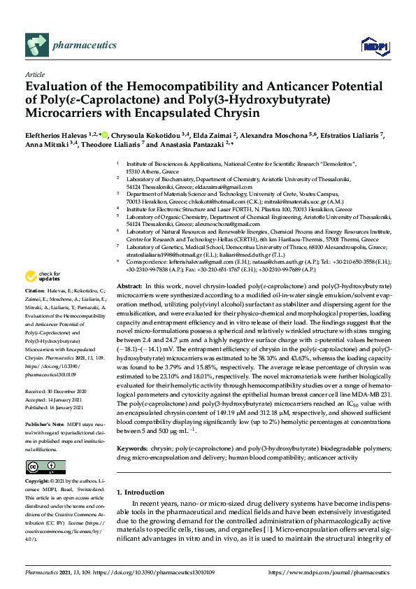
 Anastasia A Pantazaki