Received: 4 July 2019
Revised: 5 February 2020
Accepted: 11 February 2020
DOI: 10.1002/hbm.24966
RESEARCH ARTICLE
Functional magnetic resonance imaging (fMRI) item analysis of
empathy and theory of mind
Matthias G. Tholen1
| Fynn-Mathis Trautwein2
Tania Singer4 | Philipp Kanske5,6
1
Centre for Cognitive Neuroscience,
Department of Psychology, University of
Salzburg, Austria
| Anne Böckler3
|
Abstract
In contrast to conventional functional magnetic resonance imaging (fMRI) analysis
2
Edmont J. Safra Brain Research Center,
University of Haifa, Israel
3
Department of Psychology, Leibniz University
Hannover, Hannover, Germany
4
across participants, item analysis allows generalizing the observed neural response
patterns from a specific stimulus set to the entire population of stimuli. In the present
study, we perform an item analysis on an fMRI paradigm (EmpaToM) that measures
Max Planck Society, Social Neuroscience Lab,
Berlin, Germany
the neural correlates of empathy and Theory of Mind (ToM). The task includes a large
5
stimulus set (240 emotional vs. neutral videos to probe empathic responding and
Clinical Psychology and Behavioral
Neuroscience, Faculty of Psychology,
Technische Universität Dresden, Dresden,
Germany
6
Max Planck Institute for Human Cognitive
and Brain Sciences, Research Group Social
Stress and Family Health, Leipzig, Germany
240 ToM or factual reasoning questions to probe ToM), which we tested in two large
participant samples (N = 178, N = 130). Both, the empathy-related network comprising anterior insula, anterior cingulate/dorsomedial prefrontal cortex, inferior frontal
gyrus, and dorsal temporoparietal junction/supramarginal gyrus (TPJ) and the ToM
related network including ventral TPJ, superior temporal gyrus, temporal poles, and
Correspondence
Matthias G. Tholen, Centre for Cognitive
Neuroscience, Department of Psychology,
University of Salzburg, 5020 Salzburg, Austria.
Email: matthias.tholen@sbg.ac.at
anterior and posterior midline regions, were observed across participants and items.
Funding information
Austrian Science Fund, Grant/Award Number:
FWF-W1233; Bundesministerium für Bildung
und Forschung, Grant/Award Number: BMBF
FKZ 01EE1409A; Deutsche
Forschungsgemeinschaft, Grant/Award
Number: Heinz Maier-Leibnitz Prize KA
4412/1-1; FP7 Ideas: European Research
Council, Grant/Award Number: 205557
selection of the most effective items to create optimized stimulus sets that provide
Regression analyses confirmed that these activations are predicted by the empathy or
ToM condition of the stimuli, but not by low-level features such as video length, number of words, syllables or syntactic complexity. The item analysis also allowed for the
the most stable and reproducible results. Finally, reproducibility was shown in the replication of all analyses in the second participant sample. The data demonstrate (a) the
generalizability of empathy and ToM related neural activity and (b) the reproducibility
of the EmpaToM task and its applicability in intervention and clinical imaging studies.
KEYWORDS
affect sharing, anterior insula, mentalizing, social cognition, temporoparietal junction
1
|
I N T RO DU CT I O N
correlates of how we feel with (affective route) and know about
others (cognitive route). The affective route allows for sharing others'
Aiming at elucidating the mechanisms underlying social understanding,
emotions (empathy, affect sharing) (de Vignemont & Singer, 2006), for
human neuroscience research has extensively investigated the brain
example, when vicariously sharing another person's sadness or grief.
The cognitive route enables reasoning about others' mental states
Tania Singer and Philipp Kanske share senior authorship.
(Theory of Mind, ToM, mentalizing) (Frith & Frith, 2005; Premack &
This is an open access article under the terms of the Creative Commons Attribution License, which permits use, distribution and reproduction in any medium,
provided the original work is properly cited.
© 2020 The Authors. Human Brain Mapping published by Wiley Periodicals, Inc.
Hum Brain Mapp. 2020;1–18.
wileyonlinelibrary.com/journal/hbm
1
�2
THOLEN ET AL.
Woodruff, 1978), for example, when attributing another person's
physical or emotional pain yielding activity in the typical empathy and
belief, desire or intention. Several meta-analyses across different
medial parts of the ToM related neural networks, respectively. This
experimental approaches to both empathy and ToM have consistently
study did not, however, compare these results with the subject-wise
described two distinct neural networks related to these functions.
analysis published previously, which would directly show replicability of
Core regions of the empathy related network are found in the
subject- and item-wise analyses (Bruneau, Pluta, & Saxe, 2012). Interest-
anterior insula (AI), anterior cingulate/dorsomedial prefrontal cortex
ingly, Bedny et al. (2007), who studied word class processing, found dif-
(ACC/DMPFC), inferior frontal gyrus (IFG) and dorsal portions of the
ferent results for subject- and item-wise analyses, demonstrating the
temporoparietal junction/supramarginal gyrus (TPJ/SMG) (Bzdok
potential of item analysis to make theoretically important distinctions,
et al., 2012; Lamm, Decety, & Singer, 2011). The ToM related network
which in that case reconciled conflicting evidence regarding the role of
includes the ventral TPJ, anterior and posterior medial prefrontal
the prefrontal cortex in processing nouns vs. verbs (Bedny & Thompson-
cortex (MPFC), superior temporal gyrus/sulcus (STG/STS), and tempo-
Schill, 2006; Davis, Meunier, & Marslen-Wilson, 2004; Shapiro, Moo, &
ral poles (Bzdok et al., 2012; Schurz, Radua, Aichhorn, Richlan, & Per-
Caramazza, 2006; Tyler, Bright, Fletcher, & Stamatakis, 2004).
ner, 2014). Direct contrasts of both functions confirmed these
In the present study, we aimed to investigate whether item-
networks with functional (Kanske, Böckler, Trautwein, & Singer, 2015)
analyses of empathy and ToM replicate the neural networks observed
and structural neuroimaging (Eres, Decety, Louis, & Molenberghs,
with subject-wise analyses. To this end, we applied a previously vali-
2015; Valk et al., 2017; Valk, Bernhardt, Bockler, Kanske, & Singer,
dated fMRI paradigm that assesses both functions (EmpaToM)
2016). These studies show that empathizing and mentalizing engage
(Kanske et al., 2015). Empathy is probed via video stimuli with brief
distinct neural networks. Furthermore, brain regions also differ in cor-
autobiographical narrations that are highly emotionally negative or
tical thickness according to the subjects' capacity to share emotions
neutral. The negative emotional narrations included such diverse
or to reason about mental states. Importantly, even though both func-
issues as traffic accidents, involuntary pregnancy, partnership prob-
tions are essential elements of higher-level social processing, they
lems, diverse somatic and mental diseases and disorders, betrayal and
are not directly related. The independence of empathy and ToM
guilt, political violence, seeking refuge, rape, natural disaster, miscar-
processing was demonstrated on the behavioral and the neural level
riage, assault or burglary. These videos have been shown to elicit
(Kanske, Böckler, Trautwein, Parianen Lesemann, & Singer, 2016).
empathic responses on a subjective, peripheral physiological, and on a
With three notable exceptions (Bruneau, Dufour, & Saxe, 2013;
neural level. ToM reasoning is demanded in subsequent questions that
Dodell-Feder, Koster-Hale, Bedny, & Saxe, 2011; Theriault, Waytz,
either ask for the mental states of the narrator in the previous video
Heiphetz, & Young, 2017), all previous empathy and ToM investigations
or for factual reasoning about the events of the narration. The mental
used conventional functional magnetic resonance imaging (fMRI) ana-
state questions included first and second order, true and false
lyses across participants. These analyses allow generalizing the observed
beliefs, preferences and desires, irony, sarcasm, metaphors, (white)
neural response patterns from the investigated participant sample to the
lies, deception and faux pas. The empathy and ToM measures were
human population they were sampled from, if they treat subjects as
validated in several behavioral and fMRI studies through correlations
random-effect (as has become standard since the late 1990s (Friston,
and activation overlap with established empathy (Socio-affective
Holmes, & Worsley, 1999)). However, the “fixed-effect fallacy” still
Video Taks; Klimecki, Leiberg, Lamm, & Singer, 2013) and ToM tasks
applies to the item-level (Clark, 1973), that is, it is unsubstantiated to
(False Belief Task; Dodell-Feder et al., 2011, Imposing Memory Task;
claim that activation patterns observed for a sample of stimuli would
Kinderman, Dunbar, & Bentall, 1998) and additional overlap with
generalize to the population of stimuli, for instance, that the activity
meta-analytical findings (Bzdok et al., 2012; Dodell-Feder et al., 2011;
observed in an experiment eliciting emotional responses would general-
Kinderman et al., 1998; Klimecki et al., 2013). Conceptually, it is impor-
ize to the population of emotion-eliciting stimuli. Furthermore, treating
tant for social neuroscience to show that empathy related neural activ-
items as fixed could give single items with extreme responses dispropor-
ity generalizes beyond patterns only attributable to very specific
tionate weight, thereby rendering a contrast of two conditions signifi-
stimuli, and whether ToM tasks other than false-belief tasks (Dodell-
cant, just because a (possibly small) subset of items in one condition
Feder et al., 2011) also lead to generalizable brain activation. To illus-
shows very strong activity, while the majority of items shows no effect.
trate this form of generalization, as in psycholinguistics, where an item-
To overcome these problems, item analyses that treat items as random
analysis in an experiment on verb-processing allows generalizing the
are common in many behavioral fields of study and have been shown
results from the limited sample of verbs tested to the population of
to be feasible for fMRI analyses as well (Andrews-Hanna, Reidler,
verbs in that language (e.g., Bedny et al., 2007), replicating the subject-
Sepulcre, Poulin, & Buckner, 2010; Bedny, Aguirre, & Thompson-Schill,
analysis results in the EmpaToM with an item-analysis would allow gen-
2007; Dodell-Feder et al., 2011; Theriault et al., 2017; Troiani, Stigliani,
eralizing to the population of empathy-inducing and ToM-demanding
Smith, & Epstein, 2014; Yee, Drucker, & Thompson-Schill, 2010). Thus,
conversational situations. Given the breadth of the sampled situations
Theriault et al. (2017) demonstrated positive correlations between
in the EmpaToM (240 distinct videos and questions), testing generaliz-
regions in the ToM network and subjectivity ratings of metaethical judg-
ability may be challenging, but could also have particular impact.
ments. Dodell-Feder et al. (2011) replicated a subject-wise analysis with
Furthermore, a principal problem in subject-analysis is that discrep-
an item analysis showing generalizability for false-belief ToM stories.
ancies between two experimental conditions beyond the intended dif-
Bruneau et al. (2013) performed an item-analysis on brief stories of
ference are uncontrollable confounds. Item-analysis, in contrast, allows
�3
THOLEN ET AL.
specifically testing whether activations observed in a contrast of two
(ReSource Project; (Singer et al., 2016)).1 Participants were recruited
conditions are actually due to unintended low-level differences
from the general public through adverts. Recruitment of Sample 1 took
between the conditions (e.g., more or less movement when telling an
place in 2012–2013 and of Sample 2 in 2013–2014. Participants had
emotionally negative compared to a neutral story) rather than the
a very good language proficiency and were not included if they were
intended difference (e.g., negative vs. neutral emotion). As item-specific
below 20 or above 55 years of age, fulfilled the criteria for a mental or
activation patterns are obtained, they can be associated to the specific
neurological disorder (according to structured clinical interviews for
features of each item. Given that it is impossible to completely match
DSM-IV axis I and axis II disorders; Wittchen, Zaudig, & Fydrich,
emotional and neutral stimuli without erasing the difference in emo-
1997) or had any contraindication for MRI scanning. Twenty-four par-
tionality, ruling out the influence of such low-level features is a crucial
ticipants had to be excluded due to study dropout (N = 5), dropout
issue. With regard to ToM, because of the considerable overlap of ToM
from MRI measurements (N = 1), or missing data due to technical,
related activity with regions involved in language processing, particu-
scheduling, or health issues (N = 18).
larly in the temporal cortex and TPJ (Friederici, 2011; Schurz et al.,
For Sample 1, 13 participants were excluded yielding a final sam-
2014), it is critical to rule out the possibility that linguistic differences
ple of 178 participants (age mean = 40.9 years, SD = 9.5, 106 female).
account for the observed ToM effects. Dodell-Feder et al. (2011) con-
For Sample 2, 11 participants were excluded yielding a final sample of
vincingly demonstrated this for false-belief tasks, but it is important to
130 participants (age mean = 40.4 years, SD = 9.0, 72 female).
The study was approved by the Research Ethics Committee of
test whether this holds for other language-based ToM tasks as well.
Because the EmpaToM was designed to be used in extensive longi-
the University of Leipzig, number 376/12-ff and the Research Ethics
tudinal designs, it includes five parallel sets of different videos and ques-
Committee of the Humboldt University in Berlin, numbers 2013-02,
tions that allow the repeated testing of the same participants across time.
2013-29, and 2014-10. The study was registered with the Protocol
To enable usage of the EmpaToM in clinical and other settings, where
Registration System of ClinicalTrials.gov under the title “Plasticity of
only small participant samples are available or participants can be scanned
the Compassionate Brain” with the ClinicalTrials.gov Identifier:
for a very limited amount of time only, an item analysis on this large stim-
NCT01833104. All participants signed informed consent prior to
ulus set affords the chance to select the most effective items to create
participation.
stimulus sets that provide the most stable and reproducible results.
Finally, a major criticism of fMRI studies has been the limited sample size that not only reduces the likelihood to detect true effects, but
2.2
|
Stimuli and task
also reduces the chance that a statistically significant result reflects a
true effect (Button et al., 2013). Therefore, the present study made use
For details of the EmpaToM task see (Kanske et al., 2015) (Figure 1).
of a large sample of participants (N = 178) and checked for reproducibil-
Each trial started with a fixation cross (1–3 s), followed by the name
ity of the results in a second sample (N = 130).
of a person (2 s), who would speak in the subsequent video (~15 s).
In sum, applying item-analyses to an fMRI task probing empathy and
Each participant was presented with videos of 12 persons, telling four
ToM, the present study addresses several questions: (a) Will the item-
different stories each that corresponded to four conditions (2 × 2 fac-
analyses replicate the neural networks underlying empathy and ToM as
torial design, negative vs. neutral emotion, ToM vs. no ToM demands).
observed with subject-wise analyses? This would argue for generalizability
After this, participants rated the valence of their current emotional
of the observed brain activation patterns to the respective stimulus classes
state (sliding scale from negative to neutral to positive; 4 s) and how
(i.e., neutral and emotional autobiographical video narrations; factual rea-
much compassion2 they felt for the person in the previous video (slid-
soning and ToM questions, the latter involving a variety of ToM demands
ing scale from none to very much; 4 s). A second fixation cross (1–3 s)
such as irony, higher order mental state inference, false beliefs, etc.).
was followed by a multiple choice question with three response
(b) Can activity in the observed neural networks be predicted by low-level
options (one correct). These questions demanded either the attribu-
stimulus characteristics (i.e., number of sentences, words, syllables, charac-
tion of mental states or factual reasoning (ToM vs. factual reasoning).
ters, predicates, conjunctives, changes in tense, passive constructions, sub-
Participants had to respond within 14 s. For example, stories and
clauses, and the amount of motion)? (c) Does the item-analysis allow
questions, see Data S1. After a third fixation cross (0–2 s), participants
creating stimulus sets including the most effective items to provide the
were asked to rate their confidence, that their decision was done cor-
most stable and reproducible results? (d) Are all of the above described
rect (4 s) to allow assessing metacognitive abilities (Molenberghs,
results replicable in the second independent participant sample?
Trautwein, Bockler, Singer, & Kanske, 2016; Valk et al., 2016). In the
present study we focused on the main empathy and ToM measures,
that is, comparing emotional with neutral videos and ToM with factual
2
METHODS
|
reasoning questions (see (Kanske et al., 2015) for a validation of these
contrasts).
2.1
|
Participants
The total stimulus set of the EmpaToM task comprised 240 videos
and questions showing 60 different narrators in 4 conditions (see
Two samples of 191 and 141 German-speaking participants were
Figure 2). Based on this set, five parallel versions were created that
tested in the context of a large-scale longitudinal study at baseline
each contained a different set of 12 narrators in 4 conditions (yielding
�4
THOLEN ET AL.
F I G U R E 1 EmpaToM trial sequence. Emotional and neutral videos with and without ToM demands (2 × 2 design) are followed by valence
and compassion ratings, ToM and factual reasoning questions, and a confidence rating (adopted from Kanske et al. (2015)). This study
investigated the effects of subject- and itemwise analyses on the empathy and theory of mind contrasts. Empathy was tested via emotionally
negative versus neutral videos and theory of mind was tested via mental state versus factual reasoning questions. ToM, Theory of Mind
48 different videos and questions per set). The parallel sets were mat-
after emotional and neutral videos as an indicator of empathic
ched with regard to affect ratings, concern ratings, RTs, errors, confi-
responding and analyzed performance (RTs and accuracies) after ToM
dence ratings, video lengths and linguistic characteristics of the
questions as an indicator of ToM capacity. Each subject contributed
questions (number of words, characters, predicates, changes in tense,
ratings and performance measures in these conditions, averaged
complexity of the sentences [number of main and subordinate clau-
across all items. Complementarily, each item (i.e., narrator, each of
ses], number of passive sentence constructions, and number of con-
which told four different stories) contributed measures in each condi-
junctives), see (Kanske et al., 2015)). The five sets were randomly
tion, averaged across all participants.
assigned to the participants such that each set (of 48 videos and questions) was seen by a fifth of the participants in Samples 1 and 2.
2.5
2.3
|
MRI data acquisition
|
fMRI data analysis
Data preprocessing and statistical analyses were performed with
SPM 8 (http://www.fil.ion.ucl.ac.uk/spm) running in a MATLAB 7.6
Data were acquired on a 3 T MRI scanner (Siemens Magnetom Verio,
environment (Mathworks Inc., Sherbon MA). Functional images
Siemens Medical Solutions, Erlangen, Germany) using a 32 channel
were coregistered to the SPM single-subject canonical EPI image,
head coil. Functional images were acquired with a T2*-weighted
slice-time corrected and realigned to the mean image volume for
echo-planar imaging (EPI) sequence (TR = 2,000 ms; TE = 27 ms, Flip
motion correction. The high-resolution structural image was cor-
Angle 90� , matrix = 70 × 70 mm, FOV = 210 mm). Within one TR,
egistered to the SPM single-subject canonical T1 image and then to
37 axial slices of 3 mm were acquired. In addition, we collected a
the average functional image. Normalization parameters of the
high-resolution structural image (1 × 1 × 1 mm) with a T1-weighted
structural image into the Montreal Neurological Institute (MNI)
MPRAGE sequence.
space were used for spatial normalization of the functional images.
These images were resampled to isotropic 3 × 3 × 3 mm voxels and
smoothed with an 8 mm FWHM Gaussian Kernel.
2.4
|
Behavioral data analysis
The statistical analyses were performed by using the general linear model. For the subject-wise analysis, onset and duration of the
Repeated measures analyses of variance were calculated across sub-
four video types and their corresponding questions were modeled.
jects and across items. In particular, we contrasted valence ratings
These regressors were convolved by a canonical hemodynamic
�5
THOLEN ET AL.
F I G U R E 2 EmpaToM stimulus material. The overall stimulus material of the EmpaToM task contains 240 videos and questions with
60 different narrators in 4 conditions (emotional vs. neutral, ToM vs. nonToM), allocated to one of five parallel subsets. Each subset contains
12 different narrators in 4 conditions. The subsets are matched with regard to affect ratings, concern ratings, RTs, errors, confidence ratings, video
lengths, and linguistic characteristics of the questions (number of words, characters, predicates, changes in tense, complexity of the sentences,
number of passive sentence constructions, and number of conjunctives) (see Kanske et al., 2015). Subjects were randomly assigned to one of the
five subsets, so that each subset was seen by a fifth of the participants in Sample 1 (N = 178) and Sample 2 (N = 130). ToM, Theory of Mind
response function. Six regressors accounting head movement effects
contrasts between the condition differences (emotional vs. neutral
were modeled as covariates of no interest. RobustWLS Toolbox
videos, ToM vs. nonToM questions) together with the factor of sub-
(Diedrichsen & Shadmehr, 2005) was used to reduce potential noise-
groups as covariates of no interest in order to account for the depen-
artifact. Contrast images for empathy (emotional vs. neutral videos)
dencies between the 240 beta maps corresponding to the five
and ToM (ToM vs. nonToM questions) were calculated by applying
groups of participants. The main contrasts were tested with two sam-
linear weights to the parameter estimates and entered into one-
ple t-tests.
The results for the subject-wise as well as the item-wise analyses
sample t-tests for random effects analysis.
The item analyses were performed for each contrast separately
by modeling the emotional and neutral videos, and the ToM and fac-
were thresholded at p < .001 at voxel-level together with an FWE
(family-wise error) correction (p < .05) at the cluster level.
tual reasoning questions on the individual subject level. Each analysis
resulted in 48 beta maps per subject (12 narrators × 4 conditions).
The beta maps were averaged across the subjects within the five par-
2.6
|
Regression analysis
allel versions (see Figure 2) to receive one single beta map per narrator and condition. For each of the five subgroups, this method yielded
For both contrasts, regions of interest (ROI) (N = 46, 23 ToM,
48 beta maps at which each beta map comprised a mean beta value
23 empathy) were defined on the basis of the subject-wise random
across subjects at every voxel, adding up to 240 beta maps in total.
effects analyses of Sample 1 (see Table 1 for empathy, Table 2 for
For the second-level random effects analyses, we modeled the main
ToM). They were used to extract the beta values from Sample 2 for
�6
THOLEN ET AL.
T A B L E 1 Whole brain subject- and item-wise random effects results for Videos Emotional > Neutral. The results are reported at a voxel-level
threshold of p < .001 uncorrected together with an FWE-corrected cluster threshold of p < .05
MNI coordinates
T
Z
Cluster
−9
10.68
>8.21
1,027
15
45
10.04
>8.21
21
−6
8.58
7.82
−3
33
51
10.53
>8.21
9
21
57
8.69
>8.21
H
x
y
z
Inferior frontal gyrus
L
−48
39
Middle frontal
L
−42
Anterior insula
L
−36
Superior medial frontal cortex
L
Superior medial frontal
R
Subject-wise Group#1
Inferior frontal gyrus
R
51
30
−6
10.01
>8.21
Middle frontal
R
42
21
39
6.96
6.53
Anterior insula
R
30
24
−15
6.64
6.27
1,257
737
Ventral striatum
R
9
3
0
6.29
5.97
Ventral striatum
L
−6
−3
0
6.16
5.86
Caudate
L
−12
6
12
6.12
5.82
Caudate
R
12
6
12
6
5.82
0
−18
39
8.25
7.58
82
L
−54
−30
−12
6.08
5.79
26
448
Middle cingulate
Middle temporal cortex
153
TPJ-angular/supramarginal gyrus
R
63
−48
33
9.71
>8.21
Middle temporal cortex
R
60
−57
9
7.44
6.93
TPJ-angular/supramarginal gyrus
L
−54
−51
33
12.49
>8.21
599
0
−63
36
12.01
>8.21
614
L
−6
−75
−3
8.07
7.43
162
Precuneus
Lingual gyrus
Middle occipital
R
42
−84
18
5.98
5.7
30
Middle occipital
L
−39
−90
9
5.1
4.92
13
Cerebellum
L
−15
−78
−30
9.88
>8.21
186
Cerebellum
R
18
−81
−33
10.04
>8.21
219
Anterior insula
L
−39
21
−9
9.83
>8.21
767
Inferior frontal gyrus
L
−45
42
−12
8.71
7.64
Middle frontal
L
−42
18
42
10.64
>8.21
248
0
42
45
10.89
>8.21
1,239
Item-wise Group#1
Superior medial frontal cortex
Superior medial frontal
R
12
18
54
7.67
6.90
Inferior frontal gyrus
R
45
27
−12
9.33
>8.21
Anterior insula
R
30
24
−12
8.19
7.27
Middle frontal
R
39
24
39
6.68
6.14
148
59
Ventral striatum
R
9
3
0
6.33
5.86
Caudate
R
12
9
12
5.13
4.86
Caudate
L
−12
12
15
6.64
6.11
Ventral striatum
L
−6
0
−3
5.70
5.35
0
−18
39
7.00
6.39
Middle cingulate
492
54
45
Middle temporal cortex
L
−54
−30
−15
5.96
5.56
20
TPJ-angular/supramarginal gyrus
R
60
−45
36
8.02
7.15
258
Middle temporal cortex
R
48
−48
18
5.12
4.86
TPJ-angular/supramarginal gyrus
L
−54
−51
30
10.64
>8.21
412
Precuneus
L
−6
−60
33
8.87
7.74
413
Lingual gyrus
L
(Continues)
�7
THOLEN ET AL.
TABLE 1
(Continued)
MNI coordinates
H
y
z
L
−15
−78
−30
8.08
7.20
145
R
15
−78
−30
8.35
7.39
199
Inferior frontal gyrus
L
−45
36
−6
8.84
7.80
469
Anterior insula
L
−36
27
−3
7.27
6.64
Middle frontal
L
−39
15
39
7.18
6.57
106
Middle frontal
L
−36
60
−3
5.76
5.42
19
0
45
33
10.64
>8.21
758
R
15
21
63
5.18
4.93
R
Middle occipital
L
Cerebellum
Cerebellum
T
Cluster
x
Middle occipital
Z
Subject-wise Group#2
Superior medial frontal cortex
Superior medial frontal
Inferior frontal gyrus
R
45
27
3
7.31
6.67
Anterior insula
R
30
21
−15
6.50
6.03
290
Middle frontal
R
42
18
36
5.36
5.08
17
Ventral striatum
R
6
0
−3
6.00
5.62
32
Caudate
R
12
9
9
5.35
5.07
Ventral striatum
L
−6
0
0
5.84
5.49
Caudate
R
−12
6
12
5.63
5.31
0
−18
39
6.38
5.93
16
63
−51
24
7.93
7.15
153
−57
−51
33
10.36
>8.21
242
Middle cingulate
32
Middle temporal cortex
L
TPJ-angular/supramarginal gyrus
R
Middle temporal cortex
R
TPJ-angular/supramarginal gyrus
L
Precuneus
L
−6
−51
33
7.78
7.03
261
Lingual gyrus
L
−9
−75
−3
6.09
5.70
19
Middle occipital
R
Middle occipital
L
Cerebellum
L
−18
−78
−33
7.85
7.08
115
Cerebellum
R
24
−75
−33
7.90
7.12
96
Inferior frontal gyrus
L
−45
42
−6
8.52
7.50
588
Anterior insula
L
−27
24
−9
7.81
7.00
Middle frontal
L
−39
15
39
6.75
6.19
94
Superior medial frontal cortex
L
0
39
42
9.61
>8.21
823
Superior medial frontal
L
−3
33
48
9.34
>8.21
Inferior frontal gyrus
R
42
33
−3
7.90
7.06
Anterior insula
R
33
21
−15
7.43
6.72
Itemwise Group#2
283
Middle frontal
R
36
18
36
5.53
5.20
15
Caudate
R
6
−6
−12
6.91
6.32
55
Ventral striatum
L
9
0
−3
6.39
5.91
Ventral striatum
L
−6
0
0
6.03
5.62
Caudate
R
Middle cingulate
Middle temporal cortex
L
(Continues)
�8
THOLEN ET AL.
TABLE 1
(Continued)
MNI coordinates
H
TPJ-angular/supramarginal gyrus
R
Middle temporal cortex
R
TPJ-angular/supramarginal gyrus
x
y
z
T
Cluster
Z
63
−51
27
5.66
5.31
40
L
−54
−51
33
9.92
>8.21
214
Precuneus
L
−6
−51
33
6.21
5.76
70
Lingual gyrus
L
Middle occipital
R
Middle occipital
L
Cerebellum
L
−15
−78
−30
7.94
7.09
99
Cerebellum
R
15
−81
−30
7.42
6.71
108
Abbreviations: FWE, family-wise error; TPJ, temporoparietal junction/supramarginal gyrus.
the respective contrasts and consisted each of a sphere of contiguous
syntactic complexity influence activation patterns in the same cortical
voxels, 5 mm in radius. This procedure has two advantages: First, by
areas that are engaged during empathy and ToM processing.
using the ROIs from the subject-wise analysis, we might be able to
Besides to low-level features that are associated to spoken and
explain differences between item- and subject-wise analyses that are
written text, we additionally selected three general low-level features
due to low-level features. Second, the data of the regression analysis
that characterized the video material: duration of videos, motion and
is based on independently defined ROIs. To test whether the activa-
velocity of the narrator's movement. Emotionality may not only be
tions can be additionally explained by linguistic factors each item was
communicated by language and facial expression but is also facilitated
coded by at least two researchers in 9 different features. They com-
by spontaneous gestures and movements (Dick, Solodkin, & Small,
prised the following set of variables and were coded for each of the
2010). Gesture comprehension is supported by a cortical network com-
stories (spoken text, empathy contrast) and questions (written text,
prising the bilateral temporo-parietal junction, bilateral superior parietal
ToM contrast): number of words, characters, sentences, syllables
lobe, left inferior and middle frontal gyrus, and the left superior and
(as measures of the amount of spoken or written text), predicates,
middle temporal gyrus (Yang, Andric, & Mathew, 2015). Because of the
tenses, passives, conjunctives and complexity (as measures of syntac-
considerable overlap with empathy related activity, we included these
tic difficulty). Additionally, for the empathy contrast three general fea-
factors into the regression analysis to rule out that differences in the
tures were coded to characterize the video material: duration of the
video material account for the observed empathy effects.
We performed stepwise forward/backward regression analyses with
video, motion and velocity of the narrator's movement. In the following three passages, we further illustrate the choice of these features.
the item responses in the previously defined ROIs as dependent vari-
The amount of spoken or written text, for example, measured by
ables and condition and the selected features as independent variables.
the number of words, has been used as a proxy for constituent size
Stepwise regression is an iterative process of selecting and eliminating
(Goucha & Friederici, 2015; Pallier, Devauchelle, & Dehaene, 2011).
multiple variables depending on the model's best fit to the data. It is par-
These studies showed that increasing constituent size is associated
ticularly useful in cases where there are large numbers of predictors. In
with an increase of neural activation in left hemispheric cortical areas
each step, a predictor is added to the regression which most improves
such as the inferior frontal gyrus, temporo-parietal junction, superior
the fitting of the data (forward selection). To avoid overfitting, the pre-
temporal sulcus and temporal pole, regions that are also engaged dur-
dictors are excluded from the model if their contribution to predicting
ing empathy and theory of mind processing. Therefore, we tested
the outcome becomes non-significant (backward elimination). We used
whether differences in the number of words, characters, sentences or
rather strict entry and removal criteria that were based on the number
syllables can account for the observed effects in the EmpaTom task.
of predictors to account for multiple testing (theory of mind (10 predic-
Five additional features measure aspects of syntactic complexity,
tors): entry/removal: p = .005/p = .01; empathy (13 predictors): entry/
that is, number of predicates, tenses, passives, conjunctives and com-
removal: p = .0038/p = .0077). The analyses were performed on IBM
plexity (lexical diversity: type token ratio). Syntactic complexity is cor-
SPSS Statistics for Windows, version 26.0 (IBM Corp., Armonk, NY).
related with working memory load indicated by higher error rates and
longer processing times in sentence comprehension. FMRI studies
showed that this effect modulates the neural activity in the left infe-
2.7
|
Optimized sets of stimuli
rior frontal gyrus, middle frontal gyrus, and temporo-parietal junction
(Meltzer, McArdle, Schafer, & Braun, 2010; Newman, Malaia, Seo, &
The results of the item analyses were used to identify optimal sets of
Cheng, 2013) suggesting the possibility that items with higher
items which elicit the most prototypical response in both contrasts
�9
THOLEN ET AL.
T A B L E 2 Whole brain subject- and item-wise random effects results for Questions ToM > non ToM The results are reported at a voxel-level
threshold of p < .001 uncorrected together with an FWE-corrected cluster threshold of p < .05
MNI coordinates
H
x
y
z
T
Cluster
Z
Subject-wise Group#1
Rectus
R
3
57
−18
7.71
7.15
38
Superior medial frontal
L
−9
54
24
13.72
>8.21
1,185
Superior frontal
L
−9
54
33
12.34
>8.21
Superior medial frontal
R
9
57
21
11.73
>8.21
Inferior frontal gyrus
R
54
30
3
6.24
5.92
52
Inferior frontal gyrus
L
−51
24
6
10.32
>8.21
226
Inferior frontal gyrus
L
−45
27
−9
9.93
>8.21
Temporal pole
R
51
9
−33
14.68
>8.21
121
Temporal pole
L
−51
3
−30
12
>8.21
79
Postcentral
L
−54
−6
48
6.05
5.76
13
0
−15
39
8.56
7.81
50
Supplementary motor area
Middle cingulate
R
6
−24
57
5.37
5.17
10
TPJ-middle temporal
R
51
−30
−3
10.61
>8.21
640
TPJ-superior temporal
R
48
−18
−9
9.81
>8.21
TPJ-angular gyrus
R
63
−45
21
7.76
7.19
Posterior cingulate/precuneus
L
−6
−51
30
16.38
>8.21
328
TPJ-angular gyrus
L
−51
−57
24
15.81
>8.21
1,019
TPJ-middle temporal
L
−48
−30
−3
10.49
>8.21
TPJ-superior temporal
L
−60
−18
−6
9.53
>8.21
Cuneus
L
−9
−93
30
5.7
5.45
10
Cuneus
R
15
−87
39
6.11
5.82
24
Cerebellum
L
−27
−81
−36
14.65
>8.21
101
Cerebellum
R
27
−78
−33
15.82
>8.21
145
Superior medial frontal
L
−12
57
36
15.99
>8.21
1,241
Rectus
R
3
57
−18
7.97
7.12
Item-wise Group#1
Superior frontal
L
−6
54
18
14.93
>8.21
Superior medial frontal
L
−9
30
57
10.34
>8.21
Inferior frontal gyrus
R
48
30
−9
6.39
5.91
32
Inferior frontal gyrus
L
−54
24
6
11.74
>8.21
243
Inferior frontal gyrus
L
−45
30
−9
10.62
>8.21
Temporal pole
R
51
9
−33
16.30
>8.21
125
Temporal pole
L
−54
24
6
11.74
>8.21
243
Postcentral
L
0
−12
39
8.63
7.58
39
Middle cingulate
Supplementary motor area
R
TPJ-middle temporal
R
48
−30
−3
10.40
>8.21
332
TPJ-superior temporal
R
63
−51
21
6.42
5.93
71
TPJ-angular gyrus
R
66
−42
24
5.77
5.40
Posterior cingulate/precuneus
L
−9
−51
33
12.16
>8.21
253
TPJ-angular gyrus
L
−51
−54
24
15.64
>8.21
889
TPJ-superior temporal
L
−60
−15
−9
9.40
>8.21
TPJ-middle temporal
L
−48
−33
−6
8.82
7.71
(Continues)
�10
TABLE 2
THOLEN ET AL.
(Continued)
MNI coordinates
H
x
y
Cluster
z
T
Z
9
−33
16.30
>8.21
125
27
−78
−36
16.88
>8.21
135
−6
57
21
13.83
>8.21
1,008
Cuneus
L
Cuneus
R
Cerebellum
L
51
Cerebellum
R
Subject-wise Group#2
Rectus
R
Superior medial frontal
L
Superior medial frontal
R
6
57
15
12.56
>8.21
Supplementary motor area
L
−6
15
60
10.91
>8.21
Inferior frontal gyrus
R
57
27
0
5.45
5.16
15
Inferior frontal gyrus
L
−48
27
0
10.69
>8.21
206
Temporal pole
R
51
12
−27
14.12
>8.21
147
Temporal pole
L
−51
9
−30
11.66
>8.21
446
Postcentral
L
−51
−6
51
5.93
5.56
15
0
−15
39
8.55
7.60
68
446
Middle cingulate
TPJ-middle temporal
R
48
−27
−6
11.15
>8.21
TPJ-angular gyrus
R
66
−45
18
6.52
6.05
TPJ-superior temporal
R
66
−36
24
6.42
5.97
Posterior cingulate/precuneus
L
−6
−51
33
12.45
>8.21
251
TPJ-angular gyrus
L
−45
−54
24
13.41
>8.21
778
TPJ-middle temporal
L
−54
−27
−3
9.16
>8.21
TPJ-superior temporal
L
−63
−15
−15
6.43
5.98
Cuneus
L
−9
−93
30
5.69
5.36
17
Cuneus
R
Cerebellum
L
−27
−78
−36
13.97
>8.21
67
Cerebellum
R
30
−78
−36
14.07
>8.21
79
0
51
−21
7.49
6.76
42
Superior medial frontal
L
−6
54
27
16.11
>8.21
1,213
Superior medial frontal
R
6
60
15
12.49
>8.21
Superior medial frontal
L
−6
45
45
12.04
>8.21
Inferior frontal gyrus
R
54
27
0
6.45
5.96
34
Inferior frontal gyrus
L
−45
30
−6
11.88
>8.21
264
Inferior frontal gyrus
L
−51
24
6
10.28
>8.21
Temporal pole
R
51
12
−33
14.74
>8.21
164
Temporal pole
L
−48
12
−33
14.32
>8.21
97
Postcentral
L
−39
−21
21
5.68
5.33
14
Middle cingulate
L
−3
−12
39
7.18
6.52
45
Supplementary motor area
R
TPJ-middle temporal
R
45
−27
−6
11.24
>8.21
368
TPJ-superior temporal
R
60
−54
24
6.79
6.23
TPJ-angular gyrus
R
66
−42
18
6.21
5.77
Posterior cingulate/precuneus
L
−9
−51
33
12.14
>8.21
TPJ-angular gyrus
L
−48
−57
27
13.33
>8.21
Item-wise Group#2
Rectus
261
653
(Continues)
�11
THOLEN ET AL.
(Continued)
TABLE 2
MNI coordinates
z
T
Cluster
H
x
y
TPJ-superior temporal
L
−48
−33
−3
9.77
>8.21
Z
TPJ-middle temporal
L
−63
−18
−9
7.13
6.49
Cuneus
L
Cuneus
R
Cerebellum
L
−24
−78
−36
12.79
>8.21
76
Cerebellum
R
27
−78
−36
13.29
>8.21
89
Abbreviations: FWE, family-wise error; TPJ, temporoparietal junction/supramarginal gyrus.
(empathy and ToM), that is, those items that produce the greatest
The pattern of results was the same in Sample 2. For emotion
activation in the theory of mind and empathy network. More specifi-
effects, the valence ratings after emotional (M = −1.00, SD = .70) and
cally, we selected the items with the highest beta values in the experi-
neutral videos (M = .48, SD = .42), yielded significant differences in
mental conditions and the lowest beta values in the control conditions
subject- (F[1,129] = 470.18, p < .001) and item-wise analyses
for the regions of interest that were defined on the basis of the
(F[1,59] = 937.13, p < .001). Performance in ToM (M = 8,471.71 ms,
subject-wise random effects analyses of Sample 1. We identified two
SD = 1,334.42; M = 67.14%, SD = 11.85, chance level = 33.33%) and
sets, one with 48, the other with 40 videos and questions (see Data S2
nonToM questions (M = 8,563.97, SD = 1,329.51 ms; M = 57.43%,
and S3). Additionally, to allow for use in longitudinal designs, we identi-
SD = 15.30, chance level = 33.33%) resembled Sample 1. RTs did not
fied two parallel sets of stimuli, that is, two sets with 48 and two sets
differ in subject- (F[1,129] = 2.40, p > .10) and item-wise analyses
with 40 videos and questions each (see Data S4 and S5). The sets with
(F[1,59] < .81, p > .35), but accuracies were higher in the ToM than in
a reduced number of trials still reliably produce activations in the theory
the nonToM conditions in both subject- (F(1,129 = 53.96, p < .001)
of mind and empathy network, and might therefore particularly be use-
and item-wise analyses (F[1,59] = 16.08, p < .001). Again, the subject-
ful in clinical studies. The parallel sets are matched regarding on the
and item-wise analyses were perfectly in line with each other.
extent to which they recruit the respective ROIs as well as to behavioral measures (affect, concern, confidence ratings and response time,
accuracy in the questions), linguistic factors (number of words, charac-
3.2
Neuroimaging data
|
ters, sentences, syllables, predicates, tenses, passives, conjunctives, and
complexity) and general characteristics of the stimulus material (gender
3.2.1
|
Empathy
and age of narrator, movement and velocity, duration of the video).
We performed whole brain subject- and item-wise random effects
analyses, first on the data set acquired in Sample 1 (see Figure 3a,b
3
RESULTS
|
and Table 1). The results show activity in the typical empathy related neural network for emotional versus neutral videos, both
3.1
|
Behavioral data
across subjects and across items. This network includes bilateral AI,
ACC/DMPFC, IFG, and dorsal portions of TPJ/SMG. A few regions
We first analyzed data from Sample 1. To test for emotion effects, we
showed significant activity only in the subject-wise, but not the item-
compared the valence ratings after emotional (M = −1.11, SD = .61) and
wise analysis, including lingual and middle occipital gyrus, which
neutral videos (M = .43, SD = .39), which yielded significant differences in
would suggest that the activation is due to features of some specific
subject- (F[1,177] = 794.29, p < .001) and item-wise analyses (F
stimuli and that it is not generalizable. The pattern of results was the
[1,59] = 1,243.56, p < .001). To test, whether performance in ToM and
same in Sample 2 (see Figure 3c,d and Table 1).
nonToM questions differed, we compared RTs and accuracies in ToM
(M = 8,450.58 ms, SD = 1,346.38; M = 64.05%, SD = 12.31, chance
level = 33.33%) and nonToM questions (M = 8,490.93, SD = 1,272.54 ms;
3.2.2
|
Theory of mind
M = 55.05%, SD = 16.11, chance level = 33.33%). In RTs we found no significant differences in subject- (F[1,177] = .63, p > .40) and item-wise ana-
As for empathy, we first performed whole brain subject- and item-
lyses (F[1,59] < .001, p > .99). Accuracies, in contrast, were higher in the
wise random effects analyses on the data set acquired in Sample
ToM than in the nonToM conditions in both subject- (F(1,177 = 69.20,
1 (see Figure 4a,b and Table 2). All of the brain regions typically
p < .001) and item-wise analyses (F[1,59] = 14.92, p < .001), indicating that
involved in ToM were activated for ToM questions compared to fac-
the nonToM questions were slightly more difficult. Crucially, the subject-
tual reasoning questions, both across subjects and across items. These
and item-wise analyses were in line with each other for all measures.
regions include bilateral ventral TPJ, STS, temporal poles, precuneus,
�12
THOLEN ET AL.
F I G U R E 3 Consistency of the empathy related activation patterns (video: emotional > neutral) across item-wise (a) and subject-wise
(b) analyses in Sample 1 (N = 178) and in Sample 2 (N = 130) (c, d, respectively). The results show activity in the empathy related network for
emotional versus neutral videos, both across subjects and across items. This network includes anterior insula, anterior cingulate cortex/
dorsomedial prefrontal cortex, inferior frontal gyrus and dorsal portions of the temporoparietal junction (supramarginal gyrus)
and anterior MPFC. Brain regions active for subject-wise analysis and
3.2.3
|
Regression analysis
not item-wise analysis include parts of the supplementary motor area,
postcentral gyrus, and cuneus. The pattern of results was the same in
We performed stepwise forward/backward regression analyses on
Sample 2 (see Figure 4c,d and Table 2).
the data set acquired in Sample 2 for the empathy (23 ROIs) and ToM
�13
THOLEN ET AL.
F I G U R E 4 Consistency of the theory of mind related activation patterns (question: ToM > nonToM) across item-wise (a) and subject-wise
(b) analyses in Sample 1 (N = 178) and in Sample 2 (N = 130) (c, d, respectively). The results show activity in the theory of mind related network
for mental state vs. factual reasoning questions, both across subjects and across items. This network included bilateral ventral temporoparietal
junction, superior temporal sulcus, temporal poles, precuneus and anterior medial prefrontal cortex. ToM, Theory of Mind
(23 ROIs) contrast. All ROIs were independently defined by the
questions, the activity in the right cuneus is positively associated with
whole-brain subject-wise analysis of Sample 1. The results show that
the number of words and the activity in the supplementary motor
for both contrasts condition is almost the only predictor for all regions
area could not be explained by either condition or by any other stimu-
that were tested (see Table 3). For the ToM contrast, activity in the
lus characteristic. For the empathy contrast, three ROIs (left middle
left cuneus is positively associated with the number of syllables of the
temporal cortex, bilateral middle occipital cortex) could not be
�14
TABLE 3
THOLEN ET AL.
Results of the regression analyses
Coefficients
ROI
H
F
p
R
2
Predictor
β
t
p
Question ToM > non ToM
Rectus
R
12.034
.001
.093
Condition
.304
3.469
.001
Superior medial frontal
L
247.114
<.001
.677
Condition
.823
15.720
<.001
Superior frontal
L
203.853
<.001
.633
Condition
.796
14.278
<.001
Superior medial frontal
R
147.355
<.001
.555
Condition
.745
12.139
<.001
Inferior frontal gyrus
R
35.606
<.001
.232
Condition
.481
5.967
<.001
Inferior frontal gyrus
L
120.307
<.001
.505
Condition
.711
10.968
<.001
Inferior frontal gyrus
L
147.889
<.001
.556
Condition
.746
12.161
<.001
Temporal pole
R
223.881
<.001
.655
Condition
.809
14.963
<.001
Temporal pole
L
159.097
<.001
.574
Condition
.758
12.613
<.001
Postcentral
L
12.593
.001
.096
Condition
.311
3.549
.001
28.239
<.001
.193
Condition
.439
5.314
<.001
Middle cingulate
Supplementary motor area
R
None
TPJ-middle temporal
R
98.618
<.001
.455
Condition
.675
9.931
<.001
TPJ-superior temporal
R
71.325
<.001
.377
Condition
.614
8.445
<.001
TPJ-angular gyrus
R
27.034
<.001
.186
Condition
.432
5.199
<.001
Posterior cingulate/precuneus
L
107.008
<.001
.476
Condition
.690
10.344
<.001
TPJ-angular gyrus
L
148.454
<.001
.557
Condition
.753
12.184
<.001
TPJ-middle temporal
L
103.657
<.001
.468
Condition
.684
10.181
<.001
TPJ-superior temporal
L
59.992
<.001
.337
Condition
.581
7.745
<.001
Cuneus
L
15.237
<.001
.137
Syllables
.354
4.294
<.001
.207
Condition
.264
3.203
.002
Cuneus
R
16.476
<.001
.123
Words
.350
4.059
<.001
Cerebellum
L
143.531
<.001
.549
Condition
.741
11.980
<.001
Cerebellum
R
165.993
<.001
.584
Condition
.765
12.884
<.001
Inferior frontal gyrus
L
58.877
<.001
.333
Condition
.577
7.673
<.001
Middle frontal
L
18.694
<.001
.137
Condition
.370
4.324
<.001
Anterior insula
L
36.633
<.001
.237
Condition
.487
6.052
<.001
Superior medial frontal
L
59.271
<.001
.334
Condition
.578
7.699
<.001
Superior medial frontal
R
19.299
<.001
.141
Condition
.375
4.393
<.001
Inferior frontal gyrus
R
53.658
<.001
.313
Condition
.559
7.325
<.001
Middle frontal
R
13.047
<.001
.100
Condition
.316
3.612
<.001
Anterior insula
R
37.890
<.001
.243
Condition
.493
6.156
<.001
Ventral striatum
R
17.745
<.001
.131
Condition
.362
4.212
<.001
Ventral striatum
L
33.096
<.001
.219
Condition
.468
5.753
<.001
Caudate
L
12.161
.001
.093
Condition
.306
3.487
.001
Caudate
R
10.419
.002
.081
Condition
.285
3.228
.002
8.856
.004
.070
Condition
.264
2.976
.004
Video emotional > non emotional
Middle cingulate
Middle temporal cortex
L
TPJ-angular/supramarginal
R
17.682
<.001
.130
Condition
.361
4.205
<.001
Middle temporal cortex
R
15.180
<.001
.114
Condition
.338
3.896
<.001
TPJ-angular/supramarginal
L
78.740
<.001
.400
Condition
.633
8.874
<.001
12.004
.001
.092
Condition
.304
3.465
.001
Precuneus
None
(Continues)
�15
THOLEN ET AL.
TABLE 3
(Continued)
Coefficients
2
Predictor
β
.069
Condition
.264
2.968
.004
ROI
H
Lingual gyrus
L
Middle occipital
R
Middle occipital
L
Cerebellum
L
45.261
<.001
.277
Condition
.527
6.728
<.001
Cerebellum
R
39.490
<.001
.251
Condition
.501
6.284
<.001
F
p
8.807
R
.004
t
p
None
None
Abbreviations: ROI, regions of interest; ToM, Theory of Mind; TPJ, temporoparietal junction/supramarginal gyrus.
associated with any of the predictors. Low-level factors such as lin-
people express their emotions, such as gesture, body posture and
guistic or general characteristics do not show a major influence
movement or the direct observation of emotional situations, for
regarding the activation across the empathy or ToM network.
instance, of injury. Moreover, the emotional videos in the EmpaToM
paradigm are negatively valenced which also precludes generalizing to
positive empathy, that is, sharing, or joining others' positive emotions.
4
|
DISCUSSION
The theory of mind questions aim at an understanding of a person's
mental states. Stimuli that target the prediction of a person's behavior
The present study aimed to probe the generalizability and reproduc-
are not included in this task. In comparison to other tasks in theory of
ibility of the neural networks related to empathy and ToM. The results
mind, the items cannot generalize to mental state attributions that are
demonstrate replicability of subject- and item-wise analyses of both
based on action observation as in social animations (e.g., Castelli et al.,
functions with AI, ACC/DMPFC, IFG, dorsal TPJ/SMG for empathy
2000), or to conceptual knowledge about persons as in trait judg-
and ventral TPJ, STG/STS, temporal poles and anterior and posterior
ments (e.g., Mitchell et al., 2002). Consequently, the stimuli of the
midline regions for ToM, arguing for generalizability of the brain acti-
EmpaToM task do not elicit all possible forms of empathic responses
vation patterns to the respective stimulus classes. Importantly, the
and theory of mind reasoning. A more comprehensive approach to
observed activity was not predicted by low-level stimulus characteris-
generate a random sample of items that is representative for theory
tics such as the number of words or syllables, corroborating the valid-
of mind and empathy might be realized by an ecological momentary
ity of the activation patterns. Furthermore, we used the item
assessment (EMA) (Shiffman, Stone, & Hufford, 2008). This approach
information to construct stimulus sets that include the most effective
involves repeated sampling of subject's social interactions in real time
items to provide the most stable and reproducible results in future
over periodic intervals, thereby enabling a high ecological validity.
studies employing the EmpaToM paradigm. Lastly, demonstrating
Future studies could therefore arrive at stronger conclusions about
reproducibility of the findings, all of the above described results were
the precise nature of the population of items.
replicated in a second, independent participant sample.
However, given the amount of videos and questions (240 in total
The main result of the present study is the finding of consistent
for each type) and the fact that no situation was repeated, there is
activation patterns for empathy and ToM across subject- and item-
considerable breadth within this conversation type situation. Comply-
wise analyses. This consistency demonstrates that the observed net-
ing with the call for “item-analyses with a larger and more variable set
work activity is not due to idiosyncratic characteristics of (some of)
of stimuli” (Dodell-Feder et al., 2011), the present results, thus,
the utilized videos and questions, but is generalizable to the entire
expand previous reports of consistent activity for reading false-belief
populations of stimuli. One critical question here is what exactly
(20 items; (Dodell-Feder et al., 2011)) and physical or emotional pain
defines these stimulus populations. Just as the generalizability of
stories (24 items each; (Bruneau et al., 2013)).
subject-wise analyses is limited by how well the participant sample
Another critical question pertains to possible confounds due to
represents the population (e.g., the age range of 20–55 years in the
the, in general, high error rates and the differences in behavioral per-
present study precludes conclusions about empathy and ToM
formance. The EmpaToM task was explicitly designed to be hard,
processing in older adults), generalizing the results to empathy induc-
which makes it unique among other theory of mind tasks in functional
ing or ToM demanding situations needs to be done with care, consid-
neuroscience in adults. In other tasks, for example, false belief or
ering the breadth of situations covered in the applied stimuli. The
social animations, healthy participants perform typically at 100% or
shown videos were created to resemble brief episodes of a (putatively
nearly 100% accuracy. The drawback of those measurements is that
longer) complex conversation one might have with another person.
they are not sensitive to pick up improvements in performance over
The ToM questions ask for aspects of the mental state of this person
time, whereas the EmpaToM task can (Böckler et al., 2017; Trautwein
that were not overtly described. While this enables generalizing to the
et al., 2020). Given that participants were less accurate in the nonToM
empathic sharing of others' affect as conveyed in language, prosody
condition than in the ToM condition, one might think that the differ-
and facial expression, it precludes generalizing to other forms in which
ential brain activation identified with the contrast (ToM > nonToM)
�16
THOLEN ET AL.
reflects the effect of general task difficulty. However, we think this is
empathy regions. A different approach could also focus on high-level
unlikely because of the following reasons: First, prior to the fMRI
features, such as whether the ToM questions include true or false
measurements, participants were sufficiently familiarized with the task
belief, or first or second order reasoning. This approach might, there-
and the different conditions. Second, a previous study that validated
fore, be of particular importance for future research on social cogni-
the EmpaToM task with other measures of empathy and theory of
tion identifying areas with specific functions for ToM and empathy
mind did not detect any differences in accuracy (Kanske et al., 2015;
processing.
Exp. 1). In line with these results, subjects' confidence ratings, indicat-
Given the recent discussions about difficulties in replicating psy-
ing their performance evaluation, were equal across all conditions,
chological findings (Lindsay, 2015; Open Science Collaboration, 2015),
meaning the participants did not evaluate the nonToM condition as
we aimed at testing the stability of our findings in a within-study
more difficult than the ToM condition. Finally, further results of this
replication. Indeed, the results from a second independent sample cor-
study also showed that the theory of mind performance does posi-
roborated the conclusions of the first sample, that is, reproducible
tively correlate with the activity of the default mode network,
neuroimaging results in subject- and item-wise analyses that are inde-
whereas areas in the default mode network typically tend to increase
pendent
in deactivation with increasing task difficulty (e.g., Buckner, 2008).
addressing the critique of small sample sizes in many neuroimaging
from
low-level
stimulus
characteristics.
Furthermore,
Activity in a few regions observed in the subject-wise analyses
studies (Button et al., 2013), the two samples we assessed were rela-
was not present for the item-wise analyses. These include the supple-
tively large in comparison to most fMRI investigations (which mostly
mentary motor area, postcentral gyrus, and cuneus for ToM and lin-
include <40 participants) (David et al., 2013). Thus, the present study
gual and middle occipital gyrus for empathy. The results of the
lends a high degree of trustworthiness to the observed neural activa-
regression analyses could partly explain this difference by showing
tion patterns for empathy and ToM. Future studies could of course fur-
that activity in the bilateral cuneus was mainly due to the number of
ther strengthen this conclusion, for instance by probing the test–retest-
syllables and words of the theory of mind and factual reasoning ques-
reliability of the results, which has been shown to be highly variable
tions and not the condition difference itself. The lack of activation in
across brain regions and experimental paradigms (Plichta et al., 2012).
the other areas in the item-wise analyses suggests that their subject-
The specific activation patterns observed for empathy and ToM
wise activation is due to specifics of the videos and questions used,
are not only consistent across subject- and item-wise analyses, but
implying that they would not be activated by other empathy and ToM
also correspond to the typical networks associated with the two func-
stimuli. This is in line with the absence of these regions in empathy
tions in large-scale meta-analyses (Bzdok et al., 2012; Lamm et al.,
and ToM meta-analyses (Bzdok et al., 2012; Lamm, Batson, & Decety,
2011; Molenberghs, Johnson, et al., 2016; Schurz et al., 2014). An
2007; Schurz et al., 2014).
interesting aspect is that the meta-analyses suggest the existence of
FMRI item-analyses allow an item-specific estimate for the neural
core networks for empathy (AI, IFG, ACC) and ToM (TPJ, MPFC), acti-
activity in a brain region which might serve as an indicator of the
vated for all operationalizations of the respective functions, and
regions function. As the items can be characterized not only regarding
extended networks that include additional regions (for empathy:
their experimental category but also regarding multiple other features
DMPFC, dorsal TPJ/SMG; for ToM: STG/STS, temporal poles,
(e.g., constituent size, or syntactic complexity), it is possible to deter-
precuneus), when pooling across the different operationalizations.
mine which features best predict the neural response in each brain
Assuming that most experimental paradigms capture specific compo-
region (see e.g., Bruneau et al., 2013; Dodell-Feder et al., 2011). This
nent processes of full-fledged empathy or ToM (Schurz & Perner,
allowed us to test whether low-level stimulus characteristics, which
2015), the finding of activation in the extended networks for the
might confound the manipulation of empathy and ToM, have contrib-
EmpaToM suggests that the task comprehensively captures the com-
uted to some of the activations attributed to the experimental condi-
plexity of these two social capacities (as is the case for other para-
tion. The results of the regression analyses yielded the experimental
digms aiming at ecological validity (Wolf, Dziobek, & Heekeren,
condition as strongest predictor (by far) for all of the observed activa-
2010)). Furthermore, taking the independence of the neural bases of
tion clusters, demonstrating convincingly that none of the low-level
empathy and ToM into account (Kanske et al., 2015; Kanske et al.,
predictors exert major influence on the results. As it is impossible to
2016) and observing the two networks in both types of analyses here,
completely match emotional and neutral videos without erasing the
corroborates the assumption that empathy and ToM are distinct social
difference in emotionality, this is an important, reassuring finding. Also
functions, possibly serving specific purposes in social encounters, for
with regard to ToM, ruling out the possibility that linguistic character-
example, establishing the motivation for cooperation and enhancing
istics account for the ToM effects is important, because of the consid-
prosocial behavior (Kanske, Bockler, & Singer, 2017; Tusche, Bockler,
erable overlap of ToM related activity with regions involved in
Kanske, Trautwein, & Singer, 2016).
language processing, particularly in the temporal cortex and TPJ
The results of the item-analysis made it possible to select those
(Friederici, 2011; Molenberghs, Johnson, Henry, & Mattingley, 2016;
videos and questions that elicit the most prototypical responses in terms
Schurz et al., 2014) and the discussion of the intricate relationship of
of activation in the neural networks that meta-analyses have associated
ToM and language processing (de Villiers & Pyers, 2002; Ferstl & von
with empathy and ToM (Bzdok et al., 2012; Lamm et al., 2011; Schurz
Cramon, 2002). The results of the regression analyses showed that
et al., 2014) and in behavior. To avoid circularity, we selected the stimuli
low-level features do not explain the neural response in the ToM or
based on Sample 1 and tested them in the independent Sample
�17
THOLEN ET AL.
2, showing strong and consistent activation patterns across the two sam-
ENDNOTES
ples. This way, we could form several optimized stimulus sets for future
1
Please note that sample 1 in the present study is based on the same participant sample as described in Kanske et al. (2015). Importantly however, the analyses and results described in the present study are novel
and have not been described or shown elsewhere.
2
In contrast to empathy, compassion is defined as feelings of warmth and
care, including the motivation to improve the other's wellbeing (Singer &
Klimecki, 2014).
usage in specific settings. In particular, the short versions of the task
enable testing special populations with reduced attention spans, for
instance, in psychopathology (Preckel, Kanske, Singer, Paulus, & Krach,
2016) or assessing multiple tasks, including the EmpaToM, within one
session, for instance, to predict social behavior based on empathic and
ToM capabilities (Tusche et al., 2016). The optimized parallel sets could
be applied in longitudinal designs, including intervention research.
To conclude, by replicating the empathy and ToM related neural
networks across item- and subject-wise analyses and demonstrating
their independence from low-level stimulus characteristics, the present results contribute methodologically to the social neuroscience literature and add to our understanding of these social capacities as
distinct functions.
ACKNOWLEDGMENTS
This study forms part of the ReSource Project, headed by Tania Singer.
Data for this project were collected between 2013 and 2016 at the former Department of Social Neuroscience at the Max Planck Institute for
Human Cognitive and Brain Sciences Leipzig. Tania Singer (Principal
Investigator) received funding for the ReSource Project from the
European Research Council (ERC) under the European Community's
Seventh Framework Program (FP7/2007–2013) ERC grant agreement
number 205557. M.G.T. is supported by the Austrian Science Fund's
Doctoral College “Imaging the Mind” (FWF-W1233). P.K. is supported
by German Federal Ministry of Education and Research within the
ASD-Net (BMBF FKZ 01EE1409A), the German Research Council
(Heinz Maier-Leibnitz Prize KA 4412/1-1) and Die Junge Akademie at
the Berlin-Brandenburg Academy of Sciences and Humanities and the
German National Academy of Sciences Leopoldina. We wish to thank
the entire ReSource support team for help in the organization of the
study, in particular, we thank Nicole Pampus, Manuela Hoffmann and
Sylvie Neubert for help with the data acquisition. The data of this study
are available from the authors upon reasonable request.
AUTHOR CONTRIBUTIONS
Conceptualization, all authors; Data curation, F.M.T., A.B.R., P.K.; Formal analysis, M.G.T., P.K., F.M.T.; Funding Acquisition, T.S.; Investigation, F.M.T., A.B.R., P.K.; Methodology, F.M.T., A.B.R., P.K.; Project
administration, T.S., F.M.T., A.B.R., P.K.; Resources, T.S.; Supervision,
T.S., P.K.; Validation, M.G.T.; Visualization, M.G.T.; Writing—original
draft preparation, P.K., M.G.T.; Writing—review and editing, all authors.
DATA AVAI LAB ILITY S TATEMENT
The data of this study are available from the authors upon reasonable
request.
ORCID
Matthias G. Tholen
Anne Böckler
Philipp Kanske
https://orcid.org/0000-0001-8716-8807
https://orcid.org/0000-0003-1000-9052
https://orcid.org/0000-0003-2027-8782
RE FE RE NCE S
Andrews-Hanna, J. R., Reidler, J. S., Sepulcre, J., Poulin, R., & Buckner, R. L.
(2010). Functional-anatomic fractionation of the brain's default network. Neuron, 65, 550–562.
Bedny, M., Aguirre, G. K., & Thompson-Schill, S. L. (2007). Item analysis
in functional magnetic resonance imaging. NeuroImage, 35, 1093–1102.
Bedny, M., & Thompson-Schill, S. L. (2006). Neuroanatomically separable
effects of imageability and grammatical class during single-word comprehension. Brain and Language, 98, 127–139.
Böckler, A., Herrmann, L., Trautwein, F.-M., Holmes, T., & Singer, T. (2017).
Know thy selves: learning to understand oneself increases the ability
to understand others. Journal of Cognitive Enhancement, 1(2), 197–209.
https://doi.org/10.1007/s41465-017-0023-6
Bruneau, E., Dufour, N., & Saxe, R. (2013). How we know it hurts: Item
analysis of written narratives reveals distinct neural responses to
others' physical pain and emotional suffering. PLoS One, 8, e63085.
Bruneau, E. G., Pluta, A., & Saxe, R. (2012). Distinct roles of the ‘shared
pain’ and ‘theory of mind’ networks in processing others' emotional
suffering. Neuropsychologia, 50, 219–231.
Buckner, R. L., Andrews-Hanna, J. R., & Schacter, D. L. (2008). The brain's
default network. Annals of the New York Academy of Sciences, 1124(1),
1–38. https://doi.org/10.1196/annals.1440.011
Button, K. S., Ioannidis, J. P. A., Mokrysz, C., Nosek, B. A., Flint, J.,
Robinson, E. S. J., & Munafo, M. R. (2013). Power failure: Why small
sample size undermines the reliability of neuroscience. Nature Reviews
Neuroscience, 14, 365–376.
Bzdok, D., Schilbach, L., Vogeley, K., Schneider, K., Laird, A. R.,
Langner, R., & Eickhoff, S. B. (2012). Parsing the neural correlates of
moral cognition: ALE meta-analysis on morality, theory of mind, and
empathy. Brain Structure & Function, 217, 783–796.
Castelli, F., Happé, F., Frith, U., & Frith, C. (2000). Movement and mind: a
functional imaging study of perception and interpretation of complex
intentional movement patterns. NeuroImage, 12(3), 314–325.
Clark, H. H. (1973). Language as fixed-effect fallacy—Critique of language
statistics in psychological research. Journal of Verbal Learning and Verbal Behavior, 12, 335–359.
David, S. P., Ware, J. J., Chu, I. M., Loftus, P. D., Fusar-Poli, P., Radua, J., …
Ioannidis, J. P. A. (2013). Potential reporting bias in fMRI studies of the
brain. PLoS One, 8, e70104.
Davis, M. H., Meunier, F., & Marslen-Wilson, W. D. (2004). Neural
responses to morphological, syntactic, and semantic properties of single words: An fMRI study. Brain and Language, 89, 439–449.
de Vignemont, F., & Singer, T. (2006). The empathic brain: How, when and
why? Trends in Cognitive Sciences, 10, 435–441.
de Villiers, J. G., & Pyers, J. E. (2002). Complements to cognition: A longitudinal study of the relationship between complex syntax and falsebelief-understanding. Cognitive Development, 17, 1037–1060.
Dick, A. S., Solodkin, A., & Small, S. L. (2010). Neural development of networks for audiovisual speech comprehension. Brain and Language, 114
(2), 101–114.
Diedrichsen, J., & Shadmehr, R. (2005). Detecting and adjusting for artifacts in fMRI time series data. NeuroImage, 27(3), 624–634.
Dodell-Feder, D., Koster-Hale, J., Bedny, M., & Saxe, R. (2011). fMRI item
analysis in a theory of mind task. NeuroImage, 55, 705–712.
�18
Eres, R., Decety, J., Louis, W. R., & Molenberghs, P. (2015). Individual differences in local gray matter density are associated with differences in
affective and cognitive empathy. NeuroImage, 117, 305–310.
Ferstl, E. C., & von Cramon, D. Y. (2002). What does the frontomedian cortex contribute to language processing: Coherence or theory of mind?
NeuroImage, 17, 1599–1612.
Friederici, A. D. (2011). The brain basis of language processing: From
structure to function. Physiological Reviews, 91, 1357–1392.
Friston, K. J., Holmes, A. P., & Worsley, K. J. (1999). How many subjects
constitute a study? NeuroImage, 10, 1–5.
Frith, C., & Frith, U. (2005). Theory of mind. Current Biology, 15, 644–646.
Goucha, T., & Friederici, A. D. (2015). The language skeleton after dissecting meaning: A functional segregation within Broca's area.
NeuroImage, 114, 294–302.
Kanske, P., Bockler, A., & Singer, T. (2017). Models, mechanisms and moderators dissociating empathy and theory of mind. Current Topics in
Behavioral Neurosciences, 30, 193–206.
Kanske, P., Böckler, A., Trautwein, F. M., Parianen Lesemann, F. H., &
Singer, T. (2016). Are strong empathizers better mentalizers? Evidence
for independence and interaction between the routes of social cognition. Social Cognitive and Affective Neuroscience, 11, 1383–1392.
Kanske, P., Böckler, A., Trautwein, F. M., & Singer, T. (2015). Dissecting
the social brain: Introducing the EmpaToM to separate empathy and
theory of mind in brain and behavior. NeuroImage, 122, 6–19.
Kinderman, P., Dunbar, R., & Bentall, R. P. (1998). Theory-of-mind deficits
and causal attributions. British Journal of Psychology, 89, 191–204.
Klimecki, O. M., Leiberg, S., Lamm, C., & Singer, T. (2013). Functional neural plasticity and associated changes in positive affect after compassion training. Cerebral Cortex, 23, 1552–1561.
Lamm, C., Batson, C. D., & Decety, J. (2007). The neural substrate of
human empathy: Effects of perspective-taking and cognitive appraisal.
Journal of Cognitive Neuroscience, 19, 42–58.
Lamm, C., Decety, J., & Singer, T. (2011). Meta-analytic evidence for common and distinct neural networks associated with directly experienced
pain and empathy for pain. NeuroImage, 54, 2492–2502.
Lindsay, D. S. (2015). Replication in psychological science. Psychological
Science, 26, 1827–1832.
Meltzer, J. A., McArdle, J. J., Schafer, R. J., & Braun, A. R. (2010). Neural
aspects of sentence comprehension: Syntactic complexity, reversibility, and reanalysis. Cerebral Cortex, 20, 1853–1864.
Mitchell, J. P., Heatherton, T. F., & Macrae, C. N. (2002). Distinct neural
systems subserve person and object knowledge. Proceedings of the
National Academy of Sciences, 99(23), 15238–15243. https://doi.org/
10.1073/pnas.232395699
Molenberghs, P., Johnson, H., Henry, J. D., & Mattingley, J. B. (2016).
Understanding the minds of others: A neuroimaging meta-analysis.
Neuroscience and Biobehavioral Reviews, 65, 276–291.
Molenberghs, P., Trautwein, F. M., Bockler, A., Singer, T., & Kanske, P. (2016).
Neural correlates of metacognitive ability and of feeling confident: A largescale fMRI study. Social Cognitive and Affective Neuroscience, 11, 1942–1951.
Newman, S. D., Malaia, E., Seo, R., & Cheng, H. (2013). The effect of individual differences in working memory capacity on sentence comprehension: An fMRI study. Brain Topography, 26, 458–467.
Open Science Collaboration. (2015). Estimating the reproducibility of psychological science. Science, 349, aac4716.
Pallier, C., Devauchelle, A. D., & Dehaene, S. (2011). Cortical representation
of the constituent structure of sentences. Proceedings of the National
Academy of Sciences of the United States of America, 108, 2522–2527.
Plichta, M. M., Schwarz, A. J., Grimm, O., Morgen, K., Mier, D., Haddad, L., …
Meyer-Lindenberg, A. (2012). Test-retest reliability of evoked BOLD signals
from a cognitive-emotive fMRI test battery. NeuroImage, 60, 1746–1758.
Preckel, K., Kanske, P., Singer, T., Paulus, F. M., & Krach, S. (2016). Clinical
trial of modulatory effects of oxytocin treatment on higher-order
social cognition in autism spectrum disorder: A randomized, placebocontrolled, double-blind and crossover trial. BMC Psychiatry, 16, 329.
THOLEN ET AL.
Premack, D., & Woodruff, G. (1978). Does the chimpanzee have a theory
of mind? The Behavioral and Brain Sciences, 1, 515–526.
Schurz, M., & Perner, J. (2015). An evaluation of neurocognitive models of
theory of mind. Frontiers in Psychology, 6, 1610.
Schurz, M., Radua, J., Aichhorn, M., Richlan, F., & Perner, J. (2014). Fractionating theory of mind: A meta-analysis of functional brain imaging
studies. Neuroscience and Biobehavioral Reviews, 42C, 9–34.
Shapiro, K. A., Moo, L. R., & Caramazza, A. (2006). Cortical signatures of
noun and verb production. Proceedings of the National Academy of Sciences of the United States of America, 103, 1644–1649.
Shiffman, S., Stone, A. A., & Hufford, M. R. (2008). Ecological momentary
assessment. Annual Review of Clinical Psychology, 4(1), 1–32. https://
doi.org/10.1146/annurev.clinpsy.3.022806.091415
Singer, T., & Klimecki, O. M. (2014). Empathy and compassion. Current Biology, 24(18), R875–R878. https://doi.org/10.1016/j.cub.2014.06.054
Singer, T., Kok, B. E., Bornemann, B., Zurborg, S., Bolz, M., & Bochow, C. A.
(2016). The ReSource project: Background, design, samples, and measurements.
Leipzig: Max Planck Institute for Human Cognitive and Brain Sciences.
Theriault, J., Waytz, A., Heiphetz, L., Young, L. (2017) Theory of Mind network activity is associated with metaethical judgment: An item analysis. https://doi.org/10.31234/osf.io/gb5am.
Trautwein, F.-M., Kanske, P., Böckler, A., & Singer, T. (2020). Differential benefits of mental training types for attention, compassion, and theory of mind.
Cognition, 194, 104039. https://doi.org/10.1016/j.cognition.2019.
104039
Troiani, V., Stigliani, A., Smith, M. E., & Epstein, R. A. (2014). Multiple object
properties drive scene-selective regions. Cerebral Cortex, 24, 883–897.
Tusche, A., Bockler, A., Kanske, P., Trautwein, F. M., & Singer, T. (2016). Decoding
the charitable brain: Empathy, perspective taking, and attention shifts differentially predict altruistic giving. The Journal of Neuroscience, 36, 4719–4732.
Tyler, L. K., Bright, P., Fletcher, P., & Stamatakis, E. A. (2004). Neural
processing of nouns and verbs: The role of inflectional morphology.
Neuropsychologia, 42, 512–523.
Valk, S. L., Bernhardt, B. C., Bockler, A., Kanske, P., & Singer, T. (2016).
Substrates of metacognition on perception and metacognition on
higher-order cognition relate to different subsystems of the
mentalizing network. Human Brain Mapping, 37, 3388–3399.
Valk, S. L., Bernhardt, B. C., Bockler, A., Trautwein, F. M., Kanske, P., &
Singer, T. (2017). Socio-cognitive phenotypes differentially modulate largescale structural covariance networks. Cerebral Cortex, 27, 1358–1368.
Wittchen, H.-U., Zaudig, M., Fydrich, T. (1997). SKID: Strukturiertes
klinisches Interview für DSM-IV. Achse I und II. Göttingen. Hogrefe.
Wolf, I., Dziobek, I., & Heekeren, H. R. (2010). Neural correlates of social
cognition in naturalistic settings: A model-free analysis approach.
NeuroImage, 49, 894–904.
Yang, J., Andric, M., & Mathew, M. M. (2015). The neural basis of hand gesture comprehension: A meta-analysis of functional magnetic resonance
imaging studies. Neuroscience and Biobehavioral Reviews, 57, 88–104.
Yee, E., Drucker, D. M., & Thompson-Schill, S. L. (2010). fMRI-adaptation
evidence of overlapping neural representations for objects related in
function or manipulation. NeuroImage, 50, 753–763.
SUPPORTING INF ORMATION
Additional supporting information may be found online in the
Supporting Information section at the end of this article.
How to cite this article: Tholen MG, Trautwein F-M,
Böckler A, Singer T, Kanske P. Functional magnetic resonance
imaging (fMRI) item analysis of empathy and theory of mind.
Hum Brain Mapp. 2020;1–18. https://doi.org/10.1002/hbm.
24966
�
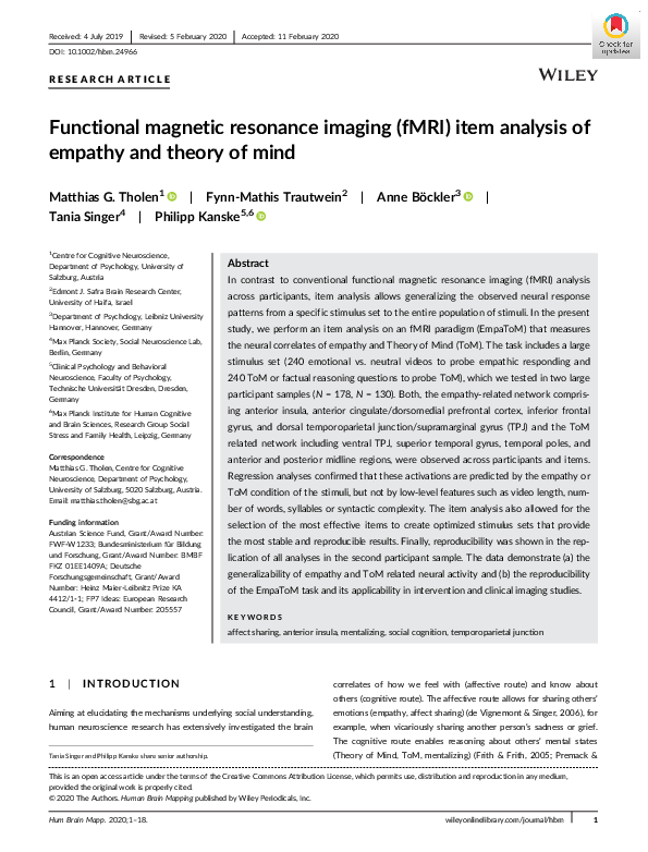
 Tania Singer