Academia.edu no longer supports Internet Explorer.
To browse Academia.edu and the wider internet faster and more securely, please take a few seconds to upgrade your browser.
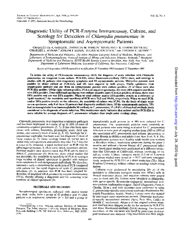
Diagnostic utility of PCR-enzyme immunoassay, culture, and serology for detection of Chlamydia pneumoniae in symptomatic and asymptomatic patients
Diagnostic utility of PCR-enzyme immunoassay, culture, and serology for detection of Chlamydia pneumoniae in symptomatic and asymptomatic patients
1994, Journal of Clinical Microbiology
Related Papers
Journal of Clinical Microbiology
Diagnostic Utility of PCR-Enzyme Imunoassay, Culture, and Serology for Detection of Chlamydia pneumoniae in Symptomatic and Asymptomatic PatientsClinical and Vaccine Immunology
Comparison of Five Serologic Tests for Diagnosis of Acute Infections by Chlamydia pneumoniae2000 •
Clinical Microbiology and Infection
Chlamydophila pneumoniae diagnostics: importance of methodology in relation to timing of sampling2009 •
Journal of clinical microbiology
Rapid detection of Chlamydia pneumoniae by PCR-enzyme immunoassay1998 •
Chlamydia pneumoniae is an important human respiratory pathogen. Laboratory diagnosis of infection with this organism is difficult. To facilitate the detection of C. pneumoniae by PCR, an enzyme immunoassay (EIA) for analysis of PCR products was developed. Biotin-labeled PCR products generated from the 16S rRNA gene of C. pneumoniae were hybridized to a digoxigenin-labeled probe and then immobilized to streptavidin-coated microtiter plates. Bound PCR product-probe hybrids were detected with antidigoxigenin peroxidase conjugate and a colorimetric substrate. This EIA was as sensitive as Southern blot hybridization for the detection of PCR products and 100 times more sensitive than visualization of PCR products on agarose gels. The diagnostic value of the PCR-EIA in comparison to cell culture was assessed in throat swab specimens from children with respiratory tract infections. C. pneumoniae was isolated from only 1 of 368 specimens tested. In contrast, 15 patient specimens were repeat...
The Journal of Infectious Diseases
Rapid Diagnosis of Respiratory Chlamydia pneumoniae Infection by Nested Touchdown Polymerase Chain Reaction Compared with Culture and Antigen Detection by EIA1997 •
2008 •
Background: Chlamydia pneumoniae causes a variety of respiratory infections and is involved in cardiovascular diseases. Diagnosis of C. pneumoniae infection currently relies on antibody detection by microimmunofluorescence (MIF), which has limited use, and is the retrospective diagnosis for acute infection. Objective: Find an effective early diagnosis of acute upper respiratory infection, or use in combination with MIF to accurately diagnose the infection by C. pneumoniae. Material and method: Direct immunofluorescence (DIF) was developed to detect C. pneumoniae in nasopharyngeal specimens obtained from patients with upper respiratory tract infection, and normal individuals. IgM and IgG antibodies against C. pneumoniae by MIF were determined for evaluation of the detected C. pneumoniae and seroconversion. Results: DIF gave positive results in 29 of 37 (78.4%) samples from 31 patients. Fifteen samples positive by DIF illustrated antibody titers interpreted as acute C. pneumoniae infection, and eight DIF positive samples showed antibody titers of chronic infection. Negative results by both DIF and MIF were found in two patients and 23 of 25 by DIF but 20 of 25 by MIF in normal subjects. Five paired sera subsequently collected from three of the 31 patients illustrated seroconversion 2-4 months after the primary specimen collection, which gave positive results by DIF but negative for antibodies. Significant association was found between C. pneumoniae detection by DIF and antibodies by MIF when analysis was done in the group of patients and normal subjects (p < 0.001; Pearson chi-square test). Conclusion: DIF could be an alternative assay for early diagnosis of C. pneumoniae infection, and may be used in combination with MIF for accurate diagnosis of acute C. pneumoniae infection.
Clinical and Vaccine Immunology
Rapid and Simple Diagnosis of Chlamydophila pneumoniae Pneumonia by an Immunochromatographic Test for Detection of Immunoglobulin M Antibodies2008 •
To evaluate a newly developed immunochromatographic test (the MySet test) for the detection of Chlamydophila pneumoniae-specific immunoglobulin M antibodies, the results obtained by the MySet test were compared with those obtained by two serological tests. The sensitivity and specificity of the MySet test were 100% and 92.9%, respectively. The MySet test is rapid and simple to use and is thought to be a useful tool for the selection of appropriate antibiotic therapy.
Journal of Clinical Microbiology
Comparison of Real-Time PCR and a Microimmunofluorescence Serological Assay for Detection of Chlamydophila pneumoniae Infection in an Outbreak Investigation2012 •
Current Medical Issues
Serological and Molecular Methods in Diagnosis of Lower Respiratory Tract Infections Caused due to Chlamydia pneumoniae2020 •
John prakash, Susmithakarunasree Perumalla, Valsan Verghese, Indira Agarwal, Indira Agarwal, P. Jaj, Kevin Plaxco
IntRoductIon Lower respiratory tract infection (LRTI) is a major cause of morbidity and mortality in children with nearly one million deaths occurring yearly worldwide. [1] Diagnosis of LRTI is generally made based on both clinical and laboratory findings. The main causes of LRTI in young children are viruses and bacteria. Although respiratory pathogens can be identified in about 25%-50% of cases of LRTI, [2-6] initial therapy is generally empiric. This is so because of the inability to determine the causative organisms in most of the patients by the time treatment is initiated. [7] One of the factors contributing to the unidentified etiology in LRTI is the difficulty in identifying atypical pathogens such as Mycoplasma pneumoniae, C. pneumoniae, Legionella spp. that do not respond to routinely used beta-lactam antibiotics for LRTI. The present study was done to determine the incidence of LRTI due to Chlamydia pneumoniae in young children. Introduction: Lower respiratory tract infections (LRTIs) continue to be a major health problem in children. Increasingly "atypical" agents such as Chlamydophila pneumoniae are being recognized as a significant cause of LRTI. The current study evaluated serological and molecular methods in detection of LRTI due to C. pneumoniae in young children. Materials and Methods: Serum and nasopharyngeal aspirate (NPA) were collected from 53 treatment-naïve children (6 months-6 years) with LRTI. Immunoglobulin M (IgM) and IgG antibodies to C. pneumoniae were detected in serum by enzyme-linked immunosorbent assay (ELISA) and microimmunofluorescence (MIF) test. Nonnested polymerase chain reaction (PCR) to detect a 183-bp fragment of the 60-kDa outer membrane protein 2 of C. pneumoniae was performed on DNA extracted from the NPA samples. Results: Of the 53 children tested, 14 (26.4%) children were diagnosed to have acute C. pneumoniae infection according to CDC guidelines. When compared with IgM MIF (reference test), PCR and IgM ELISA showed a sensitivity of 36% and 71%, respectively, and a specificity of 100%. IgG antibodies were positive in an additional 8 cases, by both MIF and ELISA, suggesting "possible" reinfection. Conclusion: This study despite its drawbacks provides evidence that C. pneumoniae is a significant cause of LRTI in young children.
RELATED PAPERS
The Power of Persuasion: A Comprehensive Guide to Advertising and Brand Communication
Colouring the Outside World with Advertising: An Exploration of Outdoor Advertising's Influence on Consumer Behavior2023 •
P. Van Nuffelen (ed.), Faces of Hellenism: Studies in the History of the Eastern Mediterranean (4th Century B.C. - 5th Century A.D.) (Studia Hellenistica 48), Leuven: Peeters, 179-197
Why tax receipts on wood? On wooden tablet archives from Ptolemaic Egypt (Pathyris)EDULINK EDUCATION AND LINGUISTICS KNOWLEDGE JOURNAL
Students’ Perception toward Implementation of Metacognitive Strategy in Higher Education2021 •
Patient Education and Counseling
The influence of patient expectations regarding cure on treatment decisions2009 •
Call Girls in Paharganj
Call Girls Service in Paharganj #DELHI.☎️ 8264348440 *@DELHI NCRSimpósio de Pesquisa Operacional e Logística da Marinha - Publicação Online
Diagnóstico Da Harmonização Das Estruturas De Controle Interno Na Marinha Do Brasil Com a Abordagem Coso2020 •
Journal of General Virology
Structural Differences between Subtype A and B Strains of Respiratory Syncytial Virus1986 •
Vol. 1 Núm. 30 (2024): Revista Científica de docencia, investigación y proyección social
Estudio comparativo del desempeño sísmico de sistemas estructurales de acero: PEM, PEAC y PAE.2024 •
2008 •
Learning and Individual Differences
The relation between academic abilities and performance in realistic word problems2020 •

 Patricia Roblin