Journal of Steroid Biochemistry and Molecular Biology 242 (2024) 106547
Contents lists available at ScienceDirect
Journal of Steroid Biochemistry and Molecular Biology
journal homepage: www.elsevier.com/locate/jsbmb
Moxibustion ameliorates ovarian function in premature ovarian
insufficiency rats by activating cAMP/PKA/CREB to promote
steroidogenesis in ovarian granulosa cells
Rui Zhao a, 1, Lingxiang Ran a, d, 1, Hanyue Yao a, Yizhi He a, Xinru Lu a, Weina Zhu b, c,
Yajie Zhang b, c, Tianyi Zhang a, Shijie Shi a, Zheng Luo a, Cairong Zhang a, *
a
Department of Acupuncture and Moxibustion, Nanjing Hospital of Chinese Medicine affiliated to Nanjing University of Chinese Medicine, Nanjing, Jiangsu 210022,
China
b
Central Laboratory, Nanjing Hospital of Chinese Medicine affiliated to Nanjing University of Chinese Medicine, Nanjing, Jiangsu 210022, China
c
Department of Biobank, Nanjing Hospital of Chinese Medicine affiliated to Nanjing University of Chinese Medicine, Nanjing, Jiangsu 210022, China
d
Department of Urology, the Second Affiliated Hospital of Soochow University, Suzhou, Jiangsu 215004, China
A R T I C L E I N F O
A B S T R A C T
Keywords:
Premature ovarian insufficiency
Moxibustion
Granulosa cells
Steroidogenesis
Premature ovarian insufficiency (POI) presents a substantial challenge to women’s physiological and psychological well-being. Hormone replacement therapy, as the preferred therapeutic approach, involves solely exogenous supplementation of estrogen. Moxibustion, a traditional Chinese external treatment, has been investigated
in our previous studies. It not only improves hormone levels and clinical symptoms in POI patients but also
safeguards ovarian reserve. This study aims to explore the regulatory mechanisms by which moxibustion
modulates hormone levels and restores ovarian function in POI. A POI rat model was established using cyclophosphamide, and moxibustion treatment was applied at acupoints "CV4" and "SP6" for a total of four courses.
Subsequently, ovaries from each group were subjected to transcriptome sequencing (Bulk RNA-seq). Target
pathways and key genes were selected through enrichment analysis and GSVA scoring, with validation using
various techniques including electron microscopy, ELISA, Western blot, and immunohistochemistry. The results
demonstrated that moxibustion restored the estrous cycle in POI rats, improved sex hormone levels, reduced the
number of atretic follicles, and increased the count of dominant follicles (P<0.05). Bulk RNA-seq analysis
revealed that moxibustion downregulated pathways associated with ovarian dysfunction, infertility, and immune
responses, upregulated pathways related to follicular development and ovarian steroidogenesis. Furthermore,
our data confirmed that moxibustion significantly increased the number of ovarian granulosa cells (GCs) and
upregulated the expression of proteins related to steroidogenesis in GCs, including FSHR, P450 arom, cAMP,
PKA, and CREB (P<0.05), with no significant effect observed on proteins related to steroidogenesis in theca cells.
These outcomes aligned with the RNA-seq results. In conclusion, these findings propose that moxibustion enhances steroidogenesis in GCs through the activation of the cAMP/PKA/CREB pathway, consequently improving
impaired ovarian function in POI rats. This study provides robust evidence supporting moxibustion as a targeted
intervention for treating POI by specifically regulating steroidogenesis in GCs.
Abbreviations: POI, premature ovarian insufficiency; CTX, cyclophosphamide; CAMP, cyclic adenosine monophosphate, PKA, protein kinase A; CREB, cAMP
response element binding protein; GCs, granulosa cells; StAR, Steroidogenic Acute Regulatory Protein; CYP19A1, P450 arom, Aromatase P450; FSH, Follicle
Stimulating Hormone; LH, Luteinizing Hormone; E2, 17β-Estradiol; AMH, anti-Mullerian hormone; T, Testosterone; CV4, Guanyuan; SP6, Sanyinjiao; HRT, Hormonal
Replacement Therapy; PBS, phosphate buffer saline.
* Corresponding author.
E-mail address: fsyy00297@njucm.edu.cn (C. Zhang).
1
Rui Zhao and Lingxiang Ran contributed equally to this work
https://doi.org/10.1016/j.jsbmb.2024.106547
Received 20 December 2023; Received in revised form 5 May 2024; Accepted 12 May 2024
Available online 14 May 2024
0960-0760/© 2024 Elsevier Ltd. All rights are reserved, including those for text and data mining, AI training, and similar technologies.
R. Zhao et al.
Journal of Steroid Biochemistry and Molecular Biology 242 (2024) 106547
1. Introduction
them, standard food and distilled water were provided ad libitum. All
animal experiments in this study were performed according to ARRIVE
guidelines[12]. The experiment protocol was approved by the Experimental Animal Ethics Committee (JHVC-IACUC-2023-B019).
Currently, there is a significant global burden of fertility-related issues. According to WHO estimates, approximately 518.6 million couples
worldwide experience infertility, with female factors contributing to
around 50%[1]. Premature ovarian insufficiency (POI) is a leading
cause, and spontaneous pregnancy rates in affected individuals range
from 5% to 10% [2]. POI, denoting ovarian dysfunction in women under
40, is characterized by abnormal menstruation (oligomenorrhea,
amenorrhea, etc.), elevated gonadotropin and fluctuating estrogen (E2)
levels, with follicular atresia being the main pathological feature [3]. E2,
the most bioactive in vivo estrogen, retards follicular atresia development and promotes follicular growth, differentiation, and development.
Hormonal Replacement Therapy (HRT) is the primary clinical choice for
POI treatment. However, its hormone supplementation has limited effects on improving ovarian function and fertility. Moreover, significant
toxic and side effects are associated with this treatment[4]. Consequently, there is an urgent need to explore new treatments aimed at
elevating endogenous estrogen levels or restoring ovarian function.
Moxibustion, a traditional Chinese medical therapy, garners widespread acceptance among patients due to its straightforward application, cost-effectiveness, efficacy, and minimal adverse effects. The
distinctive benefit of moxibustion stems from its "warm" stimulation,
luminous radiation effect and moxa combustion products. These
external stimulation facilitates the regulation of the patient’s own
function, achieving the effect of "stimulation outside and regulation
inside"[5]. Studies demonstrate that moxibustion improves hormone
levels and clinical symptoms in POI patients. Additionally, it effectively
regulates ovarian artery blood flow supply, increases diastolic blood
perfusion, and enhances ovulation and conception rates[6,7]. In comparison to HRT, moxibustion presents natural and substantial advantages in POI treatment. However, further clarification of the
moxibustion mechanism in POI treatment is needed.
Ovarian Granulosa Cells (GCs) constitute the principal functional
cells within follicles, playing a crucial role in ovarian reserve, follicular
development, maturation, and secretory function[8]. Approximately
ninety percent of estrogen in women originates from the ovary, governed by ovarian steroidogenesis-related pathways[9]. This regulation
primarily occurs in GCs and involves various proteins and enzymes[10].
While moxibustion is recognized for enhancing sex hormone levels in
POI patients, the current understanding of its relationship with
improving ovarian function, particularly steroidogenesis regulation in
GCs, remains unclear.
Bulk RNA-seq technology, a well-established method, enables the
comprehensive and rapid assessment of transcriptional activity in
various tissues or cells under different conditions. It is employed for
analyzing the expression of differentially expressed genes and has found
extensive applications in biological processes, clinical disease research,
and drug innovation[11].
In a nutshell, this study aims to investigate the potential mechanism
by which moxibustion improves ovarian function in POI rats using Bulk
RNA-seq technology. It will experimentally demonstrate the regulatory
relationship between moxibustion and GCs. The findings will offer
robust evidence for moxibustion in the clinical treatment practice of POI
and similar diseases.
2.2. Drugs and reagents
Cyclophosphamide for injection (Jiangsu Hengrui Medicine Co.,
Ltd., China); Pure moxa stick (Jiangsu Kangmei Pharmaceutical Co.,
Ltd., China); Amobarbital Sodium for Injection(Shanghai New Asia
Pharmaceutical Co., Ltd., China); Rat Follicle Stimulating Hormone
(FSH) ELISA Kit (EK17665, Signalway Antibody LLC, America), Rat antiMullerian hormone (AMH) ELISA kit (CSB-E11162r, Wuhan Huamei
BIOTECH Co., Ltd., China), Rat LH(Luteinizing Hormone) ELISA Kit,
QuicKey Pro Rat E2(Estradiol) ELISA Kit, QuicKey Pro Rat T(Testosterone)ELISA
Kit
(E-EL-R0026c、E-OSEL-R0001、E-OSEL-R0003,
Elabscience); RIPA Lysis Buffer(P0013B, Beyotime), Phosphatase Inhibitor Cocktail(B15001, Bimake), Protease Inhibitor Cocktail(B14001,
Bimake), Enhanced BCA Protein Assay Kit(P0010, Beyotime), Antibodies: StAR antibody(# 8449 S, CST), cAMP antibody (ab76238,
Abcam), PKA antibody(ab75991, Abcam), CREB antibody(# 9197 S,
CST), Aromatase antibody(# 14528 S, CST); Histone-H3 Polyclonal
antibody(17168–1-AP, Proteintech), HRP-abeled Goat Anti-Rabbit laG
(H+L)(A0208, Beyotime). Super-sensitive ECL chemiluminescent substrate(BL520B, Biosharp); FSHR Polyclonal antibody(22665–1-AP,
Proteintech).
2.3. Grouping and treatment
After all animals were arrived, we first adaptively fed them for 7
days, performed vaginal smears every morning from 9:00–11:00 to
observe the estrous cycle of rats. Then 15 female rats with normal
estrous cycle were randomly divided into three groups: CON group, CTX
group, CTX+MOX group, with 5 rats in each group. CTX group and CTX
+ MOX group were injected intraperitoneally with cyclophosphamide at
a dose of 50 mg/kg on day 8, decreasing to 8 mg/kg in later 14 days;
CON group was injected intraperitoneally with the same amount of saline daily. Meanwhile, according to the results of previous studies, we
made the following adjustments to the experimental protocol: CTX +
MOX group was given moxibustion fumigation at "Guanyuan point" and
"Sanyinjiao point (bilateral)" from 15:00–17:00 from the first day of
administration,2 – 3 cm away from the acupoint skin, 15 min per day,
5 times a week, and the experiment was terminated after 2 weeks of
maintenance treatment after the end of administration[13,14] (Fig. 1.
A).
During this period, rats were weighed daily and observed for vital
signs, such as diet, water intake, hair and activity. In addition, vaginal
smears were performed every day from 9:00–11:00, and immediately
fixed with 95% alcohol, stained with Papanicolaou. Exfoliated cells from
the vaginal epithelium of rats were observed microscopically to determine the estrous cycle (characteristics of vaginal epithelial cells in each
estrous cycle: leukocytes and nucleated epithelial cells were predominant in diestrus, nucleated epithelial cells were predominant in proestrus, keratinized epithelial cells were predominant in estrus, and
keratinized epithelial cells and leukocytes were predominant in metestrus[15]).
2. Materials and methods
2.4. Samples collection and organ index calculation
2.1. Animals and ethics
Next day after end of treatment, rats were anesthetized and samples
were collected. 2% Pentobarbital sodium was used for anesthesia at a
first dose of 0.5 mL/100 g and was increased according to the consciousness of rats (no more than 0.1 mL/time). After anesthesia, the rats
were fixed on the operating table with their stomach upwards, and blood
was collected and centrifuged (4 ◦ C, 3000 rpm, 20 min). The upper
serum was loaded into new EP tubes, labeled and stored at −80 ◦ C
6-week-old specific pathogen free (SPF) grade female SpragueDawley (SD) rats, weighing 200–220 g, unmated, were purchased
from Zhejiang Vital River Laboratory Animal Technology Co., Ltd.
(Zhejiang, China), license No. SCXK(Zhejiang)2019–0001. All animals
were kept in SPF animal room: temperature 20 – 25 ◦ C, humidity 40 –
60%, and 12 h: 12 h diurnal cycle. The bedding was changed daily for
2
R. Zhao et al.
Journal of Steroid Biochemistry and Molecular Biology 242 (2024) 106547
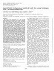
Fig. 1. Moxibustion restores the weight gain rate in POI rats. A. The diagram of constructing POI rat model and moxibustion treatment. B. CTX decreases the rate
of body weight gain in rats, moxibustion increases it in POI rats. *P<0.05, ***P<0.001,**** P<0.0001.
pending subsequent testing. The uterus and ovaries were completely
removed, the surrounding adipose tissue was removed, washed with PBS
and dried before they were weighed on a balance and photographed for
documentation. One ovary from each group was removed, approximately 1.0 mm*1.0 mm*1.0 mm of ovaries containing follicles were cut
and immersed in 2.5% glutaraldehyde solution (precooled at 4 ◦ C), and
the remaining portion was fixed in 4% paraformaldehyde solution.
Remaining ovaries were stored at −80 ◦ C.
Calculation formula of organ index: organ index = organ weight/
body weight*100%.
2.6. Transmission electron microscopy (TEM) of ovarian granulosa cells
Ovaries of 1 mm3 was fixed in 2.5% glutaraldehyde, rinsed in PBS,
fixed in 1% osmic acid, dehydrated in ethanol, embedded and polymerized in epoxy resin, sectioned in an ultramicrotome (RMC POWERTOME XL), stained with 2% uranyl acetate and 2.6% lead citrate
solution to observe the ultrastructure of ovarian tissue under transmission electron microscopy (FEI Tecnai G2 Spirit Bio TWIN), and images were collected.
2.7. ELISA of serum sex hormone levels
2.5. HE staining of ovaries and follicles counting
Serum FSH, LH, AMH, E2, and T levels were measured in strict
accordance with the instructions of the ELISA kit. Absorbance values for
each standard and sample were detected using a microplate reader (BioTek SynergyHI MFD) at 450 nm. Origin 9.0 was used to obtain the
standard curve by four-parameter fitting method, and the absorbance
values of the samples were substituted to calculate the hormone
concentration.
Ovaries were fixed in 4% paraformaldehyde solution for 24 h,
embedded in paraffin and sectioned into 4 μm sections, then stained
with hematoxylin-eosin (HE). The structure of ovaries was observed by
microscope, so that histopathological changes of ovaries were evaluated
according to the characteristics and number of follicles at each grade.
Primordial follicles: oocytes are surrounded by a layer of flat granulosa cells, the boundary of granulosa cells is not obvious, and oval
nuclei can only be seen; preantral follicles (primary follicles): the shape
of granulosa cells changes from flat to cuboidal, from one layer to
multiple layers, and radiates from the center to the periphery to form
corona radiata; antral follicles (secondary follicles): multiple layers of
columnar granulosa cells, and theca cells layer appears, and oocytes
float in follicular fluid; atretic follicles: follicles collapse, irregular
arrangement of granulosa cells, pyknosis or disappearance of oocyte cell
nuclei, or only irregular zona pellucida can be seen[16].
2.8. Bulk RNA-seq of ovaries
Total RNA was isolated using the Trizol Reagent (Invitrogen Life
Technologies), after which the concentration, quality and integrity were
determined using a NanoDrop spectrophotometer (Thermo Scientific).
Three micrograms of RNA were used as input material for the RNA
sample preparations. Sequencing libraries were generated according to
the following steps. Firstly, mRNA was purified from total RNA using
3
R. Zhao et al.
Journal of Steroid Biochemistry and Molecular Biology 242 (2024) 106547
poly-T oligo-attached magnetic beads. Fragmentation was carried out
using divalent cations under elevated temperature in an Illumina proprietary fragmentation buffer. First strand cDNA was synthesized using
random oligonucleotides and Super Script II. Second strand cDNA synthesis was subsequently performed using DNA Polymerase I and RNase
H. Remaining overhangs were converted into blunt ends via exonuclease/polymerase activities and the enzymes were removed. After
adenylation of the 3′ ends of the DNA fragments, Illumina PE adapter
oligonucleotides were ligated to prepare for hybridization. To select
cDNA fragments of the preferred 400–500 bp in length, the library
fragments were purified using the AMPure XP system (Beckman Coulter,
Beverly, CA, USA). DNA fragments with ligated adaptor molecules on
both ends were selectively enriched using Illumina PCR Primer Cocktail
in a 15 cycle PCR reaction. Products were purified (AMPure XP system)
and quantified using the Agilent high sensitivity DNA assay on a Bioanalyzer 2100 system (Agilent). The sequencing library was then
sequenced on NovaSeq 6000 platform (Illumina) Shanghai Personal
Biotechnology Cp. Ltd.
Ovaries samples are sequenced on the platform to get image files,
which are transformed by the software of the sequencing platform, and
the original data in FASTQ format (Raw Data) is generated. Sequencing
data contains a number of connectors, low-quality Reads, so we use
Cutadapt (v1.15) software to filter the sequencing data to get high
quality sequence (Clean Data) for further analysis.
All bioinformatics analyses were performed in the R (Version 4.1.3)
environment, and the “DESeq package” was used for differential
expression analysis with a data screening threshold of " |log2FoldChange
| > 1& P-value < 0.05". Filtered data were used for subsequent
enrichment analysis.
next day, after washing three times with TBST buffer, membranes were
incubated with secondary antibody horseradish peroxidase (HRP)-conjugated goat anti-rabbit IgG (1:1000) for 1 h at room temperature.
Immunoreactive bands were detected by exposing the membranes to
Clarity Western ECL chemiluminescent substrate and imaged using the
chemiluminescent gel imaging analysis system (Bio-Rad ChemiDoc XRS
+). Finally, Image J software was used to calculate the gray values of
each protein, and the target protein/Histone h3 ratio was defined as the
relative expression of the target protein.
2.12. Immunohistochemistry (IHC) staining of ovaries
Paraffin-embedded ovaries, sectioned at 4 μm thickness, were boiled
for 5 min in Tris-EDTA buffer (10 mM Tris base 1 mM disodium EDTA
dihydrate, pH 8.0) for antigen retrieval. Sections were washed three
times with PBS, blocked with goat serum and incubated with specific
antibodies against FSHR (1:600). Next, the sections were incubated with
Goat Anti-Rabbit IgG H&L HRP for 1 h. The images were captured using
an upright fluorescent microscope (Olympus BX43).
2.13. Statistical analysis
All data were analyzed and all graphics were plotted using Graphpad
Prism 8.4 (GraphPad Software, Inc, La Jolla, CA, USA) and are presented
as mean ± standard error (SEM). If data met normal distribution and
homogeneity of variance, One-way analysis of variance (ANOVA) and
Bonferroni tests for multiple comparisons were used for comparison
among multiple groups. P < 0.05 was considered statistically significant.
3. Results
2.9. GO, KEGG and GSEA enrichment analysis
3.1. Moxibustion restores the weight gain rate in POI rats
Differentially expressed genes (DEGs) were used to complete
enrichment analysis, and GO, KEGG and GSEA enrichment analysis were
mainly used in this study: The Gene Ontology (GO) system provides
structured, computable information regarding the functions of genes
and their products[17].The Kyoto Encyclopedia of Genes and Genomes
(KEGG) is a widely used database for the systematic pathway investigation of DEGs [18]; Gene set enrichment analysis (GSEA) analysis independent of DEGs, including all genes in the analysis to assess relevant
pathways and molecular mechanisms [19]. GO, KEGG and GSEA
enrichment analysis conducted based on the “ClusterProfiler” package
[20], threshold criteria is as follows: “|log2FoldChange| > 1 & P-value <
0.05”.
As shown in Fig. 1A, we used cyclophosphamide to construct POI rat
model and performed moxibustion treatment (subsequent experiments
were grouped on this basis). The results showed that the initial body
weights of rats in the three groups were similar, and then the body
weights of CON group steadily increased rapidly; compared with CON
group, the body weight gain rate in CTX group was the slowest and
fluctuating, while the body weight gain rate in CTX + MOX group was
between the two groups (Fig. 1B).
3.2. Moxibustion improves estrous cycle and reproductive system in POI
rats
2.10. GSVA pathway scoring
To verify the effect of moxibustion on the reproductive system of POI
rats, we first performed vaginal smears and analyzed the estrous cycle,
and then collected samples to complete the weighing of the ovary and
uterus, especially focused on ovarian HE staining. The results demonstrated that the estrous cycle in CTX group was apparently disturbed
compared with CON group, while it in CTX + MOX group was similar to
CON group and basically normal (Fig. 2.A, B). Compared with CON
group, the volume and organ index of the ovary and uterus became
smaller in CTX group, with slight inflammation and significant congestion, while the above parameters were better in CTX + MOX group than
in CTX group (Fig. 2.C, D). HE staining results showed that follicles were
abundant in CON group, follicles at each grade were visible, and atretic
follicles were not observed in the visual field; compared with CON
group, the number of normal follicles was decreased and the number of
atretic follicles was increased in CTX group; however, the number of
dominant follicles was significantly more in CTX + MOX group than in
CTX group (Fig. 2.E, F).
According to the results of DEGs, designated pathways were selected
for GSVA pathway scoring, and the pathway genesets were downloaded
from GSEA official website (https://www.gsea-msigdb.org/gsea/index.
jsp). GSVA pathway scoring were completed by using the “GSVA package” to show the trend of target pathway scores in the experimental
data.
2.11. Western Blot
Ovaries were lysed in RIPA buffer containing protease inhibitor and
phosphatase inhibitor cocktails, supernatants of lysates were collected
by centrifugation (12,000 rpm, 4 C, 15 min), with protein concentrations determined by BCA assay. 20 μg of protein of each sample was
mixed with loading buffer and loaded onto 10% sodium dodecyl sulfate
polyacrylamide gel electrophoresis (SDS-PAGE),then electrophoresed.
Subsequently, proteins were transferred to polyvinylidene difluoride
(PVDF) membranes, washed in TBST and blocked with 5% skim milk
for 1 h and incubated overnight at 4 ◦ C with primary antibodies
(1:1000). Histone h3 antibody was used as the internal reference. The
3.3. Moxibustion improves sex hormone levels in POI rats
To assess the effect of moxibustion on ovarian function in POI rats,
4
R. Zhao et al.
Journal of Steroid Biochemistry and Molecular Biology 242 (2024) 106547
Fig. 2. Moxibustion improves estrous cycle and reproductive system in POI rats. A-B. CTX disarranges the estrous cycle of rats, moxibustion restores the its
regularity in POI rats. C-D. CTX damages the uterine and ovarian, moxibustion ameliorates the damage in POI rats. E-F. CTX promotes follicular atresia and hinders
follicular development, moxibustion slows down follicular atresia and increases the number of dominant follicles. *P<0.05,** P<0.01,***P<0.001,**** P
<0.0001.
5
R. Zhao et al.
Journal of Steroid Biochemistry and Molecular Biology 242 (2024) 106547
we examined sex hormone levels of serum. Compared with CON group,
FSH levels increased, LH, AMH, E2 and T levels decreased, and FSH/LH
ratio increased in CTX group; compared with CTX group, FSH levels and
FSH/LH ratio decreased, LH and T levels did not change significantly,
and AMH and E2 levels increased relatively in CTX + MOX group
(Fig. 3A-F).
MOX/CON comparison were mainly: “inflammatory response”,“acute
inflammatory response”,“apoptotic process”; upregulated terms in
CTX + MOX/CON comparison were mainly “reproductive process”,“female gamete generation”,“regulation of hormone levels”
(Fig. 5A-C).
KEGG pathway enrichment results showed that: the downregulated
genes in CTX/CON comparison mainly enriched “Th1 and Th2 cell differentiation”,“Th17 cell differentiation”,“T cell receptor signaling
pathway”; the pathways upregulated in CTX/CON comparison and
downregulated in CTX + MOX/CTX comparison were mainly “Tight
junction”,“Arginine and proline metabolism”; there were 3 KEGG
pathways commonly significantly enriched in the upregulated DEGs
between the comparisons of CTX + MOX/CTX and CTX + MOX/CON,
which were of “cAMP signaling pathway”,“Calcium signaling pathway,“Ovarian steroidogenesis”; and 2 KEGG pathways commonly
significantly enriched in the downregulated DEGs in CTX + MOX/CON
comparison which were mainly involved in “HIF−1 signaling
pathway”,“IL−17 signaling pathway” (Fig. 6A-C).
GSEA enrichment results showed that: the pathways upregulated in
CTX/CON comparison and downregulated in CTX + MOX/CTX comparison were mainly “E2F targets”,“G2M checkpoint”,“MYC targets”;
the pathways downregulated in CTX/CON comparison and upregulated
in CTX + MOX/CTX comparison were mainly "“Estrogen response early”,“Interferon gamma response”; the pathways downregulated in CTX
+ MOX/CON comparison were mainly “TNFA signaling via
NFKB”,“Hypoxia”; the pathways upregulated in CTX + MOX/CON
comparison were mainly “Interferon gamma response”,“Interferon
alpha response” (Fig. 7A-C).
The above results suggest that moxibustion ameliorates POI mainly
3.4. Bulk RNA-seq results of ovaries from rats in each group
To investigate the potential mechanism of moxibustion making
impact, bulk RNA-seq was completed on the ovaries of rats in each
group. The PCA plot and clustering heatmap showed that the sequencing
samples meet the quality standards and the gene expression patterns
within group were similar and comparable (Fig. 4A, B). When the
threshold was set as “P < 0.05”, there were a huge number of DEGs in
each pairwise comparison (Fig. 4C).
3.5. GO, KEGG, GSEA enrichment analysis results of Bulk RNA-seq
After that, we performed GO, KEGG and GSEA enrichment analysis of
the DEGs. GO functional enrichment results showed that: the downregulated terms in CTX/CON comparison were mainly “T cell activation”,“T cell differentiation”,“immune system process”; the
upregulated terms in CTX/CON comparison were not significantly specific; the downregulated terms in CTX+MOX/CTX comparison were
mainly “inflammatory response”,“acute inflammatory response”,“cell death”; the upregulated terms in CTX + MOX/CTX comparison were
mainly “lipid metabolic process”,“regulation of reproductive process”,“response to steroid hormone”; the downregulated terms in CTX +
Fig. 3. Moxibustion improves sex hormone levels in POI rats. A-F. Moxibustion reduces the FSH level (A), FSH/LH ratio (C) of POI rats, increases the concentration of AMH (D), E2 (E), but has no significant effect on LH (B) and T (F) concentration. *P<0.05,**P<0.01,***P<0.01,ns indicates no significance.
6
R. Zhao et al.
Journal of Steroid Biochemistry and Molecular Biology 242 (2024) 106547
Fig. 4. Overview of bulk RNA-seq sequence alignment and data dispersion. A-B. PCA and clustering analysis show clear separation in three groups. C. Volcano
plot shows up-regulated (red color) and down-regulated (green color) significant genes in pairwise comparisons between groups.
related to cAMP signaling pathway and is largely inseparable from steroidogenesis of GCs.
ovarian GCs in POI rats, but not theca cells.
To test this, we performed IHC experiments of FSHR (the maker of
GCs[21]) on ovary sections. The results showed that the number of
FSHR-positive cells decreased in CTX group compared with CON group,
while they increased significantly in CTX + MOX group compared with
CTX group.
At the same time, the transmission electron microscopic results
showed that there were intact and clear membranes, uniform nuclear
chromatin, abundant lipid droplets and mitochondria in the GCs in CON
group; while in CTX group, there were less uniform nuclear chromatin,
less intracellular lipid droplets, and swollen mitochondria; in CTX +
MOX group, there were intact membranes, uniform nuclear chromatin,
abundant lipid droplets in the cells, and insignificant mitochondrial
swelling (Fig. 9 E).
In order to further inspect and verify the effect of moxibustion on the
function of GCs, we used western blot to detect the expression of steroidogenic key proteins in each group, and the results showed that the
relative expression of FSHR and P450 arom in CTX group decreased
compared with CON group, while their relative expression were similar
in CTX + MOX group and CON group. However, the relative expression
of StAR was not significantly different between CTX and CTX + MOX
groups (Fig. 9.F, G).
3.6. GSVA pathway scoring results of Bulk RNA-seq
To further clarify the phenotype and narrow down research scope,
we continued the analysis using GSVA pathway scoring. The results
showed that the scores of “cellular response to estrogen stimulus
pathway” and “ovulation from ovarian follicle pathway” appeared a
trend of "high-low-high" in CON group, CTX group and CTX+MOX
group, while the scores of “peroxiredoxin 2 induced ovarian failure
pathway” and “ovarian infertility pathway” showed the opposite trend
(Fig. 8A-D).
3.7. Moxibustion improves the number and function of ovarian GCs in
POI rats
The GSVA scoring results showed that the scores of "response to
follicle stimulating hormone pathway" and "regulation of steroid metabolic process pathway" appeared a trend of "high-low-high" in CON, CTX
and CTX + MOX groups, while the scores of "response to luteinizing
hormone pathway" showed the opposite trend (Fig. 9 A-C), which
indicated that moxibustion may promote the number and function of
7
R. Zhao et al.
Journal of Steroid Biochemistry and Molecular Biology 242 (2024) 106547
Fig. 5. GO functional enrichment results of Bulk RNA-seq. A-C. The biological process (BP), cellular component (CC), and molecular function (MF) enriched for
DEGs in pairwise comparisons between groups revealed by GO analyses. Upper are pathways enriched for upregulated genes and below are pathways enriched for
downregulated genes.
Notably, E2, through a negative feedback mechanism, can inhibit the
synthesis of FSH and LH, slowing the development of follicular atresia in
POI pathogenesis and improving the elevated gonadotropin state in POI
[23]. This forms the theoretical basis for HRT in POI and underscores
why our study focused on granulosa cells (GCs).
As one of the numerous somatic cells in the ovary, GCs are inseparable from follicular development and ovarian endocrine function: 1.
GCs are essential for the normal development of follicles, and it is mainly
localized in follicles, surrounding oocytes, can synthesize a variety of
hormones and necessary growth factors, and express related receptors
[24]. Under the guidance of pituitary FSH, GCs begin to proliferate,
migrate, and express FSH receptor, E2 receptor, etc., so that primordial
follicles develop into primary follicles that respond to hormones; then,
under the stimulation of FSH and E2, they successively enter the stage of
secondary follicle development, at which time GCs show explosive
proliferation, produce a large amount of E2 and induce more FSH receptor production, promote the synthesis of estrogen, so that follicles
grow rapidly and induce ovulation[25]. 2. GCs dominate ovarian
endocrine function. Estrogen production is mainly divided into two
processes: (1) Steroidogenic Acute Regulatory protein (STAR) acts as the
initial carrier of the whole process, brings cholesterol into theca cells,
and they converts cholesterol into Androstenedione (A2) and Testosterone (T); (2) The products diffuse through the basal membrane into
GCs and accumulate continuously, while Aromatase P450 (P450 arom,
CYP19A1) in GCs acts as the rate-limiting and critical enzyme in the
catalytic step, converting A2 and T into 17β-Estradiol (E2) [26]. FSHR
activates cAMP signaling to enhance CYP19A1 expression in GCs and
promote estrogen production, while estrogen also enhances FSH action
3.8. Moxibustion regulates steroidogenesis by activating cAMP/PKA/
CREB signaling pathway in ovaries of POI rats
To further explore the mechanism of moxibustion on POI, we
continued to use GSVA pathway scoring to narrow down research scope,
and the results showed that the scores of " Protein Kinase A signaling
pathway" and "CREB pathway" showed a trend of "high-low-high" in
CON, CTX, and CTX + MOX groups (Fig. 10A, B), and in combination
with the results of KEGG pathway enrichment analysis in Fig. 6, we
carried out western blot experiment of cAMP, PKA, and CREB in ovarian
protein samples. The results showed that the relative expression of the
above proteins was lower in CTX group compared with CON group,
while they were significantly increased in CTX + MOX group compared
with CTX group (Fig. 10C, D).
4. Discussion
Currently, global concern regarding fertility issues has increased,
with a growing trend of delayed conception. Reports indicate that from
1981 to 2019, the percentage of women over 30 giving birth to their first
child rose from 15% to 51%[22]. This not only raises the risk of POI but
also significantly increases fertility difficulty. Additionally, ovarian
dysfunction can expedite the aging process of various organs in women,
leading to hectic fever and night sweat, insomnia and dreaminess,
anxiety and depression, or other symptoms similar to perimenopausal
syndrome. This contradicts the contemporary pursuit of a high-quality
life for women. Consequently, POI has emerged as a critical factor
jeopardizing the physical and mental health of women at present.
8
R. Zhao et al.
Journal of Steroid Biochemistry and Molecular Biology 242 (2024) 106547
Fig. 6. KEGG pathway enrichment results of Bulk RNA-seq. A-C. Top pathways involving DEGs in pairwise comparisons between groups revealed by KEGG
analyses. On the left are pathways enriched for upregulated genes, on the right are pathways enriched for downregulated genes.
9
R. Zhao et al.
Journal of Steroid Biochemistry and Molecular Biology 242 (2024) 106547
Fig. 7. GSEA pathway enrichment results of Bulk RNA-seq. A-C. Significant pathways enrichment in GSEA of DEGs in pairwise comparisons between groups.
[27].
Up until now, moxibustion has extremely significant advantages as a
treatment for POI when HRT is accompanied by numerous side effects.
Compared with HRT, moxibustion can not only improve the hypoestrogenic state of patients, but also ameliorate the clinical symptoms of
them as a whole and restore ovarian secretion and reproductive function
[28]. The mild heat of moxibustion fire, light radiation effects, and
volatile moxa components are the stimulating characteristic; warming
yang and tonifying qi, promoting qi and activating blood circulation,
regulating blood vessels are important effects of moxibustion; the local
site of moxibustion is the direct object of moxibustion action; acupoints,
skin, and meridians are the basis for the effect of moxibustion. Guanyuan and Sanyinjiao are the main acupoints selected, both of which are
important acupoints for the treatment of gynecological diseases. Modern
10
R. Zhao et al.
Journal of Steroid Biochemistry and Molecular Biology 242 (2024) 106547
Fig. 8. Moxibustion improves female reproductive related pathways in POI rats. A-D. GSVA scoring results show promoted effect on “response to estrogen
stimulus” (A) and “ovulation” (B) pathways, antagonized effect on “ovarian failure” (C) and “infertility” (D) pathways in moxibustion on POI rats.
medical research has also confirmed that the compatibility of Sanyinjiao
with
Guanyuan
can
effectively
regulate
the
hypothalamic-pituitary-ovarian axis[13], but the related mechanism
remains to be completed.
Our team has been committed to the clinical practice and related
research of moxibustion in the treatment of POI for a long time, so we
have a lot of clinical experience and research basis. First, based on our
previous work and other studies[14,29,30], we used the cytotoxic agent
cyclophosphamide (CTX) to construct the POI rat model. Compared with
CON group, rats treated with CTX showed slower weight gain rate
(Fig. 1B), significantly disturbed estrous cycle (Fig. 2.A, B), smaller
organ index of reproductive organs (Fig. 2.C, D), increased atretic follicle counts(Fig. 2.E, F), and reduced ovarian hormone secretion (Fig. 3
A-F); response estrogen stimulus and ovulation pathways were downregulated and ovarian failure and ovarian infertility pathways were
upregulated in GSVA scoring results (Fig. 8 A-D), which indicated that
our POI model was successfully constructed.
Therefore, combined with the method previously established by our
team[13,14], we treated POI model rats with moxibustion (hereafter
referred to as CTX + MOX group). As shown in the results, the above
phenotypic parameters were effectively improved in CTX + MOX group,
and HE staining results showed that the dominant follicles increased,
indicating that moxibustion could effectively ameliorate ovarian secretion and reproductive function in POI rats (Fig. 2.E, F).
To further explore the potential mechanism of moxibustion action,
we performed bulk RNA sequencing of ovaries from each group. The
results of PCA and clustering analysis showed that the intra-group differences of samples were small and the comparability between groups
was high (Fig. 4.A, B); volcano plot results showed that the expression
patterns were significantly different between the groups at P < 0.05
(Fig. 4.C). Then, we used enrichment analysis to continue to explore,
mainly using traditional GO and KEGG enrichment, as well as GSEA
pathway enrichment and GSVA pathway scoring method which are
more reliable. The results showed that the immune response ability,
reproductive system function, T cell proliferation in CTX group were
significantly inhibited (Figs. 5, 6, 7 A-C), which was largely related to
the characteristics of cyclophosphamide-induced POI model[31], and
also similar to the pathogenesis of POI [3], which more indicated the
accuracy of our POI rat model. It is worth noting that moxibustion can
effectively improve immune system function and reduce cell death
(Figs. 5, 6, 7. A-C), which is also consistent with the mechanism of
moxibustion action in other fields [32,33]; at the same time, the
enrichment analysis results also suggest that moxibustion can also
significantly improve ovarian hormone response (Figs. 5, 6, 7.A-C),
come back to the neatly arranged and orderly GCs in MOX + CTX group
compared with CTX group in Fig. 2B, and the ovarian hormone levels
that are significantly improved in Fig. 3, it is reasonable to speculate that
moxibustion may have a more significant promoting effect on ovarian
GCs; more importantly, we noticed that cAMP signaling pathway was
significantly enriched in CTX + MOX group (Fig. 6.A-C), and this
pathway was inseparable from the steroidogenic function of GCs[34].
Therefore, combined with the known results, we hypothesized that
moxibustion may improve steroidogenic function in GCs by affecting
cAMP signaling pathway to achieve the goal of recovering ovarian
function in POI rats.
On the basis of the above results, to confirm our hypothesis, we first
completed GSVA scoring for FSH, LH, and steroid-related pathways, the
results showed that the target cells of moxibustion action were ovarian
GCs rather than theca cells (Fig. 9 A-C). Indeed, IHC staining of FSHR
(the marker of GCs) and its quantitative analysis of mean optical density
also verified this opinion, and the number of FSHR-positive cells was
significantly increased after moxibustion treatment (Fig. 9 D). The results of TEM also fully demonstrated the rescue effect of moxibustion on
the GCs in POI model (Fig. 9 E). Finally, western blot results of FSHR and
P450 arom (mainly present within GCs) and StAR (mainly present
within theca cells) also suggested that the function of GCs in POI model
was improved after moxibustion treatment, but not theca cells (Fig. 9. F,
G). Thus, we have enough evidence that moxibustion restores ovarian
function in POI by improving steroidogenic function in GCs.
Finally, based on the target cells identified in the previous data, to
further explore the mechanism of moxibustion, that is, to clarify the
changes of cAMP signaling pathway before and after moxibustion
treatment, we performed GSVA pathway scoring of cAMP signaling and
steroidogenesis pathway. In ovarian GCs, PKA and CREB are essential
for their steroidogenic function: CREB, the full name cAMP Response
Element Bound protein, whose main function is initiating aromatase
gene transcription and is essential for the activity of aromatase such as
P450 arom in GCs[34]; PKA, the full name of Protein Kinase A, directly
phosphorylates CREB, and this process is irreplaceable by other protein
kinases[35]. Because both are downstream of the cAMP signaling
pathway[34], and the cAMP signaling pathway has a clear trend in
KEGG enrichment results, we only assessed CREB and PKA signaling
pathways. The results conformed to our hypothesis, suggesting that
moxibustion may affect cAMP signaling pathway dominated by cAMP,
PKA, and CREB to act (Fig. 10. A, B). To finally confirm this opinion, we
performed western blot experiment on cAMP, PKA, and CREB proteins,
and the results demonstrated that moxibustion improved steroidogenic
function in POI rats GCs by activating cAMP/PKA/CREB signaling
pathway (Fig. 10. C, D).
In conclusion, our data demonstrate that moxibustion regulates
steroidogenesis in ovarian GCs via activation of cAMP/PKA/CREB to
upgrade P450 arom, thereby ameliorate ovarian function impairment in
POI rats. This will provide strong evidence to support the promotion and
use of moxibustion in the treatment of POI in clinical practice.
Funding information
This work was supported by the Natural Science Foundation of
Jiangsu Province (BE2020624); the Natural Science Foundation of
Nanjing University of Traditional Chinese Medicine (XZR2021051).
11
R. Zhao et al.
Journal of Steroid Biochemistry and Molecular Biology 242 (2024) 106547
Fig. 9. The promotional effect of moxibustion on the number, structure and function of ovarian GCs in POI rats. A-C. GSVA scoring results show promoted
effect on “follicle stimulating hormone” (A) and “regulation of steroid metabolic process” (B) pathways, antagonized effect on “response to luteinizing hormone”
pathway (C) in moxibustion on POI rats. D-E. IHC results show moxibustion increase FSHR positive cell. F. Transmission electron microscopy results show moxibustion repair organelles of ovarian GCs in POI rats. G-H. Western blot results show moxibustion promoted FSHR and P450 arom levels, with no significant effect on
StAR. *P<0.05,**P<0.01,***P<0.001,**** P<0.0001,ns indicates no significance.
12
R. Zhao et al.
Journal of Steroid Biochemistry and Molecular Biology 242 (2024) 106547
Fig. 10. Moxibustion regulates steroidogenesis by activating cAMP/PKA/CREB signaling pathway in ovaries of POI rats. A-B. GSVA scoring results show
promoted effect on “Protein Kinase A” (A) and “CREB” (B) pathways in moxibustion on POI rats. C-D. Western blot results show moxibustion promoted cAMP, PKA
and CREB levels. *P<0.05,**P<0.01,***P<0.001.
References
CRediT authorship contribution statement
[1] G. T, K. H, R. S, van R. F, Breaking the silence around infertility: a scoping review
of interventions addressing infertility-related gendered stigmatisation in low- and
middle-income countries, Sex. Reprod. Health Matters 31 (2023), https://doi.org/
10.1080/26410397.2022.2134629.
[2] X. Jiao, H. Zhang, H. Ke, J. Zhang, L. Cheng, Y. Liu, Y. Qin, Z.-J. Chen, Premature
ovarian insufficiency: phenotypic characterization within different etiologies,
J. Clin. Endocrinol. Metab. 102 (2017) 2281–2290, https://doi.org/
20170706140016.
[3] European Society for Human Reproduction and Embryology (ESHRE) Guideline
Group on POI, L. Webber, M. Davies, R. Anderson, J. Bartlett, D. Braat,
B. Cartwright, R. Cifkova, S. de Muinck Keizer-Schrama, E. Hogervorst, F. Janse,
L. Liao, V. Vlaisavljevic, C. Zillikens, N. Vermeulen, ESHRE Guideline:
management of women with premature ovarian insufficiency, Hum. Reprod. 31
(2016) 926–937, https://doi.org/10.1093/humrep/dew027.
[4] I. Lersten, E. Clain, N. Santoro, Use of hormone therapy in women with early
menopause and premature ovarian insufficiency, Semin Reprod. Med 38 (2020)
302–308, https://doi.org/10.1055/s-0040-1721719.
[5] Y. Yao, Z. Zhenni, C. Fengqin, L. Yufei, P. Xiangtian, X. Xiao, S. Zhiling,
Effectiveness of moxibustion alone on lumbar disc herniation: a meta-analysis of
randomized controlled trials, J. Tradit. Chin. Med. 43 (2023) 14–26, https://doi.
org/10.19852/j.cnki.jtcm.20221108.001.
[6] L.-L. Xu, X. Zhou, C.-R. Zhang, J.-B. Zhang, Effect of acupuncture on pregnancy
outcomes of frozen embryo transfer in patients with anovulatory infertility],
Zhongguo Zhen Jiu 42 (2022) 150–154, https://doi.org/10.13703/j.02552930.20210206-0002.
[7] X. Jin, J. Cheng, J. Shen, X. Lv, Q. Li, Y. Mu, H. Bai, Y. Liu, Y. Xia, Moxibustion
improves ovarian function based on the regulation of the androgen balance, Exp.
Ther. Med 22 (2021) 1230, https://doi.org/10.3892/etm.2021.10664.
[8] H.-M. Chang, J. Qiao, P.C.K. Leung, Oocyte-somatic cell interactions in the human
ovary-novel role of bone morphogenetic proteins and growth differentiation
factors, Hum. Reprod. Update 23 (2016) 1–18, https://doi.org/10.1093/humupd/
dmw039.
[9] J. Sun, L. Gan, S. Lv, T. Wang, C. Dai, J. Sun, Exposure to Di-(2-Ethylhexyl)
phthalate drives ovarian dysfunction by inducing granulosa cell pyroptosis via the
Rui Zhao: Conceptualization, Data curation, Formal Analysis,
Investigation, Methodology, Software, Validation, Visualization,
Writing – original draft, Writing – review & editing; Lingxiang Ran:
Conceptualization, Formal Analysis, Methodology, Software, Visualization, Writing – original draft, Writing – review & editing; Hanyue Yao:
Investigation, Methodology; Yizhi He: Investigation, Methodology;
Xinru Lu: Investigation, Methodology; Weina Zhu: Resources; Yajie
Zhang: Resources; Tianyi Zhang: Investigation; Shijie Shi: Investigation; Zheng Luo: Investigation; Cairong Zhang: Funding acquisition,
Project administration, Supervision.
Declaration of Competing Interest
All authors declare that they have no conflict of interest.
Data availability
Data will be made available on request.
Acknowledgements
We thank Jiangsu Health Vocational College Central Laboratory for
assistance and support with experimental animals.
13
R. Zhao et al.
[10]
[11]
[12]
[13]
[14]
[15]
[16]
[17]
[18]
[19]
[20]
[21]
Journal of Steroid Biochemistry and Molecular Biology 242 (2024) 106547
SLC39A5/NF-κB/NLRP3 axis, Ecotoxicol. Environ. Saf. 252 (2023) 114625,
https://doi.org/10.1016/j.ecoenv.2023.114625.
W. Li, Z. Sun, M. Li, B. Yue, X. Zhang, Y. Zhao, J. Wang, Exposure to fluoride from
in utero to puberty alters gonadal structure and steroid hormone expression in
offspring rats, Biol. Trace Elem. Res 201 (2023) 1261–1273, https://doi.org/
10.1007/s12011-022-03220-8.
F. Li, Y. Wang, M. Xu, N. Hu, J. Miao, Y. Zhao, L. Wang, Single-nucleus RNA
Sequencing reveals the mechanism of cigarette smoke exposure on diminished
ovarian reserve in mice, Ecotoxicol. Environ. Saf. 245 (2022) 114093, https://doi.
org/10.1016/j.ecoenv.2022.114093.
N. Percie du Sert, V. Hurst, A. Ahluwalia, S. Alam, M.T. Avey, M. Baker, W.
J. Browne, A. Clark, I.C. Cuthill, U. Dirnagl, M. Emerson, P. Garner, S.T. Holgate,
D.W. Howells, N.A. Karp, S.E. Lazic, K. Lidster, C.J. MacCallum, M. Macleod, E.
J. Pearl, O.H. Petersen, F. Rawle, P. Reynolds, K. Rooney, E.S. Sena, S.
D. Silberberg, T. Steckler, H. Würbel, The ARRIVE guidelines 2.0: Updated
guidelines for reporting animal research, PLoS Biol. 18 (2020) e3000410, https://
doi.org/10.1371/journal.pbio.3000410.
C.-R. Zhang, J.-L. Deng, W.-N. Zhu, M.-X. Miao, W.-W. Shen, S.-J. Cao, Y. Tang,
Involvement of PI 3 K/Akt/mTOR signaling in protective effects of moxibustion for
premature ovarian failure in rats, Zhen Ci Yan Jiu 43 (2018) 75–79, https://doi.
org/10.13702/j.1000-0607.170857.
C. Zhang, W. Zhu, W. Tao, W. Lin, C. Cheng, H. Deng, G. Ni, Moxibustion against
cyclophosphamide-induced premature ovarian failure in rats through Inhibiting
NLRP3-/Caspase-1-/GSDMD-Dependent Pyroptosis, Evid. -Based Complement.
Altern. Med. 2021 (2021) 1–9, https://doi.org/10.1155/2021/8874757.
S.L. Byers, M.V. Wiles, S.L. Dunn, R.A. Taft, Mouse estrous cycle identification tool
and images, PLoS One 7 (2012) e35538, https://doi.org/10.1371/journal.
pone.0035538.
E.A. McGee, A.J. Hsueh, Initial and cyclic recruitment of ovarian follicles, Endocr.
Rev. 21 (2000) 200–214, https://doi.org/2020071613545308100.
The Gene Ontology Consortium, The gene ontology resource: 20 years and still
GOing strong, Nucleic Acids Res 47 (2019) D330–D338, https://doi.org/10.1093/
nar/gky1055.
M. Kanehisa, S. Goto, KEGG: kyoto encyclopedia of genes and genomes, Nucleic
Acids Res 28 (2000) 27–30, https://doi.org/10.1093/nar/28.1.27.
A. Subramanian, P. Tamayo, V.K. Mootha, S. Mukherjee, B.L. Ebert, M.A. Gillette,
A. Paulovich, S.L. Pomeroy, T.R. Golub, E.S. Lander, J.P. Mesirov, Gene set
enrichment analysis: a knowledge-based approach for interpreting genome-wide
expression profiles, Proc. Natl. Acad. Sci. USA 102 (2005) 15545–15550, https://
doi.org/10.1073/pnas.0506580102.
G. Yu, L.-G. Wang, Y. Han, Q.-Y. He, clusterProfiler: an R package for comparing
biological themes among gene clusters, OMICS: A J. Integr. Biol. 16 (2012)
284–287, https://doi.org/10.1089/omi.2011.0118.
X. Cai, H. Fu, Y. Wang, Q. Liu, X. Wang, Depletion of GPSM1 enhances ovarian
granulosa cell apoptosis via cAMP-PKA-CREB pathway in vitro, J. Ovarian Res. 13
(2020) 136, https://doi.org/10.1186/s13048-020-00740-6.
[22] I. Glick, E. Kadish, M. Rottenstreich, Management of Pregnancy in Women of
Advanced Maternal Age: Improving Outcomes for Mother and Baby, IJWH Volume
13 (2021) 751–759, https://doi.org/10.2147/IJWH.S283216.
[23] Y.H. Haddad, R.S. Said, R. Kamel, E.M.E. Morsy, E. El-Demerdash, Phytoestrogen
genistein hinders ovarian oxidative damage and apoptotic cell death-induced by
ionizing radiation: co-operative role of ER-β, TGF-β, and FOXL-2, Sci. Rep. 10
(2020) 13551, https://doi.org/10.1038/s41598-020-70309-2.
[24] X. Zhang, R. Zhang, J. Hao, X. Huang, M. Liu, M. Lv, C. Su, Y.-L. Mu, miRNA-1225p in POI ovarian-derived exosomes promotes granulosa cell apoptosis by
regulating BCL9, Cancer Med 11 (2022) 2414–2426, https://doi.org/10.1002/
cam4.4615.
[25] E. Warzych, P. Lipinska, Energy metabolism of follicular environment during
oocyte growth and maturation, J. Reprod. Dev. 66 (2020) 1–7, https://doi.org/
10.1262/jrd.2019-102.
[26] T. Liu, Y. Huang, H. Lin, Estrogen disorders: Interpreting the abnormal regulation
of aromatase in granulosa cells (Review), Int. J. Mol. Med. 47 (2021) 73, https://
doi.org/10.3892/ijmm.2021.4906.
[27] E. Hobeika, M. Armouti, H. Kala, M.A. Fierro, N.J. Winston, B. Scoccia, A.
M. Zamah, C. Stocco, Oocyte-secreted factors synergize with FSH to promote
aromatase expression in primary human cumulus cells, J. Clin. Endocrinol. Metab.
104 (2019) 1667–1676, https://doi.org/2020071622262273200.
[28] M. Xiao, F.-X. Liang, [Research progress and thinking on the mechanism of
acupuncture in treatment of perimenopausal syndrome], Zhongguo Zhen Jiu 41
(2021) 699–702, https://doi.org/10.13703/j.0255-2930.20200608-k0005.
[29] D. Tang, X. Feng, L. Ling, W. Zhang, Y. Luo, Y. Wang, Z. Xiong, Experimental study
for the establishment of a chemotherapy-induced ovarian insufficiency model in
rats by using cyclophosphamide combined with busulfan, Regul. Toxicol. Pharm.
122 (2021) 104915, https://doi.org/10.1016/j.yrtph.2021.104915.
[30] W. Xue, F. Xue, T. Jia, A. Hao, Research and experimental verification of the
molecular mechanism of berberine in improving premature ovarian failure based
on network pharmacology, Bioengineered 13 (2022) 9885–9900, https://doi.org/
10.1080/21655979.2022.2062104.
[31] N. Spears, F. Lopes, A. Stefansdottir, V. Rossi, M. De Felici, R.A. Anderson, F.
G. Klinger, Ovarian damage from chemotherapy and current approaches to its
protection, Hum. Reprod. Update 25 (2019) 673–693, https://doi.org/10.1093/
humupd/dmz027.
[32] J. Xie, Y. Huang, H.-G. Wu, J. Li, Acupuncture and moxibustion for treatment of
Crohn’s disease: A brief review, World J. Gastroenterol. 28 (2022) 3001–3003,
https://doi.org/10.3748/wjg.v28.i25.3001.
[33] Y.-M. Zhong, L.-L. Zhang, W.-T. Lu, Y.-N. Shang, H.-Y. Zhou, Moxibustion regulates
the polarization of macrophages through the IL-4/STAT6 pathway in rheumatoid
arthritis, Cytokine 152 (2022) 155835, https://doi.org/10.1016/j.
cyto.2022.155835.
[34] T. Takahashi, K. Ogiwara, cAMP signaling in ovarian physiology in teleosts: a
review, Cell Signal 101 (2023) 110499, https://doi.org/10.1016/j.
cellsig.2022.110499.
[35] C. Stocco, Aromatase expression in the ovary: hormonal and molecular regulation,
Steroids 73 (2008) 473–487, https://doi.org/10.1016/j.steroids.2008.01.017.
14
Moxibustion ameliorates ovarian function in premature ovarian insufficiency rats by activating cAMP/PKA/CREB to promote steroidogenesis in ovarian granulosa cells
Premature ovarian insufficiency (POI) presents a substantial challenge to women's physiological and psychological well-being. Hormone replacement therapy, as the preferred therapeutic approach, involves solely exogenous supplementation of estrogen. Moxibustion, a traditional Chinese external treatment, has been investigated in our previous studies. It not only improves hormone levels and clinical symptoms in POI patients but also safeguards ovarian reserve. This study aims to explore the regulatory mechanisms by which moxibustion modulates hormone levels and restores ovarian function in POI. A POI rat model was established using cyclophosphamide, and moxibustion treatment was applied at acupoints "CV4" and "SP6" for a total of four courses. Subsequently, ovaries from each group were subjected to transcriptome sequencing (Bulk RNA-seq). Target pathways and key genes were selected through enrichment analysis and GSVA scoring, with validation using various techniques including electron microscopy, ELISA, Western blot, and immunohistochemistry. The results demonstrated that moxibustion restored the estrous cycle in POI rats, improved sex hormone levels, reduced the number of atretic follicles, and increased the count of dominant follicles (P<0.05). Bulk RNA-seq analysis revealed that moxibustion downregulated pathways associated with ovarian dysfunction, infertility, and immune responses, upregulated pathways related to follicular development and ovarian steroidogenesis. Furthermore, our data confirmed that moxibustion significantly increased the number of ovarian granulosa cells (GCs) and upregulated the expression of proteins related to steroidogenesis in GCs, including FSHR, P450 arom, cAMP, PKA, and CREB (P<0.05), with no significant effect observed on proteins related to steroidogenesis in theca cells. These outcomes aligned with the RNA-seq results. In conclusion, these findings propose that moxibustion enhances steroidogenesis in GCs through the activation of the cAMP/PKA/CREB pathway, consequently improving impaired ovarian function in POI rats. This study provides robust evidence supporting moxibustion as a targeted intervention for treating POI by specifically regulating steroidogenesis in GCs....Read more