Journal of General Virology (2006), 87, 1375–1383
DOI 10.1099/vir.0.81584-0
Micromonas pusilla reovirus: a new member of the
family Reoviridae assigned to a novel proposed
genus (Mimoreovirus)
Houssam Attoui,1 Fauziah Mohd Jaafar,1 Mourad Belhouchet,1
Philippe de Micco,1 Xavier de Lamballerie1 and Corina P. D. Brussaard2
Correspondence
Houssam Attoui
h-attoui-ets-ap@gulliver.fr or
houssam.attoui@medecine.
1
Unité des Virus Emergents EA3292, EFS Alpes-Méditerranée and Faculté de Médecine de
Marseille, 27 Boulevard Jean Moulin, 13005 Marseille cedex 5, France
2
Department of Biological Oceanography, Royal Netherlands Institute for Sea Research,
NL-1790 AB Den Burg, The Netherlands
univ-mrs.fr
Received 4 October 2005
Accepted 9 January 2006
Micromonas pusilla reovirus (MpRV) is an 11-segmented, double-stranded RNA virus isolated from
the marine protist Micromonas pusilla. Sequence analysis (including conserved termini and
presence of core motifs of reovirus polymerase), morphology and physicochemical properties
confirmed the status of MpRV as a member of the family Reoviridae. Electron microscopy showed
that intact virus particles are unusually larger (90–95 nm) than the known size of particles of
viruses belonging to the family Reoviridae. Particles that were purified on caesium chloride
gradients had a mean size of 75 nm (a size similar to the size of intact particles of members of the
family Reoviridae), indicating that they lost outer-coat components. The subcore particles had a
mean size of 50 nm and a smooth surface, indicating that MpRV belongs to the non-turreted
Reoviridae. The maximum amino acid identity with other reovirus proteins was 21 %, which is
compatible with values existing between distinct genera. Based on morphological and sequence
findings, this virus should be classified as the representative of a novel genus within the family
Reoviridae, designated Mimoreovirus (from Micromonas pusilla reovirus). The topology of the
phylogenetic tree built with putative polymerase sequences of the family Reoviridae suggested that
the branch of MpRV could be ancestral. Further analysis showed that segment 1 of MpRV was
much longer (5792 bp) than any other reovirus segment and encoded a protein of 200 kDa (VP1).
This protein exhibited significant similarities to O-glycosylated proteins, including viral envelope
proteins, and is likely to represent the additional outer coat of MpRV.
INTRODUCTION
The family Reoviridae is a large family of viruses with
genomes containing 10, 11 or 12 segments of doublestranded RNA (dsRNA). Members of the family Reoviridae
have been isolated from a wide range of mammals, birds,
reptiles, fish, crustaceans, insects, ticks, arachnids, plants
and fungi and include a total of 75 virus species, with a
further ~30 tentative species reported to date (Mertens
et al., 2005). The family currently includes 12 distinct genera,
which are Orthoreovirus, Orbivirus, Rotavirus, Coltivirus,
Aquareovirus, Cypovirus, Fijivirus, Mycoreovirus, Phytoreovirus, Oryzavirus, Seadornavirus and Idnoreovirus (Mertens
& Diprose, 2004; Mertens et al., 2005). Recently, three new
The GenBank/EMBL/DDBJ accession numbers for the sequences
described in this paper are DQ126101–DQ126111.
A supplementary table showing sequences of RdRp used in phylogenetic analysis of Micromonas pusilla reovirus is available in JGV
Online.
0008-1584 G 2006 SGM
genera were proposed to the International Committee on
Taxonomy of Viruses (ICTV) for classification of a ninesegmented insect virus, 12-segmented crustacean viruses
and an 11-segmented protist virus (described in this paper).
These proposals received preliminary support from ICTV.
The morphology of some members of the family Reoviridae
has been studied intensively (Prasad et al., 1988; Yeager et al.,
1990, 1994; Grimes et al., 1998; Gouet et al., 1999; Hill et al.,
1999; Reinisch et al., 2000; Diprose et al., 2001; Nason
et al., 2004). The virus particles have icosahedral symmetry
with a diameter of approximately 60–85 nm. They are
usually regarded as non-enveloped, although some can
acquire a transient membrane envelope during morphogenesis or cell exit (Murphy et al., 1968; Estes & Cohen,
1989; Martin et al., 1998; Mertens et al., 2000; Owens et al.,
2004). Reoviruses can contain one, two or three concentric
protein layers, identified here as ‘subcore’, ‘core’ and ‘outer
capsid’, respectively. The inner-capsid layers and proteins
are primarily involved in virus assembly and replication and
Downloaded from www.microbiologyresearch.org by
IP: 54.160.113.5
On: Mon, 06 Jun 2016 16:51:13
Printed in Great Britain
1375
H. Attoui and others
show a remarkable degree of structural conservation between
different genera, exemplified by the subcore shell, constructed from 120 molecules of a single protein (Grimes
et al., 1998; Reinisch et al., 2000; Mertens, 2004). In contrast,
the outer-capsid proteins, which are involved in virus transmission, cell attachment and penetration, show greater
variation, reflecting differences in the targeted host species,
as well as responses to immune selective pressure by ‘neutralization’ antibodies.
Members of the family Reoviridae can be subdivided into
two groups. The ‘turreted’ viruses have 12 icosahedrally
arranged projections (called turrets) situated on the surface
of the icosahedral core particle, one at each of the fivefold
axes (e.g. orthoreoviruses or cypoviruses) (Baker et al., 1999;
Hill et al., 1999). The cores of the ‘non-turreted’ viruses have
a ‘protein-bilayer’ structure, with a smooth or bristly surface
appearance (e.g. rotaviruses or orbiviruses; Grimes et al.,
1998; Baker et al., 1999; Mertens et al., 2000, 2005).
An 11-segmented dsRNA virus has been isolated from the
marine protist Micromonas pusilla (Brussaard et al., 2004)
and the isolate was originally designated Micromonas pusilla
RNA virus (MpRNAV). This virus was renamed and is
now recognized as Micromonas pusilla reovirus (MpRV).
Although 11-segmented dsRNA viruses infecting aquatic
animals are known (belonging to the genus Aquareovirus),
the isolation of a dsRNA virus from M. pusilla constitutes
the first case of isolation of a reovirus from a protist. The
polysegmented dsRNA genome of MpRV identified it as a
member of the family Reoviridae (Brussaard et al., 2004).
Among various algal species, including different strains of
M. pusilla, this virus was found to replicate only in strain
LAC38 of M. pusilla. We report here a molecular study of
MpRV. Sequence and phylogenetic analyses show clearly
that MpRV does not belong to any of the genera identified to
date.
METHODS
Virus preparation. The algal host M. pusilla (strain LAC38) was
grown in enriched artificial seawater (Harrison et al., 1980; Cottrell
& Suttle, 1991) at 15 uC under white light as described previously
(Brussaard et al., 2004). Algal suspension (20 l; 26107 cells ml21)
was infected with MpRV and incubated at 15 uC until complete lysis
occurred after 1 week.
Viruses were concentrated by ultrafiltration on Vivaflow 200
(molecular weight cut-off, 30 kDa; Vivascience), after which Tween
80 was added to a final concentration of 0?007 %. The viruses were
subsequently partially purified by removing cell debris from fresh
lysate using low-speed centrifugation at 7000 g for 30 min at 4 uC. The
supernatant was decanted and viral particles were concentrated by
ultracentrifugation at 100 000 g for 2 h at 8 uC using an SW28 rotor.
The viral pellets were resuspended in 150 ml SM buffer [0?1 M NaCl,
8 mM MgSO4, 50 mM Tris/HCl (pH 7?5), 0?005 % (w/v) glycerol;
Wommack et al., 1999] and stored at 4 uC until use.
Virus purification and electron microscopy. MpRV particles
were purified by layering the suspension onto a preformed linear
Percoll (Amersham Biosciences) gradient in a dilution buffer
1376
[150 mM NaCl, 250 mM sucrose, 1 mM MgCl2, 4 mM CaCl2,
10 mM Tris/HCl (pH 8?0)] followed by centrifugation at 110 000 g
(45 min, 10 uC). The virus band was recovered and processed as
described previously (Mohd Jaafar et al., 2005). These particles were
subsequently purified on a discontinuous caesium chloride gradient
(40/55 %) as described by Burroughs et al. (1994), at 10 uC for 2 h
at 210 000 g.
Cores of MpRV were prepared by treating 100 ml Percoll-purified virus
with 100 ml CaCl2 (3 M; final concentration, 1?5 M) (Estes & Cohen,
1989) for 30 min at 37 uC. The cores were then purified on the 40/55 %
discontinuous caesium chloride gradient as described above. MpRV
core particles were recovered at the interface, diluted with an equal
volume of 100 mM Tris/HCl (pH 8?0) and processed for electron
microscopy.
The virus was adsorbed onto Formvar/carbon-coated grids, stained
with 2 % potassium phosphotungstate for 30 s and dried prior to being
examined by electron microscopy using a Philips Morgagni 268
transmission electron microscope.
Isolation and purification of the genomic dsRNA for cloning.
Virus dsRNA was extracted from the suspended viral concentrate by
using a commercially available guanidinium isothiocyanate-based
procedure (RNA NOW reagent; Biogentex). Briefly, samples (150 ml)
were dissolved in 1 ml reagent by vigorous mixing. Chloroform
(200 ml) was added and the mixture was shaken for 1 min and kept
for 10 min on ice, followed by centrifugation at 12 000 g for 10 min
at 4 uC. The supernatant was recovered, mixed with 900 ml 100 %
2-propanol and incubated overnight at 220 uC. The RNA was pelleted
by centrifugation at 18 000 g for 10 min at 4 uC, washed with 75 %
ethanol, dried and dissolved in 50 ml water. The dsRNA was further
purified by precipitating high-molecular-mass single-stranded RNAs
in 2 M LiCl, as described elsewhere (Attoui et al., 2000a).
Cloning of the dsRNA segments. The genome segments of
MpRV were copied into cDNA, cloned and sequenced according to
the single-primer amplification technique described previously (Attoui
et al., 2000a, b). Briefly, the viral dsRNA was separated on 1 % agarose gel and purified by using an RNaid kit (Bio 101). A previously
described 39-amino-blocked oligodeoxyribonucleotide (Attoui et al.,
2000a, b) was ligated to both of the 39 ends of the purified dsRNA
segments by using T4 RNA ligase, followed by reverse transcription
and PCR amplification using a complementary primer. PCR amplicons were analysed by agarose-gel electrophoresis, ligated into the
pGEM-T cloning vector (Promega) and transfected into competent
XL-Blue Escherichia coli. Insert sequences were determined by using
M13 universal primers, a D-rhodamine DNA sequencing kit and an
ABI Prism 377 sequence analyser (Perkin Elmer).
Assays of virus replication in insect-, mammalian- and fishcell lines. The virus was inoculated into the fish-cell line FHM (fat-
head minnow), which was grown in Leibovitz’s L-15 medium at
28 uC. The inoculated FHM cells were incubated at either 20 uC (a
temperature which is closer to the growth temperature of MpRV in
M. pusilla) or 28 uC.
Other cell lines tested included mosquito-cell lines and mammaliancell lines. The mosquito-cell lines C6/36 and AA23 (both from Aedes
albopictus), A20 (Ades aegypti), AE (Aedes aegypti) and A w-albus
(Aedes w-albus) were all grown in Leibovitz’s L-15 medium at 28 uC.
The inoculated cells were incubated at either 20 or 28 uC. The
mammalian-cell lines L-929 (mouse fibroblast), BHK-21 (hamster
kidney), BGM (monkey kidney), HEp-2 (human adenocarcinoma)
and MRC5 (human embryo lung) were all grown at 37 uC in Eagle’s
minimal essential medium supplemented with 5 % fetal bovine serum.
The inoculated cells were incubated at either 37 or 32 uC (a lower
temperature, as MpRV grows at low temperature in M. pusilla).
Downloaded from www.microbiologyresearch.org by
IP: 54.160.113.5
On: Mon, 06 Jun 2016 16:51:13
Journal of General Virology 87
Mimoreovirus: novel genus within the family Reoviridae
For the purpose of adsorbing the virus to the cells, 100 ml MpRV
concentrate was added to the cell monolayers in a 25 cm2 flask and
incubated at 28 uC (mosquito or fish cells) or at 37 uC (mammalian
cells) for 1 h. The cell were washed twice with PBS and the culture
medium was added. At day 5 post-infection (p.i.), the cells were
scraped and half of the scraped-cell suspension was pelleted. The
supernatant was discarded and the RNA was extracted by using RNA
NOW (Biogentex). An aliquot of the remaining scraped-cell suspension was used to infect fresh cells in a second passage. Two more
passages were subsequently performed.
The extracted RNA was processed for agarose-gel electrophoresis and
RT-PCR, using specific MpRV primers as described below.
that these particles had lost the outermost thin layer of
protein. However, this size is similar to that described for
whole particles of other viruses of the family Reoviridae.
Particles that had been treated with CaCl2 and purified on a
Percoll gradient had a diameter of 50 nm, showing that they
had lost outer capsid proteins. They also had a smooth
outline (Fig. 1), similar to that observed for the subcores
(the pseudo-T=2, also known as modified T=1, layer) of
rotaviruses, orbiviruses and seadornaviruses (Mertens et al.,
2005; Mohd Jaafar et al., 2005), which are non-turreted
viruses.
RT-PCR of the RNA extract from the cell lines. The RNA was
copied into cDNA by using random hexanucleotide primers as
described previously (Attoui et al., 1998). Briefly, the RNA was
denatured in 15 % DMSO by heating at 99 uC for 1 min and incubated immediately on ice. Reverse transcription was performed by
using Superscript III reverse transcriptase (Invitrogen) at 42 uC. The
resulting cDNA was PCR-amplified using first-round primers
MpRVSeg2s1 (forward, positions 2259–2284: 59-CACGCGCACGCAACGTTCTTATAGAC-39) and MpRVSeg2r1 (reverse, positions
2758–2733: 59-CGTACACTGATCTAATGCGTAACATG-39) to produce an amplicon of 500 bp, and second-round primers MpRVSeg2s2
(forward, positions 2337–2362: 59-AGCTGGATTCTCATGGTCAATAGCGG-39) and MpRVSeg2r2 (reverse, positions 2636–2611:
59-CAGCGTCTGTAGCAATAACCTCGCGC-39) to produce an
amplicon of 300 bp.
Cloning of MpRV cDNA
The 11 dsRNA segments of the MpRV genome (Fig. 2) were
all cloned, sequenced and deposited in GenBank under
accession numbers listed in Table 1. The lengths of the
segments and their corresponding encoded proteins are
shown in Table 1. Analysis of the 59 and 39 non-coding
regions (NCRs) showed that all of the segments share five
conserved nucleotides at their 59 ends and six conserved
nucleotides at their 39 ends (59-GAAGA----AAAGUC-39;
Table 1). Moreover, the first and last 2 nt of all of the
segments are inverted complements.
Sequence analysis and phylogenetic comparisons. Analysis of
the MpRV sequence was performed by comparing each segment
sequence with a database constructed with all available sequences
from the family Reoviridae, using the local BLAST program implemented in the DNATools package (version 5.2.018; Rasmussen,
1995).
The predicted sequences of the proteins encoded by the 11 segments
were also analysed by using the NCBI’s online BLAST program (http://
www.ncbi.nlm.nih.gov/blast/).
For phylogenetic analysis, the putative RNA-dependent RNA polymerase (RdRp) sequence of MpRV was compared with the amino acid
sequences of putative RdRps of representative strains of viruses
representing the 12 genera of the family Reoviridae. GenBank accession
numbers are provided in Supplementary Table S1 (available in JGV
Online). Sequence alignments were performed by using the CLUSTAL W
(version 1.84) software program (Thompson et al., 1994). Phylogenetic
analyses were carried out with the software program MEGA3 (Kumar
et al., 2004) using the Poisson-correction or the gamma-distribution
algorithms and the neighbour-joining method for tree building.
Sequence analysis
A comparison of the genome sequence of MpRV with those
of characterized members of the family Reoviridae was
performed. MpRV could not be classified within any of the
existing genera of the family. In particular, MpRV could not
be assigned to either of the genera Rotavirus or Aquareovirus,
which both contain viruses with 11-segmented dsRNA
genomes. The maximal amino acid identity with aquareovirus and rotavirus proteins was found in the polymerase
gene (the most conserved gene between viruses belonging
RESULTS
Electron microscopy
The virus particles that were pelleted from supernatant or
purified on a Percoll gradient had a mean diameter of
90–95 nm, which is larger than that of any previously
described member of the family Reoviridae. Some damaged
particles (data not shown) showed an outermost thin layer
of protein (~15 nm thick) surrounding a more compact
internal structure (~75 nm).
The particles that were purified on CsCl have a mean
diameter of 75 nm (Brussaard et al., 2004), which suggests
http://vir.sgmjournals.org
Fig. 1. Electron micrographs of MpRV. Particles pelleted from
the clarified lysate of infected M. pusilla. Some particles (indicated by arrows) have a larger diameter. At the upper left
corner (inset), core particles treated with 1?5 M CaCl2 are
shown to have a smooth outline (turrets are absent). Bars,
100 nm (main image); 50 nm (inset).
Downloaded from www.microbiologyresearch.org by
IP: 54.160.113.5
On: Mon, 06 Jun 2016 16:51:13
1377
1378
81
19
25
34
457 or 22D
105
42
35
75
106
117
---AAAAGUC-39
---GAAAGUC-39
---AAAAGUC-39
---AAAAGUC-39
---AAAAGUC-39
---AAAAGUC-39
---AAAAGUC-39
---AAAAGUC-39
---AAAAGUC-39
---AAAAGUC-39
---AAAAGUC-39
---A/GAAAGUC-3§
59-GAAGAU--59-GAAGAU--59-GAAGAA--59-GAAGAA--59-GAAGAA--59-GAAGAA--59-GAAGAA--59-GAAGAA--59-GAAGAA--59-GAAGAA--59-GAAGAA--5§-GAAGAA/U--19
43
26
51
43
19
59
40
42
64
45
201 353
154 694
116 270
102 644
53 176 or 68 932D
59 002
55 411
51 690
44 315
24 690
22 258
1897
1371
1026
916
509 or 653D
521
485
458
393
236
193
Downloaded from www.microbiologyresearch.org by
IP: 54.160.113.5
On: Mon, 06 Jun 2016 16:51:13
*Calculated theoretical molecular mass.
DIn case of potential read-through.
5792
4175
3129
2833
2027
1687
1556
1449
1296
878
741
DQ126101
DQ126102
DQ126103
DQ126104
DQ126105
DQ126106
DQ126107
DQ126108
DQ126109
DQ126110
DQ126111
41?75
44?05
45?96
43?28
49?93
42?62
42?54
44?03
41?59
47?27
44?26
Terminal sequences
Terminal sequences
Length (bp)
Mass* (Da)
Length (aa)
Proteins
Segment length
(bp)
1
2
3
4
5
6
7
8
9
10
11
Consensus
It is noteworthy that VP1 was found to be related to various
haemagglutinins, such as those of the bacterial pathogens
Burkholderia spp. (amino acid identity, 20 %; similarity,
40 %; E value, 461026) and Staphylococcus spp. (amino acid
identity, 19 %; similarity, 38 %; E value, 361024) and the
G+C content
(mol%)
The BLAST search showed that VP1 exhibited significant
homology (as indicated by the E values of the BLAST program) to viral, bacterial and yeast haemagglutinins. This
analysis showed that aa 88–321 exhibited 24 % identity with
the minor capsid protein sigma-1 of orthoreoviruses (a
haemagglutinin responsible for cell attachment; GenBank
accession number AAA47276).
GenBank
accession no.
The usual size of segment 1 (Seg-1) for viruses of the family
Reoviridae is approximately 4000 bp. The Colorado tick
fever virus (CTFV) genome has previously been reported to
have the largest Seg-1 of all sequenced reoviruses (4350 bp;
Attoui et al., 2005). The Seg-1 of MpRV determined in this
study was found to be 5792 bp long, which is unusually
longer than any Seg-1 in other members of the family
Reoviridae. Sequence analysis of MpRV Seg-1 showed that it
contains a single open reading frame (ORF) encoding the
VP1 protein, which is 1897 aa long.
Segment
to distinct genera of the family Reoviridae). Amino acid
identity with aquareovirus polymerases (species Aquareovirus A and Aquareovirus C) was found to be 8–10 %,
whereas with rotaviruses polymerases (species Rotavirus A,
Rotavirus B and Rotavirus C), maximal amino acid identity
was 21 %. In both cases, these values are compatible with
those calculated for viruses belonging to distinct genera
(Attoui et al., 2002a).
Table 1. Lengths of dsRNA segments 1–11, encoded putative proteins and 59 and 39 NCRs of MpRV
Fig. 2. MpRV dsRNA genome electrophoretic profile in agarose gel. The dsRNA of MpRV was run alongside the dsRNA of
CTFV (genus Coltivirus) on a 1?2 % agarose gel in TAE buffer
for 1 h. Lane A, the genome of MpRV was separated into the
11 segments of dsRNA (Seg-1–Seg-11) constituting the virus
genome; lane B, the genome of CTFV was separated into 10
dsRNA bands constituting the 12-segmented (Seg-1–Seg-12)
dsRNA genome (segments 6 and 7 migrate as a single band
and segments 9 and 10 also migrate as a single band).
5§ NCR
3§ NCR
Length (bp)
H. Attoui and others
Journal of General Virology 87
Mimoreovirus: novel genus within the family Reoviridae
yeasts Candida albicans (identity, 20 %; similarity, 39 %; E
value, 361024) and Saccharomyces cerevisiae (identity,
20 %; similarity, 37 %; E value, 761026). VP1 also matched
the envelope proteins of viruses such as those of equine
herpesvirus gp2 (identity, 20 %; similarity, 32 %) or that
of Acholeplasma bacteriophage (identity, 26 %; similarity,
46 %).
It is interesting to note that VP1 has a high serine and
threonine content (¢11 % of each), compared with 1–7?5 %
for other amino acids. This is characteristic of glycoproteins
and, in particular, for mucin and mucin-like proteins (Byrd
& Bresalier, 2004) and cell-wall adhesins. Such serine- and
threonine-rich proteins are usually heavily O-glycosylated.
In summary, VP1 might form an extra coat at the outermost
surface.
Amino acid sequence repeats were identified within VP1.
Interestingly, each repeat was found to align best with a
protein sequence immediately N-terminal to it in VP1. The
repeated sequences were not identical to the matching
sequences. This is evocative of what has been described as
sequence duplication in viral genes, followed by distinct
evolution of the parental and the daughter repeated
sequences (Gibbs & Keese, 1995). Examples of such repeats
are shown in Fig. 3.
Segment 2 of MpRV probably encodes the viral RdRp. The
RdRp core motifs identified in the protein encoded by this
segment include the motif SG (position 801–802) and the
motif GDD (position 835–837). Interestingly, a partial
match (aa 647—962; identity, 21 %) was found between
MpRV RdRp and that of the human isolate of species
Rotavirus C (GenBank accession no. CAC44891), which is
also an 11-segmented dsRNA virus belonging to the family
Reoviridae.
The VP3 (Seg-3) of MpRV might represent the pseudoT=2 (also known as modified T=1) layer of the virus, i.e the
subcore layer. It was found to partially match (aa 229–311;
identity, 26 %) the P3(T2) of Rice dwarf virus (genus
Phytoreovirus) and the lambda-1 (T=2 protein) of Mammalian orthoreovirus MRV3 (aa 50–145; identity, 20 %).
Interestingly, lambda-1 of MRV3 possesses NTPase and
helicase activities (Mertens et al., 2005).
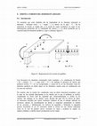
Fig. 3. Examples of contiguous repeats found in MpRV VP1.
The sequence in the top line shows the target sequence and
that in the lower line shows the matching repeat. ‘.’, Similar
residue; *, identical residue.
http://vir.sgmjournals.org
The VP5 of MpRV was found to partially match (aa 214–
318; identity, 21 %) the outer-capsid spike protein VP4
of Rotavirus A. It also showed 24 % identity to the killer
toxin protein (GenBank accession no. S51548) of yeast M28
dsRNA virus (an unclassified virus). Segment 5 shows an
ORF spanning nt 44–2005. This ORF is interrupted by an
in-frame TGA stop codon at position 1571–1573. A similar
situation has been described in segment 9 of CTFV (genus
Coltivirus), in which a read-through has been identified.
The occurrence of a read-through in segment 5 of MpRV
remains to be identified experimentally by cloning segment
5 in a eukaryotic expression vector under the control of a
strong promoter and identification of possible long and
short forms of the proteins, as has been realized for CTFV
segment 9 (Mohd Jaafar et al., 2004).
The VP7 of MpRV was found to partially match (aa 130–
209; identity, 32 %) the non-structural protein NS1 of
Cypovirus 1, whereas the VP8 of MPRV partially matched
(aa 42–66; identity, 28 %) the NSP2 of human Rotavirus A
(NSP2 has a dsRNA helix-destabilization activity, binds
RNA and is an NTPase).
The VP9 was found to partially match (aa 269–338; identity,
28 %) the segment 7-encoded protein of Nilaparvata luguens
reovirus (a fijivirus), which is a core protein and possesses a
nucleotide-binding activity (Mertens et al., 2005).
The proteins VP4, VP6, VP10, VP11 and VP12 did not show
any significant match with reovirus proteins.
To summarize, based on sequence comparisons with genomes
of members of the family Reoviridae, putative functions
deduced from the best-fit analyses were attributed to the
various MpRV proteins and the information is presented in
Table 2.
Phylogenetic analysis based on viral
polymerase sequences
It has previously been found that an amino acid identity of
¡30 % in the polymerase sequence is suitable to distinguish
between genera of the family Reoviridae (Attoui et al.,
2002a). However, there are two exceptions to this rule. The
first is the rotavirus B polymerase, which is only 22 %
identical to other rotaviruses. However, the inclusion of
Rotavirus B within the genus Rotavirus together with Rotavirus A and Rotavirus C does not rely primarily on genetic
distances between the polymerases of these viruses. It was
based on the morphological resemblance between these
viruses and the type of disease that they cause. The second
exception is the aquareoviruses and orthoreoviruses, which
are 42 % identical (Attoui et al., 2002a). This significantly
high amino acid identity is evidence of a common ancestry.
However, the distinct hosts and the different econiches of
these viruses justify their classification within two distinct
genera.
The results of the polymerase sequence analysis of MpRV are
illustrated by a radial neighbour-joining tree (Fig. 4). MpRV
Downloaded from www.microbiologyresearch.org by
IP: 54.160.113.5
On: Mon, 06 Jun 2016 16:51:13
1379
H. Attoui and others
Table 2. Putative functions of the proteins deduced from coding regions of MpRV segments
The putative functions described in this table resulted from a best-fit analysis using a database built with sequenced genomes of members
of the family Reoviridae. For details, see text. NI, No significant identity with any reovirus protein.
Segment
Protein designation
Putative function (possible location)
Similarity to
VP1
VP2
VP3
VP4
VP5
VP6
VP7
VP8
VP9
VP10
VP11
Haemagglutinin and cell attachment (outer coat)
Putative viral RNA polymerase (core)
Putative pseudo-T=2 layer (core)
Sigma-1 of orthoreoviruses
Reovirus polymerases
Pseudo-T=2 layer of phytoreoviruses
1
2
3
4
5
6
7
8
9
10
11
NI
Putative outer-coat protein (outer coat)
Possible non-structural protein
Possible non-structural protein
Putative outer layer of core (core)
NS1 of cypovirus
NSP2 of human Rotavirus A
Similarity to VP7 of fijiviruses
NI
NI
does not cluster with any of the known, characterized
members of the family Reoviridae. It clustered with neither
aquareoviruses (11-segmented), which are turreted viruses,
nor with rotaviruses (11-segmented), which are nonturreted. Rather, MpRV stands as a representative of a
separate phylogenetic group. This is confirmed by the low
amino acid identity values (5–21 %) between MpRV polymerase and those of member viruses of other genera of the
family Reoviridae.
Analysis of the replication of MpRV in insect-,
mammalian- and fish-cell lines
Analysis of RNA extracted from cells that were inoculated
with MpRV did not reveal any dsRNA profile following
agarose-gel electrophoresis. RT-PCR analysis of these extracts
using primers specific to segment 2 of MpRV was negative
upon first-round and nested PCR, demonstrating that
purified MpRV could not replicate in the tested cell lines.
DISCUSSION
When MpRV was isolated (Brussaard et al., 2004), the
identification of the 11-segmented dsRNA profile of its
genome, the morphological characteristics of the virions
and its physicochemical properties led to the classification of
MpRV as a tentative member of the family Reoviridae. The
present study provides several additional arguments for
the classification of MpRV as a full member of the family
Reoviridae. Full characterization of the dsRNA genome by
cloning and sequencing revealed that it has a total length of
25 563 bp. This is similar to the genome length of other
viruses of the family Reoviridae, which range between
~18 500 and 29 210 bp (Attoui et al., 2002b; Mertens et al.,
2005). However, segment 1 of MpRV is significantly longer
than segments 1 of other sequenced members of the family
Reoviridae.
In addition, the putative polymerase of MpRV partially
matches rotavirus polymerase and contains the signature
1380
Rotavirus VP4
NI
motifs of putative RdRps of viruses belonging to the family
Reoviridae. In compliance with the criteria defining the
family Reoviridae, analysis of the terminal sequences revealed
that all 11 segments of MpRV have conserved termini within
the 59 and 39 NCRs. Such terminal sequences are recognized
as one of the defining species parameters within the family
Reoviridae. These conserved termini in the MpRV genome
are different from any of those described previously within
the family Reoviridae, showing that MpRV does not belong
to any previously described species within this family.
The 11-segmented genome is a characteristic of rotaviruses
and aquareoviruses within the family Reoviridae. Being
isolated from an aquatic organism, it would be reasonable
to suppose that MpRV is a tentative member of the genus
Aquareovirus (infecting fishes, crustaceans and molluscs). As
an aquatic reovirus, it would be interesting to know whether
this virus can infect other aquatic species and whether it has
only one host, or whether M. pusilla might represent a vector
that transmits the virus among other aquatic species. Our
results indicated that MpRV does not replicate in fish-,
insect- or mammalian-cell lines that were tested and that it is
a member of neither the genus Aquareovirus nor the genus
Rotavirus, as MpRV has features unique among the family
Reoviridae.
Phylogenetic analysis based on the polymerase amino acid
sequence, for example, demonstrated clearly that MpRV is
not a member of the genera Rotavirus or Aquareovirus,
despite its genome being made of 11 dsRNA segments.
Amino acid identity of the polymerase of MpRV is 8–10 %
with aquareovirus polymerase and 19–21 % with rotavirus
polymerase. These are values comparable to those found
between viruses belonging to distinct genera of the family
Reoviridae. The morphology of the particles is also not
identical to that of other members of the family Reoviridae.
The diameter of whole particles purified under conditions
not affecting the integrity of the outer coat is significantly
larger (90–95 nm) than the diameters usually observed in
Downloaded from www.microbiologyresearch.org by
IP: 54.160.113.5
On: Mon, 06 Jun 2016 16:51:13
Journal of General Virology 87
Mimoreovirus: novel genus within the family Reoviridae
Fig. 4. Phylogenetic relationships of MpRV to other members of the family Reoviridae based on the sequence of the putative
RdRp. Neighbour-joining phylogenetic tree built with available polymerase sequences (using the Poisson-correction or
gamma-distribution algorithms) for representative members of 11 genera of the family Reoviridae. The abbreviations and
GenBank accession numbers are presented in Supplementary Table S1 (available in JGV Online).
viruses of the family Reoviridae. In a previous study, when
MpRV was purified on a CsCl density gradient, two virus
bands were visible (Brussaard et al., 2004). The less-dense
virus band was found to contain eight structural proteins,
including traces of a protein at approximately 200 kDa that
was absent from the denser virus band, suggesting that this
protein is removed at high ionic strength. This size is compatible with the deduced size of VP1 of MpRV. This protein
is likely to be responsible for the unusually large size of the
whole virus particles. VP1 showed similarities within its
amino-terminal sequence to Sigma-1 (a haemagglutinin and
cell-attachment protein) of MRV (genus Orthoreovirus).
http://vir.sgmjournals.org
This suggested that VP1 might represent the virus attachment protein to the cell wall of M. pusilla. VP1 was also
found to exhibit similarities to glycoproteins of viral and
non-viral agents. The high serine and threonine content of
VP1 suggests that it might be O-glycosylated, as is the case
of mucin and mucin-like proteins.
Transient envelope structures have been described for orbiviruses (Martin et al., 1998; Owens et al., 2004), coltiviruses
(Murphy et al., 1968; Attoui et al., 2002b), rotaviruses (Estes
& Cohen, 1989) and seadornaviruses (Mohd Jaafar et al.,
2005) as a consequence of budding of virus particles from
Downloaded from www.microbiologyresearch.org by
IP: 54.160.113.5
On: Mon, 06 Jun 2016 16:51:13
1381
H. Attoui and others
the cell membrane or budding into the endoplasmic reticulum during morphogenesis. If MpRV VP1 forms such a
structure, MpRV would be the first member of the family
Reoviridae to possess a constitutive additional outer coat.
This requires further investigation by techniques such as
cryoelectron microscopy.
The length of the VP1 of MpRV is unusual among sequenced
members of the family Reoviridae. The presence of many
repeats within this sequence evokes the possibility that VP1
could have arisen from amino acid fragment duplication,
which diverged afterwards. The precise mechanism and the
constraints that have driven such an evolution are unknown.
The existence of the remaining seven structural proteins in
the purified MpRV particles (Brussaard et al., 2004) is
compatible with that described for non-turreted reoviruses,
where particles are composed of seven structural proteins,
such as in seadornaviruses and orbiviruses (Mertens et al.,
2005; Mohd Jaafar et al., 2005). The MpRV particles that
were treated with CaCl2 generated structures that could
be compared with the smooth, non-turreted subcore layer
of orbiviruses, indicating that MpRV belongs to the nonturreted group within the family Reoviridae. This is further
evidence that MpRV is not a member of the genus Aquareovirus, as members of this genus are turreted viruses.
It is interesting that, within the RdRp phylogenetic tree, the
branch of MpRV dissects the tree, separating the groups of
turreted and non-turreted viruses. It has been found that
M. pusillla is evolutionarily older than the hosts of other
members of the family Reoviridae (Bhattacharya & Medlin,
1998; Doolittle, 1999; Cavalier-Smith, 2002). The location of
MpRV at the node separating the turreted and non-turreted
viruses suggests that it belongs to a third branch (although
non-turreted), which is possibly ancestral. The presence of
an additional protein coat or a potential pseudo-envelope
structure may be an ancestral character lost by other
members of the family Reoviridae or a specific adaptable
character of the reoviruses infecting protists.
Based on all of these findings, MpRV should be classified as a
member of a novel genus within the family Reoviridae. This
genus could be designated Mimoreovirus (Micromonas
pusilla reovirus). A formal proposal was made to the ICTV
session during the International Congress of Virology held
in San Francisco, CA, USA, in July 2005 to create the genus
Mimoreovirus and to designate MpRV as its type species.
This proposal received initial support and was passed for
wider consultation.
Attoui, H., Charrel, R. N., Billoir, F., Cantaloube, J.-F., de Micco, P. &
de Lamballerie, X. (1998). Comparative sequence analysis of
American, European and Asian isolates of viruses in the genus
Coltivirus. J Gen Virol 79, 2481–2489.
Attoui, H., Billoir, F., Cantaloube, J. F., Biagini, P., de Micco, P. &
de Lamballerie, X. (2000a). Strategies for the sequence determina-
tion of viral dsRNA genomes. J Virol Methods 89, 147–158.
Attoui, H., Billoir, F., Biagini, P., Cantaloube, J. F., de Chesse, R.,
de Micco, P. & de Lamballerie, X. (2000b). Sequence determination
and analysis of the full-length genome of Colorado tick fever virus,
the type species of genus Coltivirus (family Reoviridae). Biochem
Biophys Res Commun 273, 1121–1125.
Attoui, H., Fang, Q., Mohd Jaafar, F., Cantaloube, J.-F., Biagini, P.,
de Micco, P. & de Lamballerie, X. (2002a). Common evolutionary
origin of aquareoviruses and orthoreoviruses revealed by genome
characterization of Golden shiner reovirus, Grass carp reovirus,
Striped bass reovirus and golden ide reovirus (genus Aquareovirus,
family Reoviridae). J Gen Virol 83, 1941–1951.
Attoui, H., Mohd Jaafar, F., Biagini, P., Cantaloube, J.-F., de Micco,
P., Murphy, F. A. & de Lamballerie, X. (2002b). Genus Coltivirus
(family Reoviridae): genomic and morphologic characterization of
Old World and New World viruses. Arch Virol 147, 533–561.
Attoui, H., de Lamballerie, X. & Mertens, P. P. C. (2005). Coltivirus,
Reoviridae. In Virus Taxonomy. Eighth Report of the International
Committee on Taxonomy of Viruses, pp. 497–503. Edited by C. M.
Fauquet, M. A. Mayo, J. Maniloff, U. Desselberger & L. A. Ball.
London: Elsevier/Academic Press.
Baker, T. S., Olson, N. H. & Fuller, S. D. (1999). Adding the third
dimension to virus life cycles: three-dimensional reconstruction of
icosahedral viruses from cryo-electron micrographs. Microbiol Mol
Biol Rev 63, 862–922.
Bhattacharya, D. & Medlin, L. (1998). Algal phylogeny and the origin
of land plants. Plant Physiol 116, 9–15.
Brussaard, C. P. D., Noordeloos, A. A. M., Sandaa, R.-A., Heldal, M.
& Bratbak, G. (2004). Discovery of a dsRNA virus infecting the
marine photosynthetic protist Micromonas pusilla. Virology 319,
280–291.
Burroughs, J. N., O’Hara, R. S., Smale, C. J., Hamblin, C., Walton, A.,
Armstrong, R. & Mertens, P. P. C. (1994). Purification and properties
of virus particles, infectious subviral particles, cores and VP7 crystals
of African horsesickness virus serotype 9. J Gen Virol 75, 1849–1857.
Byrd, J. C. & Bresalier, R. S. (2004). Mucins and mucin binding
proteins in colorectal cancer. Cancer Metastasis Rev 23, 77–99.
Cavalier-Smith, T. (2002). The phagotrophic origin of eukaryotes
and phylogenetic classification of Protozoa. Int J Syst Evol Microbiol
52, 297–354.
Cottrell, M. T. & Suttle, C. A. (1991). Wide-spread occurrence and
clonal variation in viruses which cause lysis of a cosmopolitan,
eukaryotic marine phytoplankter, Micromonas pusilla. Mar Ecol Prog
Ser 78, 1–9.
Diprose, J. M., Burroughs, J. N., Sutton, G. C. & 8 other authors
(2001). Translocation portals for the substrates and products of a
viral transcriptase complex: the bluetongue virus core. EMBO J 20,
7229–7239.
ACKNOWLEDGEMENTS
Doolittle, W. F. (1999). Phylogenetic classification and the universal
The authors wish to acknowledge Anna Noordeloos for helping in the
preparation of MpRV. We also acknowledge Bernard Campagna and
Nicolas Aldrovandi for their excellent assistance in electron microscopy. This paper was supported by EU grant ‘Reo ID’ no. QLK2-200000143 and in part by EU grant ‘VIZIER’.
1382
REFERENCES
tree. Science 284, 2124–2128.
Estes, M. K. & Cohen, J. (1989). Rotavirus gene structure and
function. Microbiol Rev 53, 410–449.
Gibbs, A. & Keese, P. K. (1995). In search of the origins of viral
genes. In Molecular Basis of Virus Evolution, pp. 76–90. Edited by A.
Downloaded from www.microbiologyresearch.org by
IP: 54.160.113.5
On: Mon, 06 Jun 2016 16:51:13
Journal of General Virology 87
Mimoreovirus: novel genus within the family Reoviridae
Gibbs, C. H. Calisher & F. Garcı́a-Arenal. Cambridge: Cambridge
University Press.
Gouet, P., Diprose, J. M., Grimes, J. M., Malby, R., Burroughs, J. N.,
Ziéntara, S., Stuart, D. I. & Mertens, P. P. C. (1999). The highly
ordered double-stranded RNA genome of bluetongue virus revealed
by crystallography. Cell 97, 481–490.
Grimes, J. M., Burroughs, J. N., Gouet, P., Diprose, J. M., Malby, R.,
Ziéntara, S., Mertens, P. P. C. & Stuart, D. I. (1998). The atomic
structure of the bluetongue virus core. Nature 359, 470–478.
Mohd Jaafar, F., Attoui, H., de Micco, P. & de Lamballerie, X. (2004).
Termination and read-through proteins encoded by genome segment
9 of Colorado tick fever virus. J Gen Virol 85, 2237–2244.
Mohd Jaafar, F., Attoui, H., Mertens, P., de Micco, P. & de Lamballerie,
X. (2005). Structural organisation of a human encephalitic isolate of
Banna virus (genus Seadornavirus, family Reoviridae). J Gen Virol 86,
1147–1157.
Murphy, F. A., Coleman, P. H., Harrison, A. K. & Gary, G. W., Jr
(1968). Colorado tick fever virus: an electron microscopic study.
Harrison, P. J., Waters, R. E. & Taylor, F. J. R. (1980). A broad
Virology 35, 28–40.
spectrum artificial sea water medium for coastal and open ocean
phytoplankton. J Phycol 16, 28–35.
Nason, E. L., Rothagel, R., Mukherjee, S. K., Kar, A. K., Forzan, M.,
Prasad, B. V. & Roy, P. (2004). Interactions between the inner and
Hill, C. L., Booth, T. F., Prasad, B. V. V., Grimes, J. M., Mertens,
P. P. C., Sutton, G. C. & Stuart, D. I. (1999). The structure of a
outer capsids of bluetongue virus. J Virol 78, 8059–8067.
cypovirus and the functional organization of dsRNA viruses. Nat
Struct Biol 6, 565–568.
nonstructural protein in cellular pathogenesis and virus release.
J Virol 78, 6649–6656.
Kumar, S., Tamura, K. & Nei, M. (2004). MEGA3: integrated software
Prasad, B. V. V., Wang, G. J., Clerx, J. P. M. & Chiu, W. (1988). Three-
for molecular evolutionary genetics analysis and sequence alignment.
Brief Bioinform 5, 150–163.
Rasmussen, S. W. (1995). DNATools, a software package for DNA
Martin, L. A., Meyer, A. J., O’Hara, R. S., Fu, H., Knowles, N. J. &
Mertens, P. P. C. (1998). Phylogenetic analysis of African horse
sickness virus genome segment 10: sequence variation, virulence
characteristics and cell exit. Arch Virol Suppl 14, 281–293.
Mertens, P. (2004). The dsRNA viruses. Virus Res 101, 3–13.
Mertens, P. P. C. & Diprose, J. (2004). The bluetongue virus core: a
nano-scale transcription machine. Virus Res 101, 29–43.
Mertens, P. P. C., Arella, M., Attoui, H. & 41 other authors (2000).
Owens, R. J., Limn, C. & Roy, P. (2004). Role of an arbovirus
dimensional structure of rotavirus. J Mol Biol 199, 269–275.
sequence analysis. Carlsberg Laboratory, Copenhagen.
Reinisch, K. M., Nibert, M. L. & Harrison, S. C. (2000). Structure of
the reovirus core at 3.6 Å resolution. Nature 404, 960–967.
Thompson, J. D., Higgins, D. G. & Gibson, T. J. (1994). CLUSTAL W:
improving the sensitivity of progressive multiple sequence alignment
through sequence weighting, position-specific gap penalties and
weight matrix choice. Nucleic Acids Res 22, 4673–4680.
Wommack, K. E., Ravel, J., Hill, R. T., Chun, J. & Colwell, R. R.
(1999). Population dynamics of Chesapeake Bay virioplankton: total-
Reoviridae. In Virus Taxonomy: Seventh Report of the International
Committee on Taxonomy of Viruses, pp. 395–480. Edited by M. H. V.
van Regenmortel, C. M. Fauquet, D. H. L. Bishop, E. B. Carstens,
M. K. Estes, S. M. Lemon, J. Maniloff, M. A. Mayo, D. J. McGeoch,
C. R. Pringle & R. B. Wickner. New York, NY: Academic Press.
Yeager, M., Dryden, K. A., Olson, N. H., Greenber, H. B. & Baker,
T. S. (1990). Three-dimensional structure of rhesus rotavirus by
Mertens, P. P. C., Attoui, H., Duncan, R. & Dermody, T. S. (2005).
cryoelectron microscopy reconstruction. J Cell Biol 110, 2133–2144.
Reoviridae. In Virus Taxonomy: Eighth Report of the International
Committee on Taxonomy of Viruses, pp. 447–454. Edited by C. M.
Fauquet, M. A. Mayo, J. Maniloff, U. Desselberger & L. A. Ball.
London: Elsevier/Academic Press.
Yeager, M., Berriman, J. A., Baker, T. A. & Bellamy, A. R. (1994).
http://vir.sgmjournals.org
community analysis by pulsed-field gel electrophoresis. Appl Environ
Microbiol 65, 231–240.
Three-dimensional structure of the rotavirus haemagglutinin VP4 by
cryo-electron microscopy and difference map analysis. EMBO J 13,
1011–1018.
Downloaded from www.microbiologyresearch.org by
IP: 54.160.113.5
On: Mon, 06 Jun 2016 16:51:13
1383
Keep reading this paper — and 50 million others — with a free Academia account
Used by leading Academics

Sabina Passamonti
Università degli Studi di Trieste

Grum Gebreyesus
Aarhus University

Hikmet Budak
University of Nebraska Lincoln

Monica Ballarino
Università degli Studi "La Sapienza" di Roma