Academia.edu no longer supports Internet Explorer.
To browse Academia.edu and the wider internet faster and more securely, please take a few seconds to upgrade your browser.
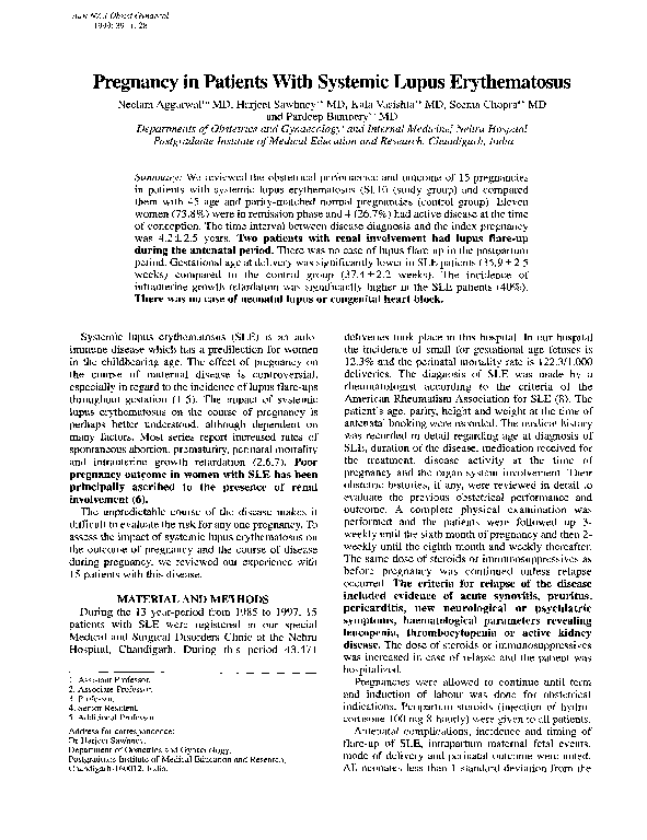
Pregnancy in Patients With Systemic Lupus Erythematosus
Pregnancy in Patients With Systemic Lupus Erythematosus
 Neelam Aggarwal
1999, Australian and New Zealand Journal of Obstetrics and Gynaecology
Related Papers
European Journal of Obstetrics & Gynecology and Reproductive Biology
Obstetrical outcome of pregnancy in patients with systemic Lupus Erythematosus. A study of 60 cases1999 •
Ghana Medical Journal
Outcome of pregnancy in patients with systemic lupus erythematosis at Korle-bu Teaching Hospital2016 •
To study maternal and fetal outcomes in Ghanaian women with systemic lupus erythematosus (SLE). Retrospective study of pregnancies in women with SLE in a single centre in Ghana. The mean age was 30.1 years and all were nulliparous. Two out of the seven pregnancies were in disease remission at the time of booking. Nephritis without renal impairment was present in 7 pregnancies (6 women). One woman developed intrapartum eclampsia. Two women had secondary antiphospholipid syndrome (APS). Two suffered early fetal losses and one late fetal loss at 32 weeks. All three who lost their fetus had uncontrolled hypertension. Six had mild flares mainly joint pains during pregnancy. There was no maternal mortality. The median gestational age at delivery was 38 weeks (range, 16 to 40 weeks) and the mean birth weight was 3017 g; the median Apgar scores were 8 and 9 at 1 and 5 minutes of life, respectively. There were no cases of intrauterine growth restriction (IUGR). There were no cases of congeni...
Sultan Qaboos University Medical Journal
Pregnancy Outcomes in Systemic Lupus Erythematosus Women2021 •
Objectives This study was conducted to assess pregnancy outcomes in women with systemic lupus erythematosus (SLE) in Oman. Methods A retrospective cohort study of 149 pregnancies in 98 women with SLE was conducted over 10 years to evaluate the impact of clinical and laboratory parameters in predicting adverse pregnancy outcomes. Results Mean maternal age was 30.6 ± 5 years ranging from 20–44 years, and the mean disease duration was 10 ± 5 years, ranging from 2–27 years. The most common maternal manifestations were joint pain in 36 (24.2%), lupus nephritis (LN) in 18 (12.08%), preeclampsia in 11 (7.4%), eclampsia in three (2%) and lupus flare in one pregnancy. The live birth rate was 139 (93.3%) with a mean gestational age of 36 ± 2 weeks ranging from 26–40 weeks. In total, 55 (39.6%) were preterm deliveries, six (4%) pregnancies ended in miscarriage, and four (2.7%) resulted in intrauterine fetal death. Intrauterine growth restriction was observed in 49 babies (35%). A significant a...
International Journal of Advances in Medicine
Successful outcome of pregnancy in a case of systemic lupus erythematosus : a case reportSystemic lupus erythematosus (SLE) is a multisystem, auto immune connective tissue disease that commonly affects women of reproductive age and may coexist with pregnancy. The autoantibodies and immune complexes lead to damage of various organs and tissues. Pregnant woman with SLE have increased risk of spontaneous abortion, preterm delivery, intrauterine growth retardation, preeclampsia, neonatal lupus, stillbirth and intrauterine fetal death. The therapeutic intervention with anticoagulants, steroids, immunosuppressive agents pose a high risk to both mother and fetus. A multidisciplinary approach and close medical, obstetrical and neonatal monitoring leads to optimal outcome. Authors describe a successful management of an antenatal patient with positive antinuclear antibody, anti-ds DNA antibody and antiphospholipid antibody with bad obstetric history. She underwent an emergency cesarean section and delivered a healthy female child.
International Journal of Reproduction, Contraception, Obstetrics and Gynecology
Systemic lupus erythematosus-a good maternal and fetal outcome2020 •
Systemic lupus erythematosus (SLE) is a multisystemic autoimmune disease which primarily affects women in their reproductive years. The fertility is generally unaffected except in women with active disease, significant impairment of renal function, or high dose corticosteroid or cyclophosphamide therapy, which can result in ovarian dysfunction. This case report elaborates on the course of the pregnancy and the favourable maternal and fetal obstetric outcome of a 28-year-old female with known case of hypothyroidism who presented with chief complaints of generalised swelling all over the body and exertional dyspnoea and was later diagnosed to be a case of focal proliferative lupus nephritis, class III (ISN/RPS) on renal biopsy done postpartum. The effect of pregnancy on maternal disease is controversial. While some studies report exacerbation of SLE during pregnancy,others have not reported increased flares. The only study on this aspect of SLE from our country did not report a flare-...
2017 •
Systemic lupus erythematosus (SLE) is an autoimmune disease most frequently found in women of childbearing age and may coexist with pregnancy. Disease exacerbation, increased fetal loss, neonatal lupus, and an increased incidence of preeclampsia are the major challenges. Its multisystem involvement and therapeutic interventions like anticoagulants, steroids, and immunosuppressive agents pose a high risk for both the mother and the fetus during the antenatal period as well as postpartum. Good multidisciplinary medical care is mandatory when detection or flare-up of SLE occurs during pregnancy. We describe the successful management of an antinuclear antibody, antiribonucleoprotein antibody, and anti-Sjogren's syndrome A (Ro) antibody positive parturient with bad obstetric history who underwent elective cesarean section and delivered a healthy child. How to cite this article Basava L, Roy P, Triveni K, Sree GS. Successful Pregnancy Outcome in a Case of Systemic Lupus Erythematosus....
Clinical Reviews in Allergy & Immunology
Pregnancy and Systemic Lupus Erythematosus: Review of Clinical Features and Outcome of 51 Pregnancies at a Single Institution2010 •
Systemic lupus erythematosus (SLE) is mainly a disease of fertile women and the coexistence of pregnancy is by no means a rare event. How SLE and its treatment affects pregnancy outcome is still a matter of debate. Assessment of the reciprocal clinical impact of SLE and pregnancy was investigated in a cohort study. We reviewed the clinical features, treatment, and outcomes of 43 pregnant SLE patients with 51 pregnancies followed from 1993 to 2007 at a tertiary university hospital. The age of patients was 28.7 ± 5.4 years and SLE was diagnosed at age of 23.0 ± 6.1 years. Previous manifestations of SLE included lupus nephritis (14 patients) and secondary antiphospholipid syndrome (11 patients). Thirty-five pregnant patients (69%) were in remission for more than 6 months at the onset of pregnancy. Patients were being treated with low doses of prednisone (29), hydroxychloroquine (20), azathioprine (five), acetylsalicylic acid (51), and low molecular weight heparin (13). Sixteen pregnancy-associated flares were documented, mainly during the second trimester (42%) and also in the following year after delivery (25%). Renal involvement was found in 11 cases (68%). Spontaneous abortion occurred in 6%, 16% had premature deliveries, and 74% were delivered at term. No cases of maternal mortality occurred. No cases of fetal malformation were recorded. There was one intrauterine fetal death and one neonatal death at 24 gestational weeks. Pregnant women with SLE are high risk patients, but we had a 90% success rate in our cohort. A control disease activity strategy to target clinical remission is essential.
Rheumatology International
Pregnancy outcomes in women with childhood-onset and adult-onset systemic lupus erythematosus: a comparative study2016 •
Proceedings of the 2009 meeting of Computer Applications in Archaeology
Rome Reborn 2.0: A Case Study of Virtual City Reconstruction Using Procedural Modeling Techniques2010 •
Rome Reborn is an international initiative launched in 1996 to create a 3D digital reconstruction of ancient Rome as it might have appeared in the year AD 320. The model consists of two categories of data: Class I features are well-attested in terms of name, function, location, and design. Class II features are less well known. The project uses "hand modeling" using tools such as AutoCad, Blender, 3DS Max, etc. to create the Class I features. In version 1.0 of the model, released in 2007, the Class II features derived from a scan of the physical model of ancient Rome created from the 1930s to 1970s under the direction of Italo Gismondi (the "Plastico di Roma Antica" in the Museum of Roman Civiization, Rome/EUR). This paper reports on version 2.0 of the model. In this version, all the scan models deriving from Gismondi's physical model were deleted and replaced by procedurally generated models using the CityEngine software developed by Pascal Mueller. Version 2.0 was released in 2008. It may be noted that version 3.0 of the model was released in 2018. Version 4.0 in 2023. These versions continue to use the same procedural models of the Class II features described in this paper. See www.flyoverzone.com for information about the virtual tour called "Flight over Ancient Rome" that utilizes version 4.0 of the Rome Reborn model.
RELATED PAPERS
Textura Ulbra
Escolástica e História: discussões historiográficas2013 •
Producción Académica
La revista de la Junta de Estudios Históricos de Santiago del Estero2011 •
Abdikarya : Jurnal Pengabdian dan Pemberdayaan Masyarakat/Abdikarya
Sosialisasi Peningkatan Pemahaman Masyarakat Sebagai Upaya Penurunan Stunting Menuju Pembangunan Berkelanjutan (SDGs)2023 •
ShikshaShastra Saurabh
Witch Allegation in Nepal: A Case Study in Tamang Community of Kavrepalanchok District2022 •
1967 •
International Journal of GEOMATE
A Numerical Groundwater Flow Model of Wadi Samail Catchment Using Modflow Software2020 •
Biophysical journal
An engineered membrane to measure electroporation: effect of tethers and bioelectronic interface2014 •
2004 •
NTU Journal of Renewable Energy
Load Frequency Control With Renewable Energy Sources Using Practical Swarm Optimization Based On PID2023 •
TYCHE – Contributions to Ancient History, Papyrology and Epigraphy
Bemerkungen zu Papyri XXVI (<Korr. Tyche> 735-753)2014 •
Antimicrobial Resistance and Infection Control
Identifying infection prevention and control gaps in healthcare facilities operating in Rivers state during the EVD outbreak in Nigeria 20142015 •
Alborz University Medical Journal
The Effect of Educational Intervention Based on BASNEF Model to Improve Interpersonal Communication Skills of Nurses2012 •
RELATED TOPICS
- Find new research papers in:
- Physics
- Chemistry
- Biology
- Health Sciences
- Ecology
- Earth Sciences
- Cognitive Science
- Mathematics
- Computer Science