Chemical Geology, 95 (1992) 347-360
Elsevier Science Publishers B.V., Amsterdam
347
[1]
Biogeochemistry of hot spring environments
3. Apolar and polar lipids in the biologically active layers of
a cyanobacterial mat
Y. Bing Zenga,~, David M. Ward b, Simon C. Brassellc and Geoffrey Eglintona
aOrganic Geochemistry Unit, School of Chemistry, University of Bristol, Bristol BS8 1TS, UK
bDepartment of Microbiology, Montana State University, Bozeman, MT 59717, USA
CDepartrnent of Geology, Stanford University, Stanford, CA 94305-2115, USA
(Received August 14, 1990; revised and accepted August 2, 1991 )
ABSTRACT
Zeng, Y.B., Ward, D.M., Brassell, S.C. and Eglinton, G., 1992. Biogeochemistry of hot spring environments, 3. Apolar and
polar lipids in the biologically active layers ofa cyanobacterial mat. Chem. Geol., 95: 347-360.
The apolar lipid, glycolipid and phospholipid components of the biologically active top 5-mm surface layers of a hot
spring cyanobacterial mat were investigated. Most of the major components could be associated with bacteria isolated
from the mat; the vertical distribution of lipids followed the known or presumed vertical distribution of these organisms.
For example, hydrocarbons (e.g., 7-methylheptadecane), phytadienes (methanolysis products from chlorophyll a) and
polar lipid fatty acids typical of mat-forming cyanobacteria maximize in the top 0-1 mm and decrease in concentration
with depth. Wax esters and octadecanol (produced upon methanolysis of bacteriochlorophyll cs) typical of Chloroflexus
aurantiacus maximized in the 1-2- and 2-4-mm intervals. Long-chain diols derived mainly from glycolipids and typical
of the aerobic heterotroph Thermomicrobium roseum maximize in the 1-2- and 2-4-mm intervals. 1-O-Alkylglycerols
derived from polar lipids and typical of anaerobic fermentative or sulphate-reducing bacteria, increase in concentration
with depth and maximize in deeper layers. The relative abundances of lipids appear to reflect the trophic structure of the
microbial community.
I. Introduction
This paper is a continuation of our collaborative investigation of lipid biomarkers in hot
spring microbial mats as model systems in
which community composition is simplified
and relatively well defined (Ward et al., 1985,
1989 ). In earlier papers of the series we investigated the apolar lipids of the cyanobacterial
mat in Octopus Spring, Yellowstone National
Park (Dobson et al., 1988), as well as polar
lipids of this and other hot spring microbial
mats of varying degrees of community com"Present address: Institute for Water Sciences, Western
Michigan University, Kalamazoo, MI 49008-5150, USA.
plexity (Zeng et al., 1992 in this issue; in this
paper referred to as Part 2). Much of our previous work has compared the composition of
extractable lipids or polar lipid components in
bulk mat samples. Here, we concentrate on the
Octopus Spring cyanobacterial mat, well-characterized with respect to many of the microorganisms which are thought to be involved in
photosynthetic formation and subsequent decomposition of the mat. Many of these microorganisms have been obtained in pure culture
and their lipid compositions have been investigated (Ward et al., 1989). The relationship
of cultivated to uncultivated mat inhabitants
of similar phylogeny is becoming increasingly
understood (Ward et al., 1990, 1992). Pro-
0009-2541/92/$05.00 © 1992 Elsevier Science Publishers B.V. All rights reserved.
�348
Y.B. ZENG ET AL.
duction and decomposition of this mat occur
principally within the top 5 m m (Ward et al.,
1987 ). We investigated this zone of biological
activity at depth intervals relevant to the distribution of microorganisms and the reactions
they catalyze in order to learn whether the vertical distribution of mat inhabitants was reflected in the distribution of the lipids they are
likely to synthesize.
2. Methods
Octopus Spring is located in the Lower Geyser Basin of Yellowstone National Park, ~ 150
m SSE of Great Fountain Geyser. Samples were
removed from a 52-55°C site along the southernmost effluent channel using a stainless-steel
coring tube (44-mm diameter). Using a spatula one core was immediately sectioned along
natural laminae into the top green layer ( ~ 01 mm; 160 mg dry weight), a reddish underlayer ( ~ 1-2 mm; 84 mg dry weight), a deepred coloured layer ( ~ 2 - 4 mm; 95 mg dry
weight) and a brown-green layer ( ~ 4-5 mm;
41 mg dry weight). Samples were immediately
frozen on dry ice for transit, lyophilized upon
return to the laboratory and kept frozen except
for a few days in transit to the U.K.
All solvents were redistilled and all glassware and materials (including sampling materials and containers) were solvent-rinsed before use. The samples were ground to powder
with a mortar and pestle before extraction using a modification of the Bligh and Dyer
(1959) method (see Part 2). The total lipid
extract was separated into apolar lipid, glycolipid and phospholipid fractions by column
chromatography (see Part 2 ) and their weights
were determined after solvent evaporation.
Following addition of internal standards, glycolipid and phospholipid fractions were subjected to methanolysis, derivatized with N,Obis (trimethylsilyl)trifluoroacetamide
(BSTFA) and analyzed by gas chromatography (GC) and gas chromatography-mass
spectrometry (GC-MS) as previously described (see Part 2 ).
3. Results
3. I. Lipid class composition
Concentrations of the various fractions, and
the total concentrations of wax ester components and polar lipid fatty acid methyl ester
(FAME) methanolysis products (estimated
TABLE 1
Compound classes of extractable lipids
Compound class
Concentration in #g g - 1 dry mat (% of total extracts )
0-1 mm
1-2 mm
2-4mm
4-5 mm
Apolarlipids*~
- Wax esters.2
10,959 ( 1 8 . 0 % )
7,462
26,027 ( 3 6 . 5 % )
20,294
28,235 ( 4 1 . 3 % )
16,254
19,231 ( 2 7 . 8 % )
8,600
Glycolipids*~
- FAME's .2
31,507 ( 5 1 . 7 % )
15,389
28,767 ( 4 0 . 4 % )
7,806
27,059 ( 3 9 . 7 % )
6,795
23,077 ( 3 3 . 3 % )
3,629
Phospholipids.1
- FAME's .2
18,493 ( 3 0 . 3 % )
8.325
16,438 ( 2 3 . 1 % )
4,219
12,941 ( 1 9 . 0 % )
3,177
26,923 ( 3 8 . 9 % )
4,597
Total extracts .3
60,959 (100%)
71,233 (100%)
68,235 (100%)
69,231 (100%)
*~Concentration determined by gravimetric method.
*:Concentration obtained by summation of GC quantitation of individual methanolysis products (Tables 2-4 ).
*3Sum of apolar lipids, glycolipids and phospholipids.
�BIOCHEMISTRYOF HOT SPRING ENVIRONMENTS,3
349
0-1mm
1.3
Y
2
20
1 -2mm
1
3
,
lB
8
. A
x
2O
2-4mm
18
4 - 5 mrn
320
._,
'
11 8
1.| .
_
. . . . .
.,
I
I ' ' ' ' 1 ' ' ' '
10
19
20
I
30
I
I
1
~
I
40
I
5O
60
RETENTION TIME (minutes)
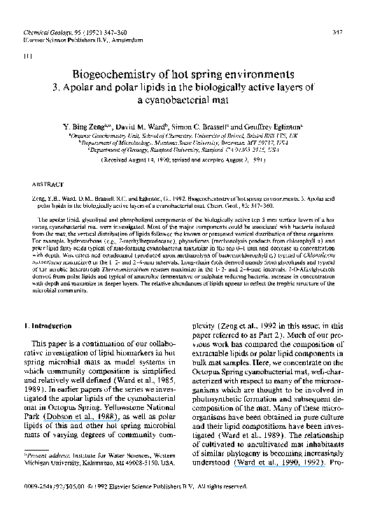
Fig. 1. Gas chromatograms of apolar lipid fractions of the biologicallyactive layers of the 52-55°C Octopus Spring cyanobacterial mat. Assignmentsand abundances of major components are given in Table 2. Minor constituents include:
15, 23, 24 = n, n-C29, -C37 and -C38 wax esters, respectively; il, i2 = internal standards (n-C23 aikane and 5a (H)-cholestane, respectively). All carboxyland hydroxylgroups were present as the TMS esters and ethers, respectively.Unlabelled
peaks represent components which could not be unambiguouslyassigned from their mass spectra.
from GC analysis), are reported in Table 1.
The glycolipid fractions (and their c o m p o n e n t
FAME's) were most a b u n d a n t in the top layer
and decreased in concentration with depth. The
phospholipid fractions ( a n d their c o m p o n e n t
FAME's) showed a similar pattern, with the
exception that concentrations were higher in
the 4 - 5 - m m layer. Apolar lipids and their
principal components, wax esters, m a x i m i z e d
in the 1-2- and 2 - 4 - m m subsurface intervals,
where they comprised ~ 4 0 % o f t h e total lipid
extracts.
3.2. A p o ~ r l ~ i d s
Gas chromatograms of apolar lipid fractions
are shown in Fig. 1 and principal components
are quantified in Table 2. In all samples wax
esters were the d o m i n a n t components. Several
series o f wax ester homologs were detected,
�350
Y.B. ZENGET AL.
TABLE 2
Concentration of major compounds in the neutral lipid fractions
Peak
Compound
label
(Fig. 1 )
Hydrocarbons:
I
2
3
4
Alcohols:
8
I1
Wax esters:
n,n-Chain:
16
17
18
19
20
21
22
Concentration (gg g-~ dry mat)
0-1 mm
1-2 mm
2-4 mm
4-5 mm
248
136
270
51
281
110
37
43
142
42
12
194
200
97
7
131
30
13
144
54
161
54
82
83
80
255
1,326
1,194
2,633
682
329
204
714
3,650
3,491
6,220
1,659
683
197
542
3,165
2,627
4,822
1,035
517
111
317
1,523
1,190
1,639
322
193
C35
29
112
157
223
91
124
517
658
867
312
112
524
519
825
211
164
651
514
793
126
C32
tr.
C34
11
47
101
73
142
128
239
n-CtTalkane
n-Ctsalkane
7Me heptadecane
phyt-l-ene
n-Cl7:o
i-C 17:o
C3o
C31
C32
C33
C34
C35
C36
/,n-Chain:
26
27
C31
28
C33
C34
29
30
/,/-Chain:
31
33
C32
tr.=trace.
with straight-chain (n,n-) ester components
predominating over branched ones. Most individual wax esters showed peak concentration in the 1-2- or 2-4-mm depth interval.
Hydrocarbon fractions contained predominantly n-C17 and n-C18 alkanes and 7Me-heptadecane in the 0-1-mm top layer, n-C17 w a s
also predominant in deeper layers, whereas
7Me-heptadecane decreased dramatically.
Phyt-l-ene, also a major hydrocarbon component, increased with depth.
Free alcohols ranging from C~5 to C,8 (maximizing at C~7) were present as minor components, n-Alkan-1-01s predominated over iso-alkan-l-ols. Phytol, which was previously
detected in a whole mat sample (see Part 2 ),
was below detection in the upper layers we investigated. Similarly, bishomohopan-32-ol, a
minor component in the whole mat sample
(see Part 2), was not detected in the individual layers.
3.3. Glycolipid fraction constituents
Gas chromatograms of the glycolipid fraction methanolysis products are presented in
Fig. 2. Major components of this fraction are
quantified in Table 3.
FAME's were abundant in the methanolysis
products of the glycolipid fractions in all lay-
�351
BIOCHEMISTRY OF HOT SPRING ENVIRONMENTS, 3
O-lmm
33 i
5
i
20
2
6
7
20
1 -2mm
2
~7
¢, 30
5
?
•
2 21
2O
2-4ram
2
I 26
8
6
•
11
16
2
17
1O
28
.L.
4-5ram
26
'
I
10
'
'
'
I
20
'
'
'
I
'
30
'
'
I
I
'
40
'
1 1 1 1 1 1 1 1 1
50
60
RETENTION TIME (minutes)
Fig. 2. Gas chromatograms of methanolysis products of glycolipid fractions obtained from the biologically active layers
of the 52-55 °C Octopus Spring cyanobacterial mat. Assignments and abundances of major components are given in Table
3. Minor constituents include: 22 = br-C22 alkane- 1,2-diol; 23, 24 = n-C16 and -Cl 7 l-O-alkylglycerol, respectively;
28 = C, 5,C~ 5 1, 2-di-O-dialkylglycerol; 30 = n-C ~7:0alcohol; 33 = phytadienes; il, i2 = internal standards ( t/-C23 alkane and
5a (H)-cholestane, respectively ). All hydroxyl groups were analyzed as the TMS derivatives.
ers. I n d i v i d u a l F A M E ' s s h o w e d different vertical distributions. T h e top layer was d o m i n a t e d by C16:0, C18:0, Cl6:l, C18:1 a n d
cyclopropyl-C~9 F A M E ' s , a n d these decreased
in c o n c e n t r a t i o n with depth. O t h e r F A M E ' s
present in relatively high c o n c e n t r a t i o n in the
top layer, such as Cis:0, C~7:0 a n d Cls:l FAME's,
showed m a x i m u m c o n c e n t r a t i o n s in the 1-2or 2 - 4 - m m layers, B r a n c h e d F A M E ' s were relatively low in c o n c e n t r a t i o n in all cases, except
for i-Cls:O which occurred only in traces in the
top layer, a n d increased in c o n c e n t r a t i o n in
subsurface layers.
CI9-C21 straight-chain a n d m o n o m e t h y -
�352
Y.B. ZENGET AL.
TABLE 3
Concentration of major methanolysis products of glycolipid fractions
Peak
label
(Fig. 2)
Compound(s)
Concentration (#g g - 1 dry mat)
0-1
mm
1-2
mm
2-4
mm
4-5 mm
Fatty acid methyl esters:
Normal chain:
I
Ci4:o
2
C15:o
3
Ct6:o
C|7:o
4
5
Ct8:o
6
Cls:l
7
Cl6:l(S)
8
C18:~(s)
85
866
5,714
497
1,311
304
832
2,783
163
1,550
2,148
594
357
476
669
795
180
1,170
1,902
633
218
248
341
778
74
322
817
339
268
62
76
870
Cyclopropyl:
9
2,574
460
57
121
99
132
75
92
tr.
43
75
I11
106
113
143
494
49
91
106
115
110
268
425
199
153
734
372
295
330
228
180
105
41
tr.
131
1,574
138
229
3,001
267
180
2,206
186
46
355
tr.
166
80
145
877
253
419
159
170
400
185
300
150
180
Cl9
Mono-methyl branched chain:
10
i-Cis:o
I1
i-Cl6:o
12
i-Ci7:o
13
a-Cl7:o
14
br-C 16:0
15
br-C~7:0
Alkane- 1,2-diols:
Normal chain:
16
Cl9
17
C2o
18
C21
Mono-methyl branched chain:
19
Cl9
20
C2o
21
C21
1-O-alkylglycerols:
Normal chain:
25
C~8
Mono-methyl branched chain:
26
Cl7
27
C18
Chlorophyll derivatives:
31
n-C~8 alcohol
32
phytadienes
tr.
tr.
tr.
tr.
tr.
-
tr.
36
-
tr.
120
1,500
(s) = sum of all isomers; tr. = trace; - - = below detection by GC and GC-MS.
lated alkane-l,2-diols, identified from their
c h a r a c t e r i s t i c m a s s s p e c t r a ( s e e P a r t 2 ), w e r e
major components in all layers, especially in
the 1-2- and 2-4-ram intervals. Diols exhib-
ited maximum concentration at the 1-2-mm
depth interval, where the major component, a
C2o b r a n c h e d d i o l , e x c e e d e d c o n c e n t r a t i o n s o f
individual FAME's.
�BIOCHEMISTRY OF HOT SPRING ENVIRONMENTS, 3
353
0 - 1 mm
$
2
1
1 -2mm
5
2-4mm
,.
~
11~
]
8I
9
16
IT Lr/1825
4-Smm
t?
7
IIlll
10
I '
2O
'
''
I
'
'
30
RETENTION
''
I
40
TIME
'
'
'
'
I '
50
'
'
'
I '
60
(minutes)
Fig. 3. Gas chromatograms of methanolysis products of phospholipid fractions obtained from the biologically active
layers of the 52-55°C cyanobacterial mat in Octopus Spring. Assignments and abundances of major components are
given in Tables 3 and 4. Minor constituents are assigned in Fig. 2. All hydroxyl groups were analyzed as the TMS derivatives.
Similarly, 1-O-alkylglycerols were also identified from their characteristic mass spectra
(see Part 2 ). A l-O-alkylglycerol with a possible methyl branched CI7 alkyl group was present in all samples and increased with depth to
become a major product of methanolysis of the
glycolipid fraction in the 2-4-ram interval.
Other CI7 and CIS straight-chain or branched
1-O-alkylglycerols were absent or present in
only trace amounts in the 0-1- and 1-2-ram
layers, but they also increased in deeper layers.
Other products of methanolysis of the glycolipid fraction may have been derived from
chlorophyll pigments which coeluted with the
glycolipid fraction on column separation.
These included phytadienes, present mainly in
�354
Y.B. ZENG ET AL.
TABLE 4
Concentration of major methanolysis products of phospholipid fractions
Peak
Compound(s)
label
(Fig. 3)
Concentration (#g g- 1dry mat)
0-1 mm
1-2 mm
2-4 mm
4-5 mm
49
273
2,800
230
1,429
199
1,075
52
399
1,361
300
567
135
308
54
250
996
289
503
58
136
75
197
1,459
340
1,171
77
219
872
341
108
211
771
258
205
tr.
345
105
193
30
184
89
183
184
123
84
230
239
24
15
17
90
54
53
41
35
36
29
26
28
94
7
306
24
180
20
180
19
tr.
tr.
15
46
72
183
99
248
Fatty acid methyl esters:
Normal chain:
I
2
3
4
5
7
8
C14:o
Cls:o
C|6:o
ClT:O
Cls:o
C16:1(s)
CIs:I(S)
Cyclopropyl:
9
Ci9
Mono-methyl branched chain:
i - e l 5:0
I0
11
12
I5
i-C16:o
i-CI7:o
br-C 17:o
Alkane- I, 2-diols:
Normal chain:
16
17
18
Ct9
C2o
C21
Mono-methyl branched chain:
20
21
25
26
C2o
C21
1-O-alkylglycerols:
n-C~s
br-C l 7
(s) =sum of all isomers; tr. =trace.
the top layer and decreasing with depth, and nC i 7 and n-C~8 alcohols, present mainly in the
1-2-, 2-4- and 4-5-mm layers and more abundant in subsurface layers.
A C~5,C~5 1,2-di-O-dialkylglycerol, identified from its mass spectrum (see Part 2), was
detected as a minor component in the 0-1-, 12- and 2-4-mm samples.
3.4. Phospholipidfraction constituents
Apart from the low abundance or absence of
alcohols and phytadienes, the products of
methanolysis of the phospholipid fraction were
similar in composition and depth distribution
to those of methanolysis of the glycolipid fraction (Fig. 3; Table 4). The predominant products were FAME's. In comparison to the glycolipid FAME's there was a lower relative
concentration of C16:l FAME and cyclopropylC19 FAME and a higher relative concentration
of i-Cls:o FAME. Also, monomethyl FAME's
were more abundant in the 0-1-mm layer and
decreased in concentration with depth. Diols
were much less abundant in the phospholipid
fraction than in the glycolipid fraction, but
�BIOCHEMISTRY OF HOT SPRING ENVIRONMENTS, 3
showed a vertical profile similar to that of diols
derived from the glycolipid fraction, maximizing in the l - 2 - m m layer, l-O-Alkylglycerol
ethers were also less abundant in the phospholipid fraction, but, as in the glycolipid fraction,
maximized in the deeper layers.
4. Discussion
4.1. Correlations between vertical distribution
of lipids and bacteria
There appear to be three distinct classes of
vertical distribution of lipids in the Octopus
Spring cyanobacterial mat bioactive zone, as
illustrated in Fig. 4. These distributions can be
interpreted in light of the many component
bacteria whose lipids have been studied, taking into account what is known of the vertical
distributions of these organisms and the turnover oflipids which is likely to occur upon burial in the mat.
The major polar lipid FAME's, C16:0 , C 1 8 : o
and C18:1, maximize in the 0-1-mm uppermost
layer and their concentrations decrease with
depth. These are the major polar lipid fatty
acids of the cyanobacterial isolate which is
thought to play a role in formation of hot spring
mats, Synechococcus lividus, when grown at
55°C (Miller, 1976; Fork et al., 1979). These
are not particularly distinctive polar lipid fatty
acids. They are, for instance, the major total
cellular fatty acids of the other cultivated mat
phototroph, the photosynthetic green nonsulfur bacterium Chloroflexus aurantiacus (Kenyon and Gray, 1974; Knudsen et al., 1982 ), and
the aerobic heterotrophic mat isolate Isophaera pallida (Giovannoni et al.,1987 ). Organisms such as these are thought to inhabit the
0-1-mm interval (Doemel and Brock, 1977 ).
Based on the vertical distributions of chlorophyll a (Bauld and Brock, 1973), oxygenic
photosynthesis (Revsbech and Ward, 1984)
and S. lividus-shaped cells (Doemel and Brock,
1977 ), cyanobacteria are restricted to the top
0-1-mm interval. Presumably they consume all
355
consume all the light available for oxygenic
photosynthesis very close to the mat surface.
which may be more diagnostic of cyanobacteria also maximize in the 0-1-mm interval and
decrease in concentration with depth below the
0-1-mm layer. Phytadienes are presumably
derived during the methanolysis of cyanobacterial chlorophyll a in the glycolipid fraction.
This inference is supported by the comparative prominence of phytadienes and n-octadecanol as methanolysis products of glycolipids
from cyanobacterial- and Chloroflexus-dominated mats, respectively (see Part 2). 7Meheptadecane is also often attributed to cyanobacterial sources (Han et al., 1968; Gelpi et al.,
1970; Blumer et al., 1971; Shiea et al., 1990).
Based on the vertical distribution of bacteriochlorophylls (Bauld and Brock, 1973),
Chloroflexus aurantiacus is thought to be more
abundant in the 1- or 2-mm undermat beneath
the cyanobacteria-dominated top layer, where
it receives infrared light suitable for its photosynthesis. Perhaps the most diagnostic lipid
biomarkers for this organism are the C28-C38
wax esters it produces (Edmunds, 1982;
Knudsen et al., 1982; Shiea et al., 1991 ) as a
significant proportion of its lipids ( Beyer et al.,
1983 ). Compounds of this type maximized in
the 1-2- and 2-4-mm depth intervals, coincident with the distribution of C. aurantiacus.
This bacterium produces a unique bacteriochlorophyll which esterifies mainly n-octadecanol (Gloe and Risch, 1978). It is thus consistent that the n-octadecanol released during
methanolysis of the glycolipid fraction, possibly derived from this bacteriochlorophyll, is
most abundant in subsurface layers.
Long-chain diols of the type found in this
study have only been reported in the hot spring
isolate Thermomicrobium roseum, an aerobic
heterotrophic bacterium (Pond et al., 1986).
The mat diols were dominated by the branched
C2o component, the most abundant diol of T.
roseum cultured at 60 °C (Pond and Langworthy, 1987). It is certainly possible that other
mat inhabitants might also produce diols, but
�356
Y.B. ZENG ET AL
A
!1-C16:0 F A M E
_n-Cl8:l FAME
phytadienes
7MeC17
mg/g TOC:
mcj/g TO<~
ug/g TOG
rng/g TO<:;
o~~~
o;
i
-!
|
3
i
i!
_n,_n-C~ wax ester
rng/g TO<3
~_
oi
br-C2o dioi
n-Cog alcohol
rncj/g TOC
ug/g TOG
o
--m
3
c.-
~
--m
c~
L
C
5"
3
L
~-%7 FAME
ug/g "[OC
br-Cl7 m o n o e t h e r
ug/g TO(]
l I I I
°i ' ? ?
5"
c~-
il=
Fig. 4. Vertical distributions of lipids in the biologically active layers of the 52-55°C cyanobacterial mat of Octopus
Spring: (A) compounds which maximize at the surface and decrease in concentration with depth; (B) compounds which
maximize in intermediate depths; and (C) compounds which increase in concentration with depth and maximize in
deeper layers.
�3 57
BIOCHEMISTRY OF HOT SPRING ENVIRONMENTS, 3
their presence in the 0-1-mm layer and maximization in the l - 2 - m m layer is consistent
with the aerobic physiology of T. roseum and
the greater abundance of oxygen in the layers
near the active zone of oxygenic photosynthesis (Revsbech and Ward, 1984).
Branched-chain FAME components of the
glycolipid and phospholipid fractions showed
two distribution patterns with depth. The major phospholipid branched FAME's, i-Cls:0, Cl6:O and -Cl7:O FAME's, were most abundant
in the 0 - l - m m layer and decreased in concentration with depth. These are characteristic
components of the phospholipids of two mat
heterotrophic isolates which are aerobic, Thermus aquaticus (Ray et al., 1971a, b), or facultatively aerobic, Bacillus stearothermophilus
(Card et al., 1969; Card, 1973). A branched
C17:o phospholipid FAME and most of the
branched glycolipid FAME's showed an increase in concentration with depth, with highest concentrations in the 2-4- or 4-5-ram layers. This might indicate a source organism of
different, possibly anaerobic, physiology. In
this regard, it is interesting that the anaerobic
fermentative mat isolate, Clostridium thermosulfurogenes, is known to produce i-C17 and C~5 fatty acyl chains (Langworthy and Pond,
1986). Interestingly, the C3o dicarboxylic acid
with "head-to-head" condensed iso-C~5 fatty
acids, which comprises a major proportion of
this organism's lipids (Langworthy and Pond,
1986), was not detected in the mat.
1-O-alkylglycerols maximized in the deeper
layers of the mat. Two mat isolates are known
to produce glycerol monoethers with alkyl
moieties comparable to those of the major mat
monoethers. One of these is the anaerobic fermenter, C. thermosulfurogenes (Langworthy
and Pond, 1986); the other is the anaerobic
sulphate reducer Thermodesulfobacterium
commune (Langworthy et al., 1983). The latter produces mainly glycerol diethers which
were not found in abundance in the mat layers,
implying that the former organism is the more
likely source of the monoethers found.
1,2-Di-O-dialkylglycerols typical of sulphate-reducing and methanogenic bacteria,
which were observed in a bulk mat sample (see
Part 2 ), were not detected in the individual mat
layers. This is presumably due to the high
trophic status and, thus, very low abundance
of these organisms and their distinctive lipids.
The vertical distribution of ether-linked isoprenoid lipids in the Octopus Spring 55 °C cyanobacterial mat was investigated previously
(Ward et al., 1985, 1987). Phytanyl and biphytanyl ethers characteristic of the only
methanogenic bacterium isolated from the mat,
Methanobacterium
thermoautotrophicum
(Tornabene and Langworthy, 1979; Tornabene et al., 1978), were low in the 0-3-mm interval, but maximized in the 3-6-mm and
deeper layers, correlating with the obligately
anaerobic nature of this organism.
Our analysis was done on samples collected
during a mid-day period of high light intensity.
As this mat undergoes diurnal change in light
and oxygen distribution (Revsbech and Ward,
1984) which might influence repositioning of
organisms in the mat, the lipid distribution
might be more dynamic than indicated by our
results. For instance, Doemel and Brock
(1977) have suggested that Chloroflexus migrates upward by positive aerotaxis at night - a possible mechanism driving upward mat accretion. Thus, the vertical distribution of
Chloroflexus lipids (e.g., wax esters, octadecanol derived from bacteriochlorophyll c~)
might maximize in the 0-1-mm interval after
a period of darkness.
4.2. Lipid abundance and trophic structure
As we have previously observed (Ward et al.,
1989; and Part 2) there seems to be a correlation between lipid patterns and abundances
and the expected abundance of organisms occupying different trophic levels in this community. The vertical distribution of lipids representative of organisms occupying different
trophic levels also follows the expected distri-
�358
Y.B. ZENG ET AL.
bution of such organisms relative to light and
oxygen gradients in the mat. Lipids in upper
layers of the mat which are likely to represent
inputs of phototrophic microorganisms (base
of the food chain, e.g., major polar lipid
FAME's, wax esters) are in greatest abundance. Lipids which occur in upper or middle
mat layers and which may reflect the inputs of
aerobic heterotrophic mat decomposers (e.g.,
diols, some branched polar lipid FAME's), and
lipids which maximize in deeper layers and
characterize anaerobic fermentative bacteria
(e.g., 1-O-alkylglycerols, some branched
FAME's) (middle of the food chain) are secondary in abundance. The least abundant lipids are those maximizing in the deepest layers
and which reflect the inputs of methanogenic
bacteria ( ~ 10 and ~ 2 5 pg g-1 biphytane and
phytane, respectively, released during ether
cleavage of the 3 - 6 - m m layer, see Ward et al.,
1987). This type of organism terminates the
consortium of microorganisms which carries
out anaerobic decomposition of the mat (top
of the food chain).
nonisoprenoid glycerol diethers. Its presence in
top layers of the mat suggests that it may originate from a phototrophic or aerobic microorganism. Isopentadecane has been released during ether cleavage of Messel oil shale kerogen
of Germany (Chappe et al., 1980) and from
polymeric organic matter subfractions of a sealoch sediment in Scotland (Eglinton, 1983). At
present, it is not known whether the diether
pentadecyl groups found in the mat are
branched.
Lipids of unassigned origin might reflect inputs of known mat isolates which are not expressed in pure cultures due to some difference
between culture and natural environment.
However, there is evidence from other lipid
analyses (Ward et al., 1985), and more recently from 16S rRNA sequence analysis
(Ward et al., 1990, 1992) that this mat contains numerous uncultivated community
members which could be the sources of some
of these lipid components.
4.3. Lipids of unknown origin
The major lipid components of the 52-55 ° C
Octopus Spring cyanobacterial mat are typical
of many bacteria which have been isolated
from this community and seem to reflect the
known or predicted distribution of these types
of microorganisms within the mat vertical profile. 7-Methylheptadecane, phytadienes derived from glycolipid fraction components
(probably chlorophyll a ) and major polar lipid
FAME's are typical of mat-forming cyanobacteria. These show a m a x i m u m in the top 0-1
m m and decrease in concentration with depth.
Wax esters and octadecanol (presumably derived from bacteriochlorophyll cs in the glycolipid fraction) are characteristic of the photosynthetic green nonsulfur bacterium, C.
aurantiacus, and maximize in the 1-2- and 24-mm intervals. Long-chain diols derived
mainly from the glycolipid fraction are typical
of the aerobic heterotrophic thermophile T. roscum and maximize in the 1-2-mm interval.
Several lipids we observed are not known to
be produced by the many bacteria which have
been isolated from the Octopus Spring cyanobacterial mat. Cyclopropyl-Cl9 FAME, derived from both glycolipid and phospholipid
fractions, is a major component of the 0 - 1 - m m
layer and decreases in concentration with
depth, suggesting a possible link with source
organisms having either phototrophic or aerobic metabolism.
Branched-chain wax esters are not known to
be produced by C. aurantiacus. However, their
similarity in carbon number and depth distribution to the straight-chain wax esters probably synthesized by this organism suggests a
c o m m o n origin.
The C l ~,C 15 1,2-di-O-dialkylglyceryl diether
is not known to be produced by T. commune,
the only mat inhabitant known to synthesize
5. Conclusions
�BIOCHEMISTRY OF HOT SPRING ENVIRONMENTS, 3
S o m e b r a n c h e d fatty acids a n d 1-O-alkylglycerols d e r i v e d f r o m p o l a r lipid f r a c t i o n s are
typical o f a n a e r o b i c f e r m e n t a t i v e isolates, such
as C. thermosulfurogenes. T h e s e increase in
c o n c e n t r a t i o n with d e p t h a n d m a x i m i z e in
d e e p e r layers. 1,2-Di-O-dialkylglycerols typical o f the u n i q u e s u l p h a t e r e d u c e r , T. comm u n e , or the m e t h a n o g e n , M. thermoautotrop h i c u m , isolated f r o m this m a t are below
d e t e c t i o n in p o l a r lipid f r a c t i o n s o f i n d i v i d u a l
layers. H o w e v e r , the vertical d i s t r i b u t i o n o f
p h y t a n e a n d b i p h y t a n e d e r i v e d f r o m an e t h e r
cleavage r e a c t i o n suggests t h a t m e t h a n o g e n i c
b a c t e r i a also increase in a b u n d a n c e in d e e p e r
layers.
T h e a b u n d a n c e o f the v a r i o u s lipid c o m p o n e n t s seems to reflect the t r o p h i c s t r u c t u r e o f
the c o m m u n i t y with lipids c h a r a c t e r i s t i c o f
p h o t o t r o p h s p r e d o m i n a t i n g o v e r lipids characteristic o f h e t e r o t r o p h s , w h i c h in t u r n pred o m i n a t e o v e r lipids c h a r a c t e r i s t i c o f b a c t e r i a
t e r m i n a t i n g the a n a e r o b i c f o o d chain.
S o m e lipids, notably, cyclopropyl-C19 F A M E
a n d C ls,Cls-di-O-dialkylglycerol ( m e t h a n o lysis p r o d u c t s o f p o l a r lipid f r a c t i o n s ) a n d
b r a n c h e d wax esters (in a p o l a r lipid fract i o n s ) , are n o t k n o w n to be p r o d u c e d b y bacteria isolated f r o m this m a t a n d r e m a i n o f unc e r t a i n origin.
Acknowledgements
Y.B.Z. was s u p p o r t e d b y S E D C a n d the
R o y a l Society o f L o n d o n . G C - M S facilities
were p r o v i d e d by the U . K . N a t u r a l E n v i r o n ment Research Council (GRC/2951
and
G R 3 / 3 7 4 8 ). We t h a n k the U.S. N a t i o n a l Scie n c e F o u n d a t i o n ( g r a n t B S R - 8 5 0 6 6 0 2 ) for
s u p p o r t i n g s a m p l e c o l l e c t i o n a n d travel, a n d
the N a t i o n a l P a r k Service for g r a n t i n g p e r m i s sion to collect samples in Y e l l o w s t o n e National Park.
References
and Brock, T.D., 1973. Ecological studies of
Chloroflexus, a gliding photosynthetic bacterium. Arch.
Mikrobiol., 92: 267-284.
Bauld, J.,
359
Beyer, P., Falk, H. and Kleinig, H., 1983. Particulate fractions from Chloroflexus aurantiacus and distribution
oflipids and polyprenoid forming activities. Arch. Microbiol., 134: 60-63.
Bligh, E.G. and Dyer, W.J., 1959. A rapid method of total
lipid extraction and purification. Can. J. Biochem.
Physiol., 37:911-917.
Blumer, M., Guillard, R.R.L. and Chase, T., 1971. Hydrocarbons of marine phytoplankton. Mar. Biol., 8:
183-189.
Card, G.L., 1973. Metabolism of phosphatidylglycerol,
phosphatidylethanolamine, and cardiolipin of of Bacillus stearotkermopkilus. J. Bacteriol., 114:1125-1137.
Card, G.L., Georgi, C.E. and Militzer, W.E., 1969. Phospholipids from Bacillus stearothermophilus. J. Bacteriol., 97: 186-192.
Chappe, B., Michaelis, W. and Albrecht, P., 1980. Molecular fossils of archaebacteria as selective degradation
products of kerogen. In: A. Douglas and J.R. Maxwell
(Editors), Advances in Organic Geochemistry. 1979.
Pergamon, Oxford, pp. 265-274.
Dobson, G., Ward, D.M., Robinson, N. and Eglinton, G.,
1988. Biogeochemistry of hot spring environments:
extractable lipids of a cyanobacterial mat. Chem. Geol.,
68: 155-179.
Doemel, W.N. and Brock, T.D., 1977. Structure, growth,
and decomposition of laminated algal-bacterial mats
in alkaline hot springs. Appl. Environ. Microbiol., 34:
433-452.
Edmunds, K.L.H., 1982. Organic geochemistry of lipids
and carotenoids in the Solar Lake microbial mat sequence. Ph.D. Thesis, University of Bristol, Bristol,
(unpublished).
Eglinton, T., 1983. The distribution of lipids in organic
matter subfractions from a Scottish sea-loch sediment. M.Sc. Thesis. University of Newcastle-uponTyne, Newcastle-upon-Tyne, (unpublished).
Fork, D.C., Murata, N. and Sato, N., 1979. Effect of
growth temperature on the lipid and fatty acid composition, and the dependence on temperature of lightinduced redox reactions of cytochrome l a n d of light
energy redistribution in the thermophilic blue-green
alga Synechococcus lividus. Plant Physiol., 63: 524-530.
Gelpi, E., Schneider, H., Mann, J. and Oro. J., 1970. Hydrocarbons of geochemical significance in microscopic algae. Phytochemistry, 9:603-612.
Giovannoni, S.J., Godchaux III, W., Schabtach, E. and
Castenholz, R.W., 1987. Cell wall and lipid composition of lsosphaera pallida, a budding eubacterium from
hot springs. J. Bacteriol.. 169: 2702-2707.
Gloe, A. and Risch, N., 1978. Bacteriochlorophyll cs, a new
bacteriochlorophyll from ChlorolTexus aurantiacus.
Arch. Microbiol., 102: 103-109.
Han, J., McCarthy, E.D. and Calvin, M., 1968. Hydrocarbon constituents of the blue-green algae Nostoc muscorum, Anacystis nidulans, Phormidium luridum and
�360
Chlorogloea fritschii. J. Chem. Soc., Sect. C, 1968:
2785-2791.
Kenyon, C.N. and Gray, A.M., 1974. Preliminary analysis of lipids and fatty acids of green bacteria and Chloroflexus aurantiacus. J. Bacteriol., 120:131-138.
Knudsen, E., Jantzen, E., Bryn, K., Ormerod, J.G. and
Sirevag, R., 1982. Quantitative and structural characteristics oflipids in Chlorobium and Chloroflexus. Arch.
Microbiol., 132:149-154.
Langworthy, T.A. and Pond, J.L., 1986. Membranes and
lipids ofthermophiles. In: T.D. Brock (Editor), Thermophiles: General, Molecular and Applied Microbiology. Wiley, New York, N.Y., pp. 107-135.
Langworthy, T.A., Holzer, G., Zeikus, J.G. and Tornabene, T.G., 1983. lso- and anteiso-branched glycerol
diethers of the thermophilic anaerobe Thermodesulfotobacterium commune. Syst. Appl. Microbiol., 4: 1-17.
Miller, L.S., 1976. Effect of carbon dioxide on pigment
and membrane content in the thermophilic blue-green
alga, Synechococcus lividus. Diss. Abstr. Int., B, 37:
1561-1562.
Pond, J.L. and Langworthy, T.A., 1987. Effect of growth
temperature on the long-chain diols and fatty acids of
Thermomicrobium roseum. J. Bacteriol., 169: 13281330.
Pond, J.L., Langworthy, T.A. and Holzer, G., 1986. Longchain diols: a new class of membrane lipids from a
thermophilic bacterium. Science, 231: 1134-1136.
Ray, P.H., White, D.C. and Brock, T.D., 1971a. Effect of
temperature on the fatty acid composition of Thermus
aquaticus. J. Bacteriol., 106: 25-30.
Ray, P.H., White, D.C. and Brock, T.D., 1971b. Effect of
growth temperature on the lipid composition of Thermus aquaticus. J. Bacteriol., 108: 227-235.
Revsbech, N.P. and Ward, D.M., 1984. Microelectrode
studies of interstitial water chemistry and photosynthetic activity in a hot spring microbial mat. Appl. Environ. Microbiol., 48: 270-275.
Shiea, J., Brassell, S.C. and Ward, D.M., 1990. Mid-chain
branched mono- and dimethyl alkanes in hot spring
cyanobaeterial mats: a direct biogenic source for
Y.B.ZENGET AL.
branched alkanes in ancient sediments? Org. Geochem., 15: 223-231.
Shiea, J., Brassell, S.C. and Ward, D.M., 1991. Comparative analysis of free lipids in hot spring cyanobacterial and photosynthetic bacterial mats and their component photosynthetic bacteria. Org. Geochem., 17:
309-319.
Tornabene, T.G. and Langworthy, T.A., 1979. Diphytanyl and dibiphytanyl glycerol ether lipids of methanogenic archaebacteria. Science, 203:51-53.
Tornabene, T.G., Wolfe, R.S., Balch, W.E., Holzer, G.,
Fox, G.E. and Oro, J., 1978. Phytanyl-glycerol ethers
and squalenes in the archaebacterium Methanobacterium thermoautotrophicum. J. Molec. Evol., 11: 259266.
Ward, D.M., Brassell, S.C. and Eglinton, G., 1985. Archaebacterial lipids in hot-spring microbial mats. Nature (London), 318: 656-659.
Ward, D.M., Tayne, T.A., Anderson, K.L. and Bateson,
M.M., 1987. Community structure and interactions
among community members in hot spring cyanobacterial mats. Symp. Soc. Gen. Microbiol., 41: 179-210.
Ward, D.M., Shiea, J., Zeng, Y.B., Dobson, G., Brassell,
S.C. and Eglinton, G., 1989. Lipid biochemical markers and the composition of microbial mats. In: Y.
Cohen and E. Rosenberg (Editors), Microbial Mats:
Physiological Ecology of Benthic Microbial Communities. Am. Soc. Microbiol., Washington, D.C., pp.
439-454.
Ward, D.M., Weller, R. and Bateson, M.M., 1990. 16S
rRNA sequences reveal numerous uncultured microorganisms in a natural community. Nature (London),
345: 63-65.
Ward, D.M., Bateson, M.M., Weller, R. and Ruff, A.L.,
1992. Ribosomal RNA analysis of microorganisms as
they occur in nature. Adv. Microbial Ecol. (in press).
Zeng, Y.B., Ward, D.M., Brassell, S.C. and Eglinton, G.,
1992. Biogeochemistry of hot spring environments, 2.
Lipid compositions of Yellowstone (Wyoming,
U.S.A. ) cyanobacterial and Chloroflexus mats. Chem.
Geol., 95:327-345 (this issue; in this paper referred
to as Part 2).
�
 Geoffrey Eglinton