CASE REPORTS
Experience with an Ultrasound Stethoscope
Eleni C. Vourvouri, MD, Don Poldermans, MD, Johan De Sutter, MD, Fabiola B. Sozzi, MD,
Paolo Izzo, MD, and Jos R.T.C. Roelandt, MD, Rotterdam, The Netherlands
Background: To test the diagnostic potential of the
SonoHeart, a battery-powered hand-held ultrasound
imaging device, in an outpatient clinic setting.
Methods: A total of 114 patients with a variety of
cardiac diseases were examined by 2 independent
cardiologists with the hand-held device using the
standard echocardiographic system (SE) as a reference. Global right ventricular (RV) and left ventricular (LV) function (scored as normal, mildly to
moderately, or severely reduced) and internal cavity
dimensions were assessed. Regional wall motion of
6 segments using a 2-point score (1 = normal wall
motion, 2 = abnormal wall motion) was evaluated in
34 patients on-line.
Results: There was a good agreement between the 2
imaging devices for evaluation of global LV (93%)
and RV function (99%), regional wall motion (90%),
dimensions of the LV (99%) and the RV (99%), and
the left (96%) and right atria (99%). Furthermore,
SonoHeart identified hypertrophic cardiomyopathy,
pericardial effusion, and abnormalities of valves.
Conclusion: The SonoHeart device allows rapid and
accurate diagnosis, whenever needed in the outpatient clinic. (J Am Soc Echocardiogr 2002;15:80-5.)
Cardiac ultrasound has become an important and
STUDY PATIENTS AND METHODS
effective noninvasive diagnostic imaging tool over
the years, and better imaging performance has been
realized with more expensive and complicated systems.To broaden the availability of ultrasound imaging and to increase its versatility in application,
small, easy-to-use, and low-cost echocardiographic
devices have recently been developed.These devices
act like an ultrasound stethoscope (from the Greek
words stethos = chest and skopein = see), being
ultraportable and providing information beyond
physical examination at the bedside.
The aim of this study was to evaluate the potential
and diagnostic accuracy of a prototype hand-held
ultrasound system (SonoHeart System, SonoSite, Inc,
Bothell, Wash) for the assessment of cardiac pathomorphology and global and regional function in an
unselected outpatient population, using the results
of high-end 2-dimensional (2D) echocardiographic
equipment for performance comparison.
From the Department of Cardiology, Thoraxcenter, Erasmus
Medical Center Rotterdam, The Netherlands.
Reprint requests: Prof Dr J.R.T.C. Roelandt, MD, Department of
Cardiology, Thoraxcenter, Erasmus Medical Centre, Dr. Molewaterplein 40, 3015 GD Rotterdam, The Netherlands. (E-mail:
roelandt@card.azr.nl).
Copyright © 2002 by the American Society of Echocardiography.
0894-7317/2002/$35.00 + 0 27/4/115268
doi:10.1067/mje.2002.115268
80
The Ultrasound Stethoscope
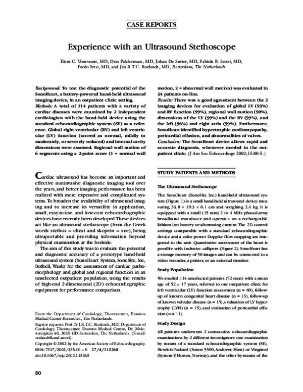
The SonoHeart (SonoSite Inc) hand-held ultrasound system (Figure 1) is a small hand-held ultrasound device measuring 33.8 × 19.3 × 6.1 cm and weighing 2.4 kg. It is
equipped with a small (15 mm) 2 to 4 MHz phased-array
broadband transducer and operates on a rechargeable
lithium ion battery or alternating current.The 2D control
settings comparable with a standard echocardiographic
device and a color power Doppler flow mapping are integrated to the unit. Quantitative assessment of the heart is
possible with inclusive callipers (Figure 2). SonoHeart has
a storage memory of 50 images and can be connected to a
video recorder, a printer, or an external monitor.
Study Population
We studied 114 unselected patients (72 men) with a mean
age of 52 ± 17 years, referred to our outpatient clinic for
left ventricular (LV) function assessment (n = 60), followup of known congenital heart disease (n = 13), follow-up
of known valvular disease (n = 15), evaluation of LV hypertrophy (LVH) (n = 15), and evaluation of pericardial effusion (n = 11).
Study Design
All patients underwent 2 consecutive echocardiographic
examinations by 2 different investigators: one examination
by means of a standard echocardiographic system (SE),
Hewlett-Packard (Sonos 5500, Andover, Mass) or Vingmed
(System V, Horten, Norway), and the other by means of the
�Journal of the American Society of Echocardiography
Volume 15 Number 1
Vourvouri et al 81
Table 1 Accuracy of left atrial size assessment (111
patients)
Ultrasound stethoscope
2D standard echo
<4 cm
4-6 cm
>6 cm
Normal
Enlarged
Severely enlarged
87
1
0
0
21
0
0
0
2
Kappa, 0.974
2D, Two-dimensional.
Table 2 Accuracy of left ventricular size assessment (111
patients)
Ultrasound stethoscope
2D standard echo
<5.5 cm
5.5-7 cm
>7 cm
Normal
Enlarged
Severely enlarged
91
0
0
1
19
0
0
0
0
Kappa, 0.969
2D, Two-dimensional
SonoHeart device. Each investigator was blinded to the
results of the other investigator.The time spent for the bedside examination with SonoHeart was always less than 5
minutes.
The patients were evaluated on-line for the following
parameters: presence of pericardial effusion, left atrial (LA)
enlargement (internal diameter >40 mm in the parasternal
long axis view), LV enlargement (end-diastolic diameter >55
mm in the parasternal long axis view),LVH (defined as a septal thickness >12 mm), right atrial (RA) enlargement (internal transverse diameter >35 mm in the 4-chamber view),
and right ventricular (RV) enlargement (end-diastolic transverse diameter >43 mm in the 4-chamber view).Also, gross
morphologic assessment of the valves was performed.
In all patients the global RV function and the LV ejection
fraction (EF), and in 34 patients, who where referred for
stress echocardiography, the regional wall motion were
estimated. With both imaging techniques, the scoring was
performed on-line during the examination procedure.
Global LV function was defined as normal when the estimated EF was greater than 55%, mildly to moderately
reduced when the estimated EF was 35% to 55%, and
severely reduced when the estimated EF was less than
35%.The scoring of the regional wall motion for testing the
efficacy and feasibility of the SonoHeart was simplified, by
dividing the LV into 6 segments (anterior, inferior, septal
posterior, septal anterior, lateral, and posterior wall) and by
using a 2-point score: 1 for normal wall motion, including
mild hypokinesia, and 2 for abnormal wall motion, including severe hypokinesia, akinesia, and dyskinesia.
Figure 1 Photograph of SonoHeart device, hand-held
ultrasound imager, used in this study.
Statistical Analysis
The agreement for segmental and global wall motion was
assessed from 2 × 2 and 3 × 3 tables with weighted kappa
statistics. Kappa values 0.4, between 0.4 and 0.75, and 0.75
or greater were considered to represent poor, fair-to-good,
and excellent agreement respectively, based on Fleiss’s
classification.1
RESULTS
Visualization
Three patients had poor echo windows with both
imaging techniques and were excluded from the
study, leaving 111 patients for analysis.
Agreement
The results for detection of LA and LV enlargement
with both examination techniques are summarized
in Tables 1 and 2.
The agreement for RV and RA size was 99%. A
thrombus in the RA was identified with both imaging techniques in 1 patient.
Pericardial effusion was correctly assessed with
SonoHeart in 11 patients and LVH (Figure 3) in 7 of
�Journal of the American Society of Echocardiography
January 2002
82 Vourvouri et al
A
B
Figure 2 Diastolic (A) and systolic (B) left ventricular long-axis views of normal subject obtained with
SonoHeart device. Measurements of diameter of left ventricle at end-diastole (A) and end-systole (B) are
shown. LA = Left atrium; LV = left ventricle; AO = aorta; RV = right ventricle; IVS = interventricular
septum.
transposition of the great arteries, and subvalvular
aortic membrane.
Evaluation of Global and Regional Left and
Right Ventricular Systolic Function
The agreement between both imaging techniques
for all 111 patients was 93% for global and 89% for
regional LV systolic function (Tables 3 and 4).
The agreement for global RV systolic function was
99%.
Interobserver Variability
Figure 3 Long-axis view of patient with hypertrophic cardiomyopathy obtained with SonoHeart device (abbreviations as in Figure 2). The arrow indicates the increased
septal thickness.
8, resulting in an agreement of 100% and 87.5%,
respectively. In 1 patient pericardiocentesis was performed under SonoHeart guidance (Figure 4).
In 20 patients, the morphologic abnormalities of
the aortic (calcification, Figure 5) and the mitral
valve (calcification, prolapse) were identified with
the SonoHeart device and confirmed by the SE.
Furthermore, the diagnosis of a chordal rupture of
the mitral valve was made by SonoHeart and verified
by the SE in a patient presenting with acute dyspnea
in the outpatient clinic.
Finally, the SonoHeart identified the morphologic
characteristics of surgically corrected congenital cardiac abnormalities in 13 follow-up patients, including
atrial septum defect (ASD) (Figure 6), ventricular
septum defect, tetralogy of Fallot, Ebstein anomaly,
There was a good correlation of the 2 independent
observers’ evaluations of regional wall motion
between the SE (81%) and the SonoHeart device
(76%).
DISCUSSION
In the late 1970s, Roelandt et al2-4 introduced the
first portable battery-powered 2D echocardiographic
instrument to be used as part of the clinical examination. Today, this vision of the past has become a
reality as a result of miniaturization and digital technology.
The SonoHeart device is easy to use, is ultraportable
with excellent imaging quality, and gives in addition
quantitative information about dimensions within the
heart. Our study demonstrates the utility of such a
device for assessing pathomorphology and function of
the heart, and the results are comparable with those
obtained by SE. We included in our study a group of
patients with corrected congenital heart disease to test
the clinical potential of the SonoHeart device in identifying the complex disease seen in these patients. It
has proven to be as reliable as the SE for the rapid
�Journal of the American Society of Echocardiography
Volume 15 Number 1
A
Vourvouri et al 83
B
Figure 4 Parasternal long-axis view of patient with pericardial effusion before (A) and after pericardiocentesis obtained with SonoHeart device (B) (abbreviations as in Figure 2; PE = pericardial effusion).
A
B
Figure 5 Parasternal (A) and apical (B) long-axis views of patient with calcified aortic valve stenosis with
SonoHeart device (abbreviations as in Figure 2).
detection or exclusion of a morphologic abnormality.
This does not indicate though that it can be substituted for the SE because most cardiac congenital diseases
require extensive 2D and Doppler analyses.
We proved that SonoHeart can become an extension of physical examination enhancing our clinical
senses and increasing the accuracy and sensitivity of
diagnosis. The ultraportability and ease of use suggest that similar data and results can be obtained in
other clinical scenarios, which are indicated in Table
5. It can be used as a screening tool for cardiac
abnormalities such as hypertrophic cardiomyopathy,
LVH, ASD, abdominal aorta aneurysm,5 mitral valve
prolapse (MVP), or enlargement of the heart. Prior
studies that used limited imaging protocols for most
of these diseases (hypertrophic cardiomyopathy,6
LVH,7-9 MVP10) have provided evidence that a limited imaging study is feasible.
The SonoHeart device can be used as a rapid diagnostic tool in emergency scenarios where rapid decisions are essential. Thus, giving immediate answers
about EF and regional wall motion abnormalities,
presence of pericardial fluid or tamponade, or the
cause of a new murmur in unstable patients, SonoHeart can play a decision-making role.
Taking into account that most of the patients are
referred for echocardiography to answer a single
clinical question (like follow-up of EF or pericardial
effusion after pericardiocentesis or search for the
source of embolism), a new strategy of limiting echo
services starts to emerge in daily clinical practice.11
Thus, a focused imaging protocol could be used to
screen for the referral disorder and to proceed to a
full study only in the discovery of any abnormality.
Such a limited echo strategy could be most effectively applied with the use of a hand-held ultrasound
device such as SonoHeart. Recently, Bruce et al5
demonstrated the efficacy and high accuracy of that
device in screening patients at risk for abdominal
aortic aneurysms.
The implementation of a limited echo or screening policy will lead to cost savings in terms of bene-
�Journal of the American Society of Echocardiography
January 2002
84 Vourvouri et al
A
B
Figure 6 A, Foreshortened apical 4-chamber view of patient with atrial septum defect of secundum type
(arrow) obtained with SonoHeart device (abbreviations as in Figure 2; RA = right atrium). B, Same view
obtained with SE system (Hewlett-Packard).
Table 3 Agreement between left ventricular ejection fraction estimated by the SonoHeart device and a standard
echocardiographic system
SonoHeart estimation of EF
Standard echo estimation of EF
>55%
35%-55%
<35%
>55%
35%-55%
<35%
61
5
0
1
31
2
0
0
11
Agreement, 93%.
Kappa, 0.871.
EF, Ejection fraction.
Number of patients: 111. The numbers inside the table express the number of patients.
Table 4 Agreement of regional wall motion analysis
between the SonoHeart and a standard echocardiographic
system
SonoHeart
Standard echo
Normal
Abnormal
Normal
Abnormal
92
15
5
92
Agreement, 90%.
Kappa, 0.80.
Number of patients: 34.
Number of segments: 204. The numbers inside the table express the number of segments.
fit to all subjects, reducing the time spent imaging
healthy patients.
Limitations
For the evaluation of regional wall motion, the LV
was divided into 6 segments and not into 16 seg-
Table 5 Clinical utility of the hand-held ultrasound device
Emergency department, CCU, and ICU
Hospital ward rounds
Outpatient clinic
Surgical theater
Cardiac catheter laboratory
Private office practice
ments, which is proposed by the American Society of
Echocardiography. Because this was an on-line
assessment and we had no recording facilities, we
limited the analysis in 6 myocardial segments. The
same segments were independently scored later
from the SE and compared with the data of the handheld device.The hand-held device cannot substitute
for the SE, but our purpose was to demonstrate the
feasibility and efficacy of the SonoHeart device as a
screening device in recognizing and distinguishing
normal from abnormal wall motion.Within this concept, the study proved that such small devices potentially could be used in acute coronary syndromes to
exclude/identify regional wall motion abnormalities.
Furthermore, because of the small number of
patients who were included, this study should be
considered as a pilot study for later wall motion
studies.
Another limitation is related to the fact that we
focused on 2D evaluation of anatomy and function of
the heart, without including the color flow modality.
Refinements in the technology of color power
Doppler flow mapping in the device since beginning
this study have made this now reliable, however.
In Europe, most of the studies are being performed by cardiologists. The SonoHeart instrument
should not be used by sonographers but by a trained
cardiologist. This study was performed by a junior
�Journal of the American Society of Echocardiography
Volume 15 Number 1
Vourvouri et al 85
staff member who had experience in echocardiography. Training and licensing for noncardiologists to
use these devices will become an important issue in
the future.
4.
Conclusion
5.
The hand-held ultrasound device, performing as a
“real” stethoscope, makes ultrasound imaging an
excellent tool immediately available for the diagnosis
and assessment of cardiac patients whenever cardiac
physical examination is indicated, improving the
health care service.
6.
7.
8.
We are grateful to Wim B. Vletter and Jackie McGhie for
their expert technical assistance and to Eric Boersma, PhD,
for statistical advice.
9.
REFERENCES
10.
1. Fleiss JL. Statistical methods for rates and proportions. 2nd
ed. New York: John Wiley; 1981.
2. Roelandt JRTC, Wladimiroff JW, Baars AM. Ultrasonic real
time imaging with a hand-held scanner, II: initial clinical
experience. Ultrasound Med Biol 1978;4:93-7.
3. Ligtvoet CM, Rijsterborgh H, Kappen L, Bom N. Real time
11.
ultrasonic imaging with a hand-held scanner, I: technical
description. Ultrasound Med Biol 1978;4:91-2.
Roelandt JRTC, Bom N, Hugenholtz P. The ultrasound cardioscope: a hand-held scanner for real-time cardiac imaging.
J Clin Ultrasound 1980;8:221-5.
Bruce CJ, Spittell PC, Montgomery SC, Bailey KR, Tajik AJ,
Seward JB. Personal ultrasound imager: abdominal aortic
aneurysm screening. J Am Soc Echoc 2000;13:674-9.
Weidenbener EJ, Krauss MD, Waller BF, Taliercio CP.
Incorporation of screening echocardiography in the preparticipation exam. Clin J Sport Med 1995;5:86-9.
Shep SG, Frohlich ED. Limited echocardiography for hypertensive left ventricular hypertrophy. Hypertension 1997;29:
560-3.
Black HR, Weltin G, Jaffe CC. Limited echocardiogram: a
modification of standard echocardiography for use in the routine evaluation of patients with systemic hypertension. Am J
Cardiol 1991;67:1027-30.
Shub C, Tajik AJ, Sheps SG. Value of two-dimensional echocardiography and Doppler examination in the assessment of
hypertensive patients: a pilot study. J Am Soc Echocardiogr
1995;8:280-4.
Kimura BJ, Scott R, Willis CL, DeMaria AN. Accuracy and
cost-effectiveness of single-view echocardiographic screening
for suspected mitral valve prolapse. Am J Medicine 2000;
108:331-3
Kimura BJ, DeMaria AN. Indications for limited echocardiographic imaging: a mathematical model. J Am Soc Echocardiogr 2000;13:855-61.
�
 Fabiola Sozzi