zyxw
zyx
zyx
zyxwvuts
zyxwvutsrqponm
zyxwvutsrqponm
zyxwvutsrqpon
Aust N Z J Obstei G y m 1 2001; 41: 1: 7 8 8 1
GYNAECOLOGICALONCOLOGY
Primary Bartholin gland carcinoma: a report of seven cases
Andreas Obermair,lJ Susanne Koller,l Alexander J Crandon,' Lewis Perrin.' and James L Nicklin'
Queensland Centrefor Gynaecological Cancel; Royal Women'sHospital.' Herston, Quolensland,Austmlia.
University Hospital Medical School Vienna, Department of Gynecology and
Vienna. Austria
SUMMARY
This study reviews our experience with 7 patients
with primary Bartholin gland cancer (BGC) treated
at the Queensland Gynaecological Cancer Centre
(QCGC) and compares this with previously p u b
lished data.
A retrospective clinicopathologic review of all
patients with primary BGC treated at QCGC from
1988 to 2OOO was performed. Of the 7 patients
treated, all underwent primary surgery and 5 of the
7 patients received radiotherapy postoperatively
All patients presented with a local swelling or a
lump.Two had associated discharge and 2 had asso
ciated pain. Of the 7 patients, 2.3 and 2 respectively
were classified as having Stage IB, I1 or I11 disease.
Five of the 7 patients had squamous cell carcinoma
(SCC), one had adenoid-cystic carcinoma and 1 had
a small-cellneuroendocrine cancer of the Bartholin
INTRODUCTION
Primary carcinoma of the Bartholin gland is a rare
malignant tumour comprising less than 1% of aU primary female genital tract tumours.l3 Because of the
low incidence, knowledge of management and factors
influencing the clinical outcome of patients with
Bartholin gland carcinoma is limlted. Consequently.
we reviewed our series of patients with primary
Bartholin gland carcinoma and report our experience
with this rive tumour in a Gynaecologfcal Cancer
Centre. We also present the flrst recorded case of
small cell carcinoma of the Bartholin gland.
gland. None of the patients with SCC developed
recurrent disease. The patient with adenoidcystic
carcinoma experienced local recurrences at 4 years
and again at 5 years and 3 months. Nine years after
primary treatment she was diagnosed with pul.
monary metastases. The patient with small-cellneuroendocrine cancer of the Bartholin @and was
considered tumour-free after operat ion. Thorough
imaging, including a CT scan of her chest, abdomen
and pelvis showed no evidence of disease. She died
1 year and three months after diagnosis from disseminated pulmonary disease.
We present the first report of small cell neuroendocrine cancer of the Bartholin gland. Therapeutic
principles in the management of vulva1 cancer at
other sites appear to be appropriate for manage
ment of BGC.
MATERIALS A N D METHODS
Patients
The records of all pattents dhgn&
with ~ r l v n ran
l
cer treated at the W<;C W e n Jnnimry izIlR nnd
March #K10 were rrvicwnf Fmm thew WM-&P H'F idrn
tUled all pntlents whose di-waw Pulnllcd the critrrln for
Bartholln gland canwr (tUW (1) the turnour art.- at
the site of the Hartholln gland. (ll)the tumoirr is m n
ststen?histologically with R p r l m r y n m p k ~ n of
t ths
Bartholin gland: (ill) them is no Pvldcnm Crc n prm-tous.
c o n m n t . or subsequent primary tumoirr d similar
histological type el-whcrr. A total of t mtlents ful
filled the crlterh for RCG and form the h i s td this
report. Petient charastcrist im arc l b t d In Table I
zyxwvuts
zyx
Treatment
Address To? correspondence
Andmas ObermaIr MD
Queensland Centre for CynaecologlcalCancer
Ned Hanlon Building. Royal Womens Hoapitai
Herston
Bueensland r10*,Australia
Andreas Obermair MD. Susanne Koller MD. Alexander J Crandon
MD. Lewis Perrin MD. James L Nicklin MD
Local treatment consisted of 8 radical hemiv-uI~w
tomy (n = 4) or of a wide local excision ( n :u
Dissection of the inguinofemoral lymph ncwlcs w a s
carried out in all but 2 of the pitrents Rea.sons for not
performing the inguinofemoral lymphadrncctom)
included medical masons (n 1) and ~pfii.~71
of the
patient (n = 1). A single patient m n p d pvrincal
�zyxwvutsrq
zyxwvut
zyxwvut
zyxwvuts
zy
zyxw
zyxwvuts
ANDREAS OBERMAIR
ET AL
radiotherapy (60 Gy) plus groin radiotherapy Three
patients received groin and pelvic radiotherapy
Radiotherapy was given as a direct perineal field (60
Gy) without a groin field (n = l), or with a groin field
(n = 3), and chemoradiation of the vulva with groin
fields was given in 1patient. Two patients did not have
any radiotherapy The 1988 International Federation of
Obstetrics and Gynaecology (FIGO) staging system for
vulvar cancer was used to classify stage of disease.
Treatment is listed in table 1.
During the study period 370 patients were treated by
the Queensland Centre for Gynaecological Cancer
(QCGC) for vulva1 carcinoma. The diagnosis of primary Bartholin gland cancer (BGC) was established in
7 of these 370 patients (1.9%). All 7 patients fulfiled
the criteria for a primary BGC and therefore none
Case
Age
Tumoursize
(mm)
.
.
1
63
2
66
mx4
3
49
35xmxm
4
71
22
5
44
65 x 40 x 30
6
45
30
7
55
45x35
14x13~4
were excluded from analysis? The mean age was 56.1
years (range 4p71 years).
The main presenting symptoms were a local
swelling or lump in all7 patients along with pain (n = 2)
or vaginal discharge (n = 2). The delay of diagnosis
ranged from 6 weeks to 2 years (Table 1). No patient was
found to have a second pathology but 1 was found to
have vulvar inlra-epithelial neoplasia 0
III on the
vulva.
Squamous cell carcinoma was noted in 5 of the 7
patients. Adenoid-cystic carcinoma was found in 1
patient and a small cell cancer of neuroendocrine origin also was noted in a single patient.
One patient (Case 3) had a history of 5 months local
swelling and underwent local excision of an encapsulated tumour with clear margins. She was diagnosed
with a small cell neuroendocrine-type caner of the
Bartholin gland on the right side with intact skin overlying the carcinoma.The histologicalfeatureswere typ
zy
zyxw
zyxwvutsrq
zyxwvut
RESULTS
Table 1 Patient characteristics
79
Duration of symptoms
before diapsosis
-
Histology
Stage
SCC
SCC
small cell,
neuroendocrine
SCC
Adenoidcystic
m
6 weeks
II
II
2 months
m
3 months
II
2 Years
IB
6 weeks
2 months
SCC
SCC
m
5 months
Follow-up
NED 5 years, 6 months
NED 6 years, 1month
Died 15months, disseminated disease
NED 3 years, 9 months
Rec # 1:4 years, local
Rec # 2: 5 years. 3 months, mom pubis
Rec # 3: 9 years, pulmonary metastam.
Alive with disease
NED 2 years
NED 5 months
NED = No evidence of disease, SCC = Squamous cell earcinoma, Rec = Recurrence
Table 2 Treatment details
Initialtreatment
surgicalmargins
Radiotherapy
Nod=
postoperative
1
Excision biopsy, radical
hemivulvectomy, bilateral
inguinofemoral node dissection
Clear but close (0.5mm)
Perineal field
Negative
2
Local radical excision,
chemoradiation
Involved
Chemoradiotherapy and fields
to perineum,both groins and pelvis
Not done
3
Local excision
Clear but close
Nil
Not done
4
Radical hemivulvectomy, bilateral
inguinofemoral node dissection
Clear but close (1.5 mm)
Field to lefl groin and hemipelvis
Left groin: 1 of 9
positive
Right groin:
negative
5
Radical local excision, bilateral
inguinofemoral node dissection
Clear
Field to vulva
Negative
6
Excision biopsy, radical hemivulvectomy, ipsilaterd inguinofemoral
node dissection
Clear
Nil
Negative
7
Excision biopsy, radical
hemivulvectomy, bilateral
inguinofemoral node dissection
Clear but close (6 nun)
Field to groins and pelvis
Lfft g r o k 3 Of 10
positive
Rightgroin:
negative
Case
�80
zyxwvut
zyxwvutsrqponm
zyxwvutsr
zyxwvutsrqp
zyxw
zyxwvutsr
ANWOG
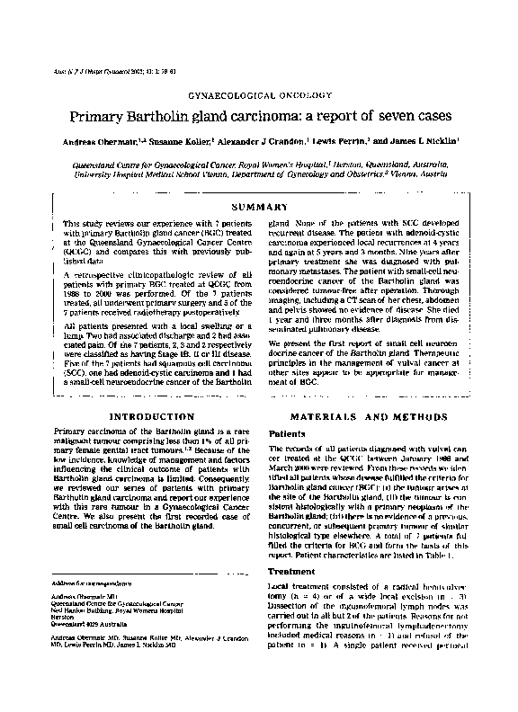
Figure 1A Small cell neuroendocrine-typecancer of the
Bartholin gland (40x).
Figure 1B Small cell neuroendocrine-typecancer of the
Bartholin gland (400~).
zyxwv
zyxwvutsrqp
zyxwvuts
ical of poorly differentiated small cell carcinoma with
neuroendocrine differentiation. The tumour displayed
sheets of poorly differentiated hyperchromatic malignant epithelial cells with scant cytoplasm and granular
chromatin. Abundant apoptotic debris and mitotic figures were seen (Figure 1). Tumour cells stained positively with Cam 5.2, neurone specific enolase and
synaptophysin, supporting the above diagnosis.
Further staging investigations included a CT scan of
the chest, the abdomen and the pelvis, as well as a bone
scan. The CT scan of the chest and the pelvis showed
some minor right inguinal lymphadenopathy which
measured 1 cm in diameter with no other enlarged
nodes and no soft tissue disease. The right inguinal
lymphadenopathy only occurred following the operation on the Bartholin gland, and therefore we considered it a postoperative reactive change. The bone scan
was normal. Full blood count and serum biochemistry
and thyroid function tests were normal. She refused all
further surgical therapy as well as adjuvant radiotherapy and chemotherapy. She died of disseminated pulmonary disease 15 months after diagnosis.
One patient (Case 5) had a 2-year history of local
swelling and had a simple excision of the Bartholin
gland showing adenoid-cystic carcinoma. Five weeks
later she underwent radical re-excision of the tumour
bed with bilateral inguinofemoral lymph node dissection. Surgical margins were clear and all nodes examined were negative. She was given postoperative
radiotherapy to the vulva consisting of 57.5 Gy in 23
fractions.
The first recurrence ocurred 4 years later in the
anterior vaginal wall and was treated by radical local
excision only. The second recurrence ocurred 1 year
and 3 months later on the mons pubis and was treated
by radical local excision and radiotherapy Nine years
after the primary treatment she presented with
breathlessness and a chest x-ray revealed multiple pulmonary metastases. She was alive with disease at the
time of preparation of this report.
DISCUSSION
This series reports our experience with BGC and presents the only known example of small cell neuroendocrine cancer of the Bartholin gland. There are
fewer than 350 cases of BGC in the entire world literature. This series of 7 patients contributes to the body
of understanding of this disease. In our series, BGC
accounted for 1.90.0of all vulvar cancers treated at the
Queensland Centre for Gynaecological Cancer
between January 1988 and March 2000. This incidence
is very similar to the 3 O t O noted by previous reports.”.“
In this series the first ever described case of a
small cell primary carcinoma of the Bartholin gland
is presented (Figure 1). Unlike earlier reports, the
majority of patients were diagnosed with a squamous
cell cancer (n = 5) with only a single case of adenoidcystic carcinoma. The diagnostic criteria described by
Wilkinson were required for a diagnosis of BGC.3That
is: (i) the tumour had to occur at the site of the
Bartholin gland: (ii) the histological type had to be
consistent with a primary Bartholin gland cancer: and
(iii) it could not be a metastatic deposit. These criteria
apply to all of the 7 patients reported in this analysis.
We did not apply the diagnostic criteria espoused
by Copeland’j and othet-~,’.~.~
which includes a ‘transition from normal gland or duct tissue to neoplastic tissue‘. This would have specifically excluded the patient
with small cell neuroendocrine cancer. This type of
malignancy is believed to arise from ubiquitous. solitary neuroendocrine-type cells scattered in the louer
genital tract. Therefore. there can be no transition
from normal to neoplastic.
Similar to the treatment of patients with vulva1
cancer generally. most authors would recommend radical excision of the primary lesion (with a margin of
at least 1 cm) plus inguinofemoral Iymphadenectomy.l
With BCG, the deep surgical margin is potentially
compromised by the proximity to the inferior pubic
ramus. Despite the extent of our surgery. surgical
�zyxwvutsrq
zyxwvutsrq
zyxwv
zyx
zyxwvutsr
zyxw
margins of > 1 cm were achieved in only 2 patients.
Four patients had clear surgical margins of < lcm,
and a single patient had an involved margin. This
would also explain the high rate of adjuvant radiotherapy used to treat the primary site. Inguinofemoral
lymphadenectomy was performed in 5 of the 7
patients with a bilateral lymphadenectomy in 4 of the
5 patients. In 1 patient (Case 6 ) only the ipsilateral
nodes were removed and proved to be negative. Pelvic
lymphadenectomywas not done in any of the patients.
In 3 patients the groin nodes were histologically
negative and they received no adjuvant radiotherapy.
In 2 patients the nodes were positive and both received
postoperative radiation to the primary site as well as
to both groins and the hemipelvis. Both patients had
SCC of the Bartholin gland and were tumour-free at 3
years and 9 months and at 5 months, respectively.
Interestingly,no patient with SCC of the Bartholin
gland experienced any form of recurrence. The
patient with small cell neuroendocrine cancer experienced early widespread systemic recurrence of disease, as is typical of this malignancy arising at other
sites. The single patient with adenoid-cystic carcinoma of the Bartholin gland had widespread
perineural invasion in the primary lesion. She developed 2 local recurrences despite radiotherapy. This
clinical behaviour is well described for this cell type.6
She developed late distant disease 9 years after initial
treatment.
In summary, we have presented 7 patients with
BGC, including the first report of small cell neuroen-
docrine cancer of the Bartholin gland. Therapeutic
principles in the management of vulval cancer at
other sites appear to be appropriate for management
of BGC.
ACKNOWLEDGEMENT
The authors would like to thank Jan Brady, QCGC for
assistance in data management.
REFERENCES
Hacker NF. Vulva Cancer, in:Berek JS. Hacker NF Eds.Practical
Gynecologic Oncology. 2nd ed. Baltimore, Williams and WiLkins
1994; 403439.
Burke TM.Eifel P. McGuire W, Wilkinson W.Vulva, In:Haskins
WJ, Perez CA, Young RC Eds. Principles and Practice of
Gynecologic Oncology. 2nd ed. Philadelphia, Lippinmtt Raven
1996; 717-751.
Wilkinson EJ. Premaligmnt and malignanttumorsof the vulva,
in Kurman RJ Ed. Blaustein’s Pathology of the female genital
tract. 4th ed. New York. Springer Verlag 1994.87-129.
C m s e n RJ.primary carcinoma of Bartholin’s gland. Am J Surg
zyx
zyxwvu
zy
1948; 75: 597-600.
Leuchter RS, Hacker NF. Voet RL, Berek JS, Townsend DE,
Lagasse LD. Primary carcinoma of the Bartholin gland a mport
of 14 cases and review of the literatun?. Obstet Gynecol1982; 60:
361368.
Copeland W, Sneige N, Gershenson DM, McGuffee VB. AWulKarim F, Rutledge FN. Bartholin gland carcinoma. Obstet
Gynecol186; 67: 794-801.
Trelford JD,Deos P H Bartholin’s gland carcinomas: five cases.
Gynecol Oncol1976: 4: 212-221.
Mossler JA, Woodard BH, Addison A, McCarty KS.
Adenocarcinoma of Bartholin‘s gland. Arch Pathol Lab Med 1980;
104: 5zs-526.
Wheelwk JB. Goplerud DR. Dunn U,Oates JF. Primary carcinoma of the Bartholin gland a report of ten cases. Obstet Gynecol
1984;63:l320424.
�
 Alex Crandon