Planta
DOI 10.1007/s00425-008-0697-1
O R I G I N A L A R T I CL E
Processing and traYcking of a single isoform of the aspartic
proteinase cardosin A on the vacuolar pathway
Patrícia Duarte · José Pissarra · Ian Moore
Received: 13 November 2007 / Accepted: 22 January 2008
Springer-Verlag 2008
Abstract Cardosin A is the major vacuolar aspartic
proteinase (APs) (E.C.3.4.23) in pistils of Cynara cardunculus L. (cardoon). Plant APs carry a unique domain, the
plant-speciWc-insert (PSI), and a pro-segment which are
separated from the catalytic domains during maturation but
the sequence and location of processing steps for cardosins
have not been established. Here transient expression in
tobacco and inducible expression in Arabidopsis indicate
that processing of cardosin A is conserved in heterologous
species. Pulse chase analysis in tobacco protoplasts indicated that cleavage at the carboxy-terminus of the PSI
could generate a short-lived 50 kDa intermediate which
was converted to a more stable 35 kDa intermediate by
removal of the PSI. Processing intermediates detected
immunologically in tobacco leaves and Arabidopsis seedlings conWrmed that cleavage at the amino-terminus of the
PSI either preceded or followed quickly after cleavage at its
carboxy-terminus. Thus removal of PSI preceded the loss
of the prosegment in contrast to the well-characterised
Electronic supplementary material The online version of this
article (doi:10.1007/s00425-008-0697-1) contains supplementary
material, which is available to authorized users.
P. Duarte · J. Pissarra
IBMC - Instituto de Biologia Molecular e Celular,
Universidade do Porto, Rua do Campo Alegre,
823, 4150-180 Porto, Portugal
P. Duarte (&) · I. Moore
Department of Plant Sciences, University of Oxford,
South Parks Rd, Oxford OX1 3RB, UK
e-mail: pduarte@ibmc.up.pt
J. Pissarra
Department of Botany, Faculty of Sciences, University of Porto,
Rua do Campo Alegre, 1191, 4150-181 Porto, Portugal
barley AP, phytepsin. PreprocardosinA acquired a complex
glycan and its processing was inhibited by brefeldin A and
dominant-inhibitory AtSAR1 or AtRAB-D2a mutants indicating that it was transported via the Golgi and that processing followed ER export. The 35 kDa intermediate was
present in the cell wall and protoplast culture medium as
well as the vacuole but the 31 kDa mature subunit, lacking
the amino-terminal prosegment, was detected only in the
vacuole. Thus maturation appears to occur only after sorting from the trans-Golgi to the vacuole. Processing or
transport of cardosin A was apparently slower in tobacco
protoplasts than in whole cells.
Keywords Arabidopsis · Dexamethasone · Nicotiana ·
Phytepsin · pOp6 LhGR · Rab1
Abbreviations
AP
Aspartic proteinase
ER
Endoplasmic reticulum
PSI
Plant speciWc insert
SAPLIP Saposin-like protein
Introduction
Aspartic proteinases (APs) (E.C.3.4.23) constitute one of
the four main classes of endoproteases, the other three
being serine, cysteine and metallo proteinases. APs are
widely present in nature, having been found in retroviruses,
bacteria, fungi, and plants as well as animals (Dunn 2002).
In spite of their widespread distribution, aspartic proteinases share common features, such as inhibition by pepstatin
A, an acidic pH optimum, and preferential cleavage of peptide bonds between residues with hydrophobic side chains.
These common features reXect the high degree of similarity
123
�Planta
between AP amino acid sequences. The tertiary structure of
these enzymes is usually bilobal, with two domains separated by a deep and large cleft where the active site is
located. Despite the overall similarity, substantial diVerences exist in catalytic properties, cellular localisation and,
consequently, in the biological functions of this family of
enzymes (Koelsch et al. 1994).
Eukaryotic aspartic proteinases are synthesised as zymogens which undergo proteolytic processing to the mature
and active form, whereas retroviral APs are synthesised as
part of a structural poly-protein (Koelsch et al. 1994). AP
precursors of eukaryotic origin are commonly made up of a
short signal peptide, which directs the protein to the endoplasmic reticulum (ER), a pro-segment (or activation segment) and a subsequent polypeptide that will compose the
mature protein (Khan and James 1998).
In the plant kingdom, aspartic proteinases have been puriWed and characterised from numerous species of gymnosperms, monocotyledons and dicotyledons (Mutlu and Gal
1999). In the Arabidopsis genome, 50 genes coding for APs
of the pepsin type were identiWed, and further assigned to
three diVerent categories: typical, nucellin-like, and atypical
proteinases (Faro and Gal 2005). Most of the available
knowledge on plant APs relates to those in the typical category and has been obtained mostly from the study of phytepsin from Hordeum vulgare (barley) seeds and cardosins
from the Xowers of Cynara cardunculus (cardoon) (Simões
and Faro 2004). Plant APs have been primarily isolated from
seeds but were also identiWed in other organs such as leaves,
roots and Xowers. Subcellularly, plant APs are mainly localised either in the vacuole/protein body or the extracellular
space (Mutlu and Gal 1999). Their biological functions,
however, are not as well assigned or characterized as those
of mammalian, microbial or viral APs (Dunn 2002). Nevertheless, plant APs have been implicated in protein processing and/or degradation in diVerent plant organs, as well as in
plant senescence, stress responses, programmed cell death
and reproduction (Simões and Faro 2004).
Cardosins are APs originally isolated from Cynara cardunculus L. (cardoon) pistils. Cardosins are expressed at
unusually high levels in cardoon Xoral organs where Cardosins A and B account for the majority of the total soluble
protein content (Ramalho-Santos et al. 1997). This is
unusual since most plant APs described to date were mainly
isolated from seeds or leaves, where they exist at much
lower concentrations (Mutlu and Gal 1999). The abundance
of cardosins in cardoon Xowers and their milk-clotting activity gives the proteins an economic signiWcance in Portugal
where the Xowers are traditionally used in cheese manufacture (Ramalho-Santos et al. 1996; Veríssimo et al. 1996).
Among plant vacuolar APs, barley phytepsin, is the best
characterised (Glathe et al. 1998; Kervinen et al. 1999;
Tormakangas et al. 2001). In keeping with other eukaryotic
123
APs, phytepsin is synthesised as a precursor which needs to
lose its signal peptide, pro-region and plant speciWc segment, to become mature (Glathe et al. 1998). Indeed,
phytepsin has been used as the model for maturation of
plant AP enzymes and for identiWcation of vacuolar sorting
determinants and routes (Glathe et al. 1998; Kervinen et al.
1999; Tormakangas et al. 2001; Pimpl et al. 2003).
The APs in plants are distinguished from those in
microbes by the presence of an additional segment of about
100 amino acids, the plant speciWc insert (PSI). The PSI
sequence shows no homology with mammalian or microbial AP domains, but is highly similar to that of saposins
and saposin-like proteins (SAPLIPs) which are lysosomal
sphingolipid-activator proteins (Simões and Faro 2004).
The maturation process of APs involves the removal of the
PSI and the Pro region to generate the mature molecules
(Mutlu and Gal 1999). Depending on the plant AP, this PSI
is either partially or completely removed during maturation.
For example, while mature phytepsin retains an N-terminal
portion of the PSI, cardosin A maturation involves the complete removal of both PSI and the Pro segment (Glathe
et al. 1998; Ramalho-Santos et al. 1998).
The biological function(s) of saposins have not yet been
completely established, however, it was already demonstrated that the correct sorting of cathepsin D, a mammalian
AP, to the lysosome, requires the association of this protein
with a saposin, namely saposin C (Zhu and Corner 1994;
Simões and Faro 2004). Additionally, the PSI of phytepsin
was shown to be essential for the correct vacuolar targeting
of this enzyme, since deletion of this region causes phytepsin’s secretion and also alters the way in which this AP
exits the ER (Tormakangas et al. 2001).
Although it is already documented that cardosin A is
extensively processed after synthesis, so far, very little is
known about the maturation process or the intracellular
location of processing events. Ramalho-Santos et al.
(1998), using antibodies raised against diVerent regions of
the protein, proposed a sequence of processing events in
maturation of cardosin A based on the analysis of two time
points in cardoon Xoral development. This sequence of
events diVers from that of phytepsin (Glathe et al. 1998) but
has not been conWrmed by direct methods. Furthermore,
nothing is known about the subcellular localisation of the
diVerent processing events to which cardosin A is subjected. The processed, mature form of cardosin A was,
however, shown to be located in the vacuoles (RamalhoSantos et al. 1997).
It is known that protein sorting to plant vacuoles is
dependent on a considerable variety of protein motifs and
this sorting can involve either traYc from the ER through
the Golgi apparatus or direct ER-to-vacuole transport since
there is evidence for the existence of a Golgi-independent
route to storage vacuoles directly from the endoplasmic
�Planta
reticulum (Levanony et al. 1992; Hara-Nishimura et al.
1998; Bassham and Raikhel 2000; Chrispeels and Herman
2000; Hadlington and Denecke 2000). The sorting pathway
followed by cardosin A from the ER towards the vacuole is
still unclear.
Transmission electron microscopy (TEM) observations
of developing cardoon pistils revealed swollen cisternae of
RER with similar appearance to cardosin A-containing vacuoles (Duarte et al. 2006). This observation led to the
hypothesis that cardosin A could reach the vacuole through
a Golgi independent route, although the presence of modiWed N-glycans indicates involvement of the Golgi complex
(Costa et al. 1997).
The work presented here further characterises the sorting
and processing of cardosin A using pulse-chase approaches
involving several expression systems in whole tissues or
protoplasts of heterologous species where other endogenous cardosins do not interfere with the analysis.
Materials and methods
Cardosin A cloning into the binary expression vectors
Standard methods were employed for molecular cloning
and bacterial culture (Sambrook et al. 1989). The original
cardosin A cDNA clone (Faro et al. 1999) was modiWed by
PCR with Platinum Pfx DNA Polymerase (Invitrogen), the
forward primer CdAFwd1: 5⬘AAAACTCGAGCCACCAT
GGGCACCTCAATCAAAGCAAACG3⬘ and the reverse
primer 5⬘CGGGTTGTATCTTAGATCGG3⬘. This introduced an optimal eukaryote translation initiation site (underlined) (Kozak 1984), two new restriction sites (Xho I and
Nco I), and a mutated nucleotide (bold) to remove a Kpn I
restriction site. The generated fragments were digested with
Ava I and Xho I, and the resulting 200-base pair (bp) fragment was inserted into the original construct pCR2.1-CdA
at Xho I and Ava I sites and conWrmed by sequencing. To
express cardosin A in tobacco leaves using Agrobacteriummediated transformation, the modiWed cardosin A cDNA
was digested with BamH I and Xho I, and further cloned
into the binary expression vector pVKH18-EN6 (Batoko
et al. 2000), between BamH I and Sal I sites. The modiWed
cardosin A cDNA was also digested with Ecl 136II and Xho
I and cloned between Sma I and Xho I restriction sites of the
binary expression vector pH-TOP for dexamethasoneinducible expression (Craft et al. 2005).
Agrobacterium-mediated transient expression
in Nicotiana tabacum
Agrobacterium-mediated transient expression of cardosin
A in Nicotiana tabacum L. cv. Petit Havana SR1 (provided
by Dr. P. Czernilofsky, MPI Cologne, Germany) was carried out as described in Batoko et al. (2000) with the following modiWcation: instead of YEB-medium, LB medium
supplemented with 25 �g/mL of kanamycin and 0.4 mM of
isopropyl-�-D-thiogalactopyranoside (IPTG) was used at all
times. For experiments requiring co-infection of more than
one construct, bacterial strains containing the constructs
were mixed before performing inWltration, with the titre of
each construct adjusted to the required Wnal OD600.
Dexamethasone treatments and GUS staining
of the Arabidopsis pOp/LhGR stable transformants
Arabidopsis thaliana L. ecotype Columbia (provided by
Dr. C. Dehio, MPI Cologne, Germany) plants of the 4c-S5
and 4c-S7 CaMV35S::LhGR activator lines were transformed and analysed according to Craft et al. (2005). Dexamethasone (Sigma-Aldrich) was dissolved at 100 mM in
dimethylsulfoxide (DMSO) and kept at ¡20°C. Typically,
20 �M dexamethasone was used for induction. All experiments were performed with T2 Arabidopsis seedlings,
induced either in liquid or solid media, as described elsewhere (Craft et al. 2005). Putative transformants were
tested for GUS expression through histochemical GUS
assays as described in JeVerson (1987), except that repeated
vacuum inWltrations were performed (up to 5£) which
resulted in more staining of internal mature leaf tissues. For
GUS staining purposes, dexamethasone induction was carried out for 2 days. For immunodetection of cardosin A,
induction periods varied from 3 to 7 days.
Protein extraction, endoglycosidase treatment,
and protein gel blot analysis
Protein extracts for endoglycosidase H (endoH) digestion
were prepared with 50–300 mg fresh weight of tobacco
infected leaf samples or Arabidopsis seedlings frozen in
liquid N2 according to the method described in Batoko et al.
(2000). EndoH digestions were carried out as described in
Batoko et al. (2000). For SDS-PAGE we used 12% acrylamide gels in the Mini-Protean 3 apparatus (Bio-Rad), and
electrophoresis was performed according to the Laemmli
method (Laemmli 1970). After electrophoresis, proteins
were transferred into polyvinylidene diXuoride (PVDF)
membranes for 1 h 30 min in transfer buVer, according to
the method described by Burnette (1981). The membrane
was blocked for 1 h at room temperature (RT) in TrisbuVered saline (TBS) containing 0.1% (v/v) Tween-20
(Sigma, Poole, UK) (TBS-T) with 5% (w/v) skimmed milk
powder, 1% (w/v) BSA (Fraction V; Roche Biochemicals)
and 0.1% (v/v) goat serum (Sigma). Rabbit anti-sera raised
against recombinant cardosin A (Faro et al. 1999),
calreticulin (gift from J. Denecke) and �-TIP (gift from
123
�Planta
A. SchaeVner) were used at 1:600, 1:10,000 and 1:200
dilutions respectively, in blocking solution, to probe the
membranes for 1 h at RT or over-night (O/N) at 4°C.
Alkaline-phosphatase conjugated secondary antibody
(Sigma) was used at 1:5,000 dilution in TBS-T for 30 min
at RT. Activity was revealed using the Sigma Fast
5-bromo-4-chloro-3-indolyl phosphate/nitroblue tetrazolium (BCIP/NBT) chromogenic reagent, according to
manufacturer’s instructions (Sigma).
Protoplast isolations
Isolation of tobacco mesophyll protoplasts was performed
using the method described by Denecke and Vitale (1995),
with some modiWcations.
Tobacco leaves were inWltrated with A. tumefaciens containing the pVKHEN6-cardosin A construct, and 24 h after
inWltration, leaf digestion was started. Leaves were treated
for 12–15 h, at RT and in the dark, with a mixture of 0.4%
(w/v) cellulase (Onozuka R10) and 0.2% (w/v)
macerozyme (Onozuka R10) dissolved in TEX medium,
supplemented with carbenicillin (200 �g/mL) and timentin
(2 �g/mL). The living protoplast fraction (Xoating) was
obtained after 2 centrifugations (100g 10 min at RT) in
TEX medium. The isolated fraction was then transferred
into a 15 mL falcon tube containing two volumes of mannitol/W5 [0.4 M D(¡)-mannitol, 1 mM D(+)-glucose,
30.8 mM NaCl, 25 mM CaCl2, 1 mM KCl, 0.3 mM Mes
(BDH Chemicals); pH 5.6–5.8] and gently mixed. Protoplasts were pelleted at 100g for 5 min at RT and the supernatant was carefully removed. TEX medium was added up to
4–5 mL, and the protoplasts were gently resuspended in
this volume. A measure of 10 �L of the cell suspension
were taken for cell counting with a haemocytometer under
a light microscope. The protoplasts resuspended in TEX
medium were allowed to Xoat freely, and the medium
underneath the cell layer was removed. The protoplast fraction was then resuspended in the appropriate TEX volume
in order to achieve the required cell concentration for the
subsequent experiments.
Arabidopsis protoplasts were isolated from 2-week-old
seedlings grown on inductive medium as described in
Zheng et al. (1997) with some modiWcations. Total seedlings were digested with 1% cellulase (Onozuka R10) and
0.25% of macerozyme (Onozuka R10) in MM buVer
(0.4 M mannitol and 20 mM Mes; pH 5.8) for 3 h in the
dark. The protoplast suspension was Wltered through a
100 �m nylon mesh and collected in 15 mL falcon tubes.
Protoplasts were pelleted at 65g for 5 min at 20°C, resuspended in 5 mL of MM buVer and centrifuged as before.
The puriWed protoplast pellet was then resuspended in the
appropriate MM volume in order to achieve the required
cell concentration for the subsequent experiments.
123
Metabolic labelling and pulse-chase experiments
Metabolic labelling of tobacco protoplasts was
performed as described by Denecke and Vitale (1995),
with some modiWcations. Protoplasts were labelled for
3 h with Trans35S-Label No-Thaw Metabolic Labelling
Reagent (ICN Biochemicals). For the labelling of
300 �L of cell suspension (ca. 1.2 £ 106 protoplasts),
20 �L of the labelling mixture (37,000 Bq/�L) diluted in
280 �L of TEX medium were used. The labelling reaction was performed in microfuge tubes kept vertically,
without agitation, at RT, and in the dark. After the labelling step, protoplasts were allowed to Xoat freely, and
the medium below the cell layer was collected and
replaced with approximately the same volume of TEX
chase medium supplemented with unlabelled 10 mM
methionine and 5 mM cysteine (Sigma). For BFA treatments the chase medium was supplemented with the
BFA solution (10 �g/mL) (Sigma).
At the appropriate chase time points, protoplasts were
resuspended and 100 �L of cell suspension taken. The protoplasts present in the suspension fraction were allowed to
Xoat freely, and the medium underneath the protoplast layer
was removed. A measure of 1 mL of 250 mM NaCl was
added before pelleting by centrifugation at 1,466g for 2 min
at 4°C. The resulting sediment was frozen in liquid N2 and
stored at ¡80°C until homogenization.
Tobacco protoplast pellets were resuspended and lysed
in four volumes of freshly prepared protoplast homogenization buVer [200 mM Tris–HCl pH 8.0, 300 mM NaCl, 1%
(v/v) Triton X-100, 1 mM EDTA] supplemented with the
appropriate volume of protease inhibitor cocktail (Sigma),
and submitted to two freeze/thaw steps. The tubes were
centrifuged at 15,493 g for 5 min at 4°C, and the colleted
supernatant was frozen in liquid N2 and stored at ¡80°C
until immunoprecipitation.
Immunoprecipitation
The protocol followed for immunoprecipitation was
described by Denecke and Vitale (1995). The primary
antibody was a rabbit antiserum raised against recombinant cardosin A (Faro et al. 1999) at a 1:200 dilution, for
2 h on ice. SDS-PAGE (see above) was followed by Wxation with a mixture of isopropanol:H2O:acetic acid
(25:65:1, by vol.) for 30 min, with agitation and in the
dark. This solution was discarded and the gel was agitated in the dark for 30 min with Amplify™ Fluorographic Reagent (Amersham Biosciences), rinsed with
deionised water for 10 min with agitation, dried on 3MM
paper, and exposed to HyperWlm™ MP (Amersham Biosciences) in a Kodak BioMax TranScreen LE (Amersham
Biosciences).
�Planta
Vacuole isolation and medium sampling
Tobacco vacuole isolation and quantiWcation of vacuole
recovery using �-mannosidase activity assays were performed according to the methods described by Hadlington
and Denecke (2001). Before loading the gel, cellular and
vacuolar protein extracts were equalized for �-mannosidase
activity. Arabidopsis vacuole isolation was performed as
described in Di Sansebastiano et al. (1998). To assay for
cardosin A secretion, the TEX medium in which the
tobacco protoplasts were incubated was collected from the
region between the Xoating protoplasts and the lysed protoplasts at the bottom of the tube and subjected to trichloroacetic acid (TCA) precipitation of proteins.
Results
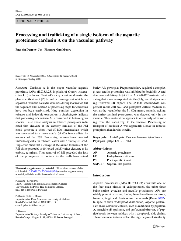
Cardosin A is stably accumulated in tobacco leaves
To follow the processing of a single cardosin isoform, a
cDNA encoding cardosin A was expressed in two diVerent
heterologous systems. In the Wrst, cardosin A was
expressed in tobacco leaf cells from an enhanced CaMV
35S promoter using Agrobacterium-mediated transient
expression (Batoko et al. 2000). This results in a rapid and
synchronous increase in gene expression that commences
approximately 30 h after inWltration of leaf tissue with the
bacteria (Zheng et al. 2005). In an alternative approach, the
pOp/LhGR dexamethasone-inducible promoter system
(Craft et al. 2005) was used to express cardosin A in
Arabidopsis seedlings.
Protein extracts made from inWltrated tobacco leaves
between 48 and 106 h post-inWltration (hpi) were analysed
by immunoblot using an antiserum raised against recombinant procardosin A. This antiserum detects epitopes in the
mature 31 kDa subunit (Fig. 1a) (Faro et al. 1999). As
shown in Fig. 1b, at 48 hpi the antibody detected a predominant 35 kDa form but a 66 kDa form was also detected.
The 66 kDa species disappeared over the subsequent 12 h,
followed by the 35 kDa form, while the 31 kDa mature
form accumulated and appeared to be stable for at least
106 hpi. These observations suggest that the 31 kDa subunit is generated by an initial cleavage at the N-terminus of
the PSI followed by removal of the amino-terminal pro segment. Cleavage at the N-terminus of the PSI must either
precede or follow closely upon cleavage at the C-terminus
as we rarely saw the 50 kDa intermediate predicted by initial cleavage of procardosin A at the C-terminus of the PSI.
To investigate cardosin A processing in Arabidopsis, we
introduced the cardosin A cDNA under control of a dexamethasone inducible promoter in plasmid pH-TOP
(Fig. 1c) (Craft et al. 2005). T2 seedlings were initially
screened for dexamethasone-inducible expression of a GUS
reporter construct borne on the same T-DNA (Fig. 1d).
Selected lines were then grown for 7 days in the presence
or absence of dexamethasone and were analysed for cardosin A expression by immunoblotting (Fig. 1e). In two GUSpositive lines, the 35 and 31 kDa forms of cardosin A were
detectable in seedlings grown on dexamethasone but were
absent from non-induced controls and from seedlings of a
GUS negative line. Thus, cardosin A can be processed to
the mature 31 kDa form in Arabidopsis seedlings as well as
in tobacco leaves. It was noticeable that after 48 h of incubation in dexamethasone, the precursor and intermediate
forms of cardosin A were already detectable, and that the
longer the induction period, the stronger was the signal for
cardosin A presence (Fig. 1f). In contrast to the tobacco
transient expression system, in Arabidopsis the mature
form of cardosin A did not predominate over the intermediate form even after 7 days of induction (Fig. 3b, bottom
panel). This may reXect either more rapid processing of the
intermediate form to the mature form in tobacco or the transient nature of the tobacco expression system.
Cardosin A expressed in both heterologous systems
is correctly sorted to the vacuole
To test whether cardosin A is ultimately targeted to the vacuole in these heterologous expression systems as it is in cardoon, protoplasts and vacuoles were prepared from
transfected tobacco leaves and Arabidopsis seedlings, and
were analysed by immunoblotting.
In preparations from tobacco leaves, the activity of the
vacuolar protein �-mannosidase was used to normalise
loading for the quantity of vacuolar protein (Fig. 2a). The
vacuolar preparations were apparently free from contaminating endomembranes as they lacked calreticulin, an abundant component of the endoplasmic reticulum (Fig. 2b).
The 31 kDa mature fragment of cardosin A was equally
abundant in the protoplasts and vacuoles indicating that the
majority of the mature protein present intracellularly is
indeed localised in the vacuole. The 35 kDa intermediate
form was also detected in the vacuole suggesting that cleavage occurs in this location to produce the 31 kDa form that
ultimately accumulates. Consistent with this, the ratio of
the 35kDa to 31 kDa form is slightly higher in the protoplast extract than in the vacuoles. Interestingly, the ratio is
signiWcantly higher still in the whole leaf samples. As discussed later, this suggests that some of the 35 kDa form
may exist in the cell wall of these samples.
As shown in Fig. 2c, in Arabidopsis seedlings, cardosin
A is also transported to the vacuole where the 35 and
31 kDa forms were both detected. Protoplast and vacuolar
preparations were normalised by probing for �-TIP
(Fig. 2d). Thus, conversion to the 31 kDa form appears to
123
�Planta
C
a
2kDa
4 kDa
Pre
Pro
31 kDa
DTG
C
CC
15 kDa
amino-terminal region
LB
b
CC
DSG
15 kDa
carboxy-terminal region
PSI
nos
RB
EN6
Hygr
CardosinA
p(A)
48hpi
54hpi
60hpi
72hpi
84hpi
96hpi
106hpi
Uninf.
66kDa
RuBisCO
35kDa
31kDa
pOp6 Promoter
c
LB
RB
(6xOp lac)
nos
Hygr
Cardosin A
GUS
p(A)
p(A)
-Dex
d
A6
A8
A4
A6
A8
+Dex
A4
- Dex
A6
e
A4
A8
A4
A4
+ Dex
A6
A6
A8
35kDa
31kDa
f
35kDa
31kDa
123
72 h
48 h
Dex
-
+
-
96 h
+
-
+
�Planta
䉳 Fig. 1 Expression of cardosin A in Nicotiana tabacum and in Arabidopsis thaliana. a Schematic representation of preprocardosin A and of
the processing events endured by the protein during maturation,
according to observations obtained while transiently expressing
cardosin A in tobacco leaves. The rabbit antiserum used in this work is
represented in purple and detects epitopes in the mature 31 kDa chain.
Pre signal peptide, Pro pro-segment, PSI plant speciWc insert. Glycans
are represented by (�). The six conserved cysteine residues present in
the PSI region of plant APs and in saposins are represented by (C). The
arrows mark preprocardosin A’s cleavage sites. b Schematic representation of the construct used for cardosin A expression in Nicotiana tabacum leaf epidermis via Agrobacterium and immunodetection of
cardosin A in tobacco samples from 48 h post-inWltration (hpi)
onwards. 48 hpi, the immature (66 kDa), intermediate (35 kDa) and
mature (31 kDa) forms of cardosin A could be observed (arrows). An
uninfected leaf was used as negative control (Uninf.). Cardosin A expressed in tobacco leaves is stably accumulated up to 5 days after inWltration. The band marked with an arrowhead is present in all samples
and corresponds to the large subunit of Rubisco (55 kDa), that cross
occur in the vacuole in Arabidopsis as well as in tobacco
cells. The relative abundance of the 31 and 35 kDa forms
was similar in vacuole and protoplast fractions, consistent
with the view that each species is resident in the vacuole.
The higher relative abundance of the 35 kDa form in Arabidopsis vacuoles compared to tobacco leaf vacuoles may
reXect slower processing in at least some Arabidopsis vacuoles or the sustained delivery of new 35 kDa precursor molecules to the vacuole together with turn over of the 31 kDa
form in the induced Arabidopsis seedlings.
Cardosin A N-glycans are complex and acquire partial
Endoglycosidase H (EndoH) resistance during transport
Cardosin A has two potential N-linked glycosylation sites
(at Asn 70 and Asn 363, Fig. 1a), and in cardoon extracts
both of these are occupied by oligosaccharides that are predominantly resistant to removal by endoglycosidase H
indicative of Golgi processing (Costa et al. 1997).
To determine whether cardosin A travels through the
Golgi when expressed in tobacco cells, transfected tissue
was analysed for endoH resistance at various times after
inoculation (Fig. 3a). At 48 hpi the 66 kDa procardosin A
form was visible and was converted by endoglycosidase H
treatment to two species of approximately 64 and 62 kDa
that are consistent with removal of one or two glycans,
respectively (Fig. 3a, 48 hpi). Thus, one of the two glycans
was endoglycosidase H resistant on approximately 50% of
cardosin A molecules indicating that at least 50% of molecules had passed through the Golgi and that procardosin A
can reach the Golgi prior to the Wrst cleavage. At 48 hpi, the
single glycan on the 35 kDa intermediate was also at least
50% endoglycosidase H resistant. The endoglycosidase H
resistant proportion of the 66 and 35 kDa forms was similar
at this time point so it is possible that the glycan on the
15 kDa form remained largely unmodiWed in this system.
reacts with the anti-recombinant cardosin A antiserum. The unspeciWc
labelling of Rubisco occurred in all Western blots and will be marked
in all Wgures. c Schematic representation of the construct used for the
generation of the pop/LhGR Arabidopsis stable transformants with
dexamethasone (Dex) inducible cardosin A expression. d GUS staining of T2 seedlings showing dexamethasone-induced GUS activity.
The GUS-positive lines (A4 and A6) were tested for cardosin A
expression upon addition of dexamethasone (e). Line A8 was negative
for GUS staining and was used as a negative control in further experiments. e Immunodetection of cardosin A in pOp/LhGR Arabidopsis
seedlings treated with dexamethasone. Samples were collected after
7 days of induction. The intermediate (35 kDa) and mature (31 kDa)
forms of cardosin A were detected in the two GUS-positive lines (A4
and A6) but were absent from the non-induced controls and from seedlings of the GUS-negative line A8. f After ca. 48 h of Dex induction,
cardosin A reaches levels detectable by western blotting. Immunodetection of cardosin A in Arabidopsis seedlings becomes more evident
with increased induction periods
At later time-points, the proportion of resistant molecules
increased and was approximately the same for the 35 and
31 kDa forms (Fig. 3a, 84 and 106 hpi).
Similar observations were made with Arabidopsis seedlings following induction of cardosin A expression with
dexamethasone (Fig. 3b). Three days after induction, cardosin A was found principally in the 35 kDa form, most of
which was resistant to endoglycosidase H activity (Fig. 3b,
d induction). After 5–7 days of induction, when signiWcantly more cardosin A had accumulated and the 31 kDa
form, most but not all of each form was resistant to endoglycosidase H (Figs. 3b, 5d, 7d induction).
Inhibition of ER-Golgi traYc leads to an accumulation
of procardosin A
To test the conclusion that cardosin A is transported to the
vacuole via the Golgi, we asked whether its transport was
dependent on two GTPases that are required for normal
ER-to-Golgi transport in tobacco epidermal cells (Andreeva et al. 2000; Batoko et al. 2000; Saint-Jore et al. 2002).
Dominant inhibitory forms of NtSAR1 and AtRAB-D2a
(ARA5, AtRab1b) inhibit the function of the respective
GTPases and inhibit ER to Golgi traYc of secreted, Golgilocalised, and vacuolar markers (Andreeva et al. 2000;
Batoko et al. 2000; Zheng et al. 2005). It is hypothesised
that NtSAR1 is required for COPII vesicle formation at the
ER and AtRAB-D2a for accurate vesicle targeting to the
Golgi.
Cardosin A was co-expressed with dominant inhibitory
AtRAB-D2a[NI] and NtSar1[HL] mutants. AtRAB-D2a[NI]
clearly slowed the processing of the 66 kDa form of procardosin A which was still present at 72 hpi although
almost absent by 48 hpi in controls (Fig. 4a). The appearance of the 31 kDa mature form was also delayed though
interestingly the accumulation of the 35 kDa form was
123
�Planta
a
Leaf Unif
Leaf CdA
Ppts
䉳 Fig. 2 Intracellular location of cardosin A in Nicotiana tabacum and
Vac
*
66kDa
35kDa
35kDa
b
Leaf
Cardosin A
-
Ppts
+
-
Vac
-
+
+
*
35kDa
31kDa
Calreticulin
c
Ppts
Dex
Vac
-
+
-
+
*
66kDa
31kDa
Ppts
d
+
Vac
-
+
-
a-TIP
barely aVected. Similar results were obtained with a dominant inhibitory mutant of a second member of the plant
Rab-D group (Duarte et al. unpublished observations). The
dominant inhibitory NtSar1[HL] mutant also delayed the
processing of procardosin A up to 72 hpi (Fig. 4b) though
its eVect was less pronounced than that of AtRAB-D2a[NI].
These results show that normal processing of procardosin A
is dependent on RAB-D2 and Sar1 activity, consistent with
transport to the Golgi in COPII-coated vesicles. It is also
consistent with the view that the Wrst processing event
occurs after export from the ER. As discussed later, the failure of either mutant to completely inhibit processing of pro-
123
cardosin A may simply reXect the probability of coexpression in the transfected cell population.
Brefeldin A inhibits the processing of cardosin A
in tobacco protoplasts
35kDa
Dex
in Arabidopsis thaliana. a Cardosin A expressed in tobacco leaves is
correctly sorted to the vacuole. Samples were collected 7 days after
inWltration and an uninfected leaf (Leaf Uninf) was used as negative
control. The intermediate (35 kDa) and mature (31 kDa) forms of cardosin A were observable in leaf (Leaf CdA), protoplast (Ppts) and vacuolar (Vac) extracts. The asterisk labels the BSA (67 kDa) present in
the vacuole buVer which cross-reacts with the anti-recombinant cardosin A antiserum. This was previously tested running a lane with vacuole buVer only (results not shown). The unspeciWc labelling of BSA
occurred in all lanes with vacuolar extracts and will be marked when
applicable, although it will not be referred again throughout this paper.
b Samples from tobacco leaves transfected with cardosinA (+) or from
mock transfections (¡) were collected 7 days after inWltration. The
intermediate (35 kDa) and mature (31 kDa) forms of cardosin A were
observable in leaf, protoplast (Ppts) and vacuolar (Vac) extracts. The
bottom panel shows an immunoblot of the ER marker calreticulin, with
approximately 60 kDa (arrow), which demonstrates the absence of
non-speciWc endomembranes in the vacuolar prep. c Immunodetection
of cardosin A in protoplasts (Ppts) and in vacuoles (Vac) isolated from
Arabidopsis pOp/LhGR seedlings showed that most of the intracellular
protein is located in the vacuolar compartment. Samples were collected
after 7 days of induction and the intermediate (35 kDa) and mature
(31 kDa) forms of cardosin A (arrows) were speciWcally detected in
the protoplasts and vacuoles isolated from dexamethasone treated
seedlings. d Immunodetection of the vacuolar marker �-TIP (� isoform
of the tonoplast intrinsic protein) with approximately 29 kDa (arrow)
was performed for all Arabidopsis extracts
To further test the involvement of the Golgi in procardosin
A transport and processing we investigated the eVect of the
fungal metabolite brefeldin A (BFA). In tobacco epidermal
and cultured cells, this drug causes the Golgi to fuse with
the ER inhibiting further anterograde transport of secreted
and vacuolar markers (Matsuoka et al. 1995; Boevink et al.
1998; Saint-Jore et al. 2002). Since BFA can be applied
homogeneously to populations of protoplasts, the eVect of
this inhibitor on processing of cardosin A was followed by
pulse-chase analysis of protoplasts from inWltrated leaves
(Fig. 5).
As shown in Fig. 5 at the end of the 3 h labelling period,
corresponding to 54 hpi, almost all cardosin A was in the
66 kDa precursor form. In control protoplasts, processing to
the 35 kDa form was detected within 3 h of chase and was
almost complete by 18 h. In contrast, in samples treated with
brefeldin A there was no detectable conversion of the 66 kDa
cardosin A precursor to the 35 kDa intermediate during the
18 h chase (Fig. 5). During the chase period, we also detected
the accumulation of a 50 kDa band that was much reduced
from the BFA treated lanes. This species was not detected in
immunoprecipitation from control transfections (Suppl. Fig. 1)
so it appears to be a processing intermediate of cardosin A
�Planta
a
Nicotiana tabacum
Arabidopsis thaliana
b
48hpi
EndoH
-
3d induction
+
-
66kDa
35kDa
33kDa
+
-
72hpi
+
-
secGFP
+
+
35kDa
31kDa
A6 +Dex
-
60hpi
b
5d induction
EndoH
35kDa
33kDa
84hpi
+
60hpi
+
66kDa
31kDa
-
48hpi
-
AtRab-D2a[NI]
62-64kDa
31kDa
EndoH
a
A6 +Dex
+
NtSAR1[HL]
-
Sar1 wt Sar1 HL
72hpi
-
Uninf.
Sar1 wt Sar1 HL
66kDa
35kDa
35kDa
33kDa
31kDa
33kDa
31kDa
29kDa
29kDa
35kDa
31kDa
A6 +Dex
106hpi
EndoH
35kDa
31kDa
-
7d induction
+
EndoH
33kDa
35kDa
29kDa
31kDa
-
+
33kDa
29kDa
Fig. 3 Cardosin A expressed in Nicotiana tabacum and in Arabidopsis thaliana exhibited partial EndoH resistance. a Extracts of tobacco
leaf tissues expressing cardosin A were incubated either with (+) or
without (¡) endoH and were analysed by SDS-PAGE and immunoblotting with the anti-recombinant cardosin A antiserum. Samples
were obtained 48, 84 and 106 h post-inWltration. The extracts tested for
endoH resistance are the same as in Fig. 1b. 48 hpi the precursor and
intermediate forms of cardosin A (arrows; 66 and 35 kDa) appear as
partially resistant to endoH digestion. At later stages the intermediate
and mature forms of the protein (arrow; 35 and 31 kDa) show almost
complete resistance to endoH action. b Protein extracts were obtained
from Arabidopsis pOp/LhGR seedlings subjected to diVerent induction
periods with Dex (3, 5 and 7 days), incubated either with (+) or without
(¡) endoH and further analysed by SDS-PAGE and immunoblotting
with the anti-recombinant cardosin A antiserum. After 5 days of Dex
induction, the intermediate and mature forms of cardosin A (arrows;
35 and 31 kDa) have signiWcantly accumulated and both appeared as
partially resistant to endoH digestion
that was not detected by immunoblotting of leaf extracts.
Unexpectedly, however, we did not detect any of the mature
31 kDa form (Fig. 5) even if the chase was extended to 24 h,
corresponding to 78 hpi (data not shown).
The 35 kDa form of cardosin A is secreted from protoplasts
Previous fractionation data (Fig. 2) had suggested that a
portion of the 35 kDa intermediate may be secreted during
transient expression in the tobacco leaf. To investigate the
sorting of cardosin A sorting in tobacco leaf cells, protoplasts expressing cardosin A were isolated from previously
Fig. 4 EVects of dominant-inhibitory mutants of diVerent GTPases involved in ER-Golgi traYcking over cardosin A transport and processing. a Co-expression of cardosin A in tobacco leaves with the AtRabD
dominant-negative mutant AtRab-D2a[NI] delayed the transport and
processing of the AP. Cardosin A and secGFP were expressed alone
(¡), or in the presence of the mutant (+). Expression of secGFP alone
was the negative control in these experiments. Samples were collected
48 hpi onwards; 60 and 72 hpi, accumulation of the precursor form of
cardosin A (arrow; 66 kDa) was still evident in the cases where the
mutant was present (+). In comparison to the controls, the overall
processing of cardosin A was clearly delayed in the samples where
co-expression of this AP and the dominant-inhibitory mutant was performed. b Co-expression of cardosin A alone (¡), with the wt
(SAR1wt), or with the dominant inhibitory mutant form of NtSAR1
(Sar1 [H74L]). Samples were collected 60 and 72 hpi and an uninfected leaf (Uninf.) was used as negative control; 72 hpi, accumulation
of the precursor form of cardosin A (arrow; 66 kDa) was still clearly
visible but only in samples where the mutant form of NtSAR1[HL] was
present
inWltrated tobacco leaves. Protoplast preparation was completed at 48 hpi at which time a sample of the medium and
the protoplast fraction was taken. The same samples were
taken 6 h later and all four were analysed by immunoblot.
Figure 6a and b show that cardosin A is detected in the
medium fraction within 6 h (54 hpi) indicating that a portion of the total protein synthesised is indeed secreted. The
absence of the 66 kDa precursor in the medium fraction at
54 hpi when it is abundant in the protoplasts argues against
release of cardosin A to the medium through rupturing of
protoplasts. The presence of the 35 kDa form in the
medium indicates that secretion from the protoplasts can
occur before processing to the 31 kDa form.
Cardosin A processing is slower in protoplasts
The observation that secretion occurs before processing to
the 31 kDa form is consistent with the view that processing
123
�Planta
0hr
-
BFA
3hr
-
+
18hr
-
+
+
66kDa
35kDa
Fig. 5 EVects of the fungal metabolite BFA over cardosin A transport
and processing. Protoplasts (1.2 £ 106) were labelled with 35S for 3 h
(pulse) and cardosin A was immunoprecipitated with anti-recombinant
cardosin A antiserum from samples collected at the indicated timepoints of chase, in the presence and absence of BFA. 18 h into the
chase period, and in the presence of BFA, all of cardosin A appeared
in the precursor form (arrow; 66 kDa). Conversely, at the same timepoint and in the absence of BFA, cardosin A processing was not
inhibited and most of cardosin A molecules were at an intermediate
processing stage (arrow; 35 kDa). In this set of experiments an additional band with 50 kDa (white block arrow) appeared, consistent with
the cleavage of cardosin A at the C-terminus of the PSI (see Fig. 1a)
a
b
Protoplasts
48hpi
54hpi
67hpi
Medium
48hpi
73hpi
54hpi
66kDa
35kDa
35kDa
Fig. 6 Cardosin A expressed in tobacco endures partial secretion.
a Immunodetection of cardosin A in tobacco protoplasts transiently
expressing cardosin A. Samples were collected at the indicated timepoints after inWltration. The precursor (65 kDa) and intermediate
(35 kDa) forms of cardosin A were present intracellularly at all the
tested time-points. b Immunodetection of cardosin A in the protoplast
medium. Extracellular accumulation of the 35 kDa intermediate was
evident. Altogether, the absence of the 66 kDa precursor and presence
of the 35 kDa intermediate indicate that most probably cardosin A is
secreted as the later form
occurs after the Golgi, in the vacuole or a prevacuolar compartment. However, this interpretation was complicated by
the unexpected observation that whenever cardosin A processing was monitored in protoplasts either by pulse-chase
(Fig. 5) or Western blotting (Fig. 6), the mature 31 kDa
vacuolar form was never observed even at time points when
its presence would be expected in intact cells in inWltrated
leaves (Fig. 1b). For example in inWltrated leaves, there was
signiWcant conversion of the 35 kDa form to the 31 kDa
form over 12 h (see Fig. 1b) whereas in the protoplast system no conversion was observed between 3 h, when the
35 kDa form was already visible, and 18 h (Fig. 5). Thus,
transport of cardosin A to the vacuole, its subsequent processing, or stability within the vacuole, appear to be
aVected by the protoplasting process. To investigate this
123
further approximately half the tissue from inWltrated leaves
were subjected to 16 h protoplasting from 53 to 69 hpi
while the other half was subjected to rapid protoplasting
between 63 and 68 hpi. A sample of intact leaf was taken at
67 hpi. Each sample was analysed by immunoblot for cardosin A processing (Fig. 7). In the sample subjected to protoplasting at 53 hpi, the 66 kDa form was still visible at
69 hpi and none of the mature form was visible (Fig. 7a). In
the intact leaf, the 66 kDa form was already gone at 67 hpi
and the major species was the mature 31 kDa form
(Fig. 7b). Similarly, in samples subjected to processing at
63 hpi, the predominant form of cardosin A in the protoplasts was the mature 31 kDa form (Fig. 7c). Thus, protoplasting appears to reduce the rate at which cardosin A is
processed to the mature form but does not prevent the
detection of pre-existing mature form in the vacuole. Again
the protoplasts prepared at 63–68 hpi showed a higher ratio
of 31 kDa to 35 kDa forms than the intact leaf tissue, consistent with the hypothesis that much of the 35 kDa form in
the intact leaf is apoplastic.
Discussion
Here we show that cardosin A, a soluble vacuolar aspartic
proteinase originally isolated from Cynara cardunculus
(Veríssimo et al. 1996), is correctly targeted to the vacuoles
and highly stable when expressed in two distinct heterologous systems: tobacco leaf epidermis and Arabidopsis
seedlings. Furthermore, our observations indicate that the
processing of cardosin A in these two expression systems
proceeds similarly, showing that the targeting and processing of cardosin A can occur through conserved mechanisms.
In cardoon, cardosin A is synthesised as a preproenzyme
of 66 kDa which is translocated into the RER where the
signal peptide (Pre) is excised and the protein becomes glycosylated at its two predicted N-glycosylation sites (Asn70
and Asn 363) (Costa et al. 1997). Procardosin A then
undergoes a sequence of proteolytic events to generate the
mature form of the enzyme (Ramalho-Santos et al. 1998).
In cardoon, the analysis is complicated by the presence of
at least Wve isoforms of vacuolar and secreted cardosins
(Pimentel et al. 2006).
In this work, the processing of a single cardosin A isoform was followed in two heterologous expression systems
and the resulting data suggested that cleavage at the N-terminus of the PSI preceded removal of the prosegment, producing the 35 kDa intermediate and the mature 31 kDa
forms, respectively. As the 50 kDa intermediate predicted
by initial cleavage of procardosin A at the C-terminus of
the PSI was rarely seen, cleavage at the N-terminus of the
PSI must either precede or closely follow upon cleavage at
�Planta
a
Ppts
early
b
Leaf
intact
c
66 kDa
66 kDa
35 kDa
35 kDa
35 kDa
31 kDa
31 kDa
31 kDa
66 kDa
Ppts
late
Fig. 7 Cardosin A processing is slower in protoplasts than in whole
tissues. a–c Immunodetection of cardosin A in tobacco leaves and protoplasts, transiently expressing cardosin A. a Samples were collected
53 hpi and isolation of protoplasts was carried out with a long digestion
of leaves (ca. 16 h). Only the precursor (66 kDa) and intermediate
(35 kDa) forms of cardosin A (arrows) are present. b Leaf sample collected 67 hpi. Both intermediate (35 kDa) and mature (31 kDa) forms
of cardosin A (arrows) are present in the leaf extract. c Samples were
collected 63 hpi and protoplasting was performed with a short digestion period (5 h). In the protoplast fraction, the mature form of cardosin
A (31 kDa) is the predominant (arrow)
the C-terminus. Interestingly, by pulse chase analysis in
tobacco protoplasts, where the transport or processing of
cardosin A appeared to be slower than in intact cells, we
did observe a 50 kDa intermediate that is consistent with
cleavage at the C-terminus of the PSI. This form accumulated during the Wrst 3 h of chase but was diminished at
18 h relative to the 35 kDa form, so it is likely to be an
early intermediate. Thus, it may be that cleavage at the
C-terminus of the PSI can occur before cleavage at its
N-terminus but if so these two events must follow closely
relative to the subsequent removal of the prosegment.
The results obtained in this study support the sequence
of processing events for cardosin A which had been previously inferred by Ramalho-Santos et al. (1998) from the
analysis of cardoon extracts. In contrast to procardosin A,
the Wrst processing steps of prophytepsin are cleavage
downstream of the prosegment and cleavage within the PSI
(Glathe et al. 1998). Removal of the PSI to generate the
mature forms is delayed by 24 h. For phytepsin it was
described that the presence of the prosegment is essential
for the control of unwanted proteolysis by this protein (Kervinen et al. 1999). The propetide and the N-terminus of the
protein are anchored in the active site cleft by ionic interaction between Lys11 and the catalytic aspartates 36 and 223
determining inactivation (Kervinen et al. 1999). This interaction is destabilised at lower pH, found in the vacuolar
compartment, leading to enzyme activation. The role of an
internal anchor to active site aspartates, assumed in animal
APs by Lys36 p (pepsin numbering) of the propeptide, is
played by Lys11 in the mature part of prophytepsin
(Kervinen et al. 1999). Homology studies between plant
APs suggest that this is the case for plant APs in general,
however, cardosin A does not Wt in this picture since it does
not possess a Lys11 homologue (Kervinen et al. 1999).
Nevertheless, the late removal of the prosegment in cardosin A, presumably upon arrival to vacuole, suggests that
this region may also take part in an inactivation strategy in
cardosin A. In fact, it was already described that for recombinant cardosin A to be active, all it takes is the partial
removal of the prosegment (Castanheira et al. 2005). Our
results, obtained in vivo, diVer however to the ones
described by Castanheira et al. (2005) on the processing of
recombinant procardosin A in vitro, that show the partial
excision of the pro segment prior to the incomplete removal
of the PSI. The authors suggest that complete maturation of
cardosin A in vivo requires the action of additional proteases or exopeptidases (Castanheira et al. 2005). Prophytepsin processing was also shown to be incomplete in vitro and
markedly distinct to the maturation pattern observed in the
in vivo situation (Glathe et al. 1998).
Our results indicate that not all plant APs are processed
similarly but the maturation process can be conserved in
heterologous species. It is worth noting too that in contrast
to the extensively studied phytepsin which is degraded in
the vacuoles of tobacco protoplasts (Tormakangas et al.
2001), cardosin A is stable for several days in vacuoles of
tobacco leaves, tobacco protoplasts, and Arabidopsis seedlings so it may be a better marker for such studies than
phytepsin.
In cardoon, both glycans become mostly resistant to
removal by endoglycosidase H (Costa et al. 1997) but in
tobacco and Arabidopsis extracts acquisition of endoglycosidase H resistance is less complete. In tobacco, the single
glycan on the 31 kDa subunit is eYciently converted to the
resistant form, particularly at later stages of transient
expression, though sensitive forms always remain. The
observation that not all of the N-glycans on the 31 kDa subunit acquire endoH resistance in either heterologous system
is in agreement to what had been previously described for
the homologous system (Costa et al. 1997). The 66 kDa
procardosin A is converted by endoglycosidase H to a mixture of 64 and 62 kDa forms in tobacco leaves indicating
that this form does reach the Golgi but that only one of the
two glycans can be rendered endoglycosidase H resistant.
As the proportion of 64 and 62 kDa endoglycosidase H
digestion products of procardosin A is similar to the proportion of resistant to sensitive forms of the 35 kDa intermediate at the corresponding time point, the simplest
interpretation is that the glycan on the 15 kDa subunit of
cardosin A remains unprocessed in the tobacco leaf system.
In contrast, in cardoon pistils, the N-glycan on the 15 kDa
subunit is almost fully converted to the endoglycosidase H
resistant form.
123
�Planta
Several observations indicate that processing of procardosin A occurs only after the protein has been exported from
the ER. Firstly, endoglycosidase H resistant forms of procardosin A can be detected. Secondly, the 66 kDa procardosin A does not undergo any processing in protoplasts
treated with brefeldin A which causes the ER and Golgi to
fuse and inhibits further anterograde transport (Saint-Jore
et al. 2002). Third, the processing of cardosin A is inhibited
by co-expression with dominant inhibitory forms of Sar1,
and Rab-D2 GTPases that inhibit ER to Golgi traYc via
diVerent mechanisms (Andreeva et al. 2000; Batoko et al.
2000; Takeuchi et al. 2000; Phillipson et al. 2001).
Although it cannot be excluded, it seems unlikely that these
disparate inhibitors of membrane traYc would all act additionally on an ER processing activity that converts procardosin A to its intermediate forms. Finally, we observed the
35 kDa intermediate in the vacuole fraction suggesting that
the Wnal step in processing occurs in this compartment.
The presence of endoglycosidase-H-sensitive forms of
the 35 and 31 kDa forms of cardosin A together with the
failure of the dominant-inhibitory Rab GTPases to completely inhibit processing of procardosin A could be interpreted as evidence for processing within the ER. However,
we propose that a simpler explanation, consistent with the
other data, is that conversion to the endoglycosidase H
resistant form is simply ineYcient, particularly when rates
of traYc are high and that the mutant proteins result in only
partial inhibition of transport simply because not all cells
are co-infected by bacteria harbouring the cardosin A and
inhibitory GTPase constructs. In support of this interpretation, it should be noted that cardosin A was expressed using
a particularly high bacterial titre (OD600 0.3) to aid immunodetection while the inhibitory GTPase mutants were
expressed using a 10-fold lower bacterial titre.
The relatively high expression levels employed in this
study may also explain why some cardosin A was missorted to the apoplast in the tobacco transient expression
system, as sorting to the vacuole is presumably a saturable
receptor-mediated process (daSilva et al. 2005). Similar
observations were made with protoplasts expressing
phytepsin (Tormakangas et al. 2001; daSilva et al. 2005).
The higher ratio of 35:31 kDa form in whole tissue vs. protoplasts suggests that it is the 35 kDa form which is
secreted, consistent with the view that the 31 kDa form
arises after the vacuolar and secretory pathways diverge at
the Golgi. Attempts to isolate pure apoplastic Xuid from
intact tobacco leaf tissue were unsuccessful but the 31 and
35 kDa forms could be detected in such extracts at similar
abundance to the intact leaf (data not shown).
Although we found that the protoplast incubation
medium contained only the 35 kDa form, we also found
that processing to the 31 kDa form was inhibited in protoplasts. Therefore, the protoplast system does not allow us to
123
establish whether the 31 kDa species is also secreted. It is
not clear whether the slower processing of cardosin A in
protoplasts reXects a speciWc property of this molecule or a
wider eVect of protoplasting on the traYcking or proteolytic activities of tobacco cells. An alternative possibility
that is consistent with much of the data is that cardosin A is
Wrst secreted as the 35 kDa intermediate and targeted to the
vacuole only after subsequent endocytosis from the apoplast. In this view, protoplasting would inhibit the subsequent endocytosis by dilution of the secreted 35 kDa
species or by interfering with endocytosis directly. While
we cannot exclude such a mechanism; we note that the
66 kDa ER-localised precursor persists longer in transfected protoplasts than intact leaves, arguing that the rate of
transport is indeed slower in protoplasts.
The PSI domain is present only in plant APs and its role
is yet to be determined. Its presence does not appear to be
essential for the maintenance of AP enzymatic activity
(Asakura et al. 2000; Tormakangas et al. 2001), however,
some data point to a possible role for the PSI in the correct
vacuolar sorting of some plant APs (Mutlu and Gal 1999;
Tormakangas et al. 2001; Simões and Faro 2004). This is
the case for barley phytepsin and for the soybean aspartic
proteinase soyAP2, but not for soyAP1 in which deletion of
the PSI has no eVect on the vacuolar targeting (Terauchi
et al. 2006).
The PSI sequence shows no homology with mammalian
or microbial APs, but is highly similar to that of saposins
and saposin-like proteins (SAPLIPs) (Simões and Faro
2004),
lysosomal
sphingolipid-activator
proteins.
Tormakangas et al. (2001) have shown that deletion of the
PSI domain of phytepsin, a model vacuolar AP, causes
eYcient secretion of the truncated version of the protein.
Phytepsin’s PSI is highly homologous to saposin C. In
mammalian cells, it has been demonstrated that a portion of
the synthesized prosaposin is associated with newly synthesized procathepsin D in the ER (Zhu and Corner 1994) and
both the proteins have been reported to be associated with
the membrane during their biosynthesis (Rijnboutt et al.
1991). The formation of the prosaposin–procathepsin D
complex could explain the proposed M6P-independent
lysosomal targeting of cathepsin D mediated by saposin C
(Glickman and Kornfeld 1993). Based on these facts Tormakangas et al. (2001) suggested that the targeting of
phytepsin to the plant vacuole may resemble events in the
mammalian system, with the exception that in plants the
saposin-like PSI and phytepsin are encoded as a single precursor. Although it is yet to be conWrmed that deletion of
cardosin A’s PSI domain would cause secretion of the
protein, Egas et al. (2000) showed that the PSI of procardosin A induces the release of content of vesicles with
membranes containing acidic phospholipids in a similar
way to saposin C. Therefore, it has been suggested that
�Planta
procardosin A binding to membranes is accomplished via
the PSI while being a part of the precursor protein (Egas
et al. 2000). The fact that the 35 kDa form was detected in
the vacuole of both species used in this study and also in the
cell wall of tobacco indicates that the PSI segment is covalently attached at the late Golgi and in most if not all cardosin A molecules delivered to the vacuole.
In conclusion, we have shown that cardosin A can be targeted to the vacuoles in diverse heterologous systems indicating that conserved mechanisms are likely to be involved.
Processing of cardosin A is also conserved in the native and
heterologous species but it appears to diVer from that of
phytepsin. As the cardosin family contains vacuolar and
secreted forms, heterologous expression of these proteins
and mutant derivatives may elucidate the targeting signals
and vesicular transport pathways that direct plant aspartic
proteases to their destinations.
Acknowledgments We are grateful to Jürgen Denecke (University
of Leeds, UK) for the gift of anti-calreticulin antibody, to Tony
SchaeVner (GSF Research Centre, München, Germany) for the antiAt-�-TIP antibody and to Sandro Vitale for advice on pulse chase and
immunoprecipitation. We thank an anonymous reviewer for constructive
comments that improved the manuscript. This research was supported
by the Portuguese Science and Technology Foundation— Fundação
para a Ciência e a Tecnologia (FCT), project POCTI/BME/39765/
2001. The corresponding author, Patrícia Duarte, was beneWciary of a
PhD grant from FCT.
References
Andreeva A, Zheng H, Saint-Jore C, Kutuzov M, Evans D, Hawes C
(2000) Organization of transport from endoplasmic reticulum to
Golgi in higher plants. Biochem Soc Trans 28:505–512
Asakura T, Matsumoto I, Funaki J, Arai S, Abe K (2000) The plant
aspartic proteinase-speciWc polypeptide insert is not directly related to the activity of oryzasin 1. Eur J Biochem 267:5115–5122
Bassham D, Raikhel N (2000) Unique features of the plant vacuolar
sorting machinery. Curr Opin Cell Biol 12:491–495
Batoko H, Zheng H, Hawes C, Moore I (2000) A Rab1 GTPase is required for transport between the endoplasmic reticulum and Golgi
apparatus and for normal Golgi movement in plants. Plant Cell
12:2201–2218
Boevink P, Oparka K, Cruz SS, Martin B, Beterridge M, Hawes C
(1998) Stacks on tracks: the plant Golgi apparatus traYcs on an
actin/ER network. Plant J 15:441–447
Burnette W (1981) Western blotting: electrophoretic transfer of proteins from sodium dodecyl sulphate polyacrylamide gels to
unmodiWed nitrocellulose and radiographic detection with antibody and radioiodinated protein A. Anal Biochem 112:195–203
Castanheira P, Samyn B, Sergeant K, Clemente JC, Dunn BM, Pires E,
Van Beeumen J, Faro C (2005) Activation, proteolytic processing, and peptide speciWcity of recombinant cardosin A. J Biol
Chem 280:13047–13054
Chrispeels M, Herman E (2000) Endoplasmic reticulum-derived compartments function in storage and as mediators of vacuolar
remodeling via a new type of organelle, precursor protease vesicles. Plant Physiol 123:1227–1233
Costa J, Ashford D, Nimtz M, Bento I, Frazão C, Esteves C, Faro C,
Kervinen J, Pires E, Veríssimo P, Wlodawer A, Carrondo M
(1997) The glycosilation of the aspartic proteinases from barley
(Hordeum vulgare L.) and cardoon (Cynara cardunculus L.). Eur
J Biochem 243:695–700
Craft J, Samalova M, Baroux C, Townley H, Martinez A, Jepson I,
Tsiantis M, Moore I (2005) New pOp/LhG4 vectors for stringent
glucocorticoid-dependent transgene expression in Arabidopsis.
Plant J 41:899–918
daSilva LLP, Taylor JP, Hadlington JL, Hanton SL, Snowden CJ, Fox
SJ, Foresti O, Brandizzi F, Denecke J (2005) Receptor salvage
from the prevacuolar compartment is essential for eYcient vacuolar protein targeting. Plant Cell 17:132–148
Denecke J, Vitale A (1995) The use of protoplasts to study protein synthesis and transport by the plant endomembrane system. Method
Cell Biol 50: 335–348
Di Sansebastiano G, Paris N, Marc-martin S, Neuhaus J (1998) SpeciWc accumulation of GFP in a non-acidic vacuolar compartment
via a C-terminal propetide-mediated sorting pathway. Plant J
15:449–457
Duarte P, Figueiredo R, Pereira S, Pissarra J (2006) Structural characterization of the stigma-style complex of Cynara cardunculus
(Asteraceae) and immunolocalization of cardosins A and B during Xoral development. Can J Bot 84:737–749
Dunn B (2002) Structure and mechanism of the pepsin-like family of
aspartic peptidases. Chem Rev 102:4431–4458
Egas C, Lavoura N, Resende R, Brito R, Pires E, Lima M, Faro C
(2000) The saposin-like domain of the plant aspartic proteinase
precursor is a potent inducer of vesicle leakage. J Biol Chem
275:38190–38196
Faro C, Gal S (2005) Aspartic proteinase content of the Arabidopsis
genome. Curr Protein Pept Sci 6:493–500
Faro C, Ramalho-Santos M, Vieira M, Mendes A, Simões I, Andrade
R, Veríssimo P, Lin X, Tang J, Pires E (1999) Cloning and characterization of cDNA encoding cardosin A, an RGD-containing
plant aspartic proteinase. J Biol Chem 274:28724–28729
Glathe S, Kervinen J, Nimtz M, Li G, Tobin G, Copeland T, Ashford
D, Wlodawer A, Costa J (1998) Transport and activation of the
vacuolar aspartic proteinase phytepsin in barley (Hordeum vulgare L.). J Biol Chem 273:31230–31236
Glickman JN, Kornfeld S (1993) Mannose-6-phosphate independent
targeting of lysosomal-enzymes in I-cell disease B-lymphoblasts.
J Cell Biol 123:99–108
Hadlington J, Denecke J (2000) Sorting of soluble proteins in the
secretory pathway of plants. Curr Opin Plant Biol 3:461–468
Hadlington J, Denecke J (2001) Transient expression, a tool to address
questions in plant cell biology. In: Hawes C, Satiat-Jeunemaitre B
(eds) Plant cell biology: a practical approach. Oxford University
Press, Oxford, pp 107–126
Hara-Nishimura I, Shimada T, Hatano K, Takeuchi Y, Nishimura M
(1998) Transport of storage proteins to protein storage vacuoles is
mediated by large precursor-accumulating vesicles. Plant Cell
10:825–836
JeVerson R (1987) Assaying chimeric genes in plants: the GUS gene
fusion system. Plant Mol Biol Rep 5:387–405
Kervinen J, Tobin G, Costa J, Waugh D, Wlodawer A, Zdanov A
(1999) Crystal structure of plant aspartic proteinase prophytepsin:
inactivation and vacuolar targeting. EMBO J 18:3947–3955
Khan A, James M (1998) Molecular mechanisms for the conversion
of zymogens to active proteolytic enzymes. Protein Sci 7:815–
836
Koelsch G, Mares M, Metcalf P, Fusek M (1994) Multiple functions of
pro-parts of aspartic proteinase zymogens. FEBS Lett 343:6–10
Kozak M (1984) Compilation and analysis of sequences upstream
from the translational start site in eukaryotic mRNAs. Nucleic
Acids Res 12:857–872
Laemmli U (1970) Cleavage of structural proteins during the assembly
of the head of bacteriophage T4. Nature 277:680–685
123
�Planta
Levanony H, Rubin R, Altschuler Y, Galili G (1992) Evidence for a
novel route of wheat storage proteins to vacuoles. J Cell Biol
119:1117–1128
Matsuoka K, Bassham D, Raikhel N, Nakamura K (1995) DiVerent
sensitivity to wortmannin of 2 vacuolar sorting signals indicates
the presence of distinct sorting machineries in tobacco cells. J Cell
Biol 130:1307–1318
Mutlu A, Gal S (1999) Plant aspartic proteinases: enzymes on the way
to a function. Physiol Plant 105:569–576
Phillipson B, Pimpl P, daSilva L, Crofts A, Taylor J, Movafeghi A,
Robinson D, Denecke J (2001) Secretory bulk Xow of soluble proteins is eYcient and COPII dependent. Plant Cell 13:2005–2020
Pimentel C, Pires E, Faro C, Rodrigues-Pousada C (2006) Genomic
organization and functional analysis of Cynara cardunculus L.
aspartic protease gene family. FEBS J 273:PP1071
Pimpl P, Hanton S, Taylor J, Pinto-da Silva L, Denecke J (2003) The
GTPase ARF1p controls the sequence-speciWc vacuolar sorting
route to the lytic vacuole. Plant Cell 15:1242–1256
Ramalho-Santos M, Veríssimo P, Faro C, Pires E (1996) Action on bovine �(s1-casein of cardosins A and B, aspartic proteinase from the
Xowers of the cardoon Cynara cardunculus L. Biochim Biophys
Acta 1297:83–89
Ramalho-Santos M, Pissarra J, Veríssimo P, Pereira S, Salema R, Pires
E, Faro C (1997) Cardosin A, an abundant aspartic proteinase,
accumulates in protein storage vacuoles in the stigmatic papillae
of Cynara cardunculus L. Planta 203:204–212
Ramalho-Santos M, Veríssimo P, Cortes L, Samyn B, Van Beeumen J,
Pires E (1998) IdentiWcation and proteolytic processing of procardosin A. Eur J Biochem 255:133–138
Rijnboutt S, Aerts H, Geuze HJ, Tager JM, Strous GJ (1991) Mannose
6-phosphate-independent membrane association of cathepsin-D,
glucocerebrosidase, and sphingolipid-activating protein in Hepg2
cells. J Biol Chem 266:4862–4868
Saint-Jore C, Evins J, Batoko H, Brandizzi F, Moore I, Hawes C (2002)
Redistribution of membrane proteins between the Golgi apparatus
123
and endoplasmic reticulum in plants is reversible and not dependent on cytoskeletal networks. Plant J 29:661–678
Sambrook J, Fritsch E, Maniatis T (1989) Molecular cloning: a laboratory manual. CSHL Press, Cold Spring Harbor
Simões I, Faro C (2004) Structure and function of plant aspartic proteinases. Eur J Biochem 271:2067–2075
Takeuchi M, Ueda T, Sato K, Abe H, Nagata H, Nakano A (2000)
A dominant negative mutant of Sar1 GTPase inhibits protein
transport from the endoplasmic reticulum to the Golgi apparatus
in tobacco and Arabidopsis cultured cells. Plant J 23:517–525
Terauchi K, Asakura T, Ueda H, Tamura T, Tamura K, Matsumoto I,
Misaka T, Hara-Nishimura I, Abe K (2006) Plant-speciWc insertions in the soybean aspartic proteinases, soyAP1 and soyAP2,
perform diVerent functions of vacuolar targeting. J Plant Physiol
163:856–862
Tormakangas K, Hadlington J, Pimpl P, Hillmer S, Brandizzi F, Teeri
T, Denecke J (2001) A vacuolar sorting domain may also inXuence the way in which proteins leave the endoplasmic reticulum.
Plant Cell 13:2021–2032
Veríssimo P, Faro C, Moir A, Lin Y, Tang J, Pires E (1996) PuriWcation, characterization and partial aminoacid sequencing of two
new aspartic proteinases from fresh Xowers of Cynara cardunculus L. Eur J Biochem 235:762–768
Zheng HQ, Wang GL, Zhang L (1997) Alfalfa mosaic virus movement
protein induces tubules in plant protoplasts. Mol Plant Microbe In
10:1010–1014
Zheng HQ, Camacho L, Wee E, Henri BA, Legen J, Leaver CJ, Malho
R, Hussey PJ, Moore I (2005) A Rab-E GTPase mutant acts
downstream of the Rab-D subclass in biosynthetic membrane
traYc to the plasma membrane in tobacco leaf epidermis. Plant
Cell 17:2020–2036
Zhu Y, Corner G (1994) Intermolecular association of lysosomal protein precursors during biosynthesis. J Biol Chem 269:3846–3851
�
 José Pissarra