Academia.edu no longer supports Internet Explorer.
To browse Academia.edu and the wider internet faster and more securely, please take a few seconds to upgrade your browser.
Tyrosine residues in phospholipase Cγ2 essential for the enzyme function in B-cell signaling
 Jeronimo Bravo
2001
Free related PDFsRelated papers
Free PDF

Expression and Function of Tec, Itk, and Btk in Lymphocytes: Evidence for a Unique Role for Tec
Molecular and Cellular Biology, 2004
Free PDF

BANK regulates BCR-induced calcium mobilization by promoting tyrosine phosphorylation of IP(3) receptor
The EMBO journal, 2002
B-cell activation mediated through the antigen receptor is dependent on activation of protein tyrosine kinases (PTKs) such as Lyn and Syk and subsequent phosphorylation of various signaling proteins. Here we report on the identification and characterization of the B-cell scaffold protein with ankyrin repeats (BANK), a novel substrate of tyrosine kinases. BANK is expressed in B cells and is tyrosine phosphorylated upon B-cell antigen receptor (BCR) stimulation, which is mediated predominantly by Syk. Overexpres sion of BANK in B cells leads to enhancement of BCR-induced calcium mobilization. We found that both Lyn and inositol 1,4,5-trisphosphate receptor (IP(3)R) associate with the distinct regions of BANK and that BANK promotes Lyn-mediated tyrosine phosphorylation of IP(3)R. Given that IP(3)R channel activity is up-regulated by its tyrosine phosphorylation, BANK appears to be a novel scaffold protein regulating BCR-induced calcium mobilization by connecting PTKs to IP(3)R. Because...
Free PDF
Regulation of antigen receptor function by protein tyrosine kinases
Progress in Biophysics and Molecular Biology, 1999
Free PDF
The Direct Recruitment of BLNK to Immunoglobulin Couples the B-Cell Antigen Receptor to Distal Signaling Pathways
Molecular and Cellular Biology, 2002
Free PDF

Signal transduction by the high-affinity immunoglobulin E receptor FcεRI: Coupling form to function
Advances in Immunology, 2001
Free PDF

The role of adaptor proteins in lymphocyte activation
Molecular Immunology, 2004
Free PDF

Subcellular localization of Grb2 by the adaptor protein Dok-3 restricts the intensity of Ca2+ signaling in B cells
The EMBO Journal, 2007
Spatial and temporal modulation of intracellular Ca2+ fluxes controls the cellular response of B lymphocytes to antigen stimulation. Herein, we identify the hematopoietic adaptor protein Dok-3 (downstream of kinase-3) as a key component of negative feedback regulation in Ca2+ signaling from the B-cell antigen receptor. Dok-3 localizes at the inner leaflet of the plasma membrane and is a major substrate for activated Src family kinase Lyn. Phosphorylated Dok-3 inhibits antigen receptor-induced Ca2+ elevation by recruiting cytosolic Grb2, which acts at this location as a negative regulator of Bruton's tyrosine kinase. This leads to diminished activation of phospholipase C-gamma2 and reduced production of soluble inositol trisphosphate. Hence, the Dok-3/Grb2 module is a membrane-associated signaling organizer, which orchestrates the interaction efficiency of Ca2+-mobilizing enzymes.
Free PDF

THE JOURNAL OF BIOLOGICAL CHEMISTRY
© 2001 by The American Society for Biochemistry and Molecular Biology, Inc.
Vol. 276, No. 51, Issue of December 21, pp. 47982–47992, 2001
Printed in U.S.A.
Tyrosine Residues in Phospholipase C␥2 Essential for the Enzyme
Function in B-cell Signaling*
Received for publication, August 8, 2001, and in revised form, October 9, 2001
Published, JBC Papers in Press, October 17, 2001, DOI 10.1074/jbc.M107577200
Rosie Rodriguez, Miho Matsuda, Olga Perisic‡, Jeronimo Bravo‡, Angela Paul, Neil P. Jones,
Yvonne Light, Karl Swann§, Roger L. Williams‡, and Matilda Katan¶
From the Cancer Research Campaign Centre for Cell and Molecular Biology, Chester Beatty Laboratories, the Institute of
Cancer Research, Fulham Rd., London SW3 6JB, United Kingdom, the ‡Medical Research Council Laboratory of
Molecular Biology, Medical Research Council Centre, Hills Rd., Cambridge CB2 2QH, United Kingdom, and the
§Department of Anatomy and Developmental Biology, University College, Gower St., London WC1 6BT, United Kingdom
The hydrolysis of phosphatidylinositol 4,5-bisphosphate by
phosphoinositide-specific phospholipase C occurs in response to
a large number of extracellular signals (reviewed in Refs. 1– 4).
Four families of mammalian phosphoinositide-specific phospholipase C (PLC),1 PLC (1–4), PLC␥ (␥1, ␥2), PLC␦ (␦1␦4), and PLC⑀, have been described. Each family is characterized by the distinct domain organization and type of signaling
* This work was supported by an Institute of Cancer Research studentship (to R. R.), a Cancer Research Campaign grant (to M. K.), and
a Medical Research Council grant (to R. L. W.). The costs of publication
of this article were defrayed in part by the payment of page charges.
This article must therefore be hereby marked “advertisement” in accordance with 18 U.S.C. Section 1734 solely to indicate this fact.
¶ To whom correspondence should be addressed. Tel.: 44 207 352
8133; Fax: 44 207 352 3299; E-mail: matilda@icr.ac.uk.
1
The abbreviations used are: PLC, phospholipase C; BCR, B-cell
receptor; PBS, phosphate-buffered saline; GFP, green fluorescent protein; EGF, epidermal growth factor; GST, glutathione S-transferase;
SH2 and SH3, Src homology 2 and 3, respectively; PH, pleckstrin
homology.
pathways that regulate enzyme activity.
PLC␥ isoforms are mainly regulated through receptors with
intrinsic tyrosine kinase activity (e.g. growth factor receptors)
or receptors (such as B- and T-cell antigen receptors) that are
linked to the activation of nonreceptor tyrosine kinases
through a complex signaling network (3–5). The two isoforms of
PLC␥ have distinct tissue distributions; whereas PLC␥1 is
expressed ubiquitously, the pattern of expression of PLC␥2 is
characterized by high levels in cells of hematopoietic origin.
Transgenic studies suggested that the biological function of
these isoforms is reflected in their cellular distribution. Thus, a
deficiency in PLC␥1 is embryonic lethal in mice (6), whereas
homozygous disruption of PLC␥2 allowed normal development
but resulted in functional and signaling disorders in a subset of
cell types including B-cells, platelets, and mast cells (7).
The importance of PLC␥2 in signaling in B-cells has not only
been documented in experiments using transgenic animals deficient in PLC␥2 (7) but also by studies of a chicken B-cell
lymphoma cell line (DT40) (reviewed in Refs. 8 and 9) with the
property of extraordinarily high frequency of homologous recombination when DNA constructs are introduced into the
cells. Generation of a number of targeted mutations in specific
genes in DT40 cells provided valuable information about signaling components linking the activation of the B-cell receptor
(BCR) to an increase in intracellular calcium concentrations.
Using this system, it has been found that protein-tyrosine
kinases from Src, Tec (e.g. Btk), and Syk/ZAP70 families are
essential signaling components of the BCR pathway (10, 11). In
addition, an adapter BLNK (B-cell linker protein), inositol
1,4,5-trisphosphate receptors, and PLC␥2 itself were required
for calcium responses triggered by the BCR (12–15). Although
each of these components may have more than one function and
could be integrated in different pathways in B-cells, the current
model (8, 9) suggests that the Src family kinase Lyn interacts
with BCR and becomes activated upon the receptor aggregation. Activation of Syk kinase results in phosphorylation of
BLNK that could provide binding sites for PLC␥2 and a number of other proteins. Syk, together with Btk, has also been
implicated in phosphorylation of PLC␥2, which, through inositol 1,4,5-trisphosphate production, results in calcium mobilization. A similar pathway seems to be involved in calcium responses to oxidative stress after exposure of B-cells to hydrogen
peroxide (16 –18). It has been reported that the BCR complex
and tyrosine kinases Syk, Lyn, and Btk, are components required for calcium responses. In addition, phosphorylation of
several protein components, including BLNK and PLC␥2, has
been described.
Despite extensive genetic dissection of B-cell signal transduction, it has not been shown which tyrosine kinase(s) directly
47982
This paper is available on line at http://www.jbc.org
Downloaded from http://www.jbc.org/ by guest on March 23, 2016
Phospholipase C␥ (PLC␥) isoforms are regulated
through activation of tyrosine kinase-linked receptors.
The importance of growth factor-stimulated phosphorylation of specific tyrosine residues has been documented for PLC␥1; however, despite the critical importance of PLC␥2 in B-cell signal transduction, neither the
tyrosine kinase(s) that directly phosphorylate PLC␥2
nor the sites in PLC␥2 that become phosphorylated after
stimulation are known. By measuring the ability of human PLC␥2 to restore calcium responses to the B-cell
receptor stimulation or oxidative stress in a B-cell line
(DT40) deficient in PLC␥2, we have demonstrated that
two tyrosine residues, Tyr753 and Tyr759, were important
for the PLC␥2 signaling function. Furthermore, the double mutation Y753F/Y759F in PLC␥2 resulted in a loss of
tyrosine phosphorylation in stimulated DT40 cells. Of
the two kinases that previously have been proposed to
phosphorylate PLC␥2, Btk, and Syk, purified Btk had
much greater ability to phosphorylate recombinant
PLC␥2 in vitro, whereas Syk efficiently phosphorylated
adapter protein BLNK. Using purified proteins to analyze the formation of complexes, we suggest that function of Syk is to phosphorylate BLNK, providing binding
sites for PLC␥2. Further analysis of PLC␥2 tyrosine residues phosphorylated by Btk and several kinases from
the Src family has suggested multiple sites of phosphorylation and, in the context of a peptide incorporating
residues Tyr753 and Tyr759, shown preferential phosphorylation of Tyr753.
Important Tyrosine Residues in PLC␥2
EXPERIMENTAL PROCEDURES
Generation of DT40 Cell Lines Expressing Human PLC␥2—For the
expression of human PLC␥2 in DT40 cells, the full-length cDNA (22)
was subcloned into the pApuro vector as described previously (23). The
original sequencing data contained a sequencing error close to the C
terminus; the corrected frame of the amino acid sequence shows good
alignment with the rat PLC␥2 sequence in this region (Fig. 1B).
The mutations of tyrosine residues, Y753F, Y759F, and Y753F/
Y759F, were generated using a two-stage PCR-based overlap extension
method and introduced into pApuro/PLC␥2 construct. The plasmids
were used for stable transfection of DT40/PLC␥2⫺ cells (15) as described previously for the wild type PLC␥2 (23). Briefly, the linearized
constructs of PLC␥2 were introduced into the cells by electroporation
(950 V, 25 microfarads, ⬁ ⍀), and puromycin (0.35 g/ml) was added to
the medium. 10 –12 days after the selection, colonies were picked, and
the puromycin selection was repeated for 5– 8 days. Subsequently, the
puromycin-resistant colonies were grown in normal medium (RPMI
1640 medium supplemented with 10% (v/v) fetal bovine serum (Life
Technologies, Inc.) and 1% (v/v) chicken serum (Life Technologies)), and
the expression of PLC␥2 was confirmed by Western blotting.
Analysis of PLC␥2 Phosphorylation and Calcium Responses in DT40
Cell Lines after Stimulation—DT40 cell lines (DT40 PLC␥2-deficient
cells and these cells stably transfected with either the wild type or
Y753F, Y759F, and Y753F/Y759F mutants of human PLC␥2) were
stimulated by the addition of either anti-chicken IgM or H2O2. Typically, a 6 ⫻ 106-cell aliquot of these cell lines was stimulated with 10
g/ml goat anti-chicken IgM (M4) (Universal Biologicals) or 2 mM H2O2
at 37 °C for up to 5 min in 200 l of PBS. The cell pellet was resuspended in 200 l of lysis buffer (1% Triton, 150 mM NaCl, 10 mM Tris,
pH 7.4, 1 mM EDTA, 1 mM EGTA, 0.2 mM Na3VO4, 0.5% Nonidet P-40,
protease inhibitor mixture (Roche), and phosphatase inhibitor mixture
(Sigma)), and the cells were lysed by incubation for 30 min at 4 °C. The
supernatant was removed and added to anti-PLC␥2 antibody-protein G
complexes (prepared by mixing 2 g of anti-PLC␥2 antibody (Santa
Cruz Biotechnology, Inc., Santa Cruz, CA) with 9 l of protein G
(Roche)) and incubated at 4 °C for 1.5 h. After the incubation, immunocomplexes were washed, resuspended in SDS gel loading buffer, and
subjected to SDS-PAGE (7.5% polyacrylamide gels) and subsequent
Western blotting. For detection of PLC␥2 in immunocomplexes or in cell
extracts and membrane fractions (prepared as described in Ref. 14), the
anti-PLC␥2 antibody (1:4000; Santa Cruz Biotechnology) was used,
while the detection of PLC␥2 phosphorylation present in immunocomplexes was performed using anti-phosphotyrosine antibody (1:1000;
Transduction Laboratories). After incubation with the secondary antibody (anti-rabbit or anti-mouse Ig horseradish peroxidase-linked whole
antibody from Amersham Biosciences, Inc., diluted 1:3000), the visualization was performed using the enhanced chemiluminescence (ECL)
system (Amersham Biosciences). Total protein transferred to polyvinylidene difluoride membrane was stained using Amido Black solution.
For detection of BLNK in PLC␥2 immunoprecipitates, mouse anti-
chicken antibody (described in Ref. 14) was used for Western blotting;
the same protein was detected using anti-phosphotyrosine antibody.
For measurements of intracellular calcium concentrations in DT40
cells, a cell suspension containing 5 ⫻ 106 cells was loaded with 2 M
Fluo-3 AM (Molecular Probes, Inc., Eugene, OR) in RPMI medium for
1 h at room temperature. The cells were washed with PBS, resuspended
in RPMI medium, and stimulated with 10 g/ml M4 antibody or 2 mM
H2O2, and their calcium mobilization was simultaneously measured at
40 °C, with constant stirring in a LS-50B fluorimeter (PerkinElmer Life
Sciences). The excitation wavelength was 490 nm, and emission was
monitored at a wavelength of 535 nm.
Immunofluorescence Confocal Microscopy—Localization of PLC␥2
constructs containing a GFP tag was analyzed after transfection of
A431 cells, exactly as described previously (23), using EGF for stimulation. Similar experiments were performed with A20 B-cells, microinjected with the PLC␥2 plasmids. The images were recorded before and
after stimulation of A20 cells with anti-IgG2a (30 g/ml).
Constructs for Expression of Recombinant Proteins—For expression
of the full-length PLC␥2 protein containing the His6 tag at the C
terminus, the DNA fragment encoding the PLC␥2 sequence (with the
PCR-generated tag) was subcloned into baculovirus vector pVL1393
(Pharmingen) using the XbaI site. The fragment of PLC␥2 with the
Y753/Y759F mutations was also subcloned into pVL1393 using EcoRI
and XbaI sites. The PLC␥2 construct for baculovirus expression, encoding a deletion of 1187–1265 amino acid residues, was also made as a
His6 tag protein at the C terminus. A region encoding a PLC␥-specific
array of domains of PLC␥2 (␥2SA) (amino acids 468 –919) was subcloned from the pEGFP/␥2SA construct described previously (23) into
bacterial expression vector pGEX-2T (Amersham Biosciences) using the
BglII and EcoRI site. After generation of Y753F/Y759F mutations by
PCR in the pEGFP/␥2SA construct, the same strategy was used to
subclone the fragment into pGEX-2T vector. Both ␥2SA constructs
contained a GST tag at the N terminus. Expression of pEGFP/␥2SA
constructs incorporating either the wild-type sequence or Y753F/Y759F
mutations was analyzed in A431 cells, and translocation of encoded
GFP fusion proteins was monitored as described previously (23).
The constructs for expression of different protein-tyrosine kinases
(Btk, Syk, Lck, Fyn, and Src) and the adapter protein BLNK, using
baculovirus expression, incorporated PCR-generated His6 tag in frame
with the N terminus of the protein. The full-length cDNA encoding
human Btk was subcloned into pVL1393 using the BglII and BamHI
sites. To generate a truncated Btk (⌬213-Btk), a fragment encoding
residues 214 – 659 was produced by PCR and cloned as a BamHI/NotI
fragment into pVL1393 cut with the same enzymes. The construct
encoding GST-Syk fusion protein described previously (13) was used to
make a truncated catalytically active version of Syk (⌬318-Syk). The
region encoding amino acid residues 319 – 635 was amplified using PCR
with a forward primer encoding a BamHI site upstream of the initiation
codon and a reverse primer encoding a NotI site downstream from the
stop codon. Following digestion with BamHI and NotI, the PCR product
was cloned into the pVL1393 baculovirus transfer vector cut with the
same enzymes. The His6-BLNK construct, containing an N-terminal
His6 tag (Met-Asp-His6) attached to residue 2 of human BLNK, was
cloned in pVL1393 using the previously described BLNK plasmid (13).
Constructs for baculovirus expression of Src family kinases were made
by subcloning the cDNAs (24) encoding activated Src (Y527F Src),
activated Fyn (Y531F Fyn), and activated Lck (Y525F Lck) into the
baculovirus transfer vector pVLplink.2.2 All viruses were constructed
using Baculogold genomic DNA (Pharmingen), according to the manufacturer’s instructions.
Expression and Purification of Recombinant Proteins—Insect (Sf9)
cells, grown at 27 °C in shaker flasks in TNM-FH medium (supplemented with 10% (v/v) fetal calf serum, 1⫻ lipid mixture (Life Technologies),
5 mM glutamine, penicillin (50 IU/ml), streptomycin (50 g/ml), and
fungizone (0.25 g/ml)), were infected with baculoviruses for 48 –56 h.
For purification of His6-tagged PLC␥2, BLNK, ⌬318-Syk, and the fulllength Btk and ⌬213-Btk, cell pellets were sonicated in PBS supplemented with 5% glycerol and 2 mM imidazole (40 ml of buffer/liter of
original Sf9 culture) and centrifuged at 100,000 ⫻ g. The supernatant
was loaded onto a 5-ml nickel column equilibrated in 20 mM Tris-HCl,
pH 8.0, 50 mM KH2PO4, pH 8.0, 0.4 M NaCl, 5% glycerol, and 15 mM
imidazole. The column was washed first with the same buffer and then
with a buffer containing 50 mM KH2PO4, pH 8.0, 0.1 M (NH4)2SO4, 5%
glycerol, and 20 mM imidazole, followed by elution in a buffer containing
10 mM HEPES, pH 7.5, 5% glycerol, 100 mM (NH4)2SO4, 100 mM EDTA,
2
R. Marais, unpublished results.
Downloaded from http://www.jbc.org/ by guest on March 23, 2016
phosphorylate PLC␥2 or which sites in PLC␥2 become phosphorylated in response to BCR activation or oxidative stress.
Similarly, the relative importance of specific tyrosine residues
for signaling function of PLC␥2 has not been clarified. More
generally, the molecular mechanism of activation of PLC␥ and
the role of phosphorylation in this process is not well understood. Previous studies of PLC␥ phosphorylation have been
mainly restricted to PLC␥1 in signaling through growth factor
receptors (19 –21). These studies revealed multiple phosphorylation sites, not all of which appear to be functionally critical at
least in the context of a specific signaling pathway.
To analyze phosphorylation and importance of specific tyrosine residues in PLC␥2, we used DT40 cell lines stimulated by
BCR cross-linking or by oxidative stress. In experiments where
the human wild-type and mutated PLC␥2 constructs were
tested for reconstitution of calcium responses in DT40 PLC␥2⫺
cells, two tyrosine residues have been identified as important
for PLC␥2 phosphorylation and activation in B-cells. Further
experiments, using purified protein components, implicated
tyrosine kinase Btk and possibly some kinases from the Src
family in direct phosphorylation of PLC␥2 and suggested that
the requirement for Syk kinase in PLC␥2 activation mainly
involves phosphorylation of the adapter protein BLNK.
47983
47984
Important Tyrosine Residues in PLC␥2
Downloaded from http://www.jbc.org/ by guest on March 23, 2016
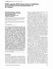
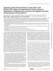
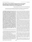
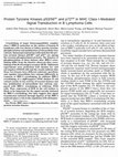
FIG. 1. Domain organization and sequence similarity between PLC␥1 and PLC␥2. A, PLC␥1 and PLC␥2 share the same domain
organization including the N-terminal PH domain, EF-hand domain, catalytic domain, and the C2 domain. In addition, and specific to the PLC␥
family, they have a specific array of domains (␥SA) inserted through a loop in the catalytic domain comprising the “split PH domain,” two SH2
domains and one SH3 domain. Residues at the boundaries for all domains are indicated for PLC␥2, for two regions located between the second SH2
(C-SH2) domain and the SH3 domain (region I), and for the C terminus (region II). B, alignment of mammalian PLC␥1 and PLC␥2 sequences
(ClustalW 1.81) is shown for regions I and II, where the main tyrosine phosphorylation sites in PLC␥1 have been mapped. These tyrosine residues
in PLC␥1 (771, 783, and 1254) are boxed. Conserved tyrosine residues between PLC␥1 and PLC␥2 within the C-SH2/SH3 linker are indicated by
the arrows. Accession numbers are as follows: human PLC␥1, P19174; rat PLC␥1, P10686; bovine PLC␥1, P08487; human PLC␥2, P16885; rat
PLC␥2, P24135.
pH 8.0, and increasing concentrations of imidazole. For purification of
Src family kinases Fyn, Src, and Lck, a buffer containing 25 mM Tris,
pH 7.5, 0.1% (v/v) Triton X-100, 1 mM dithiothreitol, and complete
protease inhibitors (Roche Molecular Biochemicals) was used. The cells
were lysed by sonication and subjected to centrifugation at 100,000 ⫻ g
for 1.5 h at 4 °C. The supernatant was added to Probond nickel resin
(Invitrogen) equilibrated in 20 mM HEPES, pH 8.0, 400 mM NaCl, 5%
(v/v) glycerol, 1 mM 2-mercaptoethanol, and 5 mM imidazole, and incubation was carried out for 1.5 h at 4 °C. The resin was washed with the
equilibration buffer, and bound proteins were eluted with the same
buffer containing increasing concentrations of imidazole. The His6-Src
family kinases were eluted with 100 mM (Fyn), 60 mM (Src), or 30 mM
Important Tyrosine Residues in PLC␥2
47985
(Lck) imidazole in 20 mM Hepes, pH 8.0. Proteins were either bufferexchanged into the appropriate assay buffer using a NAP-5 (Sephadex
G-25) column (Amersham Biosciences) or subjected to further purification. PLC␥2 was further purified on a heparin-Sepharose column (Amersham Biosciences) followed by gel filtration on a Superdex 200 16/60
column (Amersham Biosciences). His6-BLNK and ⌬213-Btk were further purified on a 5-ml HiTrapQ column (Amersham Biosciences) followed by gel filtration on a Superdex 200 16/60 column, while ⌬318-Syk
was purified by gel filtration on a Superdex 75 16/60. For purification of
GST-Syk, the supernatant was incubated with glutathione-Sepharose
(Amersham Biosciences) equilibrated in PBS, and bound protein was
isolated by centrifugation at 4,000 ⫻ g for 5 min.
The ␥2SA domains were expressed as GST fusion proteins in Escherichia coli. After induction with 0.2 mM isopropyl-1-thio--D-galactopyranoside (Calbiochem), cells were grown for 18 h at 20 °C. Cell pellets
were resuspended in (8 ml/liter of original culture) PBS supplemented
with 2 mM dithiothreitol, 1 mM EDTA, 1% (v/v) Triton X-100 with
complete protease (Roche) and phosphatase inhibitors (Sigma), lysed by
sonication, and subjected to centrifugation (10,000 ⫻ g for 10 min). The
supernatant was added to glutathione-Sepharose and incubated at 4 °C
for 45 min. After extensive washing with PBS, the fusion protein was
eluted with 50 mM Tris, pH 8.0, supplemented with 10 mM reduced
glutathione (Sigma) and 0.1% (v/v) Triton X-100.
In Vitro Assays for Analysis of Protein Phosphorylation, Formation of
Protein Complexes, and PLC Activity—For phosphorylation reaction in
vitro, purified preparations of PLC␥2 (5 g), ␥2SA proteins (1–5 g), or
synthetic peptides (10 –30 g) (Genosphere Biotechnologies) were used
as a substrate for the purified protein kinases (0.1– 0.5 g). The reaction
mixture contained 50 mM Tris, pH 8.0, 2 mM MnCl2, 2 mM MgCl2, 1 mM
Na3VO4, 50 M ATP, 2 mM dithiothreitol and, when specified, also
included 1–5 Ci of [␥-32P]ATP. Reactions were carried at 30 °C for
20 –30 min (or longer, when indicated) and terminated by the addition
of 4⫻ SDS loading buffer, and protein was subjected to SDS-PAGE (7.5
and 10% polyacrylamide for PLC␥2 and ␥2SA proteins, respectively) or
a 10 –20% gradient of polyacrylamide (Invitrogen) for the peptides).
Further analysis was by Western blotting using anti-phosphosphotyrosine antibody as described above or, when [␥-32P]ATP was included in
reaction mixtures, using a PhosphorImager (Storm 860; Molecular Dynamics, Inc., Sunnyvale, CA) or scintillation counting of extracted peptides. Kinetic analysis of PLC␥2 and BLNK phosphorylation by Syk and
Btk was performed in the presence of 5 Ci of [␥-32P]ATP and quantitated using a PhosphorImager. For measurements of initial velocity
(V0), it was verified that reaction rates were linear with respect to time
for all concentrations of substrates. Values for V0 were expressed as
PhosphorImager units/min of incubation time/mg of kinase (units/min/
mg). Apparent Km and Vmax values were determined by plotting results
as the double reciprocal Lineweaaver-Burk plot.
For analysis of interactions between PLC␥2 and phosphorylated or
nonphosphorylated BLNK, 8 g of each protein was incubated for 40
min at 4 °C in a reaction mixture containing 20 mM Tris, pH 7.5, 150 mM
Downloaded from http://www.jbc.org/ by guest on March 23, 2016
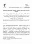
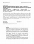
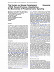
FIG. 2. Restoration of calcium responses in PLC␥2-deficient DT40 cells by human PLC␥2. A, calcium responses in Fluo-3 AM-loaded
cells were analyzed in the wild-type DT40 cells (wt, top), PLC␥2-deficient DT40 cells (PLC␥2⫺, middle), and a cell line generated by stable
transfection of human PLC␥2 into PLC␥2-deficient DT40 cells (PLC␥2⫺/h PLC␥2, bottom) after stimulation with the M4 antibody. B, stimulation
of different DT40 cell lines was carried out using the M4 antibody (dark shaded bars) or hydrogen peroxide (light shaded bars) as described for A,
and the region under the peak of calcium responses was quantitated. The measurements were performed in duplicates. The DT40 cell lines were
as follows: the wild-type DT40 (lane 1); PLC␥2-deficient DT40 cells (lane 2); and PLC␥2-deficient DT40 cells transfected with either the wild-type
human PLC␥2 (lane 3), PLC␥2 mutant Y753F (lane 4), PLC␥2 mutant Y759F (lane 5), or the double mutant PLC␥2 Y753F/Y759F (lane 6). C, the
wild type (lane 1) or Y753F/Y759F double mutant of PLC␥2 (lane 2) was expressed as His6-tagged constructs using a baculovirus system (left panel).
The activity of the wild-type (middle panel) and PLC␥2 Y753F/Y759F mutant (right panel) proteins was measured over the range of calcium
concentrations using an in vitro assay for phosphatidylinositol 4,5-bisphosphate hydrolysis.
47986
Important Tyrosine Residues in PLC␥2
NaCl, 1 mM EDTA, and 1 mM dithiothreitol and subjected to gel filtration on a Superose 12 PC 3.2/3.0 column (Amersham Biosciences) using
the same buffer. The conditions used to phosphorylate BLNK were as
described for PLC␥2, except that GST-Syk bound to glutathione-Sepharose was used; after incubation, BLNK was separated from the enzyme
by centrifugation.
Analysis of interaction between PLC␥2 and nonphosphorylated or
phosphorylated BLNK was also performed by band shift on 12.5%
polyacrylamide native PHAST gels (Amersham Biosciences) after incubation of protein components at 25 °C for 15 min. To ensure full phosphorylation of BLNK for this analysis, BLNK was prepared from Sf9
cells co-infected with Syk, further phosphorylated by Syk in vitro, and
purified using chromatography steps described above.
Phospholipase C activity was measured using detergent-mixed micelles containing sodium cholate and [3H]phosphatidylinositol 4,5bisphosphate at different concentrations of free calcium, as previously
described (25).
Separation of Peptides and Mass Spectrometry—Separation of peptides was carried out using RPC C2/C18 C2.1/10 (Amersham Biosciences) or 5 C18 300A (Phenomenex) reverse phase columns, and
elution was performed by increasing concentrations of MeCN in 0.1%
trifluoroacetic acid.
All mass spectra were acquired in reflector mode using a VoyagerDETM STR BioSpectrometryTM work station fitted with a 337-nm nitrogen laser. All samples were prepared using the dried droplet method
with freshly prepared ␣-cyano-4-hydroxycinamic acid at 10 mg/ml in
50% MeCN, 0.1% trifluoroacetic acid.
RESULTS
Calcium Responses in DT40 Cell Lines after B-cell Receptor
and Hydrogen Peroxide Stimulation—PLC␥2 is an essential
component in calcium signaling triggered either by the stimulation of BCR (8, 9) or, as described below (Fig. 2B), by stress
responses to hydrogen peroxide in DT40 cells. The stimulation
of the PLC␥2 activity in these cells by both agonists is accompanied by phosphorylation of the enzyme at tyrosine residues
(15, 17). To analyze which tyrosine residues may be involved in
activation of PLC␥2 in these systems, the sequences of PLC␥1
and PLC␥2 from two regions were compared. The first region
corresponds to linker between the C-terminal SH2 (C-SH2)
domain and the SH3 domain (within the “specific array of
domains” unique to the PLC␥ family (␥SA)), and the second
region is located at the C terminus of PLC␥ (Fig. 1). In PLC␥1,
two phosphorylated residues have been mapped to region I
(Tyr771 and Tyr783) and one residue (Tyr1254) within region II.
However, only one of these residues, Tyr783, appears to be
critical for the enzyme signaling function after platelet-derived
growth factor stimulation (21). The amino acid sequence alignment of mammalian PLC␥1 and PLC␥2 enzymes shows conservation of Tyr783, which corresponds to Tyr759 in PLC␥2 (Fig.
1B). The sequence around this residue, however, is not strictly
conserved. Residues Tyr771 and Tyr1254 seem to be unique for
PLC␥1. Analysis of sequence similarity between PLC␥1 and
PLC␥2 has also revealed that another tyrosine residue within
the C-SH2/SH3 linker, Tyr775 in PLC␥1 and Tyr753 in PLC␥2,
is conserved.
To analyze the role of conserved tyrosine residues within the
region I for PLC␥2 signaling in B-cells, stable cell lines were
generated by transfection of human PLC␥2 into PLC␥2-deficient DT40 cells. As shown in Fig. 2A, human PLC␥2 containing the wild-type sequences restored calcium responses to BCR
Downloaded from http://www.jbc.org/ by guest on March 23, 2016
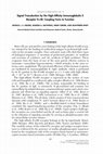
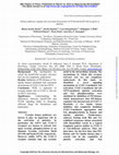
FIG. 3. Tyrosine phosphorylation of PLC␥2 in different DT40 cell lines. A, the phosphorylation of PLC␥2 was analyzed in the
PLC␥2-deficient DT40 cells stably transfected with human PLC␥2 (PLC␥2⫺/h PLC␥2). The analysis was performed before (lane 1) and 2 and 5 min
after stimulation with M4 (lanes 2 and 3). The phosphorylation was also analyzed before (lane 4) and 2 and 5 min after stimulation by hydrogen
peroxide (lanes 5 and 6). PLC␥2 from various cell extracts was isolated by immunoprecipitation and analyzed by Western blotting using either
anti-phosphotyrosine antibody (PY, top panels) or antibody to PLC␥2 (PLC␥2, bottom panels). B, the PLC␥2-deficient DT40 cells (␥2⫺) (lane 1) and
these cells stably transfected either with the wild-type human PLC␥2 (wt) (lanes 2 and 3) or PLC␥2 mutants Y753F/Y759F (lanes 4 and 5), Y753F
(lanes 6 and 7), and Y759F (lanes 8 and 9) were analyzed for PLC␥2 phosphorylation. After immunoprecipitation using anti-PLC␥2 antibody,
Western blotting was performed using anti-phosphotyrosine antibody (PY, top panel). The PLC␥2 protein was visualized on the same nitrocellulose
membrane by Amido Black staining (PLC␥2, bottom panel). C, immunoprecipitation of the wild-type PLC␥2 (lanes 1 and 2) and PLC␥2
Y753F/Y759F mutant (lanes 3 and 4) from the stable DT40 cell lines was performed as described for A and B. The presence of BLNK in the
immunoprecipitates from stimulated (lanes 1 and 3) and unstimulated cells (lanes 2 and 4) was analyzed by Western blotting.
Important Tyrosine Residues in PLC␥2
stimulation (bottom panel) to levels similar as measured in the
wild-type DT40 cells (top panel) but lacking in PLC␥2-deficient
cells (middle panel). In addition to the wild-type PLC␥2, the
constructs incorporating mutations Y753F, Y759F, and Y753F/
Y759F were also used to generate stable DT40 cell lines
(PLC␥2⫺/wtPLC␥2, PLC␥2⫺/PLC␥2 Y753F, PLC␥2⫺/PLC␥2
Y759F, and PLC␥2⫺/PLC␥2 Y753F/Y759F). Expression of
PLC␥2 in all cell lines was initially analyzed by Western blotting, and clones expressing similar amounts of PLC␥2 were
selected for further study. Immunoprecipitation confirmed that
these cell lines expressed similar amounts of human wild-type
or mutant PLC␥2 (see Fig. 3B, bottom panel). The selected cell
lines were analyzed for calcium responses to stimulation by
either the M4 antibody, which binds to BCR, or to hydrogen
peroxide (Fig. 2B). In all DT40 cell lines, the M4 antibody and
hydrogen peroxide had a similar effect on calcium responses.
Both agonists stimulated calcium responses in the wild-type
DT40 cells and PLC␥2⫺/wtPLC␥2. In contrast, DT40 cell lines
PLC␥2⫺/PLC␥2 Y753F, PLC␥2⫺/PLC␥2 Y759F, and PLC␥2⫺/
PLC␥2 Y753F/Y759F showed no calcium responses (in addition
to those seen in PLC␥2-deficient cells). These results indicate
that Tyr753 and Tyr759 are essential for PLC␥2 function in
B-cells.
To confirm the possibility that the mutation of tyrosine residues had an effect on PLC␥2 signaling function, rather than by
causing more general changes in the catalytic properties of this
PLC, the wild-type and PLC␥2 Y753F/Y759F mutant were expressed using a baculovirus system, and the purification based
on the presence of His6 tag was carried out. Preparations of pure
proteins (Fig. 2C, left panel) were analyzed for PLC activity in
vitro using conditions measuring basal catalytic activity. Under
similar conditions, measurements of PLC␥1 activity gave the
same values for the enzyme isolated from nonstimulated and
stimulated cells (26). In this assay, the wild-type and PLC␥2
Y753F/Y759F mutant had similar specific activities (in the range
of 120 –180 mol/mg). Measurements of PLC activity over the
range of calcium concentrations (Fig. 2C, middle and right panels) also demonstrated similar calcium dependence with the
highest activity at 5–10 M. These data demonstrated an intact
function of the PLC␥2 Y753F/Y759F catalytic domain and suggested the importance of Tyr753 and Tyr759 residues in the
context of the BCR signal transduction and stress responses.
Further evidence ruling out gross changes in protein structure
and correct folding is presented in Figs. 3C and 4, demonstrating
that Y753F/Y759F mutation did not affect interaction with
BLNK or the ability of PLC␥2 to translocate to the plasma membrane, previously shown to require functional SH2 domains (23).
Phosphorylation of PLC␥2 in DT40 Cell Lines—The tyrosine
phosphorylation of PLC␥2, endogenously present in the wildtype DT40 cell, has previously been observed after stimulation
by the M4 antibody or hydrogen peroxide (15, 17). In the
experiments shown in Fig. 3A, the phosphorylation of human
PLC␥2 in the DT40 PLC␥2⫺/wtPLC␥2 cell line could also be
detected 2 and 5 min following stimulation with either M4
(right panel) or hydrogen peroxide (left panel). The stimulation
in the presence of hydrogen peroxide appeared to be more
potent and particularly prominent after 5 min of stimulation.
Phosphorylation of PLC␥2 was also analyzed in different
DT40 cell lines: PLC␥2⫺, PLC␥2⫺/wtPLC␥2, PLC␥2⫺/PLC␥2
Y753F, PLC␥2⫺/PLC␥2 Y759F, and PLC␥2⫺/PLC␥2 Y753F/
Y759F. Essentially the same data were obtained using either
M4 or hydrogen peroxide to stimulate the DT40 cells. As illustrated in Fig. 3B for hydrogen peroxide stimulation, the tyrosine phosphorylation of PLC␥2 present in immunoprecipitates
obtained using anti-PLC␥2 antibody was clearly seen for the
wild-type PLC␥2 (lane 2), PLC␥2 Y753F (lane 6), and PLC␥2
Y759F (lane 8) proteins. The PLC␥2 with the double mutation
of tyrosine residues, Y753F/Y759F (lane 5), did not contain any
detectable phosphotyrosine residues.
An attempt was made to map tyrosine residues in PLC␥2
that become phosphorylated after stimulation of DT40 cells.
Analysis of tryptic peptides from tyrosine-phosphorylated
PLC␥2 demonstrated that several peaks (resolved by reversephase chromatography) contained tyrosine-phosphorylated
peptides. Further analysis of the peak fractions by mass spectroscopy revealed that masses corresponding to phosphorylated
peptides containing Tyr753 and Tyr759 were present under two
of these peaks. However, the fractions contained a mixture of
peptides, and phosphorylation of Tyr753 and Tyr759 was not
confirmed by sequencing due to limiting amounts (data not
shown).
Further analysis of PLC␥2 Y753F/Y759F mutant in DT40
cell has suggested that, as previously shown for the wild type
PLC␥2 (13–15), it could also interact with BLNK (Fig. 3C) and
the plasma membrane (Fig. 4). Whether or not levels of the
mutant in glycolipid-enriched microdomains were comparable
with that of the wild-type was not demonstrated conclusively
due to the background presence of PLC␥2 in the absence of
stimulation (data not shown). Nonetheless, the translocation of
a PLC␥2 construct incorporating the Y753F/Y759F mutation in
a system previously used to demonstrate a requirement for
functional SH2 domains (23) was clearly demonstrated (Fig.
4B). Using the same approach, translocation of the Y753F/
Y759F mutant was also confirmed in A20 B-cells (data not
shown).
Downloaded from http://www.jbc.org/ by guest on March 23, 2016
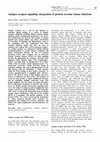
FIG. 4. Analysis of the association of PLC␥2 with isolated membrane fractions and membrane localization in cells. A, the presence of PLC␥2 in a membrane fraction isolated from unstimulated
DT40 cells stably transfected with the wild-type PLC␥2 (WT) (lane 1) or
from these cells stimulated with M4 for 2 or 5 min (lanes 2 and 3) was
analyzed by Western blotting. The same analysis was performed 5 min
after stimulation to compare DT40 cell lines expressing the wild-type
(WT) (lane 4) and Y753F/Y759F mutant (Y753F/Y759F) (lane 5) PLC␥2.
B, constructs encoding GFP fusion proteins of PLC␥2 (GFP-␥2SA) incorporating either the wild-type sequences (WT, top panel) or Y753F/
Y759F mutation (Y753F/Y759F, bottom panel) were expressed in A431
cells and analyzed before (⫺, left panels) or after (⫹, right panels)
stimulation. A similar experiment in A20 B-cells resulted in translocation of both constructs (the wild type and Y753F/Y759F) but with more
uneven plasma membrane appearance.
47987
47988
Important Tyrosine Residues in PLC␥2
The data presented in Fig. 3 have demonstrated that the
residues Tyr753 and Tyr759 are not only important for calcium
signaling function in B-cells (Fig. 2B) but also for phosphorylation of the entire PLC␥2 protein. Although the mapping of
tyrosines phosphorylated in vivo has not shown this conclusively, the loss of phosphorylation observed for the Y753F/
Y759F mutant suggests that these are the tyrosine residues
that become phosphorylated in response to stimulation. Furthermore, this phosphorylation could be a requirement for
phosphorylation of other tyrosine residues, which may be present in other regions of PLC␥2.
Role of Btk and Syk in Phosphorylation and Complex Formation in Vitro—Genetic studies of B-cell signal transduction
have demonstrated that nonreceptor tyrosine kinases from at
least three families, Src, ZAP-70/Syk, and Tec, were important
for an increase in intracellular calcium (8, 9). The tyrosine
kinases Syk and Btk, present in DT40 and other B-cells, have
been considered to directly phosphorylate PLC␥2. Previous
analysis of PLC␥2 phosphorylation in cells after overexpression
of a particular tyrosine kinase (27, 28), however, has not been
conclusive, since the possibility that this tyrosine kinase could
contribute to PLC␥2 phosphorylation through activation of another tyrosine kinase(s) endogenously present in the cell could
not be ruled out. Furthermore, concentrations of tyrosine kinases or the PLC␥2 substrate could not be controlled in those
experiments. To circumvent these problems, purified proteins
(prepared either as His6 or GST fusion proteins) were used in a
phosphorylation assay in vitro (Fig. 5). While Btk was able to
use purified PLC␥2 protein as a substrate, phosphorylation of
this PLC by Syk kinase was much lower (Fig. 5, B and C).
Using the same preparation of Syk kinase, autophosphorylation (Fig. 8A) and phosphorylation of purified BLNK protein
(Fig. 4B, bottom panel) could be demonstrated clearly. Essentially the same results were obtained using a purified GST
fusion protein of the full-length Syk as with a truncated, catalytically active His6-tagged protein. Syk kinase could also phosphorylate ␥SA of PLC␥1 at tyrosine residue 783, as demonstrated using a specific antibody to this phosphorylation site
(data not shown). When longer incubation times and increased
concentrations of PLC␥2 were used (Fig. 5C, bottom panel) or
when greater amounts of purified kinases were included in the
reaction (data not shown), phosphorylation of PLC␥2 by Syk
could also be measured. Detailed kinetic analysis, directly comparing phosphorylation of PLC␥2 by Syk and Btk, is illustrated
in Fig. 5D and has allowed calculation of apparent Km values
and relative values for Vmax. The difference between Km values
was about 2-fold (50.0 M for Btk and 83.3 M for Syk), and the
difference between values for Vmax (expressed as units/min/mg)
was about 7-fold (6.6 for Btk and 1.1 for Syk). The kinetic
analysis was extended to phosphorylation of BLNK by Syk
(Fig. 5E), demonstrating even greater differences between
phosphorylation of BLNK and PLC␥2 by Syk than when the
two kinases were compared for PLC␥2 phosphorylation. An
apparent Km value for phosphorylation of BLNK by Syk was
43.2 M (compared with 83.3 M with PLC␥2 as a substrate),
and Vmax was 37.1 units/min/mg, about 40 times greater than
PLC␥2 phosphorylation (1.1 units/min/mg).
Analysis of phosphorylation by Btk was also performed using
a synthetic peptide incorporating Tyr753 and Tyr759 residues,
745
MERDINSLYDVSRMYVDPSE764, designated as peptide 1
Downloaded from http://www.jbc.org/ by guest on March 23, 2016
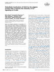
FIG. 5. In vitro phosphorylation of PLC␥2 and BLNK recombinant proteins by purified tyrosine kinases Btk and Syk. A, His6-PLC␥2
(lane 1) and His6-BLNK (lane 2) were expressed using a baculovirus system, and the purified proteins were analyzed by SDS-PAGE. B, purified
PLC␥2 (top panel) and BLNK (bottom panel) proteins were used as a substrate, in an in vitro phosphorylation assay, in the presence of purified
Btk (lane 1) or Syk (lane 2). Tyrosine phosphorylation was analyzed by Western blotting using anti-phosphotyrosine antibody. C, time course of
PLC␥2 phosphorylation by Btk (top) and Syk (bottom), visualized as in B. D, kinetic analysis of PLC␥2 phosphorylation by Syk and Btk, performed
in the presence of [32P]ATP (see “Experimental Procedures”). E, kinetic analysis of BLNK phosphorylation by Syk, performed as in D.
47989
Important Tyrosine Residues in PLC␥2
(Figs. 1B and 6A). This peptide was phosphorylated, separated
from nonphosphorylated peptide, and analyzed by mass spectrometry, demonstrating an increase in mass (by 80, from
2420.00 to 2499.98) corresponding to the phosphorylation of
one tyrosine residue. When two additional peptides incorporating either the Y753F (peptide 2) or Y759F (peptide 3) mutation
were used, it was shown that Tyr753 was phosphorylated in
preference to Tyr759 (Fig. 6B). Further phosphorylation studies
in vitro using the wild type and Y753F/Y759F mutant in the
context of the full-length PLC␥2 demonstrated phosphorylation
of both proteins (data not shown). Thus, additional Btk phosphorylation sites, outside the region represented by the peptide, are present in PLC␥2. Some of the additional sites could
be within the ␥2SA protein (containing 24 tyrosine residues), as
suggested in Fig. 8D.
Interaction of purified PLC␥2 and BLNK has been analyzed
by gel filtration (Fig. 7A) and band shift on native gels (Fig. 7B)
and demonstrated that phosphorylation of BLNK by Syk resulted in incorporation of PLC␥2 in high molecular weight
complexes. These data are consistent with previous observations of co-immunoprecipitation of these proteins after B-cell
stimulation (13, 14). When purified preparation of PLC␥2
Y753F/Y759F mutant protein was tested, it was also incorporated into a complex with phosphorylated BLNK in this in vitro
assay (Fig. 7B, right panel).
In Vitro Phosphorylation of PLC␥2 by Various Tyrosine Kinases—In addition to Syk and Btk, several other nonreceptor
tyrosine kinases from the Src family were tested for their
ability to phosphorylate PLC␥2 (Fig. 8, A and B). It has been
previously reported that partially purified preparations of several of these kinases could phosphorylate PLC␥2 in vitro (29).
The Src family kinases used in our study included Src, Lck, and
Fyn, and all contained a mutation (corresponding to Y527F in
Src) known to prevent phosphorylation and inhibition by other
tyrosine kinases in cells (30). The proteins were expressed
using a baculovirus system and contained a His6 tag for purification. Like Syk and Btk, all Src kinases were autophosphorylated in vitro (Fig. 8A). Also, all Src kinases, like Btk, phosphorylated PLC␥2 (Fig. 8B). Thus, among the tyrosine kinases
tested at the specific conditions, only Syk kinase was unable to
efficiently phosphorylate the full-length PLC␥2.
Since the mutagenesis of tyrosine residues identified Tyr753
and Tyr759 as important for PLC␥2 signaling function and
tyrosine phosphorylation in stimulated DT40 cells, ␥2SA pro-
tein (which includes these tyrosine residues) was also used as
a substrate. ␥2SA encoding the wild-type sequences and the
protein incorporating Y753F/Y759F mutations were expressed
as GST fusion proteins (Fig. 8C). When the panel of proteintyrosine kinases (Syk, Btk, Src, Lck, and Fyn) was used with
the wild-type ␥2SA as a substrate, Btk and Lck phosphorylated
this protein better than other kinases. Further comparison of
these kinases using both the wild-type and Y753F/Y759F ␥2SA
demonstrated that the mutation abolished phosphorylation by
Lck but not with Btk (Fig. 8D). This demonstrates that Lck can
phosphorylate one or both of these tyrosine residues in PLC␥2.
The studies of phosphorylation of Tyr753 and Tyr759 were also
performed in the context of a synthetic peptide corresponding
to residues 745–764 in PLC␥2 (peptide 1) (Fig. 8E). Phosphorylation of the peptide by Syk, Btk, Lck, Fyn, and Src was
analyzed in a reaction mixture containing [␥-32P]ATP, and the
peptide was separated from other components by SDS-PAGE.
When low concentrations of the enzymes (0.1 g) and short
incubation times (20 min) were used, Lck was clearly the most
efficient tyrosine kinase from the panel (Fig. 8E). Purified
preparations of Lyn, prepared as a GST fusion protein, could
phosphorylate the peptide to levels comparable with Btk and
Fyn but not Lck (data not shown). Analyses of the peptide
phosphorylated by Lck by mass spectrometry revealed phosphorylation of only one tyrosine residue in the peptide (an
increase of the peptide mass by 80, from 2420.00 to 2500.14).
Further analysis using peptides with either Tyr753 or Tyr759
replaced by phenylalanine identified Tyr753 as the main site
phosphorylated by Lck (data not shown).
DISCUSSION
Phosphorylation of both PLC␥1 and PLC␥2 has been well
documented for the majority of cellular systems where the
activation of PLC␥ isoforms takes place (3–5). However, phosphorylation sites and the importance of specific tyrosine residues that become phosphorylated have been analyzed only for
PLC␥1 in cells stimulated through growth factor receptors.
Within a complex profile of PLC␥1-phosphorylated peptides,
obtained after EGF stimulation, two main tyrosine-phosphorylated residues have been mapped as Tyr771 and Tyr1254, and one
minor site has been found to correspond to Tyr783 (19). More
recently, the use of phosphospecific antibodies to Tyr(P)783
confirmed phosphorylation of this site in stimulated cells
(23, 31). Similar patterns of phosphorylation have been seen
Downloaded from http://www.jbc.org/ by guest on March 23, 2016
FIG. 6. Phosphorylation of peptides incorporating Tyr753 and Tyr759 of PLC␥2 by Btk. A, sequences of the peptides designated as
peptides 1, 2, and 3. B, time course of phosphorylation of peptide 1, linear over 60 min (left panel) and phosphorylation of peptides 2 and 3, analyzed
after 60 min of incubation and a prolonged exposure (right panel). The phosphorylation reaction was carried out in the presence of [32P]ATP. After
separation by SDS-PAGE, the gel was subjected to analysis using a PhosphorImager. Relative intensity of the area containing the peptide was
analyzed and expressed as PhosphorImager units.
47990
Important Tyrosine Residues in PLC␥2
Downloaded from http://www.jbc.org/ by guest on March 23, 2016
FIG. 7. Formation of complexes containing purified PLC␥2 and BLNK. A, formation of protein complexes was analyzed by gel filtration
chromatography using preparations of PLC␥2 and either nonphosphorylated BLNK (top panel) or BLNK isolated after phosphorylation by Syk in
vitro (bottom panel). The phosphorylation was analyzed by Western blotting with anti-phosphotyrosine antibodies (lanes indicated as PY). When
nonphosphorylated BLNK was used in the incubation reaction with PLC␥2, the elution of BLNK (homo-oligomers, about 200,000 kDa) and PLC␥2
(a monomer, about 150,000 kDa) was as observed when each component was analyzed individually. Formation of complexes with phosphorylated
BLNK resulted in the presence of PLC␥2 protein not only in fractions corresponding to a monomer (*) but also in fractions corresponding to high
molecular weight proteins (**). B, phosphorylation of BLNK and complex formation with PLC␥2 were analyzed by band shift on 12.5%
polyacrylamide native gels. In the left panel, nonphosphorylated BLNK (lane 1) shows a lower mobility than BLNK phosphorylated by Syk (lane
2). Lane 3 shows migration of PLC␥2 C2 (protein lacking the C-terminal sequences after the C2 domain of PLC␥2). After incubation with an excess
of purified phosphorylated BLNK, PLC␥2 is shifted completely. In the right panel, a complex formation between PLC␥2 mutant Y753F/Y759F
(PLC␥2 FY) (lane 2) and phosphorylated BLNK (lane 1) was analyzed. PLC␥2 Y753F/Y759F is completely shifted into a complex (lane 3).
after stimulation of fibroblasts with platelet-derived growth
factor and in several other systems (3–5, 21). These (Tyr771,
Tyr783, and Tyr1254) and some additional sites have been identified after in vitro phosphorylation of purified PLC␥1 by EGF
receptor kinase (20). Interestingly, mutational studies have
revealed that only Tyr783 was critical, while other residues had
less impact on PLC␥1 function when tested in platelet-derived
growth factor signaling (21), demonstrating that not all phos-
phorylation sites may be functionally important. Taking into
account the complexity of the phosphorylation pattern and
possible functional redundancy, the studies of PLC␥2 described
here focused on a mutagenesis approach based on information
obtained for the PLC␥1 isoform. Comparison of PLC␥1 and
PLC␥2 sequences has revealed that of three tyrosine residues
in the loop region between the C-SH2 and the SH3 domain,
only two are conserved (Tyr753 and Tyr759 in PLC␥2, the latter
Important Tyrosine Residues in PLC␥2
47991
corresponding to phosphorylation site Tyr783 in PLC␥1), while
there is no conservation of sequences in the C-terminal region,
including residue Tyr1254 in PLC␥1 (Fig. 1). Our mutagenesis
analysis of tyrosines in PLC␥2 within the C-SH2/SH3 loop
region demonstrated that both Tyr753 and Tyr759 are required
to restore calcium signaling in DT40 cells deficient in PLC␥
(Fig. 2B). Thus, the conserved residue corresponding to Tyr759
in PLC␥2 and Tyr783 in PLC␥1 is important for the function of
both isoforms. The other conserved residue (753 in PLC␥2/775
in PLC␥1) has not been mutated in PLC␥1 and has not been
identified as one of the major phosphotyrosine sites in response
to EGF stimulation. Further studies are required to establish
whether or not this site is functionally important in any of a
number of different signaling pathways leading to phosphorylation of PLC␥1.
Comparison between properties of a double mutant within
the C-SH2/SH3 loop region in PLC␥1 (Y771F/Y783F, where
Tyr771 is unique for PLC␥1) observed in previous studies (21)
with the PLC␥2 Y753F/Y759F double mutant in the same
region described here (Figs. 2 and 3) reveals several similarities. For example, both proteins (PLC␥1 Y771F/Y783F and
PLC␥2 Y753F/Y759F) retained full in vitro catalytic activity.
Also, when the function of these proteins has been analyzed in
the context of platelet-derived growth factor signaling for
PLC␥1 and in B-cell signaling for PLC␥2, these mutations not
only inhibited generation of inositol 1,4,5-trisphosphate and
calcium mobilization but also abolished phosphorylation of the
PLC␥ protein. In the case of PLC␥1, it has been shown that the
Y771F/Y783F mutation resulted in a loss of not only phosphorylation in the C-SH2/SH3 loop region but also phosphorylation
of Tyr1254 at the C terminus. Since the phosphorylation profile
of PLC␥2 in stimulated B-cells also appears to be complex, it is
possible that the double mutation Y753F/Y759F in PLC␥2
could have a similar effect on other potential phosphorylation
sites. It has been speculated that the main impact of tyrosine
phosphorylation on the function of PLC␥ isoforms could be to,
through conformational changes, increase the access of the
enzyme to phosphatidylinositol 4,5-bisphosphate present in the
plasma membrane and in this way result in a higher rate of
substrate hydrolysis (3–5). However, these conformational
changes in the C-SH2/SH3 loop region may also be required to
expose additional phosphorylation sites.
Genetic analysis of DT40 cells has suggested the importance
of several nonreceptor tyrosine kinases for PLC␥2-mediated
calcium signaling (8, 9). However, it has not been established
which of these enzymes could phosphorylate PLC␥2 directly.
Downloaded from http://www.jbc.org/ by guest on March 23, 2016
FIG. 8. In vitro phosphorylation of PLC␥2 recombinant proteins by purified tyrosine kinases. A, different tyrosine kinases, containing
the His6 tag, were expressed in a baculovirus system and purified. An aliquot of each protein preparation was incubated in a phosphorylation
reaction, and autophosphorylation was analyzed by Western blotting using anti-phosphotyrosine antibody. The protein-tyrosine kinases were Src
(lane 1), Fyn (lane 2), Lck (lane 3), Btk (lane 4), and Syk (lane 5). B, purified PLC␥2 protein was used as a substrate for various tyrosine kinases.
Phosphorylation of PLC␥2 was analyzed by Western blotting using anti-phosphotyrosine antibody. The protein-tyrosine kinases used to phosphorylate PLC␥2 were Src (lane 1), Fyn (lane 2), Lck (lane 3), Btk (lane 4), and Syk (lane 5). C, GST fusion proteins encoding ␥2SA (residues 468 –920),
containing the wild-type sequences (lane 1) or the double mutation Y753F/Y759F (lane 2), were isolated from bacterial extracts and analyzed by
SDS-PAGE. D, GST-␥2SA proteins bound to glutathione-Sepharose were used as a substrate for Btk (top panel) or Lck (bottom panel).
Phosphorylation was analyzed by Western blotting using anti-phosphotyrosine antibody. The wild-type ␥2SA (lane 1) and the double mutation
Y753F/Y759F (lane 2) were used. E, the phosphorylation reaction was carried out in the presence of [32P]ATP. After separation by SDS-PAGE, the
peptide 1 (marked by the asterisk) was visualized by GelCode Blue Stain (left panel), and the gel was subjected to analysis using a PhosphorImager
(right panel). The relative intensity of the area containing the peptide was analyzed after incubation in the absence of a tyrosine kinase (lane 1)
and after phosphorylation in the presence of Syk (lane 2), Btk (lane 3), Lck (lane 4), Fyn (lane 5), or Src (lane 6). Quantitation of the data is
presented in the bottom panel. Relative intensities are expressed as PhosphorImager units.
47992
Important Tyrosine Residues in PLC␥2
regulation of PLC␥2 were further assessed. Direct phosphorylation of PLC␥2 by Btk is observed; however, the role of Syk may
not be to phosphorylate PLC␥2 directly but to provide docking
phosphotyrosine sites on the adapter protein BLNK, essential
in B-cell signaling.
Acknowledgments—We are grateful to A. Chan for the GST-Syk and
Myc-BLNK constructs, S. Watson and J. Wilde for the synthetic peptides (peptides 2 and 3) and a construct of Btk, L. Stephens for purified
GST-Lyn, T. Kurosaki for antibody to chicken BLNK, and M. Ellis for
assistance in preparing GST-␥2SA constructs. We are especially grateful to H. Paterson for studies involving microinjection and confocal
microscopy.
REFERENCES
1.
2.
3.
4.
5.
6.
7.
8.
9.
10.
11.
12.
13.
14.
15.
16.
17.
18.
19.
20.
21.
22.
23.
24.
25.
26.
27.
28.
29.
30.
31.
32.
33.
34.
35.
36.
Williams, R. L., and Katan, M. (1996) Structure 4, 1387–1394
Katan, M. (1998) Biochim. Biophys. Acta 1436, 5–17
Rebecchi, M. J., and Pentyala, S. N. (2000) Physiol. Rev. 80, 1291–1335
Rhee, S.-G. (2001) Annu. Rev. Biochem. 70, 281–312
Carpenter, G., and Ji, Q.-S. (1999) Exp. Cell. Res. 253, 15–24
Ji, Q.-S., Winnier, G. E., Niswender, K. D., Horstman, D., Wisdom, R.,
Magnuson, M. A., and Carpenter, G. (1997) Proc. Natl. Acad. Sci. U. S. A.
94, 2999 –3003
Wang, D., Feng, J., Wen, R., Marine, J.-C., Sangster, M. Y., Parganas, E.,
Hoffmeyer, A., Jackson, C. W., Cleveland, J. L., Murray, P. J., and Ihle,
J. N. (2000) Immunity 13, 25–35
Kurosaki, T., Maeda, A., Ishiai, M., Hashimoto, A., Inabe, K., and Takata, M.
(2000) Immunol. Rev. 176, 19 –29
Kurosaki, T., and Tsukada, S. (2000) Immunity 12, 1–5
Takata, T., Sabe, H., Hata, A., Inazu, T., Homma, Y., Nukuda, T., Yamamura,
H., and Kurosaki, T. (1994) EMBO J. 13, 1341–1349
Takata, M., and Kurosaki, T. (1996) J. Exp. Med. 184, 31– 40
Miyaka, T., Maeda, A., Yamazawa, T., Hirose, K., Kurosaki, T., and Lino, M.
(1999) EMBO J. 18, 1303–1308
Fu, C., Turck, C. W., Kurosaki, T., and Chan, A. C. (1998) Immunity 9, 93–103
Ishiai, M., Kurosaki, M., Pappu, R., Okawa, K., Ronko, I., Fu, C., Shibata, M.,
Iwamatsu, A., Chan, A. C., and Kurosaki, T. (1999) Immunity 10, 117–125
Takata, M., Homma, Y., and Kurosaki, T. (1995) J. Exp. Med. 182, 907
Tomlinson, M. G., Woods, D. B., McMahon, M., Wahl, M. I., Witte, O. N.,
Kurosaki, T., Bolen, J. B., and Johnston, J. A. (2001) BMC Immunol. 2,
4 –15
Qin, S., Stadtman, E. R., and Chook, P.-B. (2000) Proc. Natl. Acad. Sci. U. S. A.
97, 7118 –7123
Qin, S., Inazu, T., Takata, M., Kurosaki, T., Homma, Y., and Yamamura, H.
(1996) Eur. J. Biochem. 236, 443– 449
Wahl, M. I., Nishibe, S., Kim, J.-W., Kim, H., Rhee, S.-G., and Carpenter, G.
(1990) J. Biol. Chem. 265, 3944 –3948
Kim, J.-W., Sim, S.-S., Kim, U.-H., Nishibe, S., Wahl, M. I., Carpenter, G., and
Rhee, S.-G. (1990) J. Biol. Chem. 265, 3940 –3943
Kim, H.-K., Kim, J.-W., Zilberstein, A., Margolis, B., Kim, J.-G., Schlessinger,
J., and Rhee, S.-G. (1991) Cell 65, 435– 441
Sultzman, L., Ellis, C., Lin, L.-L., Pawson, T., and Knopf, J. (1991) Mol. Cell.
Biol. 11, 2018 –2025
Matsuda, M., Paterson, H. F., Rodriguez, R., Fensome, A. C., Ellis, M. V.,
Swann, K., and Katan, M. (2001) J. Cell Biol. 153, 599 – 612
Mason, C. S., Springer, C. J., Cooper, R. G., Superti-Furga, G., Marshall, C. J.,
and Marais, R. (1999) EMBO J. 18, 2137–2148
Ellis, M. V., James, S. R., Perisic, O., Downes, P., Williams, R. L., and Katan,
M. (1998) J. Biol. Chem. 273, 11650 –11659
Wahl, M. I., Jones, G. A., Nishibe, S., Rhee, S.-G., and Carpenter, G. (1992)
J. Biol. Chem. 267, 10447–10456
Hashimoto, S., Iwamatsu, A., Ishiai, M., Okawa, K., Yamadori, T., Matsushita,
M., Baba, Y., Kishimoto, T., Kurosaki, T., and Tsukada, S. (1999) Immunology 94, 2357–2364
Yamamoto, T., Matsuda, T., Junicho, A., Kishi, H., Yoshimura, A., and
Muraguchi, A. (2001) FEBS Lett. 491, 272–278
Liao, F., Shin, H.-S., and Rhee, S.-G. (1993) Biochem. Biophys. Res. Commun.
191, 1028 –1033
Blume-Jensen, P., and Hunter, T. (2001) Nature 411, 355–365
Yu, H., Fukami, K., Itoh, T., and Takenawa, T. (1998) Exp. Cell Res. 243,
113–122
Corey, S. J., and Anderson, S. M. (1999) Blood 93, 1–14
Sattethwaite, A., and Witte, O. N. (1996) Annu. Rev. Immunol. 14, 131–154
Campbell, M. A., and Sefton, B. M. (1992) Mol. Cell. Biol. 12, 2315–2321
Fluckiger, A. C., Li, Z., Kato, R. M., Wahl, M. I., Ochs, H. D., Longnecker, R.,
Kinet, J. P., Witte, O. N., Scharenberg, A. M., and Rawlings, D. J. (1998)
EMBO J. 17, 1973–1985
Scharenberg, A. M., El-Hillal, O., Fruman, D. A., Beitz, L. O., Li, Z., Lin, S.,
Gout, I., Cantley, L. C., Rawlings, D. J., and Kinet, J. P. (1998) EMBO J. 17,
1961–1972
Downloaded from http://www.jbc.org/ by guest on March 23, 2016
This was examined here (Figs. 5, 6, and 8) using purified
preparations of PLC␥2 constructs and various tyrosine kinases
with an emphasis on Btk and Syk, both essential for PLC␥2
signaling.
The role of Btk in B-cell signaling has been extensively
studied. B-cells deficient in Btk and stable cell lines where the
wild-type or different Btk mutants have been transfected into
these deficient cells have been assessed for calcium signaling
and PLC␥2 phosphorylation (11, 16, 27, 35, 36). While the
calcium responses in Btk⫺ cells were abolished, in most reports
only reduction in PLC␥2 phosphorylation has been observed,
suggesting the involvement of additional tyrosine kinases in
phosphorylation of this PLC. The in vitro phosphorylation
study using purified components described here demonstrated
that Btk could directly phosphorylate PLC␥2, including an
important residue, Tyr753 (Figs. 5, 6, and 8). Our studies have
also shown that additional sites are phosphorylated by Btk in
vitro. However, the identity of all sites remains to be established, together with their physiological relevance. Furthermore, studies of Btk have also suggested that the role of this
protein in calcium signaling could be more complex than a
requirement for PLC␥2 tyrosine phosphorylation. Mutations in
the Btk PH and SH2 domains as well as a mutation affecting
the catalytic activity resulted in a loss of signaling function, as
measured by restoration of calcium responses in DT40 Btk⫺
cells (11). While the Btk PH domain could be involved in critical
membrane binding interactions, it is possible that the Btk SH2
and/or SH3 domains provide important sites for a formation of
a signaling complex. It has been reported recently that a tyrosine kinase-inactivating mutation (in the active site and different from the nonactive site mutation affecting the catalytic
activity in a preceding study) did not abolish the function of Btk
in calcium signaling (16). This further emphasizes the potential scaffolding role of Btk and the possibility that the important tyrosine residues phosphorylated by Btk, and possibly
other critical residues in PLC␥2, could also be phosphorylated
by another kinase. Surprisingly, the studies using a panel of
different tyrosine kinases (Fig. 8) have identified Lck, an Src
family kinase where a link to B-cell signaling was not confirmed in all studies (10, 32, 33, 34), as a tyrosine kinase that
can efficiently phosphorylate a peptide incorporating Tyr753
and Tyr759 residues of PLC␥2.
Protein-tyrosine kinase Syk has also been implicated in Bcell signaling and shown to be required for both PLC␥2 phosphorylation and calcium responses (10). It has been shown that
the essential adapter protein BLNK, forming complexes with a
number of signaling components including PLC␥2, needs to be
phosphorylated by Syk in order to bind other proteins (13, 14).
Therefore, the role of Syk in calcium responses could be to
phosphorylate both PLC␥2 and BLNK or to phosphorylate only
BLNK, thereby enabling formation of signaling complexes. The
data presented here (Fig. 5) show that Syk does not efficiently
phosphorylate PLC␥2, but it does phosphorylate BLNK. Furthermore, phosphorylation of BLNK by Syk, in the absence of
additional components, could be sufficient to provide docking
sites for direct binding of PLC␥2 (Fig. 7).
In summary, we identified tyrosine residues Tyr753 and
Tyr759 as important for activation and tyrosine phosphorylation of PLC␥2 in B-cells. Based on this observation, the roles of
various tyrosine kinases that genetic analysis has implicated in
Tyrosine Residues in Phospholipase Cγ2 Essential for the Enzyme Function in B-cell
Signaling
Rosie Rodriguez, Miho Matsuda, Olga Perisic, Jeronimo Bravo, Angela Paul, Neil P.
Jones, Yvonne Light, Karl Swann, Roger L. Williams and Matilda Katan
J. Biol. Chem. 2001, 276:47982-47992.
originally published online December 14, 2001
Access the most updated version of this article at doi:
Alerts:
• When this article is cited
• When a correction for this article is posted
This article cites 36 references, 17 of which can be accessed free at
http://www.jbc.org/content/276/51/47982.full.html#ref-list-1
Downloaded from http://www.jbc.org/ by guest on March 23, 2016
Click here to choose from all of JBC's e-mail alerts
Free related PDFsRelated papers
Vav3 Modulates B Cell Receptor Responses by Regulating Phosphoinositide 3-Kinase Activation
Journal of Experimental Medicine, 2002
Free PDF

PI3K in lymphocyte development, differentiation and activation
Nature Reviews Immunology, 2003
Free PDF

TEC FAMILY KINASES IN T LYMPHOCYTE DEVELOPMENT AND FUNCTION*
Annual Review of Immunology, 2005
Free PDF


Free PDF

Functional Cloning of Src-like Adapter Protein-2 (SLAP-2), a Novel Inhibitor of Antigen Receptor Signaling
Journal of Experimental Medicine, 2001
Free PDF


Epstein-Barr virus LMP2A signaling in statu nascendi mimics a B cell antigen receptor-like activation signal
Cell Communication and Signaling, 2012
Free PDF


Src kinase-mediated signaling in leukocytes
Journal of leukocyte biology, 2000
Free PDF
Non-T Cell Activation Linker (NTAL): A Transmembrane Adaptor Protein Involved in Immunoreceptor Signaling
Journal of Experimental Medicine, 2002
Free PDF

The molecular requirements for LAT-mediated differentiation and the role of LAT in limiting pre-B cell expansion
European Journal of Immunology, 2004
Free PDF

IP 3 receptors: some lessons from DT40 cells
Immunological Reviews, 2009
Free PDF

Ikaros has a crucial role in regulation of B cell receptor signaling
European Journal of Immunology, 2006
Free PDF

Free PDF

Protein tyrosine phosphorylation in T cell signaling
Frontiers in Bioscience, 2002
Free PDF
Protein Tyrosine Kinases p53/56lynand p72sykin MHC Class I-Mediated Signal Transduction in B Lymphoma Cells
Experimental Cell Research, 1998
Free PDF
Functions of Bruton's tyrosine kinase in mast and B cells
Journal of leukocyte biology, 1999
Free PDF
The tyrosine kinase Syk is required for light chain isotype exclusion but dispensable for the negative selection of B cells
European Journal of Immunology, 2004
Free PDF


Antigen-Specific B-Lymphocyte Activation
Critical Reviews in Immunology, 2003
Free PDF

- Find new research papers in:
- Physics
- Chemistry
- Biology
- Health Sciences
- Ecology
- Earth Sciences
- Cognitive Science
- Mathematics
- Computer Science