BB
ELSEVIER
Biochimica et Biophysica Acta 1272 (1995) 113-118
Biochi~ic~a
et
BiophysicaA~ta
Cirrhosis of the human liver: an in vitro 31p nuclear magnetic resonance
study
Simon D. Taylor-Robinson
E. Louise Thomas a, Janet Sargentoni a, Claude D. Marcus a,
Brian R. Davidson c, Jimmy D. Bell
a,b.*,
~' NMR Unit Royal Postgraduate Medical School, Hammersmith Hospital, Du Cane Road, London WI2 0NN, UK
h Department of Gastroenterology, Royal Postgraduate Medical School, Hammersmith Hospital, Du Cane Road, London WI20NN, UK
c Unit'ersitv Department c~fSurgery, The Royal Free Hospital and School c~fMedicine. Hampstead, London NW3 2QG, UK
Received 7 November 1994: revised 28 March 1995: accepted 16 May 1995
Abstract
Human livers with histologically proven cirrhosis were assessed using in vitro 31p NMR spectroscopy. Spectra were compared with
those from histologically normal livers and showed significant elevations in phosphoethanolamine (PE) and phosphocholine (PC) and
significant reductions in glycerophosphorylethanolamine (GPE) and glycerophosphorylcholine (GPC). There were no significant
differences in spectra from livers with compensated and decompensated cirrhosis. These results help to characterise the alterations in
membrane metabolism in cirrhosis of the liver.
Keywords: NMR, ~rp: Cirrhosis; Phospholipid; Human; Liver
1. Introduction
The human liver responds to injury in broadly the same
way, irrespective of the original causal agent [1]. Persistent
alcohol abuse, viruses such as hepatitis B and hepatitis C,
genetic disorders including haemochromatosis, Wilson's
disease and a~-antitrypsin deficiency, cholestatic conditions such as primary biliary cirrhosis and primary sclerosing cholangitis, certain drugs and autoimmune diseases all
may provoke a series of events that ultimately lead to
cirrhosis or irreversible liver damage [2].
Cirrhosis of the liver is a diffuse process, characterised
by the formation of fibrous tissue and regrowth of hepatocytes in an abnormal nodular pattern [3]. Current assess-
Abbreviations: FID, free induction decay; GPC, glycerophosphorylcholine: GPE, glycerophosphosphorylethanolamine; MDP, methylene
diphosphonate; NMR, nuclear magnetic resonance; NTP. nucleotide
triphosphates; PC, phosphocholine; PCA, perchloric acid; PCr, phosphocreatine; PDE, phosphodiesters; PE, phosphoethanolamine; Pi, inorganic
phosphate; PME, phosphomonoesters.
* Corresponding author. Fax: +44 181 7403038.
0925-4439/95/$09.50 © 1995 Elsevier Science B.V. All rights reserved
SSDI 0 9 2 5 - 4 4 3 9 ( 9 5 ) 0 0 0 7 4 - 7
ment methods of the functional state of liver injury in
cirrhosis are not entirely satisfactory, usually depending on
a severity index obtained from a collection of laboratory
parameters and clinical findings [4-7].
Nuclear magnetic resonance (NMR) spectroscopy is a
non-invasive technique, which can be used to provide
localised biochemical information on hepatic metabolic
processes in vivo. A typical 3~p NMR spectrum of the
human liver in vivo contains resonances which may be
assigned to phosphomonoesters (PME), phosphodiesters
(PDE), inorganic phosphate (Pi) and nucleotide triphosphates (NTP) [8-12].
The PME and PDE resonances in hepatic spectra are
multicomponent and the constituents cannot as yet be
completely resolved at the magnetic field strengths employed in human in vivo NMR studies, despite the use of
proton-decoupling techniques [13]. The PME resonance
includes contributions from cell membrane precursors [14]
and glycolytic intermediates [15]. The PDE resonance is
also composite, containing information from cell membrane breakdown products [14] and from endoplasmic
reticulum [ 16].
Previous human in vivo NMR studies have reported on
�114
S.D. Taylor-Robinson et al. / Biochimica et Biophysica Acta 1272 (1995) 113-118
the elevation in P M E / A T P and the reduction in P D E / A T P
with increasing functional severity of cirrhosis [17,18].
However, the underlying metabolic abnormalities responsible for these observations have not been fully investigated.
In vitro NMR techniques on human tissue extracts have
been successfully used to study the metabolite changes
responsible for the in vivo PME and PDE signals in
hepatic tumours and normal liver [ 15,1 9]. Although Menon
and colleagues [18] reported on in vitro NMR findings
from a small number of livers from patients with chronic
liver disease, no systematic approach has been applied to
the characterisation of the cirrhotic liver.
Therefore, the aim of this study was to characterise the
metabolic changes observed by in vitro 3Jp NMR in
cirrhosis of the liver. The results are discussed in the
context of previous in vivo hepatic 31p NMR findings.
Table 1
Laboratory data on the patients from whom the liver samples were
collected
Tissue type
Serum
bilirubin
(~mol/l)
(5-17) +
Plasma
albumin
(g/l)
(35-50) +
Prothrombin
time
(s)
(12-14) +
Pugh's
score ~
(5-15)
Compensated
(n = 10)
Decompensated
(n = 15)
177
(35-460)
143
(34-388)
40
(31-48)
31
(22-40)
14
(13-16)
17
(13-25)
7
(6-7)
10
(8-12)
Data are means (range values).
Pugh's score [4] = functional severity of cirrhosis. Score < 7, compensated cirrhosis. Score > 8, decompensated cirrhosis.
+ limits of reference range.
All information was obtained preoperatively, on the day of liver transplantation.
2. Materials and methods
2.2. Reference data
Standard percutaneous liver biopsies do not yield enough
tissue for in vitro NMR studies, and therefore samples of
cirrhotic liver were taken during surgery for orthotopic
hepatic transplantation. Liver tissue was obtained from 25
patients with histologically proven cirrhosis. Ten patients
(40%) had primary biliary cirrhosis, seven (28%) post-viral
cirrhosis, six (24%) primary sclerosing cholangitis, one
(4%) Wilson's disease and one (4%) alcoholic cirrhosis.
The severity of liver dysfunction was assessed using the
Pugh's score [4], obtained from clinical and biochemical
data, acquired on the day of liver transplantation. This is
the standard scoring system, which is used clinically,
grading liver injury from 5 (best function) to 15 (worst
function), taken from information comprising serum bilirubin, plasma albumin levels, prothrombin time and the
presence/severity of ascites and hepatic encephalopathy.
The 25 liver samples were categorised into two groups:
functionally compensated cirrhosis with a Pugh's score
_< 7 (n = 10) and functionally decompensated cirrhosis
with a Pugh's score > 8 (n = 15)(Table 1).
Permission for this study was obtained from the Ethics
Committees of the Royal Postgraduate Medical School,
London, and the Royal Free Hospital and School of
Medicine, London. All patients provided written, informed
consent.
Reference data were obtained from wedge biopsy samples of liver, taken from 6 patients undergoing iaparotomy
for surgical treatment of pancreatitis. In each case, contiguous samples of liver tissue were found to be histologically normal on examination [15].
2.3. Tissue extract preparation
The wet weight of each sample was between 560 mg
and 2310 mg. Twelve per cent perchloric acid (PCA) was
added to the still-frozen samples, in a ratio of 5 m l / g of
liver tissue. Each sample was ground down under liquid
nitrogen with a mortar and pestle and then allowed to
thaw, before centrifugation at 3000 rpm for 10 min. The
supernatant was separated, neutralized with 3 M KOH,
freeze-dried and reconstituted in D20. The pH was readjusted to 7.5, after the addition of 100 mmol/l of EDTA to
chelate any paramagnetic metal ions present. Absolute
quantification of metabolites was achieved by adding
known amounts of methylene diphosphonate (MDP)
a n d / o r phosphocreatine (PCr) to the perchloric acid extracts. These acted as internal reference standards for
chemical shift assignments of the resonances observed.
2.1. Sample collection
2.4. NMR methods
Two investigators were present in the operating theatre
to obtain tissue samples from each recipient liver. In every
case, 6 - 8 representative sugar lump sized pieces of liver
were freeze-clamped in liquid nitrogen with minimum
possible ischaemic time (2-7 min). This was performed ex
vivo within 3 min of hepatectomy in 22 cases. All samples
were stored separately in a liquid nitrogen dewar until
further processed.
All NMR spectroscopy measurements were performed
at room temperature. Proton-decoupled 31p NMR spectra
were obtained using a high resolution NMR spectroscopy
system (operating at l l.7T), from the perchloric acid
extracts of liver tissue, with 16 K data points and a 45 °
pulse angle applied at intervals of 1 s. Corrections for T t
relaxation were made using samples run with a repetition
time of 20 s. Metabolites were assigned using the methods
�S.D. Taylor-Robinson et al. / Biochimica et Biophysica Acta 1272 (1995) 113-118
we have previously described [15]. The chemical shift of
each metabolite was found and subsequently confirmed by
the use of 'spiking' with known compounds [15].
115
MDP
(a)
PME
Pi
2.5. Data processing
The free induction decay (FID) was zero filled to 32 K
and Fourier transformed after line-broadening of 5 Hz.
Peak areas for PE, PC, GPE, GPC, MDP a n d / o r PCr were
obtained, using the NMR1 ® spectral processing program
(New Methods Research, E. Syracuse, USA) on a SUN
SPARCstation l0 (Sun Microsystems, Mountain View,
CA, USA). The data were fitted to Lorentzian functions.
PDE
PCr
PDE
I
I
I
-20
-15
-10
NTP/NDP
-5
I
I
I
I
I
0
5
10
15
20
ppm
pi
(a)
(b)
PME
¢'I
Pi
l~ISr
1
I
5
I~rI'P/NI)P
I
8
I
-5
ppm
I
I
6
I
-10
-15
GPC
(b)
GPE
I
5
ppm
I
4
I
3
I
2
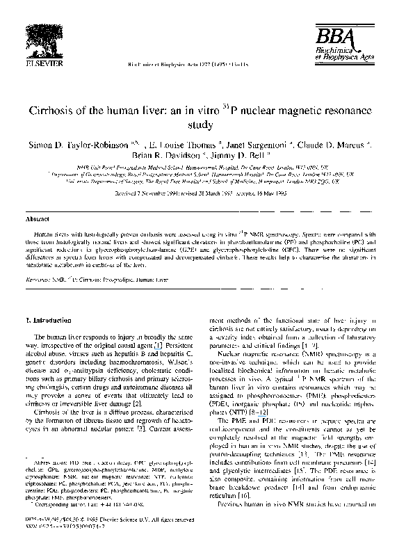
Fig. 2. Typical proton decoupled ~lp NMR spectrum of perchloric acid
extract from liver tissue with histologically proven cirrhosis. (a) Full
spectrum, (b) PME and PDE regions. Abbreviations:PME, phosphomonoesters; PDE, phosphodiesters; NAD, NADH+ NAD; NTP, nucleotide
triphosphates; NDP, nucleotide diphosphate: PE, phosphoethanolamine:
PC, phosphocholine; GPE, glycerophosphorylethanolamine:GPC, glycerophosphorylcholine: PCr (phosphocreatine) and MDP (methylene
diphosphonate) were added as internal reference standards.
2.6. Statistical analysis
I
7
6
I
I
5
4
ppm
3
Fig. I. Typical proton-decoupled ~ P NMR spectrum of perchloric acid
extract prepared from histologicallynormal liver tissue. (a) Full spectrum;
(b) PME and PDE regions. Abbreviations: PME, phosphomonoesters;
PDE, phosphodiesters; NAD, NADH+NAD; NTP, nucleotide triphosphates; NDP, nucleotide diphosphate; PE, phosphoethanolamine; PC,
phosphocholine; GPE, glycerophosphorylethanolamine; GPC, glycerophosphorylcholine; PCr (phosphocreatine) was added as an internal
reference standard. This figure is modified from Bell et al. [15].
Since the data were not normally distributed, non-parametric statistical analysis was applied. Values for metabolite concentrations in the patient and reference populations
were compared using the Mann-Whitney U-test. A P-value
of < 0.05 was considered significant. All metabolite concentrations are quoted as mean values + 1 standard deviation.
3. Results
A typical 31p NMR spectrum from a PCA extract of
normal liver contains resonances arising from PME, PDE,
�S.D. Taylor-Robinson et al. / Biochimica et Biophysica Acta 1272 (1995) 113-118
116
Table 2
Concentrations of metabolites obtained from in vitro 3~p N M R spectra from histologically normal and cirrhotic liver tissue
Tissue type
Metabolite concentrations ( / ~ m o l / g wet weight)
Normal liver (n = 6)
All cirrhosis (n = 25)
Compensated cirrhosis (n = 10)
Decompensated cirrhosis (n = 15)
PE
PC
GPE
0.16 + 0.03
1.04 + 0.75 ~
1.28+0.70 ~
0.88 ± 0.76 J
0.16 + 0.04
0.41 + 0.37 b
0.38_+0.22 ~
0.44 ± 0.45 ~
2.35
0.29
0.27
0.30
GPC
±
+
±
±
0.46
0.37 ~
0.31 ~
0.42 d
2.46 + 0.37
0.14 ± 0.26 ~
0.13±0.12 c
0.14 ± 0.29 d
Data are mean values _ 1 S.D.
Significant difference from the reference population: ~ P < 0.0005, b p < 0.05, c p < 0.0001, d p < 0.001, e p < 0.01.
NTP, NDP and Pi (Fig. 1). The PME region of the
spectrum consists of over 10 resonances, including signal
from PE, PC, AMP,2,3-DPG, coenzyme A, glucose 6phosphate, glycerol l-phosphate, 3-phosphoglycerate and
ribose 5-phosphate [15-19]. The PDE region contains two
major resonances, GPE and GPC [15,19-21].
Most of these resonances vary markedly with ischaemia
and it was therefore only sensible to quantify the more
stable compounds, namely PE and PC from the PME
region and GPE and GPC from the PDE region of the
spectrum [22-24].
The signal intensity of the PE and PC resonances was
increased and the GPE and GPC resonances reduced in
spectra from liver with histologically proven cirrhosis (Fig.
2) when compared to spectra from histologically normal
liver. The metabolite concentrations ( / z m o l / g wet weight
of liver tissue) are summarised in Table 2.
All cirrhotic livers showed significantly higher PE (1.04
+ 0.75 vs 0.16 + 0.03; P < 0.0005) and PC concentrations
(0.41 + 0.37 vs 0.16 + 0.04; P < 0,05) and significantly
lower GPE (0.29 + 0.37 vs 2.35 + 0.46; P < 0.005) and
GPC concentrations (0.14 + 0.26 vs 2.46___ 0.37; P <
0.0001) than normal tissue (Table 2).
There was no significant difference between PE, PC,
GPE and GPC concentrations from livers with functionally
compensated cirrhosis and those from livers from functionally decompensated cirrhosis (Table 2).
There were regional variations in metabolite concentrations when liver samples from different areas of the same
liver were analysed. Table 3 illustrates these variations in
metabolite levels in a patient with compensated cirrhosis.
There was no correlation between individual biochemi-
Table 3
In vitro 31p NMR: Variations in metabolite concentrations obtained from
different regions of the same liver
Metabolite concentration
Region 1
Region 2
Region 3
Region 4
PE (0.09-0.24) *
PC (0.11-0.23) *
GPE ( 1.79-2.71 ) ~
GPC (2.09-2.83) *
1.34
0.43
0
0
1.76
0.77
0
0
3.72
0.91
1.00
0
1.85
1.13
0.15
0
All values expressed as / x m o l / g wet weight of liver tissue.
* Reference range.
cal indices (serum bilirubin, plasma albumin and prothrombin time) or clinical parameters of liver dysfunction
(presence of ascites and hepatic encephalopathy), measured on the day of the transplant operation, and PE, PC,
GPE and GPC concentrations from the liver extracts.
4. Discussion
This study used in vitro 3 1 p NMR to describe the
changes in aqueous soluble membrane components in livers with histologically proven cirrhosis, compared to normal human liver tissue.
Several human in vivo 31p NMR studies of the liver
have shown abnormalities in PME, P M E / A T P , P M E / P D E
and P D E / A T P in patients with cirrhosis [17,18,25-27].
Two of these studies have correlated the functional severity of liver injury in cirrhosis with an elevation in
P M E / A T P and a reduction in P D E / A T P [17,18].
Our study attempted to investigate the underlying
metabolic changes responsible for these in vivo spectral
appearances in man. Unfortunately, a limitation of human
tissue character±sat±on by in vitro methods is the unavoidable period of ischaemia during biopsy collection. Only
quantification of PE, PC, GPE and GPC was attempted, as
the other metabolites that comprise the PME and PDE
peaks are known to alter radically from the in vivo situation during periods of ischaemia [15,19]. Hachisuka and
colleagues [28] noted that in rat liver subjected to prolonged periods of ischaemia beyond 30 min, PC and PE
were relatively stable, while GPE and GPC decreased.
However, post-mortem studies of human brain and animal
liver have indicated that the levels of PE, PC, GPE and
GPC are not significantly affected by periods of ischaemia
of up to one hour [22-24]. In our study much shorter
periods of ischaemia were encountered. Twenty-two of the
25 tissue samples from cirrhotic liver were collected within
3 min of hepatectomy, while in the three tissue samples the
ischaemic period was up to 7 min.
Comparison of the 3 1 p NMR spectra of PCA extracts
from cirrhotic liver and histologically normal tissue showed
increased concentrations of PE and PC and decreased
concentrations of GPE and GPC from the diseased tissue.
�S.D. Taylor-Robinson et al. / Biochimica et Biophysica Acta 1272 (1995) 113-118
Regional variations in metabolite concentrations were observed from samples obtained from different areas of each
individual liver.
Our results suggest that increased concentrations of PE
and PC may be responsible for elevation in P M E / A T P
observed in vivo [17,18,27]. Similarly, the reduction of
P D E / A T P seen in vivo [17,18] may be explained, at least
in part by the reduction in GPE and GPC which we have
noted. Endoplasmic reticulum is also an important component of the PDE resonance in vivo [16,29], but its relative
contribution in the human cirrhotic liver is unclear and
requires further study.
The predominant contribution of PC and PE are as
intermediates on the pathway of phospholipid biosynthesis
[14]. GPE and GPC are phospholipid breakdown products
[14]. Increased PE [30-33] and PC [34] have been observed in the regenerating rat liver and in other conditions
of rapid cellular proliferation, such as in hepatic tumours
[15,19,35]. Lymphomatous infiltration of the liver is also
associated with elevated PE levels [35].
The hallmark of cirrhosis is abnormal regrowth of liver
tissue in a nodular pattern. This occurs in the presence of
increased fibroblastic activity [3]. The increase in PE and
PC in our study may therefore be due to increased cell
turnover as the cirrhotic liver attempts to regenerate. Either
hepatocyte regeneration or the laying down of fibrous
tissue, during the cirrhotic process, may be responsible for
this phenomenon.
GPE and GPC levels are reduced in rapidly proliferating cells [15,19,32,33] and, in conditions of increased cell
turnover such as the failing cirrhotic liver, it may be
reasonable to expect reduced levels of these cell membrane
degradation products.
Unlike the in vivo studies where there was an elevation
in P M E / A T P and P D E / A T P , correlated with the functional severity of liver injury [17,18], there was no statistical difference between metabolite levels from functionally
compensated and functionally decompensated cirrhotic
liver in our study. This may partially reflect the arbitrary
nature of the clinical grading system [4], which is subject
to a number of extrahepatic influences. Furthermore, the
regional variation in metabolites concentrations that we
observed within each individual liver highlights the fact
that cirrhosis is not a uniform process. Therefore, the lack
of distinction between liver samples from patients with
compensated and decompensated cirrhosis may also be a
reflection of the varying composition of these tissue samples.
Further studies correlating in vivo 3Jp N M R spectral
abnormalities with in vitro 3Jp N M R appearances and
electron microscopy o f liver tissue to assess the N M R
contribution o f endoplasmic reticulum are required. However, the results of this study suggest that the changes in
PE, PC, GPE and GPC are responsible, to a large extent,
for the P M E / A T P and P D E / A T P abnormalities seen in
patients with cirrhosis of the liver.
1t7
Acknowledgements
This study was supported by the U.K. Department of
Health and the Medical Research Council.
W e would like to thank the MRC Biomedical N M R
Centre, Mill Hill and Birkbeck College, ULIRS for their
NMR and technical support; Brenda Hayter, Linda Selves
and Debbie Marshall, the liver transplant coordinators at
the Royal Free Hospital, London, for their assistance in
sample collection and Dr David Menon for helpful advice.
References
[1] Sherlock, S. and Dooley, J. (1993) Diseases of the Liver and Biliary
System, pp. 357-369, Blackwell Scientific Publications, Oxford.
[2] Erlinger, S. and Benhamou, J.-P. (1991) in: Oxlbrd Textbook of
Clinical Hepatology (Mclntyre, N., Benhamou, J.-P., Bircher, J.,
Rizzetto, M. and Rodes, J., eds), pp. 380-390, Oxford University
Press, Oxlbrd.
[3] Rojkind, M. and Greenwel, P. (1991) in: Oxlord Textbook of
Clinical Hepatology (Mclntyre, N., Benhamou, J.-P., Bircher, J.,
Rizzetto, M. and Rodes, J., eds), pp. 357-369. Oxford University
Press, Oxford.
[4] Pugh, R.N.H., Murray-Lyon, I.M., Dawson, J.L., Pietroni, M.C. and
Williams, R. (1972) Br. J. Surg. 60, 646-649.
[5] Shapiro, J.M., Smith, H. and Schaffner, F. (1979) Gut 20, 137-140.
[6] Albers, I., Hartmann, H., Bircher, J. and Creutzfeldt, W. (1989)
Scand. J. Gastroenterol. 24, 269-276.
[7] Dickson, E.R., Grambsh, P.M., Fleming, T.R., Fischer, L.D. and
Langworthy, A. (1989)Hepatology 10, 1-7.
[8] Oberhaensli, R.D., Hilton Jones, D., Bore, PJ.. Hands, L.J.. Rampling, R.P. and Radda, G.K. (1986) Lancet ii, 8-11.
[9] Cox, I.J., Bryant, D.J., Collins, A.G., George. P., Harman, R.R.,
Hall, A.S., Hodgson, HJ.F., Khenia, S., McArthur, P., Spencer,
D.H. and Young, I.R. (1988) J. Comput. Assist. Tomogr. 12,
369-376.
[10] Meyerhoff, D.J., Boska, M.D., Thomas, A.M. and Weiner, M.W.
(1989) Radiology 173, 393-400; erratum (1990) Radiology 176,
584.
[11] Angus, P.W., Dixon, R.M., Rajagopalan B, Ryley N.G., Simpson,
K.J., Peters, T.J., Jewell, D.P. and Radda, G.K. (1990) Clin. Sci. 78,
33-38.
[12] Oberhaensli, R., Rajagopalan B., Galloway, G.J., Taylor, D.J. and
Radda, G.K. (1990) Gut 31,463-467.
[13] Lenkinski, R.E. (1989) Invest. Radiol. 24, 1034-1038.
[14] Ruiz-Cabello, J. and Cohen, J.S. (1992) NMR Biomed. 5, 226-233.
[15] Bell, J.D., Cox, I.J., Sargentoni, J., Peden, C.J.. Menon, D.K.,
Foster, C.S., Watanapa, P., Iles, R.A. and Urenjak, J. (1993)
Biochem. Biophys. Acta 1225, 71-77.
[16] Murphy, E.J., Rajagopalan, B., Brindle, K.M. and Radda, G.K.
(1989) Magn. Reson. Med. 12, 282-289.
[17] Munakata, T., Griffiths, R.D., Martin, P.A., Jenkins, S.A., Shields,
R. and Edwards, R.H.T. (1993) NMR Biomed. 6. 168-172.
[18] Menon, D.K., Sargentoni, J., Taylor-Robinson, S.D., Bell, J.D., Cox,
l.J., Bryant, D.J., Coutts, G.A., Rolles, K., Burroughs, A.K. and
Morgan, M.Y. (1995) Hepatology 21,417-427.
[19] Cox, l.J., Bell, J.D., Peden, C.J., lies, R.A., Foster, C.S., Watanapa,
P. and Williamson, R.C.N. (1992) NMR Biomed. 5, 114-120.
[20] Iles, R.A., Stevens, A.N. and Griffiths, J.R. (1982) Prog. Nucl.
Magn. Spectrosc. 15, 49-200.
[21] Cohen, S.M. (1983)J. Biol. Chem. 258, 14294-14308.
[22] Dawson, R.M.C. (1955) Biochem. J. 60, 325-328.
[23] Perry, T.L., Hansen, S., Berry, K., Mok, C. and Lesk, D. (1971) J.
Neurochem. 18, 521-528.
�118
S.D. Taylor-Robinson et al. / Biochimica et Biophysica Acta 1272 (1995) 113-118
[24] Perry, T.L., Hansen, S. and Gandham, S.S. (1981) J. Neurochem.
36, 406-412.
[25] Cox, I.J., Menon, D.K., Sargentoni, J., Bryant, D.J., Collins, A.G..
Coutts, G.A., Iles, R.A., Bell, J.D., Benjamin, I.S., Gilbey, S.,
Hodgson, H.J.F. and Morgan, M.Y. (1992) J. Hepatol. 14, 265-275.
[26] Meyerhoff, D.J., Karczmar, G.S. and Weiner, M.W. (1989) Invest.
Radiol. 24, 908-984.
[27] Rajanayagam, V., Lee, R.R., Ackerman, Z., Bradley, W.G. and
Ross, B.D. (1992) J. Magn. Reson. Imag. 2, 183-190.
[28] Hachisuka, T., Nakayama, S., Tomita, T. and Takagi, F. (1992) J.
Surg. Res. 53, 251-256.
[29] Bates, T.E., Williams, S.R. and Gadian, D.G. (1989) Magn. Resort.
Med. 12, 145-150.
[30] Ferrari, V. and Harkness, R.D. (1954) J. Physiol. 124, 443-463.
[31] Murphy, E.J., Brindle, K., Rorison, C.J., Dixon, R.M., Rajagopalan,
B. and Radda, G.K. (1992) Biochim. Biophys. Acta 1135, 27-34.
[32] Morikawa, S., Inubushi, T., Kitoh, K., Kidoh, C. and Nozaki, M.
(1992) Biochim. Biophys. Acta 1117, 251-257.
[33] Farghali, H., Rilo, H., Zhang, W., Simplaceanu, V., Gavaler, J.S.,
Ho, C. and van Thiel, D.H. (1994) Lab. Invest. 70, 418-425.
[34] van Noorden, C.J., Vogels, I.M. and Houtkooper, J.M. (1988) Cell
Biochem. Funct. 6, 53-60.
[35] Dixon, R.M. and Tian, M. (1993) Biochim. Biophys. Acta 1181,
111-121.
�
 Simon Taylor-robinson