ORIGINAL ARTICLE
Extending the TIME concept:
what have we learned
in the past 10 years?*
David J Leaper, Gregory Schultz, Keryln Carville, Jacqueline Fletcher,
Theresa Swanson, Rebecca Drake
Leaper DJ, Schultz G, Carville K, Fletcher J, Swanson T, Drake R. Extending the TIME concept: what have we learned
in the past 10 years? Int Wound J 2012; 9 (Suppl. 2):1–19
ABSTRACT
The TIME acronym (tissue, infection/inflammation, moisture balance and edge of wound) was first developed more
than 10 years ago, by an international group of wound healing experts, to provide a framework for a structured
approach to wound bed preparation; a basis for optimising the management of open chronic wounds healing by
secondary intention. However, it should be recognised that the TIME principles are only a part of the systematic and
holistic evaluation of each patient at every wound assessment. This review, prepared by the International Wound
Infection Institute, examines how new data and evidence generated in the intervening decade affects the original
concepts of TIME, and how it is translated into current best practice. Four developments stand out: recognition of
the importance of biofilms (and the need for a simple diagnostic), use of negative pressure wound therapy (NPWT),
evolution of topical antiseptic therapy as dressings and for wound lavage (notably, silver and polyhexamethylene
biguanide) and expanded insight of the role of molecular biological processes in chronic wounds (with emerging
diagnostics and theranostics). Tissue: a major advance has been the recognition of the value of repetitive and
maintenance debridement and wound cleansing, both in time-honoured and novel methods (notably using NPWT
and hydrosurgery). Infection/inflammation: clinical recognition of infection (and non infective causes of persisting
inflammation) is critical. The concept of a bacterial continuum through contamination, colonisation and infection
is now widely accepted, together with the understanding of biofilm presence. There has been a return to topical
antiseptics to control bioburden in wounds, emphasised by the awareness of increasing antibiotic resistance.
Moisture: the relevance of excessive or insufficient wound exudate and its molecular components has led to the
development and use of a wide range of dressings to regulate moisture balance, and to protect peri-wound skin, and
optimise healing. Edge of wound: several treatment modalities are being investigated and introduced to improve
epithelial advancement, which can be regarded as the clearest sign of wound healing. The TIME principle remains
relevant 10 years on, with continuing important developments that incorporate new evidence for wound care.
Key words: Chronic wounds • Debridement • Infection • Inflammation • Moisture balance • TIME • Wound bed preparation
INTRODUCTION
The TIME acronym was first developed more
than 10 years ago, by an international group
of wound healing experts, to provide a
framework for a structured approach to wound
bed preparation (1). This concept was adopted
from a principle used in plastic surgery to
ensure optimal preparation of a recipient
Authors: DJ Leaper, MD, ChM, FRCS, FACS, FLS, Section of Wound Healing, Institute for Translation, Innovation, Methodology and
Engagement, Cardiff University, Cardiff, UK; G Schultz, PhD, Department of Obstetrics and Gynecology, Institute for Wound Research,
University of Florida, Gainesville, FL, USA; K Carville, RN, STN(Cred), PhD, Silver Chain Nursing Association & Curtin University, Osborne
Park, Western Australia; J Fletcher, MSc, BSc, PGCE, RN, FHEA, Institute for Translation, Innovation, Methodology, and Engagement,
Cardiff University, Cardiff, UK; T Swanson, RN, NPWM, AA/Dip Nursing (USA), CC(WNDM), PGC(Periop), PGDip HSc (Nursing), Masters HSc
(Nursing), International Wound Infection Institute, South West Healthcare, Warrnambool, Victoria, Australia; R Drake, BSc, London, UK
Address for correspondence: Prof. David J Leaper, Section of Wound Healing, Institute for Translation, Innovation, Methodology,
and Engagement, Cardiff University, Cardiff CF14 4XN, UK
E-mail: profdavidleaper@doctors.co.uk
*Sponsored by Smith & Nephew Wound Management.
2012 The Authors
International Wound Journal 2012 Blackwell Publishing Ltd and Medicalhelplines.com Inc
1
�Extending the TIME concept
wound bed before split thickness skin grafting,
and which was deemed to be a relevant framework for optimising the management of open
chronic wounds healing by secondary intention. The framework was therefore termed
‘wound bed preparation’ and was subsequently published in 2003 by Schultz et al. (1).
Since then the TIME acronym has been widely
used as a practical guide for the assessment
and management of chronic wounds. The clinical observations and interventions relating to
wound bed preparation are grouped into four
areas, all of which need to be addressed at each
wound assessment:
• Tissue: assessment and debridement of
non viable or foreign material (including host necrotic tissue, adherent dressing material, multiple organism-related
biofilm or slough, exudate and debris) on
the surface of the wound.
• Infection/inflammation: assessment of the
aetiology of each wound, need for topical
antiseptic and/or systemic antibiotic use
to control infection and management of
inappropriate inflammation unrelated to
infection.
• Moisture imbalance: assessment of the
aetiology and management of wound
exudate.
• Edge of wound: assessment of non advancing or undermined wound edges (and
state of the surrounding skin).
The TIME acronym was first presented at
the 2003 annual meeting of the European
Wound Management Association and has since
been cited frequently in wound management
papers, guidelines, protocols and consensus
documents, in addition to being included in
several other formats such as practical teaching aids and product formulary tools. Although
certain aspects of the TIME acronym have been
considered by some to be problematic (which
are discussed later), it has generally been found
to be a useful tool. Nevertheless, the TIME principles should always be considered as part of a
systematic and holistic evaluation of the patient
and their healing environment (Figure 1).
Since the TIME acronym was developed,
there have been several developments in
wound healing science, notably in the fields
of molecular and biological research, and
in the development, introduction and use
2
of new wound management therapies. Four
developments stand out:
• Recognition of the presence of biofilms
in chronic wounds has increased exponentially. Although still the source of
much debate and discussion, biofilms are
now known to have a significant negative
influence in chronic wounds, and the management and eradication of biofilms is an
integral part of wound healing.
• Increasing use of negative pressure wound
therapy (NPWT), which has had an
expanding influence in the treatment of
several wound types, including acute surgical wounds as well as chronic wounds.
• Evolution of a number of topical antimicrobial treatments (particularly silver and
other antiseptic dressings).
• Expanded insight into the molecular biology of wounds and the role of proteases
and pro-inflammatory markers in chronic
wounds, which has led to the continuing
emergence of a range of diagnostic and
theranostic devices.
In response to these developments and a
decade of new evidence found in the literature,
the International Wound Infection Institute has
re-examined the TIME acronym and the principles of wound bed preparation to determine
its validity for current best practice. The original table from the 2003 publication (1) has been
evaluated in the context of these new developments, and a new version has been produced,
detailing important developments that affect
the principles of TIME.
TIME – TISSUE
Over the past decade, there have been considerable developments in wound care technology; in particular, the devices or therapies
used for wound debridement, such as lowfrequency ultrasound, hydrosurgery devices,
larvae and enzymatic agents. Furthermore,
there is increased understanding of the role that
debridement plays in the treatment of wound
bioburden and infection, biofilm management, and subsequent maintenance of moisture
balance.
Debridement
Necrotic, non viable tissue and excessively
colonised, multiple organism-related biofilm
2012 The Authors
International Wound Journal 2012 Blackwell Publishing Ltd and Medicalhelplines.com Inc
�Extending the TIME concept
Patient environment
Tissue debridement
Epithelial edge
Wound bed
preparation
Inflammation
Infection
Moisture balance
Surrounding skin
Cost
benefit &
QoL issues
Therapeutic services
environment
Holistic &
systemic
evaluation
Healing environment
Figure 1. The TIME concept as part of the overall patient evaluation (created by David Leaper & Dianne Smith, with thanks to
Caroline Dowsett for the original concept of the Care Cycle).
or slough, exudate and debris are common in
chronic non healing wounds and are known
to delay healing, provide a focus for infection, exacerbate the inflammatory response and
impede optimal progression of wound granulation, contraction and epithelialisation. The
removal of this material is therefore considered
to be beneficial in stimulating healthy tissue to
heal (2–4). The methods of debridement are
summarised in Table 1 (5–11).
A number of guidelines and recommendations on wound bed preparation have been
published following publication of the first
concept of TIME. The Debridement Performance Index was published in 2002 and was
shown to be an independent predictor of
successful wound closure. It assesses callus
removal, undermining of the wound edges and
wound bed necrotic tissue (12). A wound bed
score (WBS) system has been developed (13),
which provides a more general assessment
of the wound and wound bed preparation.
It scores the following clinical parameters
(from 0 to 2): healing edges (wound edge
effect), presence of eschar, greatest wound
depth/granulation tissue, amount of exudate,
oedema, peri-wound skin inflammation, periwound callus and/or fibrosis, and presence of
a pink/red wound bed. A total score of 16 can
be achieved, and a significantly higher WBS can
be expected in wounds that go on to achieve
full closure, than in those that fail to heal.
Recommendations by another expert
panel (14) propose the use of maintenancedebridement for removal of tissue in the wound
bed when it is colonised with an excessive
bacterial burden. The aim is to help maintain the wound in a healing mode, and it is
recommended that maintenance-debridement
should be performed if the wound is not showing evidence of closure – even if the wound
bed appears clinically ‘healthy’.
A list of top tips for wound debridement (5)
recommends that specified procedures and
principles be adhered to when undertaking commonly used methods of debridement
(Box 1). Before beginning any debridement
procedure, the clinical practitioner is encouraged to ensure that the patient understands the
procedure, and the patient’s consent should be
obtained.
Wound Cleansing
Two recent Cochrane reviews have summarised methods that are used for wound
cleansing. The first reviewed wound cleansing
for pressure ulcers, and concluded that there is
limited evidence to support the use of a saline
spray containing aloe vera, silver chloride and
decyl glucoside in these wounds, but could find
no strong evidence to support the use of any
particular solution or technique for cleansing
pressure ulcers (15). The second review concluded that there is no evidence that using tap
2012 The Authors
International Wound Journal 2012 Blackwell Publishing Ltd and Medicalhelplines.com Inc
3
�Extending the TIME concept
Table 1 Methods of debridement
Type of debridement
Autolytic debridement
• Moistens necrotic tissue, allowing
degradation by host enzymes (2,5)
Methods used
Occlusive or semi-occlusive dressings (i.e. hydrocolloids) or hydrogels (2,5,6)
Hypertonic saline and honey, dressings promote autolytic debridement by
osmosis (7)
Polyacrylate, activated by Ringer’s solution (8)
Some antiseptics (silver, honey and iodine-based products) can also be used as
autolytic debriding agents
Enzymatic debridement
• Frequent dressing changes needed
• Slow but specific
• May be used with other debridement
strategies
Collagenase/papain: not available worldwide (papain has been discontinued,
as have streptokinase/streptodornase & fibrinolysin
desoxyribonuclease) (9,10)
Mechanical debridement (5)
• Non specific but gives fast results
• Can be painful & harm viable tissue
Hydrosurgery or wound cleansing debridement – wound cleansing 4–14 psi
Hydrosurgical 15 000 psi (11)
Whirlpool debridement
Recently developed debriding pads with monofilaments which allegedly retain
dead tissue and bacteria
Ultrasound debridement (5): Two types: contact and non contact
Ultrasound probe – agitates the wound bed directly; works by cavitation and
acoustic streaming
Atomised saline – gas-filled bubbles explode at the wound bed lifting necrotic
tissue and bacterial cells
Larval (maggot) therapy (5)
• Selective microdebridement
Lucilia sericata, Phaenicia sericata and Lucilia cuprina used
Sharp debridement (5)
• Not selective
• Risks of bleeding & tissue damage
For removal of necrotic/septic tissue using scalpel & scissors
Surgical debridement (5)
• Surgeon or advanced practitioner
• Not selective
• Risks of bleeding & tissue damage
For large-scale removal of necrotic/septic tissue using scalpel & scissors – by a
skilled practitioner only
Chemical debridement
Antiseptics (octenidine, silver, povidone iodine and chlorhexidine, PHMB)
Older debridement agents can be painful & have toxic effects on
healthy tissue, but can also be effective when used for limited
periods of time
water to clean a wound increases the risk of
wound infection, and that there is no strong
evidence to suggest that wound cleansing
decreases infection or promotes healing (16).
This review was updated in 2012 (17), but
no new studies were identified as eligible for
inclusion. However, in this update, the authors
concluded that there is some evidence that
using potable tap water to clean a wound may
reduce infection, and that it is likely to be as
safe as sterile water or saline. Nonetheless, caution should be exercised in the use of tap water
in immune-compromised patients, particularly
if the water might be non potable (18). The use
4
of non cytotoxic antiseptic irrigants for wound
cleansing is widely practiced but the evidence
base for their use is weak and requires further
research.
Negative pressure wound therapy
The use of NPWT, or vacuum-assisted wound
therapy, has become increasingly prominent in
wound management. Negative pressure, when
applied to the wound via a sealed foam or
gauze dressing, facilitates wound drainage,
and reduces oedema and the bioburden
of microorganisms, while increasing wound
perfusion. Recent developments have revealed
2012 The Authors
International Wound Journal 2012 Blackwell Publishing Ltd and Medicalhelplines.com Inc
�Extending the TIME concept
Box 1
TOP TIPS FOR DEBRIDEMENT (5)
• Environment
• Ensure that the room chosen for
treatment is suitable, with adequate
disposal facilities
• The room should include privacy,
adequate lighting and positioning
capacity
• Close doors and windows to prevent
cross-contamination
• Basic equipment should be provided,
for example, scalpel, forceps, curette,
sharp scissors
• Wound inspection
• Carry out a thorough inspection of
the wound bed
• Focus on the material in the wound
bed that is to be removed
• Ensure that no structures such as ligaments or blood vessels are involved
with the tissue to be removed
• Consider patient and wound condition plus goal of treatment
• Ensure that the appropriate debridement method is selected for the volume of tissue to be removed
• Competency
• Ensure that the debridement method
selected falls within the clinician’s
training and competency
that NPWT may loosen slough and necrosis,
and facilitate sharp debridement (19), although
caution is recommended when tissue is
more than 20% devitalised. The combination
of NPWT with several other debridement
methods has been demonstrated to support
TIME principles, as it expedites removal of
exudate and infective material and promotes
granulation tissue formation, contraction and
epithelialisation (20).
TIME – Tissue. What has changed? The
original TIME table indicated that non viable
tissue, multiple organism-related biofilm
or slough, exudate and debris signifies a
defective wound bed that needs debridement
to restore successful wound healing. This
principle has not changed, although some
of the practices used to facilitate this
have changed over the intervening years.
Advances in debridement technology such as
low-frequency ultrasound, hydrosurgery and
add-on use of NPWT devices with existing
technology have led to more efficacious
outcomes, as have advances in traditional non
surgical debridement methods such as larval
and enzymatic debridement. The practice of
repetitive or maintenance-debridement for
the management of static chronic wounds
has also improved outcomes.
TIME – INFECTION/INFLAMMATION
Inflammation is a physiological response to
wounding and is required for wound healing
to progress. However, excessive or inappropriate inflammation, often in the presence of
infection, may have serious consequences for
the patient. Chronicity or the stalling of healing
in wounds may be due to persistent inflammation (2,18). Wounds that do not progress
beyond an inflammatory phase often demonstrate an increased activity of proteases such
as matrix metalloproteinases (MMPs) and elastase, as well as the persistence of inflammatory
cells. Prolonged degradation of the extracellular matrix and suppression of growth factors
may also hinder wound healing. The presence
of wound biofilm may further inhibit downregulation of the immune response, causing systemic debilitation, unless adequately disrupted
and treated (21). Elimination or reduction of
prolonged inflammation revitalises tissue healing, reduces exudate and is usually associated
with a reduction in bioburden. It is important that the clinician can confidently distinguish signs and symptoms of inflammation
related to normal physiological healing from
those related to excessive inflammation caused
by underlying adverse aetiologies and infection. The clinician should, however, be aware
that inflammation may also be the result of
a number of non infective, autoimmune diseases, such as systemic lupus erythematosus,
rheumatoid arthritis, vasculitis or scleroderma,
or due to an inflammatory condition such as
inflammatory bowel disease where pyoderma
gangrenosum may result. Their recognition
and management is beyond the scope of this
article.
2012 The Authors
International Wound Journal 2012 Blackwell Publishing Ltd and Medicalhelplines.com Inc
5
�Extending the TIME concept
The signs and symptoms of infection may
be subtle or non specific (Box 2) – so care
should be taken to ensure that they are recognised (22). All wounds are potentially subject to exogenous and endogenous microbial
contamination. The microbial bioburden in a
wound can range from contamination, colonisation or critical colonisation and ultimately
to local and systemic infection if not appropriately controlled (Table 2). It has been suggested
that this progression is also influenced by the
presence of maturing bacterial biofilm in the
wound (23).
The clinician needs to be aware of the signs
and symptoms of localised, spreading (such
as cellulitis and lymphangitis) and systemic
infection. The classic signs of infection are
usually obvious in acute or surgical wounds
in otherwise healthy patients. When patients
are immunosuppressed or malnourished, however, or have comorbidities such as diabetes
mellitus, anaemia, renal or hepatic impairment,
malignancy, rheumatoid arthritis, morbid obesity or arterial, cardiac and respiratory disease,
these signs of infection may be more subtle.
An increase in pain and wound size in chronic
wounds are probably the two most useful predictors (24). The decision to use systemic or topical antibiotics should be carefully considered
in light of the risk of antimicrobial resistance,
but topical antiseptic dressings might prove to
be valuable prophylactic measures in patients
where infection is suspected – particularly as
more recent evidence suggests that they may
prevent attachment, as well as maturation, of
biofilm (25).
Biofilms
A biofilm is a complex microbial community, consisting of bacteria embedded in a
protective matrix of sugars and proteins (glycocalyx). Biofilms are known to form on the
surface of medical devices and are also found
in wounds (21,23,26). Biofilms provide a protective effect for the microorganisms embedded within them, improving their tolerance
to the host’s immune system, antimicrobials
and environmental stresses. Biofilm communities interact with host tissue resulting in
stable attachment, sustainable nutrition and
a parasitic relationship (21,23,26,27). The bacteria in biofilms have considerable phenotypic
and genotypic diversity (21).
6
Box 2
MADE-EASY GUIDELINE FOR
SIGNS OF INFECTION IN
CHRONIC WOUNDS (22)
General signs
• Malaise
• Appetite loss
Local wound signs
•
•
•
•
•
•
•
•
•
•
Increased discharge
Delayed healing
Wound breakdown
Pocketing at the base of the wound
Epithelial bridging
Unexpected pain or tenderness
Friable granulation tissue
Discolouration of the wound bed
Abscess formation
Malodour
Biofilms first form a reversible attachment to
the wound surface, which may then become
permanent with bacterial differentiation and
further accumulation of the protective glycocalyx. Biofilm structures have been recognised
in biopsies, using scanning electron and confocal microscopy, in 60% of chronic wounds
and 6% of acute wounds (28). Biofilms are a
major contributing factor to persistent, chronic
inflammatory changes in the wound bed, and
it is likely that almost all chronic wounds contain biofilm communities on at least part of the
wound bed (21,23,26). They are a problem in
wounds because of the chronic inflammatory
response that they stimulate, which benefits
the organisms in the biofilm. Mature biofilms
also shed biofilm fragments, planktonic bacteria and microcolonies, which can disperse to
form new biofilm colonies, with the risk of local
or distant invasive infection.
The recommended treatment for managing
biofilms is a combination strategy to reduce
the biofilm burden and prevent it reconstituting itself. Once the biofilm has been disrupted,
it reconstitutes itself via a metabolically active,
growth phase of the microorganisms present
and is more vulnerable to treatment agents
during this stage. It is important to understand
the genetics of the biofilm using molecular
diagnostic methods, thereby allowing therapy to be more specifically targeted. The
2012 The Authors
International Wound Journal 2012 Blackwell Publishing Ltd and Medicalhelplines.com Inc
�Extending the TIME concept
Table 2 Overview of the wound infection continuum
Contamination
Colonisation
Critical colonisation/localised infection
Spreading infection
Systemic infection
Bacteria do not multiply or cause clinical problems
Bacteria multiply but wound tissues are not damaged
Bacteria multiply to the extent that healing is impaired & wound tissues damaged
May also mean that biofilm communities are present in the wound bed
Bacteria spread from wound, causing problems in nearby healthy tissue (cellulitis and
erythema)
Bacteria spread from wound, causing infection throughout the body (systemic
inflammatory response, sepsis and organ dysfunction)
use of frequent aggressive debridement, longduration high-dose systemic antibiotics, selective biocides and combinations of antibacterial
biofilm agents are major strategies in biofilmbased wound care (Box 3) (21,23,26). There is
evidence that silver-containing dressings can
be useful in preventing biofilm reformation.
However, their efficacy has been found to be
variable, with silver-impregnated charcoal and
alginate-carboxymethylcellulose-nylon dressings not being able to prevent biofilm formation (29).
It is not possible to categorically state when
a wound is biofilm-free, because there is a lack
of definitive clinical signs and available laboratory tests. The most likely clinical indicator is
progression of healing, with reduction in exudate and slough. Standard clinical microbiology tests are not optimised to adequately measure biofilm bacteria; the most reliable method
of detecting microbial biofilm is by using specialised microscopy. A simple diagnostic is
eagerly awaited. The clinician’s judgement is
vital when deciding how to manage wounds
that contain a suspected biofilm. It is important to frequently reassess the wound and also
to practice a holistic approach to the patient’s
health to promote healing. Antibiofilm agents
(such as silver, PHMB, iodine and honey
dressings) are recommended for treatment
of wounds containing biofilm or suspected
biofilm, but wounds must be regularly assessed
on a patient-by-patient basis (26).
Are biofilms visible?
Although the existence of wound biofilms is
accepted, there is still much discussion about
their visibility to the naked eye (30). It has been
suggested that the opaque material seen on
chronic wounds may be biofilm that reforms
after removal, and may indicate the presence of critical colonisation that precedes overt
Box 3
SUGGESTED STRATEGIES FOR
REMOVAL AND PREVENTION OF
BIOFILM
1. Biofilm removal
• Physical disruption (aggressive/
sharp debridement is generally
agreed to be the best method of
removing biofilm)
• Regular debridement to reduce the
biofilm potential for regrowth
• Accompanied by vigorous physical cleansing (such as irrigation or
ultrasound)
Some products are thought to aid physical
cleansing by facilitating removal of biofilm and
debris, and disturbing biofilm (for example,
PHMB is thought to be effective in disrupting
biofilm due to its surfactant component).
2. Prevention of biofilm reconstitution
• Rational dressing use to prevent
further wound contamination
• Use of a topical broad-spectrum
antimicrobial (silver, iodine, honey,
PHMB) to kill planktonic microorganisms
• Change to a different antimicrobial if there is a lack of progress
infection. Wound biofilm, if it is visible to the naked eye, may therefore also
represent an assessment tool in managing
chronic wounds (31). However, this evidence is
entirely conjectural and biofilm will continue to
need confocal or scanning electron microscopy
or molecular technologies for definition. The
appeal for a diagnostic is clear.
2012 The Authors
International Wound Journal 2012 Blackwell Publishing Ltd and Medicalhelplines.com Inc
7
�Extending the TIME concept
Managing wound colonisation with
microorganisms
Prudent use of modern antiseptic-impregnated
dressings or irrigants may reduce microorganisms on the wound surface and in biofilms.
The concerns relating to traditional antiseptics and their toxicity to host tissue have been
widely discussed (32), but the prevailing clinical view is that it is appropriate to use most
contemporary antiseptic solutions and dressings, in accordance with the manufacturer’s
instructions or local protocols.
Antimicrobials
The term ‘antimicrobial’ is used broadly
to describe disinfectants, antiseptics and
antibiotics. The main reason for using antimicrobials in wound care is to prevent or treat
infection, and thereby facilitate the wound
healing process. Unlike antibiotics, disinfectants and antiseptics have broad-spectrum
antimicrobial activity, and microbial resistance
is rare, particularly in human pathogens. However, antibiotics have a selective antimicrobial
activity, and microbial resistance to antibiotics
is a serious concern (33–35). Colonisation and
infection in chronic wounds are usually due to a
mixed population of microorganisms. To select
the most appropriate antimicrobial therapy,
accurate diagnosis of the infecting organisms
is vital, especially when antibiotics are being
used, as microbial sensitivities can also be used
to guide the best therapy choice. Diagnosis can
be performed by tissue biopsy or by swab culture; in particular, high accuracy has been seen
when using the Levine technique (36,37).
Microbial resistance
Microbial resistance to antibiotics is of increasing concern (34). The major difference between
antibiotics and antiseptics is that antibiotics
work more specifically, allowing bacteria an
opportunity to mutate and form resistance,
whereas antiseptics work at all levels of cell
biology, so bacterial resistance is less likely
to occur. The activity of topical antimicrobial agents has been tested against multi-drug
resistant (MDR) bacteria isolated from burn
wounds. No susceptibility of topical antimicrobial agents was found to be associated with
MDR isolates; mafenide acetate was the most
effective agent against Gram-negative bacteria,
and silver also had moderate efficacy (38). No
8
silver resistance has been found in a collection
of bacterial strains tested from 349 clinical and
170 non clinical isolates from humans, meat
and production animals (39). The use of topical antibiotics is not generally recommended as
they further increase the induction of resistance
and allergy.
Topical antiseptic dressings are recommended for the following (22):
• Prevention of infection in patients who are
considered to be at an increased risk.
• Treatment of localised wound infection.
• Local treatment of wound infection in
cases of local spreading or systemic
wound infection, in conjunction with
systemic antibiotics.
Use of antiseptic dressings should be continued for 14 days (the ‘2-week rule’) and the
need for further topical antimicrobial therapy
should then be reassessed (40). Use of antiseptic dressings should be considered for those
patients at high risk of infection, or for the
early treatment of locally infected wounds
(cellulitis, lymphangitis or erythema), and discontinued if these signs of spreading or local
infection resolve. However, if signs of infection
persist, use of a systemic antibiotic is warranted and should be prescribed in accordance
with microbiological wound swab culture or
blood culture results and sensitivities. Empirical treatment with broad-spectrum antibiotics
may be commenced following clinical diagnosis, but specific antibiotic regimens should be
prescribed once the infecting organisms and
their antibiotic sensitivities have been identified. Concurrent use of topical antiseptic
dressings and debridement may reduce the
local wound bioburden.
Silver dressings
Silver has a long history of use as a topical
antimicrobial in wound care – from historical
application directly to wounds in its solid
form to the modern day application of silver salt solutions such as silver nitrate for
wound cleansing and creams or ointments
such as silver sulfadiazine (SSD) (40). Metallic silver (Ag0 ) is relatively inert, but when
exposed to moisture, highly reactive silver ions
(Ag+ ) are released, which avidly bind to tissue
proteins and cause structural changes in bacterial cell walls and intracellular and nuclear
2012 The Authors
International Wound Journal 2012 Blackwell Publishing Ltd and Medicalhelplines.com Inc
�Extending the TIME concept
membranes. This antimicrobial action, enacted
through the ionised Ag+ ion, forms strong
complexes with essential bacterial metabolic
pathways, rendering them unworkable and
leading to microbial death.
Several silver-containing dressings are available to manage wound bioburden and are
available in a number of different forms:
• Elemental: silver metal, nanocrystalline
silver.
• Inorganic: silver oxide, silver phosphate,
silver chloride, silver sulphate, silvercalcium-sodium phosphate, silver zirconium compound, SSD.
• Organic: silver-zinc allantoinate, silver
alginate, silver carboxymethylcellulose.
Silver is incorporated into dressings either
as a coating, within the dressing itself, as
part of the dressing, or as a combination of these agents. Dressings incorporating
nanocrystalline technology donate a high sustained release of Ag+ ions at the wound surface.
Silver salts are associated with minimal toxicity when applied topically, and there have
been no substantiated clinical reports of silver
toxicity. Nanocrystalline silver dressings have
been associated with improved wound healing (41,42). In a clinical study, nanocrystalline
silver dressings, under four-layer compression bandages, promoted healing in patients
with recalcitrant chronic venous leg ulcers
(VLUs). The VLUs were not clinically infected
but treatment was found to reduce bioburden and neutrophil-related inflammation (43).
Nanocrystalline silver dressings have also been
found to promote healing with reduced levels of MMPs, in a porcine model of wound
infection (44).
Silver alginate dressings have been revealed
to have broad antimicrobial activity against
wound isolates grown in both the biofilm
and non biofilm states (45), and to rapidly
decrease bacterial viability with >90% of the
bacterial and yeast cells, on the silver alginate
dressing tested, being no longer viable after
16 hours (46).
However, not all research has been supportive of silver dressing use. The multicentre,
prospective, randomised controlled VULCAN study examined the efficacy and costeffectiveness of antimicrobial silver dressings
in treating VLUs by comparing silver dressings
with non antimicrobial, low-adherent control
dressings. No statistically significant difference
in healing was found between the two dressing types, and it was concluded that there
was a lack of benefit from silver dressings (47).
Following this, a review article expressed the
opinion that the evidence base supporting silver dressing use was weak and that it was
difficult to justify the amount spent by the NHS
on silver dressings (48). A negative impact
on the perception and use of silver dressings
resulted, leading to restrictions in their availability for clinical use (40). Further reviews
have asserted that the VULCAN study has
a number of flaws, the main one being that
the silver dressings were not used as recommended (49–51). Others have commented that
antimicrobial dressings, including silver, are
key components of the management of patients
with wound infection and that failure to use
these products in appropriate cases may put
patients at risk (22,40).
Iodine dressings
Iodine-based preparations have a long history
of use in surgery and wound care. Elemental
iodine is toxic to tissues, but in its povidone iodine (PVP-I) and cadexomer iodine
forms, which are both iodophores, it is not (52).
There is evidence, including that from a recent
Cochrane review, to suggest that wound healing rates are higher with cadexomer iodine
than with standard care (52,53), and while its
antimicrobial properties are well known, several studies have indicated that cadexomer
iodine may potentially be effective against
biofilms. Staphylococcus aureus and its related
glycocalyx were not detected in the vicinity
of cadexomer iodine beads in a mouse dermis
wound model (54), and cadexomer iodine has
been found to be effective against Pseudomonas
aeruginosa biofilm in a porcine skin model (55).
A further study has demonstrated that cadexomer iodine penetrated biofilms more effectively than either silver or polyhexamethylene
biguanide (PHMB) (56).
PHMB dressings
The antiseptic PHMB has been in general
use for more than 50 years, but has now
been introduced for management of bioburden
in wounds as PHMB-impregnated dressings
or gels and solutions for wound irrigation. The active compound is effective in
2012 The Authors
International Wound Journal 2012 Blackwell Publishing Ltd and Medicalhelplines.com Inc
9
�Extending the TIME concept
both decreasing bacterial load and preventing bacterial penetration of the dressing, which
reduces infection and prevents further infection. PHMB also appears to have low toxicity
to human tissue and does not promote bacterial resistance (50,57,58). Treatment with a
polyhexanide-containing biocellulose dressing
has been revealed to remove bacterial burden
significantly faster than silver dressings (59).
now the type and behaviour of microorganisms in the wound, and the options for their
control that is of particular interest. When
biofilm microorganisms behave in a different
way to their planktonic phenotype, the action
of certain topical antimicrobial agents such
as PHMB, iodine, silver and honey needs
to be better understood, so that these agents
may be effectively used in conjunction with
debridement to control wound biofilm.
Honey
Medical-grade honey dressings are non toxic,
‘natural’ and easy to use; they are available as hydrocolloid, alginate, synthetic tulle
or gel-based dressings and promote autolytic
debridement by osmosis, while maintaining a
moist wound environment (8). Patients with
VLUs have been revealed to have increased
healing, lower infection and more effective
desloughing when treated with honey dressings compared with controls (60). Application
of honey also reduces or removes wound
malodour (8,61). Honey is hygroscopic, can
dehydrate bacteria, and its high sugar content causes inhibition of bacterial growth
with improvement of wound healing through
anti-inflammatory effects and reduction in
oedema and wound exudate (62). There is also
experimental evidence that honey may disrupt
or prevent biofilm formation (8,63,64).
Surfactants
Surfactants lower the surface tension of a liquid, allowing it to spread more easily; they also
lower the interfacial tension between two liquids. Surfactant action in wounds facilitates the
separation of loose, non viable material on the
wound surface and has potential for preventing
and managing biofilm. Several combinations
of surfactant and products with antimicrobial activity have been developed (PHMB
and undecylenamidopropyl betaine; octenidine dihydrochloride and phenoxyethanol;
octenidine and ethylhexylglycerin) and are
used clinically for skin disinfection (65,66).
TIME – Infection and inflammation. What
has changed? The original TIME table recommended that the removal of infected foci in the
wound bed lowers inflammatory cytokines
and protease activity and helps create bacterial balance and control of inflammation.
This remains the case 10 years on, but it is
10
TIME – MOISTURE
Excessive or insufficient exudate production
may adversely affect healing. Excessive exudate and odour may significantly affect the
patient’s quality of life. Exudate characteristics are important, and any alteration such as
increasing bioburden or autolysis of necrotic
tissue may indicate a change in wound status.
Updated recommendations for exudate management focus on the selection of appropriate
dressings or devices (18,67).
There are differences in composition between
acute and chronic wound fluid. Acute wound
fluid is rich in leukocytes and nutrients,
whereas chronic wound fluid has high levels
of proteases and pro-inflammatory cytokines
and elevated levels of MMPs, which decrease
as healing progresses (68,69). The increased
proteolytic activity of chronic wound exudate is thought to inhibit healing by damaging
the wound bed, degrading the extracellular matrix and aggravating the integrity of
the peri-wound skin (67), while the high levels of cytokines promote and prolong the
chronic inflammatory response seen in these
wounds (69).
Appropriate wound moisture is required
for the action of growth factors, cytokines
and cell migration – too much exudate can
cause damage to the surrounding skin, too
little can inhibit cellular activities and lead
to eschar formation, which inhibits wound
healing. Biofilm formation has also been linked
to poor exudate management (31), based on the
reasoning that wound exudate is a potentially
important nutrient source for wound biofilm.
Rapid removal of wound exudate has been
revealed to facilitate wound healing, although
not all patients showed a reduction in wound
bacteria (70).
The volume and viscosity of exudate should
be considered when choosing a dressing,
2012 The Authors
International Wound Journal 2012 Blackwell Publishing Ltd and Medicalhelplines.com Inc
�Extending the TIME concept
as some dressings are better for managing
excessive exudate, while others are better for
managing viscous exudate. The most widely
used methods for managing excessive exudate
are absorbent dressings and topical NPWT.
Dressings should maintain an appropriate
moisture balance and avoid maceration or
desiccation of the wound bed. Improved
healing was found in a pooled analysis of three
trials following the use of hydrogel dressings
compared with gauze as standard care in
diabetic foot ulcers (DFUs). It is not clear,
however, whether this was achieved as a result
of autolytic debridement or hydration of the
wound bed (71). The ideal dressing for patient
comfort and convenience is one that is not
bulky, not painful to change and reduces the
number of dressing changes needed. It should
also be effective therapeutically, and in terms
of cost, should prevent leakage and maceration
and be easy to apply and remove (69). It
is also important to protect the peri-wound
skin around chronic wounds; the increased
proteolytic activity of chronic wound exudate
can cause skin damage, and excess moisture
may cause maceration and erosion. Dressing
sensitivity or allergy is also an important
consideration, and the peri-wound skin should
be monitored for signs of this (67,69).
TIME – EDGE OF WOUND (ALSO
KNOWN AS EPITHELIAL EDGE
ADVANCEMENT)
Negative pressure wound therapy
EMT delivers a continuous or pulsed electromagnetic field, which allegedly induces tissue
healing and cell proliferation, although the
exact mechanism is unclear. Pulsed EMT consists of short-duration pulses, which has the
advantage of protecting tissues from damage
by the heat generated by continuous fields.
EMT has been used to treat VLUs, but a
Cochrane review concluded that there is no
high-quality evidence to support the hypothesis that EMT speeds healing in VLUs. However,
the same review suggested that further studies
are needed to explore the effects of EMT as an
adjunct to compression therapy or in patients
who cannot undergo compression therapy (72).
Use of NPWT is particularly valuable in
optimising the ‘M’ element of the TIME
concept, as it provides a closed moist wound
healing environment in patients with highly
exuding wounds. It is particularly effective
in removing viscous exudate, but frequent
dressing changes may be painful (67,69).
TIME – Moisture. What has changed?
Excessive wound fluid severely affects patient
well-being and wound healing. Exudate regulation has been the cornerstone of chronic
wound management since the 1960s, with
moisture balance as the goal. Over the past
10 years, the main focus has been in two
core areas – developing ways to understand
and improve the moisture management of
dressings and the role of NPWT in removing
and containing large amounts of exudate.
Research into the components of wound exudates, and their relationship to wound healing
and infection in particular, continues.
The final component of the TIME acronym
is probably the one that has led to the most
debate with regard to what the ‘E’ represents
and how it fits in with the other components of
the TIME concept. If wound bed preparation is
satisfactory, the closure of chronic wounds can
be expedited by the use of split thickness skin
grafts or biological skin replacements. Assessment of wound edges can indicate whether
wound contraction and epithelialisation is progressing, and confirm either the effectiveness of
the wound treatment being used or the need for
re-evaluation. An increasing range of treatment
modalities are proposed to improve wound
healing and thus influence the ‘edge’ effect.
These therapies include electromagnetic therapy (EMT), laser therapy, ultrasound therapy,
systemic oxygen therapy and NPWT.
The clinician should also consider the
condition of the peri-wound skin in assessing
wound contraction, as dry or macerated wound
edges may affect the ability of the wound to
contract.
Developments in managing ‘edge
of wound’
Electromagnetic therapy
Laser therapy
Low-level laser therapy, such as such as helium
neon (HeNe) or gallium arsenide (GaAs) gas
lasers, has been used to treat wounds, based on
the hypothesis that this may enhance cellular
proliferation or migration. A Cochrane review
of laser therapy for VLUs concluded that there
2012 The Authors
International Wound Journal 2012 Blackwell Publishing Ltd and Medicalhelplines.com Inc
11
�Extending the TIME concept
is no evidence of either benefit or no benefit
from using laser therapy on VLUs (73).
Ultrasound therapy
Ultrasound therapy, generated in the megaHertz or kiloHertz range, provides mechanical
energy that is thought to alter cellular activity. Until recently, megaHertz therapy was
used to treat sclerotic peri-wound skin. There
has been a recent shift towards use of lowfrequency ultrasound in the kiloHertz range
for healing in bone and tissue, which is considered to promote vascular vasodilation and
debridement (74). Several types of commercial low-frequency ultrasound therapy devices
are available, with differing mechanisms
of action.
Systemic oxygen therapy and wound healing
Oxygen is considered to have a vitally important role in wound healing, particularly in the
inflammatory and proliferative phases. A 2011
review on the role of oxygen in wound healing
concludes that supplementing treatment with
oxygen (either breathed by mask or hyperbaric therapy) may improve angiogenesis,
reduce infection rates and facilitate improved
healing (75). Further evaluation, however, is
required before it can be recommended for
clinical use in wound healing.
NPWT
The use of NPWT has been revealed to stimulate granulation tissue formation and wound
closure (76–78). One study has demonstrated
that tissue changes varied at three layers
within the wound – each responding differently under NPWT. The most superficial layer
developed granulation tissue, while the two
deeper layers demonstrated a decreased proliferation rate and clearance of chronic inflammatory markers and oedema, with tissue
stabilisation (76). NPWT has been found to
lead to significantly reduced tissue infiltration
of CD68+ macrophages and reduced IL-1β and
TNFα expression in skin-grafted free muscle
flaps. There was also a reduction in interstitial
oedema formation, which improved the microcirculation and reduced tissue damage (79). A
number of studies have also demonstrated
effective use of NPWT in wounds that have
bacterial colonisation or reveal active signs of
12
infection (80–84). Increased evidence supports
the value of NPWT in treating hard-to-heal
wounds (2); when compared with advanced
moist wound therapy in DFUs, a greater
proportion of wounds achieved closure with
NPWT (85). Amputations, secondary to DFUs,
also revealed faster postoperative healing
when treated with NPWT, compared with controls (86). However, a systematic review and
meta-analysis of 21 studies found no clear evidence that wounds heal either better or worse
with NPWT compared with conventional treatment (87), although the authors conceded that
NPWT may have a positive effect on wound
healing. Overall, study evidence supports
improved wound closure with NPWT (78,85).
TIME – Edge of wound. What has changed?
Epithelial edge advancement and an improved
state of the surrounding skin (which was not
discussed in the original TIME document) is
the clearest sign of healing, and a 20–40%
reduction in wound area after 2 and 4 weeks
of treatment is seen as a reliable predictive
indicator of healing (69). Various wound
modalities for stimulating wound healing
have been introduced; further knowledge of
their role and contraindications is warranted.
‘E’ is also a reminder of the importance of
evaluation as there is a sense that, after each
specific clinical intervention (debridement,
infection control or moisture management),
a return to the wound should be made with
an assessment of wound closure. The original
TIME table supports this by suggesting that
if the wound is not responding, a reassessment should be made with consideration of
other adjunctive or corrective therapies.
Psychosocial issues
Patients with chronic wounds have been shown
to suffer associated stress and anxiety. As well
as the negative impact on patient well-being,
stress and anxiety can also have a negative
impact on wound healing (88). A questionnaire
survey, investigating the prevalence of mood
disorders among patients with acute and
chronic wounds, found that pain (particularly
associated with dressing changes), lack of
control over treatment, and living with slowhealing chronic wounds caused stress and
anxiety (88). Suggested, non pharmacological
therapies for relieving pain in patients with
2012 The Authors
International Wound Journal 2012 Blackwell Publishing Ltd and Medicalhelplines.com Inc
�Extending the TIME concept
chronic wounds include cognitive behavioural
therapy, hypnosis, acupuncture, distraction
and meditation and prayer (89).
DISCUSSION
Although the major principles of the TIME
wound bed preparation table remain the same,
they are facilitated by many new developments
(Table 3):
Tissue: non viable dead tissue and
bacterial-related slough and debris
Debridement remains the quickest and most
efficient method of removing these materials. Clinicians have a variety of debridement
methods to choose from, depending on the
individual requirements of the patient and the
skill set of the practitioner. Autolytic debridement is most likely to be used in conjunction
with other debridement methods, and can
also be used alone if a slower, more conservative option is preferred. Newer modalities
such as low-frequency ultrasound and hydrosurgical debridement may be selective, but
require advanced clinician knowledge and further testing for appropriate and efficacious use.
Regardless of which debridement option is
chosen, healing potential and outcome goals
must be determined before commencing with
debridement.
Infection or inflammation
Infection and inflammation remain the major
challenge to healing, particularly in chronic
wounds. However, knowledge of the inflammatory process and its role in chronic wounds
has increased since the TIME acronym was
first developed. It is now known that reducing excessive inflammation can revitalise
tissue with reduction in exudate and in
the risk of infection. The understanding of
biofilms – what they are and how to detect
them – has improved considerably. Wound
biofilm presents a clinical conundrum, and
how to detect it remains a major issue; a diagnostic method for biofilm detection is required.
Although a number of dressings such as silver, honey, cadexomer iodine and possibly
PHMB have revealed some efficacy in disrupting biofilm, it is generally agreed that the best
way to disrupt biofilm is by debridement. Once
the biofilm has been disrupted, it is then possible to implement treatment with antiseptic
agents, while the biofilm is more vulnerable
to antimicrobials, and prevent its reformation (90). The use of antiseptic dressings and
wound irrigants has been more widely reintroduced and represents another area that has
revealed a great deal of growth. The increased
recent use of antiseptics also probably reflects
the concerns regarding antibiotic resistance,
whereas concerns about microbial resistance to
antiseptics appear unfounded.
Moisture imbalance
Understanding of wound moisture balance has
increased, and clinicians are more aware of the
importance of maintaining an appropriate level
of wound moisture, as well as the differences
between acute and chronic wound fluid. There
are more dressings available to ‘intelligently’
manage exudate, and some of its contained
constituents that can adversely affect wound
healing. NPWT has also proved to be an
increasingly valuable tool, particularly with its
extension for wound management to the home
environment.
Edge of wound
There have been considerable developments
in the means of facilitating wound healing,
with greater use of NPWT and new therapies,
such as EMT, laser and ultrasound therapy.
The original recommendation made by Schultz
and colleagues in 2003, of a holistic approach
with treatment of the whole patient, remains
just as valid today. Causes of poor or delayed
healing, and patient factors that might impede
or facilitate healing, must be reconsidered
at every assessment. What is new, however,
is the raised awareness of patient concerns
and the active effort to promote patients
to act as advocates for their own care and
concerns. One of the treatment modalities
which has revealed the most development
and interest since the TIME acronym was first
developed is NPWT. From its introduction
as a simple means of removing exudate and
facilitating wound closure, it now also appears
to have effects on biofilm reduction and to be
effective in infected and hard-to-heal wounds.
It may also facilitate wound debridement when
used in combination with other debridement
methods.
Using the TIME concept in practical wound
care raises further questions – for example,
2012 The Authors
International Wound Journal 2012 Blackwell Publishing Ltd and Medicalhelplines.com Inc
13
�14
Clinical observations
Tissue
2012 The Authors
International Wound Journal 2012 Blackwell Publishing Ltd and Medicalhelplines.com Inc
Infection/inflammation
WBP
Developments
Debridement
New methods
• Low-frequency ultrasound
• Hydrosurgery
• Debriding wipes
Advances in use of existing methods
• Larvae
• Autolytic (honey and hydrogels)
• Use of enzymes (collagenase)
• Sharp/surgical (new guidelines)
• Chemical (antiseptics, i.e. silver and PHMB)
NPWT – as add-on with existing debridement methods
Microbicidal irrigation solutions
Biofilm
• Improved understanding of biofilms and their role in non healing wounds
• Management – combination strategy to disrupt biofilm and prevent reconstitution
(debridement and antiseptic agents)
• Detection of biofilm
Use of Polymerase Chain Reaction (PCR)/pyrosequencing techniques to identify bacteria/fungi in
wounds
Improved understanding of the role of persistent inflammation in chronic/stalled wounds
• Role of MMPs and other proteases (diagnostics and inhibitors)
• Role of biofilms in promoting wound inflammation
• Increased use of antiseptic agents
• Role of nanocrystalline silver as an anti-inflammatory
• Combination of surfactants with antimicrobials – biofilm disruption
• NPWT combined with instillation of microbicidal solutions to reduce levels of planktonic and
biofilm bacteria
• Alternative use of new or existing agents – for example, using nanocrystalline silver to
dampen down inflammation
• Improved healing of wounds treated with custom formulations of topical antibiotics/antiseptics
based on bacterial profiles
Wound cleansing
Bacterial balance
Persistent inflammation
Managing infection/inflammation
Factors to consider
Use of maintenance debridement
Considerations around safe practice
• Knowledge
• Skills
• Competence
• Evidence of efficacy
Increased bacterial tolerance to
topical/systemic agents
Mixed flora living synergistically
Quiescent state of some bacteria in biofilms
reduces effectiveness of antibiotics
Diagnostic for biofilm detection needed
Diagnostic tests – when and how often?
Point-of-care detection
Review of appropriate antimicrobials
Rotation of products
Microbial resistance (particularly to
antibiotics)
Extending the TIME concept
Table 3 Summary table of new developments within the TIME concept
�Dressing selection – what do we need to
consider?
• Absorption
• Retention
• Patient comfort
• Bacterial pool
• Skin sensitivity or allergy
Revisiting existing therapies
Alternative use of products, for example,
using NPWT to splint wounds
Role of diagnostics/theranostics
Improved awareness of need to maintain appropriate moisture levels
Improved understanding of exudate composition – differences between acute and chronic wound fluid
• Damaging proteolytic activity of chronic wound fluid
Relationship of exudate with bacterial burden and biofilm formation
Selection of appropriate dressings or devices for exudate management (i.e. new super-absorbers)
Greater emphasis on moisture management
NPWT – for removal and containment of large exudate volumes
Epithelial edge advancement
Improved state of surrounding skin
Evaluation – check whether wound is closing
Use of NPWT to encourage contraction
Adjunct therapies (EMT, laser, ultrasound, systemic oxygen therapy)
Edge of wound
Moisture balance
Exudate
Moisture
Clinical observations
Table 3 (Continued)
WBP
Developments
Factors to consider
Extending the TIME concept
how moist should a wound be? Knowledge of
exudate management has improved considerably but, as discussed above, that question cannot be answered simply but depends on many
factors, including the type of wound, its location and the type of exudate associated with it.
Wound infection and patient comfort are also
important considerations, as are other issues
relating to patient needs. Clinicians have developed an increased awareness of psychosocial issues relating to wound care – chronic
wounds in particular can cause patient stress
and anxiety, not just in relation to pain, but
in the complexities of caring for a non healing wound, and concerns about social aspects
such as appearance and malodour. Clinicians,
industry, research and health care organisations often focus on complete wound healing
as a key outcome measure, while people living
with a chronic wound may have different priorities. This criticism has also occasionally been
levelled at the TIME acronym itself, focussing
as it does on the wound bed, as opposed to
patient-centred concerns. More emphasis may
need to be undertaken within the TIME framework to encompass patient-centred concerns
and promote a holistic approach to patient
well-being in wound care.
The TIME acronym could be redefined from
its first, assessment stage to become a second,
management stage consisting of treatment,
implementation, monitoring and evaluation:
i. Treatment: An appropriate treatment
plan is important, based on the objectives of care to be achieved, and the
objectives of the original TIME framework.
ii. Implementation: Agreed treatment plans
should be implemented consistently
for optimal, effective objectives with
evaluation of outcomes.
iii. Monitoring: This should include detection of any local or systemic adverse
events and ensure that clinical practice
and products used achieve the best performance.
iv. Evaluation: All treatments should be
regularly and objectively evaluated to
include, for example, a wound healing
curve, a validated pain assessment tool,
a debridement index or other symptom
measurement, and assessment of impact
on quality of life.
2012 The Authors
International Wound Journal 2012 Blackwell Publishing Ltd and Medicalhelplines.com Inc
15
�Extending the TIME concept
CONCLUSION
Complete and timely wound closure is the
main objective of all aspects of wound care,
although this is not always possible. Chronic
wounds, in particular, present a challenge
to effective wound care. Since the TIME
acronym was first published a decade ago, the
understanding of wound bed preparation and
the inflammatory and infective pathways has
increased considerably, as have available treatment options. Of necessity, most clinical guidelines represent ‘work in progress’ because of
the continuous changing and understanding
of wound pathology, healing and therapeutic
agents. Although the basic principles of the
TIME concept have not changed greatly since
its first inception, the application of these principles has expanded, with developments in
knowledge and interventions for wound management. It is important to consider that, while
the TIME concepts provide a valuable framework for wound assessment and management,
they are also inextricably linked.
So, 10 years on – is TIME still relevant to
clinical practice? Although there are many new
developments in the field of wound therapy
and our understanding of wounds, the basic
concepts of tissue, infection/inflammation,
moisture and edge of wound still remain
important in guiding clinical practitioners in
their approach to wound management.
ACKNOWLEDGEMENTS
The Wound Infection Institute is an international
group of clinicians, scientists and other stakeholders
committed to produce original work that contributes
towards research, education and evidence in
wound infection while remaining an independent
multinational organisation.
The authors are grateful to the committee of the International Wound Infection
Institute for their comments and enhancements to this article. The committee members
are Terry Swanson – Chair (Australia), David
Armstrong (USA), Joyce Black (USA), Keryln
Carville (Australia), Jose Contreras Ruiz (Mexico), Marc Despatis (Canada), Val EdwardsJones (UK), Jacqui Fletcher (UK), Georgina
Gethin (Ireland), Jenny Hurlow (USA), David
Keast (Canada), Patricia Larsen (USA), David
Leaper (UK), Heather Orsted (Canada), Greg
Schultz (USA), Dianne Smith (Australia), Geoff
Sussman (Australia) and Richard White (UK).
16
The authors would also like to extend their
thanks to the members of the International
Advisory Board on Wound Bed Preparation,
who developed the original TIME principles,
and to Keith Harding (UK), chair of the International Wound Infection Institute, 2007–2012.
DJL paid speaker or received research grants
from BBraun, Convatec, Smith & Nephew,
Ethicon, Coloplast, Systagenix; GS received
research grants from KCI, Health Point, Smith
& Nephew, and Hollister Wound Care in
2011–2012; KC attended education events or
presented for Smith & Nephew during 2012; JF
paid consultancy for KCI, Systagenix, BBraun
and Medi in 2011–2012; TS attended education events or presented for Coloplast,
Smith & Nephew, Convatec, Independence
Australia, and Mölnlycke during 2012; RD
undertakes freelance writing and editorial
assignments.
REFERENCES
1 Schultz GS, Sibbald RG, Falanga V, Ayello EA,
Dowsett C, Harding K, Romanelli M, Stacey MC,
Teot L, Vanscheidt W. Wound bed preparation:
a systematic approach to wound management.
Wound Repair Regen 2003;11:1–28.
2 Ousey K, McIntosh C. Understanding wound
bed preparation and wound debridement. Br
J Community Nurs 2010;15:S22–8.
3 Steed DL, Donohoe D, Webster MW, Lindsley L.
Effect of extensive debridement and treatment on
the healing of diabetic foot ulcers. Diabetic Ulcer
Study Group. J Am Coll Surg 1996;183:61–4.
4 Walcott RD, Kennedy JP, Dowd SE. Regular
debridement is the main tool for maintaining
a healthy wound bed in most chronic wounds. J
Wound Care 2009;18:54–6.
5 Leak K. Ten top tips for debridement. Wounds Intl
2012;3:21–3.
6 Smith F, Dryburgh N, Donaldson J, Mitchell M.
Debridement for surgical wounds. Cochrane
Database Syst Rev 2011;(5);CD006214.
7 Weller C, Sussman G. Wound dressings update. J
Pharmacy Pract Res 2006;36:318–24.
8 Fleck CA, Chakravarthy D. Newer debridement
methods for wound bed preparation. Adv Skin
Wound Care 2010;23:313–5.
9 Falanga V. Wound bed preparation and the role
of enzymes: a case for multiple actions of
therapeutic agents. Wounds 2002;14:47–57.
10 Stotts NA. Wound infection: diagnosis and management. In: Morison MJ, Ovington LG, Wilkie
K, editors. Chronic wound care: a problem based
approach. Edinburgh: Mosby, 2004:101–16.
11 Granick MS, Posnett J, Jacoby M, Noruthun S,
Ganchi PA, Datiashvili RO. Efficacy and costeffectiveness of a high-powered parallel waterjet
for wound debridement. Wound Repair Regen
2006;14:394–7.
2012 The Authors
International Wound Journal 2012 Blackwell Publishing Ltd and Medicalhelplines.com Inc
�Extending the TIME concept
12 Saap LJ, Falanga V. Debridement performance
index and its correlation with complete closure
of diabetic foot ulcers. Wound Repair Regen
2002;10:354–9.
13 Falanga V, Saap LJ, Ozonoff A. Wound bed score
and its correlation with healing of chronic
wounds. Dermatol Ther 2006;19:383–90.
14 Falanga V, Brem H, Ennis WJ, Wolcott R, Gould
LJ, Ayello EA. Maintenance debridement in the
treatment of difficult-to-heal chronic wounds.
Recommendations of an expert panel. Ostomy
Wound Manage 2008;(Suppl):2–13. Quiz14–5.
15 Moore ZE, Cowman S. Wound cleansing for
pressure ulcers. Cochrane Database Syst Rev
2005;(4);CD004983.
16 Fernandez R, Griffiths R. Water for wound
cleansing. Cochrane Database Syst Rev 2008;(1);
CD003861.
17 Fernandez R, Griffiths R. Water for wound
cleansing. Cochrane Database Syst Rev 2012;2:
CD003861.
18 Sibbald RG, Goodman L, Woo KY, Krasner DL,
Smart H, Tariq G, Ayello EA, Burrell RE,
Keast DH, Mayer D, Norton L, Salcido RS.
Special considerations in wound bed preparation
2011: an update. Adv Skin Wound Care
2011;24:415–36. Quiz 437–8.
19 Riley S, Tongue J, Strokes S, Jefferies L. Using
negative pressure wound therapy as an aid
to debridement. Poster presentation. Harrogate:
Wounds UK, 2009.
20 Davis J. Combining wound debridement modalities
with negative pressure wound therapy. Abstract
#123; WOCN Society 38th Annual Conference, 2006.
21 Wolcott RD, Dowd S, Kennedy J, Jones CE.
Biofilm-based wound care. Adv Wound Care
2008;1:311–6.
22 Vowden P, Vowden K, Carville K. Antimicrobial
dressings made easy. Wounds Intl 2011;2(1).
23 Percival S, Bowler P. Biofilms and their potential
role in wound healing. Wounds 2004;16:234–40.
24 Gardner SE, Frantz RA, Dobbeling BN. The
validity of the clinical signs and symptoms used
to identify localised chronic wound infection.
Wound Repair Regen 2001;9:178–86.
25 Rhoads DD, Wolcott RD, Percival SL. Biofilm in
wounds: management strategies. J Wound Care
2008;17:502–8.
26 Phillips PL, Wolcott RD, Fletcher J, Schultz GS.
Biofilms made easy. Wounds Intl 2010;1(3).
27 Wolcott RD, Rhoads DD, Dowd SE. Biofilms and
chronic wound inflammation. J Wound Care
2008;17:333–41.
28 James GA, Swogger E, Wolcott R, Pulcini E,
Secor P, Sestrich J, Costerton JW, Stewart PS.
Biofilms in chronic wounds. Wound Repair
Regen 2008;16:37–44.
29 Driffield K, Woodmansey E, Floyd H. The use of
silver-containing dressings to prevent biofilm
formation by single and mixed bacterial flora.
Poster presentation #285, EWMA, 2008.
30 White RJ, Cutting KF. Wound biofilms – are they
visible? J Wound Care 2012;21:552–3.
31 Hurlow J, Bowler PG. Potential implications of
biofilm in chronic wounds: a case series. J Wound
Care 2012;21:109–19.
32 Drosou A, Falabella A, Kirsner RS. Antiseptics
on wounds: an area of controversy. Wounds
2003;15:149–66.
33 Leaper D. Topical antiseptics in wound care: time
for reflection. Int Wound J 2011;8:547–9.
34 Leaper D. Editorial: European Union antibiotic
awareness day. Relevance for wound care
practitioners Int Wound J 2010;7:314–5.
35 Venous leg ulcers: infection diagnosis and microbiology investigation. Quick reference guide
for primary care; 2010. Revision. Association
of Medical Microbiologists/Health Protection
Agency.
36 Kirsner R. Infection and chronic wounds. Wound
healing perspectives 2006;3:1–2.
37 Gardner SE, Frantz RA, Saltzman CL, Hillis SL,
Park H, Scherubel M. Diagnostic validity of three
swab techniques for identifying chronic wound
infection. Wound Repair Regen 2006;14:548–57.
38 Glasser JS, Guymon CH, Mende K, Wolf SE,
Hospenthal DR, Murray CK. Activity of topical
antimicrobial agents against multidrug-resistant
bacteria recovered from burn patients. Burns
2010;36:1172–84.
39 Jakobsen L, Andersen AS, Friis-Møller A, Jørgensen
B, Krogfelt KA, Frimodt-Møller N. Silver resistance: an alarming public health concern? Int
J Antimicrob Agents 2011;38:454–5.
40 Leaper D, Ayello EA, Carville K, Fletcher J, Keast D,
Lindholm C, Martinez JLL, Mavanini SD, McBain
A, Moore Z, Opasanon S, Pina E. Appropriate
use of silver dressings in wounds. International
Consensus Document. Wounds Int 2012.
41 Miller CN, Newall N, Kapp SE, Lewin G,
Karimi L, Carville K, Gliddon T, Santamaria
NM. A randomized-controlled trial comparing
cadexomer iodine and nanocrystalline silver on
the healing of leg ulcers. Wound Repair Regen
2010;18(4):359–67.
42 Fong J, Wood F. Nanocrystalline silver dressings in wound management: a review. Int J
Nanomedicine 2006;1(4):441–449.
43 Sibbald RG, Contreras-Ruiz J, Coutts P, Fierheller M,
Rothman A, Woo K. Bacteriology, inflammation,
and healing: a study of nanocrystalline silver
dressings in chronic venous leg ulcers. Adv Skin
Wound Care 2007;20:549–58.
44 Wright JB, Lam K, Buret GA, Olson EM, Burrell
ER. Early healing events in a porcine model
of contaminated wounds: effects of nanocrystalline silver on matrix metalloproteinases, cell
apoptosis and healing. Wound Repair Regen
2002;10:141–51.
45 Percival S, Slone W, Linton S, Okel T, Corum L,
Thomas JG. The antimicrobial efficacy of a silver
alginate dressing against a broad spectrum of
clinically relevant wound isolates. Int Wound J
2011;8:237–43.
46 Hooper SJ, Percival SL, Hill KE, Thomas DW, Hayes
AJ, Williams DW. The visualisation and speed
of kill of wound isolates on a silver alginate
2012 The Authors
International Wound Journal 2012 Blackwell Publishing Ltd and Medicalhelplines.com Inc
17
�Extending the TIME concept
47
48
49
50
51
52
53
54
55
56
57
58
59
60
18
dressing. Int Wound J 2012; Mar 8. [Epub ahead
of print].
Michaels JA, Campbell B, King B, Palfreyman
SJ, Shackley P, Stevenson M. Randomized
controlled trial and cost-effectiveness analysis
of silver-donating antimicrobial dressings for
venous leg ulcers (VULCAN trial). Br J Surg
2009;96:1147–56.
Iheanacho I. Silver dressings: do they work? Drugs
Ther Bull 2010;48:38–42.
White R, Kingsley A. Silver dressings in the light of
recent clinical research: what can be concluded?
Wounds UK 2010;6:157–8.
Barrett S, Battacharyya M, Butcher M, Enoch S,
Fumarola S, Gray D, Stephen Haynes J, EdwardsJones V, Leaper D, Strohal R, White R, Wicks G,
Young T. PHMB and its potential contribution to
wound management. Aberdeen, UK: Wounds,
2010.
Leaper D, Drake R. Should one size fit all? An
overview and critique of the VULCAN study on
silver dressings. Int Wound J 2011;8:1–4.
Sibbald RG, Leaper DJ, Queen D. Iodine made easy.
Wounds Int 2011;2(2).
O’Meara S, Al-Kurdi D, Ovington LG. Antibiotics
and antiseptics for venous leg ulcers. Cochrane
Database Syst Rev 2008;(1):CD003557.
Akiyama H, Oono T, Saito M, Iwatsuki K. Assessment of cadexomer iodine against Staphylococcus
aureus biofilm in vivo and in vitro using confocal laser scanning microscopy. J Dermatol
2004;31:529–34.
Phillips PL, Yang Q, Sampson E, Schultz G. Effects
of antimicrobial agents on an in vitro biofilm
model of skin wounds. Adv Wound Care
2010;1:299–304.
Schultz GS, Phillips P, Yang Q, Sampson E.
Microbicidal effects of wound dressings on
mature bacterial biofilm on pig skin explants,
Helsinki, Finland. Poster presentation P142,
EWMA, 2009.
Dissemond J, Assadian O, Gerber V, Kingsley A,
Kramer A, Leaper DJ, Mosti G, Piatkowski de
Grzymala A, Riepe G, Risse A, Romanelli M,
Strohal R, Traber J, Vasel-Biergans A, Wild T,
Eberlein T. Classification of wounds at risk and
their antimicrobial treatment with polihexanide:
a practice-oriented expert recommendation. Skin
Pharmacol Physiol 2011;24:245–55.
Best practice statement. The use of topical antiseptic/antimicrobial agents in wound management.
2nd edition. London: Wounds UK, 2011.
Eberlein T, Haemmerle G, Signer M, Haemmerle
G, Signer M, Gruber Moesenbacher U, Traber J,
Mittlboeck M, Abel M, Strohal R. Comparison of
PHMB-containing dressing and silver dressings
in patients with critically colonised or locally
infected wounds. J Wound Care 2012;21:12–20.
Gethin G, Cowman S. Manuka honey vs. hydrogel – a prospective, open label, multicentre, randomised controlled trial to compare desloughing
efficacy and healing outcomes in venous ulcers.
J Clin Nurs 2009;18:466–74.
61 Molan PC. The evidence supporting the use of
honey as a wound dressing. Int J Low Extrem
Wounds 2006;5:40–54.
62 Simon A, Traynor K, Santos K, Blaser G, Bode U,
Molan P. Medical honey for wound care – still
the ‘latest resort’? Evid Based Complement
Alternat Med 2009;6:165–73.
63 Maddocks SE, Lopez MS, Rowlands RS, Cooper
RA. Manuka honey inhibits the development
of Streptococcus pyogenes biofilms and causes
reduced expression of two fibronectin binding
proteins. Microbiology 2012;158(Pt 3):781–90.
64 Alandejani T, Marsan J, Ferris W, Slinger R,
Chan F. Effectiveness of honey on Staphylococcus
aureus and Pseudomonas aeruginosa biofilms.
Otolaryngol Head Neck Surg 2009;141:114–8.
65 Vanscheidt W, Harding K, Téot L, Siebert J.
Effectiveness and tissue compatibility of a 12week treatment of chronic venous leg ulcers with
an octenidine based antiseptic – a randomized,
double-blind controlled study. Int Wound J
2012;9:316–23.
66 Romanelli M, Dini V, Barbanera S, Bertone MS, Brilli
NTC. Evaluation of the efficacy and tolerability
of a solution containing undecylenamidopropylbetaine and polihexanide (Prontosan) in controlling the bacterial burden of chronic wounds
during wound bed preparation. Poster presentation. SAWC, San Diego, CA, 2008.
67 Romanelli M, Vowden K, Weir D. 2010: Exudate
management made easy. Wounds Int 1(2). URL
http://www.woundsinternational.com [accessed
on 21 May 2012].
68 Trengove NJ, Stacey MC, MacAuley S, Bennett
N, Gibson J, Burslem F, Murphy G, Schultz
G. Analysis of the acute and chronic wound
environments: the role of proteases and their
inhibitors. Wound Repair Regen 1999;7:442–52.
69 Dowsett C. Exudate management: a patient-centred
approach. J Wound Care 2008;17:249–52.
70 Wolcott RD. The effect of a hydroconductive
dressing on the suppression of wound biofilm.
Wounds 2012;24:132–7.
71 Edwards J, Stapley S. Debridement of diabetic foot ulcers. Cochrane Database Syst Rev
2010;(1):CD003556.
72 Aziz Z, Cullum NA, Flemming K. Electromagnetic
therapy for treating venous leg ulcers. Cochrane
Database Syst Rev 2011;(3):CD002933.
73 Fleming K, Cullum NA. Laser therapy for
venous leg ulcers. Cochrane Database Syst Rev
2000;(2):CD001182.
74 Ennis WJ, Valdes W, Gainer M, Meneses P.
Evaluation of clinical effectiveness of MIST
ultrasound therapy for the healing of chronic
wounds. Adv Skin Wound Care 2006;19:437–46.
75 Chambers AC, Leaper DJ. Role of oxygen in wound
healing: a review of evidence. J Wound Care
2011;20:160–4.
76 Bassetto F, Lancerotto L, Salmaso R, Pandis L,
Pajardi G, Schiavon M, Tiengo C, Vindigni V.
Histological evolution of chronic wounds under
negative pressure therapy. J Plastic Reconstr
Aesthet Surg 2011;65:91–9.
2012 The Authors
International Wound Journal 2012 Blackwell Publishing Ltd and Medicalhelplines.com Inc
�Extending the TIME concept
77 Dini V, Miteva M, Romanelli P, Bertone M,
Romanelli M. Immunohistochemical evaluation
of venous leg ulcers before and after negative
pressure wound therapy. Wounds 2011;23:
257–66.
78 Suissa D, Danino A, Nikolis A. Negative-pressure
therapy versus standard wound care: a metaanalysis of randomized trials. Plast Reconstr Surg
2011;128:498e–503e.
79 Eisenhardt SU, Schmidt Y, Thiele JR, Iblher N,
Penna V, Torio-Padron N, Stark GB, Bannasch
H. Negative pressure wound therapy reduces the
ischaemia/reperfusion-associated inflammatory
response in free muscle flaps. J Plast Reconstr
Aesthet Surg 2011;65:640–9.
80 Ngo QD, Vickery K, Deva AK. The effect of topical
negative pressure on wound biofilms using an
in vitro wound model. Wound Repair Regen
2012;20:83–90.
81 Simek M, Kalab M, Hajek R, Grulichova J, Tobbia P,
Zalesak B, Lonsky V. Topical negative pressure in
the treatment of deep sternal infection following
cardiac surgery: five year results of first-line
application protocol. EWMA J 2011;11:P38.
82 Singh K, Anderson E, Harper JG. Overview and
management of sternal wound infection. Semin
Plast Surg 2011;25:25–33.
83 Wallin AM, Boström L, Ulfvarson J, Ottosson C.
Negative pressure wound therapy – a descriptive study. Ostomy Wound Manage 2011;57:
22–9.
84 Shweiki E, Gallagher KE. Negative pressure
wound therapy in acute, contaminated wounds:
documenting its safety and efficacy to support
current global practice. Int Wound J 2012; 15 Mar.
[Epub ahead of print].
85 Blume PA, Walters J, Payne W, Ayala J, Lantis J.
Comparison of negative pressure wound therapy
using vacuum-assisted closure with advanced
moist wound therapy in the treatment of diabetic
foot ulcers. Diabetes Care 2008;31:631–6.
86 Armstrong D, Lavery LA. Diabetic Foot Study
Consortium. Negative pressure wound therapy
after partial diabetic foot amputation. Lancet
2005;366:1704–10.
87 Peinemann F, Sauerland S. Negative pressure
wound therapy – systematic review of randomized controlled trials. Dtsch Arztebl Int
2011;108:381–9.
88 Upton D, Solowiej K. Mood disorders in patients
with acute and chronic wounds: a health professional perspective. J Wound Care 2012;21:42–8.
89 White R. A multinational survey of the assessment
of pain when removing dressings. Wounds UK
2008;4:1–6.
90 Wolcott RD, Rumbaugh KP, James G, Schultz G,
Phillips P, Yang Q, Watters C, Stewart PS,
Dowd SE. Biofilm maturity studies indicate
sharp debridement opens a time-dependent therapeutic window. J Wound Care 2010;19:320–8.
2012 The Authors
International Wound Journal 2012 Blackwell Publishing Ltd and Medicalhelplines.com Inc
19
�
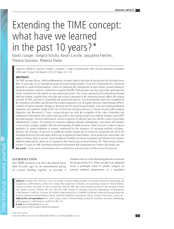
 Gregory Schultz