Leukemia (2001) 15, 1448–1450
2001 Nature Publishing Group All rights reserved 0887-6924/01 $15.00
www.nature.com/leu
Investigation on the role of the ATM gene in chronic myeloid leukaemia
JV Melo1, A Kumberova1, AG van Dijk2, JM Goldman1 and MR Yuille2
1
Department of Haematology, Imperial College School of Medicine, Hammersmith Hospital, London; UK and 2Academic Department of
Haematology and Cytogenetics, Institute of Cancer Research, Sutton, UK
Chronic myeloid leukaemia (CML) is characterised by an indolent, chronic phase (CP) preceding an acute transformation to
blast crisis (BC). While the BCR-ABL fusion oncogene is
strongly implicated in the CP, the molecular changes underlying BC are largely unknown. The ataxia telangiectasia gene,
ATM, is a candidate gene for this transformation because the
complex karyotypes associated with BC of CML suggest that
DNA double-strand break repair is defective and because the
ABL pathway involves the interaction between the Abl and the
Atm proteins. We performed a mutational analysis for ATM in
CML using genomic DNA from 14 CML cell lines and 59 CML
patients in BC. No clearly deleterious nucleotide changes were
observed. A new polymorphism C4138T was discovered which
results in a non-conservative amino acid substitution (H1380Y).
This variant lies in the Atm recognition motif for the Abl protein.
While ATM is unlikely to contribute substantially to CML,
further investigation of the H1380Y substitution should clarify
whether it has any functional effect. Leukemia (2001) 15,
1448–1450.
Keywords: chronic myeloid leukaemia; ataxia telangiectasia; blast
crisis; mutation; genomic instability
Introduction
Chronic myeloid leukaemia (CML) is a myeloproliferative disorder that classically evolves in three clinical stages: a chronic
phase (CP) with a relatively benign phenotype, an accelerated
phase (AP) characterised by increasing refractoriness to therapy, and a final acute transformation or blast crisis (BC) which
is usually unresponsive to treatment and invariably fatal. The
onset of CML is caused by the generation of the BCR-ABL
oncogene in a t(9;22)(q34;q11) chromosomal translocation.
The leukaemogenic nature of the encoded Bcr-Abl fusion protein relies on the fact that it has a constitutively activated tyrosine kinase which ultimately leads to a deregulated proliferation, decreased cell adherence and reduced apoptotic
response to mutagenic stimuli.1 The latter feature is believed
to underlie the invariable evolution of the disease to BC, by
facilitating the emergence of additional deleterious mutations.
However, the known abnormalities in RAS, p53, RB and
p16,2–5 account for less than 30% of the BC.3 The search for
other possible causative mutations is naturally focused on
genes that are relevant to the control of cell cycle, differentiation or apoptosis, and which have been associated with the
genesis of other malignancies.6,7
Ataxia telangiectasia, a recessive disorder due to mutations
in the ATM gene, is characterised by genomic instability and
by early onset of solid and haematological malignancies.8,9
After X-irradiation, the Atm protein directly interacts with the
Abl protein via 10 amino acids encoded by sequence within
exon 3010 and then phosphorylates Abl at Ser 465.11 At the
same time, the Atm protein interacts with and phosphorylates
Correspondence: JV Melo, Dept of Haematology – ICSM, Hammersmith Hospital, Ducane Road, London W12 0NN, UK; Fax: + 44
(0)20 8742 9335
Received 26 February 2001; accepted 18 May 2001
p53 Ser15 via residues encoded within ATM exons 61 to
65.12,13 This Atm protein kinase activity also has Chk2 as a
substrate.14 The last 10 exons encode a region called the PIKlike domain that shows extensive homology to phosphatidylinositol 3- and 4-kinases.15
Sporadic forms of some of the haematological malignancies
that are prevalent in A-T patients are mutated at ATM. In T
cell prolymphocytic leukemia (T-PLL) half of cases acquire
mutations and there is a clustering of missense mutations in
exons 50 to 65.16 ATM is also mutated in a B cell non-Hodgkin’s lymphoma subtype and B cell chronic lymphocytic leukemia.17–19 The number of mutations reported so far is small
but consistent with clustering in exons 50 to 65.
ATM is a candidate gene in CML because the complex
karyotypes associated with BC of CML suggest that there is
genomic instability and because the ABL pathway is strongly
implicated in CML and this pathway involves the interaction
between the Abl and the Atm proteins. We therefore performed a mutation analysis of two regions of the ATM gene in
CML cases and cell lines: the region involved in the interaction with Abl and the mutational cluster region comprising the
last 15 exons of ATM.
Materials and methods
Study population
Fourteen cell lines established from the blast crisis (BC) of
CML were investigated: K562, KCL22, KYO1, KU812,
LAMA84, BV173, EM3, EM2, NALM1, CML-T1, AR230, SD1,
TOM1 and MEG-01. These lines20 were either purchased from
cell repository banks (American Type Culture Collection,
Rockville, MD, USA; European Collection of Cell Cultures,
Winchester, UK; or German Collection of Microorganisms
and Cell Cultures, Braunschweig, Germany) or kindly donated
by their originators. Cells were grown in RPMI1640 (GibcoBRL, Paisley, UK) supplemented with penicillin, streptomycin,
L-glutamine and 10% fetal calf serum.
Peripheral blood leucocytes from 59 patients with CML in
BC and 83 healthy adult volunteers were obtained by red cell
lysis. For some of the latter group, buccal epithelial cells from
saline mouthwash were used instead of or in addition to blood
leucocytes. The BC leucocyte suspensions were further fractionated by density centrifugation and the mononuclear cells
containing 85–95% of blasts were isolated for the mutation
analysis. All samples were obtained after informed consent.
Mutation detection
DNA was extracted by conventional methods and 50 ng was
used in polymerase chain reaction (PCR) amplification using
primers designed for exons 27 to 33 and exons 50 to 65. Exons
are numbered according to Uziel et al.21 Primer sequences
were in the flanking introns (except for reverse primer for exon
�CML and the ATM gene
JV Melo et al
65), and their design was based on the sequence deposited
as GenBank accession number U82828. Forward and reverse
primers (5⬘–3⬘) were as follows: exon 27 = CTT AAC ACA
TTG ACT TTT TGG and GTA TGT GTG TTG CTG GTG AG;
exon 28 = GCT GAT GGT ATT AAA ACA GTT T and GTT
ATA TCT CAT ATC ATT CAG G; exon 29 = TGC CTT TTG
AGC TGT CTT GA and AGA CAT TGA AGG TGT CAA CCA
A; exon 30 = TGA ACA AAA CTT TTT AAA ACG ATG AC
and AGA AGG AAT GTT CTA TTA TTA AAC TCA; exon 31
= CCG AGT ATC TAA TTA AAC AAG and CAG GAT AGA
AAG ACT GCT TAT; exon 32 = CCA GAA CTT ACT GGT
TGT TGT TG and AAA ACA CTC AAA TCC TTC TAA CAA
T; exon 33 = TTC GCA ACG TTA TGG TGG TAT and TGC
TAG AGC ATT ACA GAT TTT TG; exon 50 = GGG CAG TTG
GGT ACA GTC AT and GTA ACA ATG TTT CAC TCC ACC
C; exon 51 = CGT GGG TTG GAC AAG TTT G and TAA GCC
GAC CTT TAG AGC TCC; exon 52 = TTT CCC TGG GAT
AAA AAC CC and TAC ACG ATT CCT GAC ATC AAG G;
exon 53 = CCA CTT GTG CTA ATA GAG GAG C and TTC
CAT TTC TTA GAG GGA ATG G; exon 54 = TGC AGG CAT
ACA CGC TCT AC and CCA GCC TTG AAC CGA TTT TA;
exon 55 = AAA GGC ACC TAA GTC ATT GAC G and GGG
AAT GTT GAA GCC ATC AG; exon 56 = CTT GAC CTT CAA
TGC TGT TCC and TGC CAA TAT TTA GCC AAT TTT G;
exon 57 = CAC ATC GCA TTT GTT TCT CTG and CAA AAT
CCC AAA TAA AGC AGA A; exon 58 = ATT GGT TTG AGT
GCC CTT TG and ATT ATG AAT ATG GGC ATG AGC C;
exon 59 = AGG TCA ACG GAT CAT CAA ATG and AGC TGT
CAG CTT TAA TAA GCC A; exon 60 = ATC CTG TTC ATC
TTT ATT GCC C and CAA AAA TAA AAC CTG CCA AAC A;
exon 61 = CTC AAC ATG GCC GGT TAT G and CAA ACA
ACA TTC CAT GAT GAC C; exon 62 = TGA GGA AGG CAG
CCA GAG and GTG CAA AGA ACC ATG CCC; exon 63 =
TTG ACA ACT TGG TGT GTA ACG and GCC ACA TCC CCC
TAT GTT AA; exon 64 = TCC CCC ATC AAC TAC CAT GT
and GAA CAG TTT AAA GGC CTT GGG; exon 65 = CAA
GGC CTT TAA ACT GTT CAC C and TTG GCA GGT TAA
AAA TAA AGG C. The PCR volume was 10 l, with 0.1 Ci
P-32dCTP in a 96-well plate, using the Hybaid Touchdown
sub-ambient apparatus (Hybaid, Ashford, UK). Where a PCR
product was longer than 250 bp, it was digested with an
appropriate restriction enzyme to yield two or three fragments
prior to single-strand conformation polymorphism (SSCP).
PCR product in 50% formamide was heat-denatured, snapchilled and fractionated for 6 h at room temperature at 15 W
on a 6% polyacrylamide gel containing 10% glycerol. The gel
was dried down on a sheet of 3MM paper and visualised by
autoradiography. Some samples gave rise to abnormal bands.
These samples were re-amplified with the original primers,
subcloned in the TOPO-TA vector (Invitrogen, Groningen,
The Netherlands) following the manufacturer’s protocol, and
sequenced with M13 primers and the Big Dye kit (Applied
Biosystems Warrington, UK) on an ABI Prism 377 DNA
Sequencer (Applied Biosystems). Nucleotides (nt) are identified by counting from the first base of the first codon of ATM
cDNA (GenBank accession number U33841, nucleotide 190).
1449
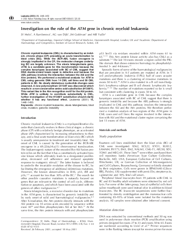
Figure 1
Autoradiogram of SSCP mutation detection for ATM exon
31. Labelled PCR products were digested with EcoRI and fractionated
by SSCP. The arrow indicates an extra band seen in one CML patient
in blast crisis and in chronic phase (BC1 and CP1, respectively) and
in a second CML patient in blast crisis.
Figure 2
Analysis of ATM C4138T by MlnI digestion of exon 30
PCR product. Lane 1: 123 bp DNA ladder (Gibco BRL, Paisley, UK).
Lanes 2 and 3: undigested products. Lanes 4–13: products digested
with MlnI. Vertical arrow (lane 10) indicates a heterozygote.
one of these two patients a DNA sample from diagnosis at CP
was shown to have the same additional band.
Sequencing of the abnormal PCR products identified
C4138T (H1380Y) in NALM1, and C4258T (L1420F) in the
two patients. C4138T results in the loss of an MnlI site and
so can be detected by its resistance to restriction (Figure 2).
Screening of 78 healthy persons identified one heterozygote
with C4138T. SSCP analysis of 83 healthy persons for exon 31
identified one individual heterozygous for C4258T (Table 1).
Discussion
This study was undertaken to assess the contribution of ATM
to the blastic transformation of CML by mutation detection
analysis of two key regions of ATM. DNA from 73 samples
was examined for mutations in ATM exons encompassing the
Table 1
Exon
Polymorphisms in the Abl-binding region of ATM in CML
Nucleotide and
amino acid change
Results
DNA was prepared from 14 CML cell lines and 59 patients in
BC of CML, amplified with primers for ATM exons 27–33 and
exons 50–65 and subjected to SSCP gel fractionation.
Additional bands abnormal in position were detected in exon
30 for NALM1 and in exon 31 in two patients (Figure 1). In
Allelic counts (%)
CML
General population
30
C4138T
H1380Y
C = 145 (99.3)
T = 1 (0.7)
C = 165 (99.4)
T = 1 (0.6)
31
C4258T
L1420F
C = 144 (98.6)
T = 2 (1.4)
C = 165 (99.4)
T = 1 (0.6)
Leukemia
�CML and the ATM gene
JV Melo et al
1450
Abl-binding region and the region most strongly implicated in
sporadic haematological malignancy.
No clearly deleterious nucleotide changes were detected in
the CML samples. However, a non-conservative amino acid
change H1380Y (C4138T) was discovered that lies directly
within the Abl-binding motif DPAPNPPHFP in exon 30 and
two samples had a nearby rare polymorphism, C4258T, causing a non-conservative amino acid change (L1420F) that has
been reported previously.22,23 Both nucleotide changes
detected in the CML samples appeared to have arisen on
one allele.
It is likely that C4138T is a germline variant since the same
variant was detected in a panel of healthy persons. However,
there is evidence that some ATM germline variants recur as
somatic mutations.24 It will be of interest to determine if either
H1380Y or L1420F affect the interaction of the Abl with the
Atm protein. It will also be of interest to test the frequency of
these probable polymorphisms in a fully matched population.
In conclusion, this study has provided evidence that tends
to exclude mutations at ATM from playing any role in CML or
its progression. These results were obtained by SSCP mutation
detection, a technique that was validated by the identification
of known and novel rare polymorphisms in both the CML and
the normal populations studied. Moreover, the same assay has
been used previously to identify frequent sequence changes
in T-PLL25,26 and some B cell malignancies.27 The molecular
basis for the complex karyotypes detected in BC of CML
remains unexplained. Future candidates for investigation
should include genes which when mutated are associated
with genomic instability and elevated risk of myeloid
malignancy.
Acknowledgements
We acknowledge support from the Kay Kendall Leukaemia
Trust and the Leukaemia Research Fund. AGvD was a student
on placement from the University of Manchester. We thank
PS Bradshaw for critically reading the manuscript.
8
9
10
11
12
13
14
15
16
17
18
19
20
21
22
References
1 Deininger MW, Goldman JM, Melo JV. The molecular biology of
chronic myeloid leukemia. Blood 2000; 96: 3343–3356.
2 LeMaistre A, Lee MS, Talpaz M, Kantarjian HM, Freireich EJ, Deisseroth AB, Trujillo JM, Stass SA. Ras oncogene mutations are rare
late stage events in chronic myelogenous leukemia. Blood 1989;
73: 889–891.
3 Feinstein E, Cimino G, Gale RP, Alimena G, Berthier R, Kishi K,
Goldman J, Zaccaria A, Berrebi A, Canaani E. p53 in chronic myelogenous leukemia in acute phase. Proc Natl Acad Sci USA 1991;
88: 6293–6297.
4 Ahuja HG, Jat PS, Foti A, Bar Eli M, Cline MJ. Abnormalities of
the retinoblastoma gene in the pathogenesis of acute leukemia.
Blood 1991; 78: 3259–3268.
5 Sill H, Goldman JM, Cross NC. Homozygous deletions of the p16
tumor-suppressor gene are associated with lymphoid transformation of chronic myeloid leukemia. Blood 1995; 85: 2013–2016.
6 Bose S, Goldman JM, Melo JV. Mutations of the BCL10 gene are
not associated with the blast crisis of chronic myeloid leukaemia.
Leukemia 1999; 13: 1894–1896.
7 Steer EJ, Goldman JM, Cross NCP. Mutations of the transcription
Leukemia
23
24
25
26
27
factor AML1/CBFA2 are uncommon in blastic transformation of
chronic myeloid leukaemia. Leukemia 2001; 15: 476–477.
Meyn MS. Ataxia-telangiectasia, cancer and the pathobiology of
the ATM gene. Clin Genet 1999; 55: 289–304.
Taylor AM, Metcalfe JA, Thick J, Mak YF. Leukemia and lymphoma in ataxia telangiectasia. Blood 1996; 87: 423–438.
Shafman T, Khanna KK, Kedar P, Spring K, Kozlov S, Yen T, Hobson K, Gatei M, Zhang N, Watters D, Egerton M, Shiloh Y, Kharbanda S, Kufe D, Lavin MF. Interaction between ATM protein and
c-Abl in response to DNA damage (see comments). Nature 1997;
387: 520–523.
Baskaran R, Wood LD, Whitaker LL, Canman CE, Morgan SE, Xu Y,
Barlow C, Baltimore D, Wynshaw Boris A, Kastan MB, Wang JY.
Ataxia telangiectasia mutant protein activates c-Abl tyrosine kinase
in response to ionizing radiation. Nature 1997; 387: 516–519.
Canman CE, Lim DS, Cimprich KA, Taya Y, Tamai K, Sakaguchi
K, Appella E, Kastan MB, Siliciano JD. Activation of the ATM kinase by ionizing radiation and phosphorylation of p53. Science
1998; 281: 1677–1679.
Banin S, Moyal L, Shieh S, Taya Y, Anderson CW, Chessa L, Smorodinsky NI, Prives C, Reiss Y, Shiloh Y, Ziv Y. Enhanced phosphorylation of p53 by ATM in response to DNA damage. Science
1998; 281: 1674–1677.
Matsuoka S, Rotman G, Ogawa A, Shiloh Y, Tamai K, Elledge SJ.
Ataxia telangiectasia-mutated phosphorylates Chk2 in vivo and in
vitro. Proc Natl Acad Sci USA 2000; 97: 10389–10394.
Savitsky K, Sfez S, Tagle DA, Ziv Y, Sartiel A, Collins FS, Shiloh
Y, Rotman G. The complete sequence of the coding region of the
ATM gene reveals similarity to cell cycle regulators in different
species. Hum Mol Genet 1995; 4: 2025–2032.
Yuille MR, Coignet LJ. The ataxia telangiectasia gene in familial
and sporadic cancer. Rec Res Cancer Res 1998; 154: 156–173.
Schaffner C, Stilgenbauer S, Rappold GA, Dohner H, Lichter P.
Somatic ATM mutations indicate a pathogenic role of ATM in Bcell chronic lymphocytic leukemia. Blood 1999; 94: 748–753.
Vorechovsky I, Luo L, Dyer MJ, Catovsky D, Amlot PL, Yaxley JC,
Foroni L, Hammarstrom L, Webster AD, Yuille MA. Clustering of
missense mutations in the ataxia-telangiectasia gene in a sporadic
T-cell leukaemia. Nat Genet 1997; 17: 96–99.
Stankovic T, Weber P, Stewart G, Bedenham T, Murray J, Byrd PJ,
Moss PA, Taylor AM. Inactivation of ataxia telangiectasia mutated
gene in B-cell chronic lymphocytic leukaemia. Lancet 1999; 353:
26–29.
Drexler HG. The Leukemia–Lymphoma Cell Line Facts Book. London: Academic Press, 2000.
Uziel T, Savitsky K, Platzer M, Ziv Y, Helbitz T, Nehls M, Boehm
T, Rosenthal A, Shiloh Y, Rotman G. Genomic Organization of
the ATM gene. Genomics 1996; 33: 317–320.
Vorechovsky I, Rasio D, Luo L, Monaco C, Hammarstrom L,
Webster AD, Zaloudik J, Barbanti-Brodani G, James M, Russo G.
The ATM gene and susceptibility to breast cancer: analysis of 38
breast tumors reveals no evidence for mutation. Cancer Res 1996;
56: 2726–2732.
Izatt L, Greenman J, Hodgson S, Ellis D, Watts S, Scott G, Jacobs
C, Liebmann R, Zvelebil MJ, Mathew C, Solomon E. Identification
of germline missense mutations and rare allelic variants in the
ATM gene in early-onset breast cancer. Genes Chromosomes Cancer 1999; 26: 286–294.
Stankovic T, Taylor AM, Yuille MR, Vorechovsky I. Recurrent ATM
mutations in T-PLL on diverse haplotypes: no support for their
germline origin. Blood (in press).
Vorechovsky I, Luo L, Dyer MJ, Catovsky D, Amlot PL, Yaxley JC,
Foroni L, Hammarstrom L, Webster AD, Yuille MA. Clustering of
missense mutations in the ataxia-telangiectasia gene in a sporadic
T-cell leukaemia. Nat Genet 1997; 17: 96–99.
Stilgenbauer S, Schaffner C, Litterst A, Liebisch P, Gilad S, BarShira A, James MR, Lichter P, Dohner H. Biallelic mutations in
the ATM gene in T-prolymphocytic leukemia. Nat Med 1997; 3:
1155–1159.
Schaffner C, Stilgenbauer S, Rappold GA, Dohner H, Lichter P.
Somatic ATM mutations indicate a pathogenic role of ATM in Bcell chronic lymphocytic leukemia. Blood 1999; 94: 748–753.
�
 Martin Yuille