See discussions, stats, and author profiles for this publication at: https://www.researchgate.net/publication/273782512
Ataxia with oculomotor apraxia type 2: not
always an easy diagnosis
Article in Neurological Sciences · March 2015
DOI: 10.1007/s10072-015-2119-z · Source: PubMed
CITATIONS
READS
0
64
5 authors, including:
Andrea Mignarri
Alessandra Tessa
44 PUBLICATIONS 262 CITATIONS
154 PUBLICATIONS 2,098 CITATIONS
Università degli Studi di Siena
SEE PROFILE
Università di Pisa
SEE PROFILE
Antonio Federico
Università degli Studi di Siena
533 PUBLICATIONS 8,927 CITATIONS
SEE PROFILE
All content following this page was uploaded by Andrea Mignarri on 27 March 2015.
The user has requested enhancement of the downloaded file. All in-text references underlined in blue are added to the original document
and are linked to publications on ResearchGate, letting you access and read them immediately.
Neurol Sci
DOI 10.1007/s10072-015-2119-z
LETTER TO THE EDITOR
Ataxia with oculomotor apraxia type 2: not always an easy
diagnosis
A. Mignarri • A. Tessa • A. Federico •
F. M. Santorelli • Maria Teresa Dotti
Received: 24 December 2014 / Accepted: 19 February 2015
Ó Springer-Verlag Italia 2015
Dear Sirs,
Autosomal recessive cerebellar ataxias (ARCAs) represent
a large number of diseases, the most frequent being
Friedreich’s ataxia (FRDA), autosomal recessive spastic
ataxia of Charlevoix-Saguenay (ARSACS), ataxia with
vitamin E deficiency (AVED), mitochondrial ataxias,
ataxia-telangiectasia (AT), and ataxia with oculomotor
apraxia type 1 and 2. Ataxia with oculomotor apraxia type
2 (AOA2) is caused by mutations in SETX, which encodes
a DNA/RNA helicase with multiple complex roles in DNA
and RNA metabolism. Disease onset is usually between 7
and 25 years. AOA2 is characterized by ataxia, occasional
oculomotor apraxia, cerebellar atrophy affecting predominantly the vermis, axonal sensory-motor neuropathy,
and increased a-fetoprotein (AFP) serum level [1, 2].
Here, we describe two AOA2 patients presenting cerebellar atrophy in association with posterior fossa cystic
malformations and mutations in SETX. Of note, both patients manifested AFP elevation and polyneuropathy
2 years after clinical onset.
Two Italian siblings, a 12-year-old boy (FB) and a
10-year-old girl (CB) born to healthy non-consanguineous
parents, were referred to us because of mild gait disturbances and incoordination since they were 9 years. Both
siblings had developed normally with an unremarkable
prenatal and perinatal clinical history. In both children,
A. Mignarri A. Federico M. T. Dotti (&)
Unit of Neurology and Neurometabolic Disorders, Department
of Medicine, Surgery and Neurosciences, University of Siena,
Siena, Italy
e-mail: dotti@unisi.it
A. Tessa F. M. Santorelli
Unit of Molecular Medicine, IRCCS Stella Maris, Pisa, Italy
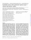
neurological examination revealed cerebellar ataxia, oculomotor apraxia, slight limb dysmetria, hypotonus, and decreased deep tendon reflexes. Brain magnetic resonance
imaging (MRI) showed cerebellar atrophy and cystic malformations of the posterior fossa, including retrocerebellar
arachnoid cyst in one case (FB) and enlargement of cisterna
magna and fourth ventricle in the other sib (CB) (Fig. 1).
Electromyography (EMG) and nerve conduction study
(NCS) were normal in both subjects. Routine blood tests
were unremarkable. Serum assay of AFP (\7 ng/ml), immunoglobulins, vitamin E, and lactic acid was also normal.
Molecular analyses for the expanded GAA tract in the first
intron of FXN and for mutations in coding exons of the
SACS and APTX genes were all normal. Patients underwent
yearly clinical re-evaluation including brain MRI, EMG/
NCS, and AFP assessment. A slow progression of ataxia was
clinically observed, while brain MRI did not detect changes
over time. At 2-year follow-up, serum AFP dosage first revealed increased values (7.3 ng/ml in FB; 14.6 ng/ml in
CB), and NCS documented mild sensory axonal neuropathy.
Novel findings prompted us to reconsider clinical presentation and seek mutations in SETX; the two siblings harbored
the heteroallelic c.839-2A[G and c.6461C[T on the paternal and maternal alleles, respectively. The c.839-2A[G
affects an almost invariable AG consensus sequence at the
donor splice site, it was absent in 200 Italian heathy chromosomes, and it is considered to be damaging in silico based
on splicing site prediction tools (e.g., Splice Site Prediction,
http://www.fruitfly.org/seq_tools/splice.html; HSF, http://
www.umd.be/HSF/; and Netgene2, http://www.cbs.dtu.dk/
services/NetGene2/). Unfortunately, no tissue was available
to test experimentally effects on mRNA splicing in vitro.
The c.6461C[T predicts a novel missense p.Thr2154Met,
affecting a conserved residue in the AAA domain. In
silico analyses (PolyPhen2, http://genetics.bwh.harvard.
123
Neurol Sci
Fig. 1 a, b Brain MRI (a T1 sagittal, b FLAIR axial) of FB shows
cerebellar atrophy and retrocerebellar (right [ left) arachnoid cyst in
the posterior fossa. c, d Brain MRI (c T1 sagittal, d FLAIR axial) of
CB reveals cerebellar atrophy, expanded cisterna magna, and mild
enlargement of the fourth ventricle
edu/pph2/; MutPred, http://mutpred.mutdb.org/) suggest
that the p.Thr2154Met is probably damaging. The mutation
was absent in 300 Italian control chromosomes and not
listed in large collection of human exome studies (http://evs.
gs.washington.edu/EVS/; http://exac.broadinstitute.org/).
The expanding set of genes and the ever growing list of
clinical phenotypes associated with new etiologies emerging in the clinical use of exome sequencing make differential diagnosis of ARCAs a diagnostic challenge. Clinical
evaluation and family history are essential but brain MRI,
EMG/NCS, and selected laboratory analyses are often required to get clues as for a specific diagnosis and prioritize
gene testing. Measurement of serum AFP is considered a
simple and reliable tool in the diagnostic workup: AFP is
almost invariably increased in patients with AT (range
50–900 ng/ml) and AOA2 (range 10–100 ng/ml), and
within the normal range in other ARCAs [3]. Results
gathered in large cohorts of molecularly defined AOA2
patients [1, 2] have shown that AFP concentrations are
elevated ([7 ng/ml) in 99 % of the cases, and levels are
stable during the course of the disease. Moreover, cerebellar atrophy and peripheral neuropathy are almost invariable features of the clinical syndrome, occurring in
95–100 % of patients with AOA2 [1, 2].
In our patients, laboratory and instrumental data were
initially misleading and caused a considerable diagnostic
delay. Indeed, the contemporary absence of both AFP increase and peripheral neuropathy is to consider exceptional. Only during the follow-up, 2 years after
neurological advice had been seeked, pathological serum
AFP levels and NCS were disclosed. Thus, our experience
suggests to re-evaluate laboratory parameters periodically
during follow-up of ARCA patients when suggestive
clinical manifestations are present. This could address the
123
Neurol Sci
proper molecular diagnosis before embarking in more demanding deep sequencing studies. The late appearance of
sensory axonal neuropathy occurred in our patients is intriguing: while marked cerebellar atrophy is present since
onset and does not seem to undergo substantial changes
over time, damage of peripheral axons may be time dependent, and NCS could be useful for monitoring the
progression of the disease. One additional comment
emerges by reconsidering the neuroimaging of our patients.
MRI evidence of posterior fossa malformations has been
reported in congenital cerebellar hypoplasias and ARSACS
[4, 5]. Our neuroimages enlarge the spectrum of MRI
findings associated with AOA2. Therefore, presence of
posterior fossa malformations in addition to cerebellar atrophy should not lead to rule out SETX mutations.
In conclusion, our case study suggests that AOA2
should not be excluded in patients with ataxia and cerebellar atrophy initially lacking AFP increase and/or
polyneuropathy, since these alterations may emerge later as
disease progresses.
Conflict of interest
of interest.
The authors declare that they have no conflict
References
1. Anheim M, Monga B, Fleury M et al (2009) Ataxia with
oculomotor apraxia type 2: clinical, biological and genotype/
phenotype correlation study of a cohort of 90 patients. Brain
132:2688–2698
2. Nanetti L, Cavalieri S, Pensato V et al (2013) SETX mutations are
a frequent genetic cause of juvenile and adult onset cerebellar
ataxia with neuropathy and elevated serum alpha-fetoprotein.
Orphanet J Rare Dis 8:123
3. Schieving JH, de Vries M, van Vugt JM et al (2014) Alphafetoprotein, a fascinating protein and biomarker in neurology. Eur
J Paediatr Neurol 18:243–248
4. Steinlin M (1998) Non-progressive congenital ataxias. Brain Dev
20:199–208
5. Synofzik M, Soehn AS, Gburek-Augustat J et al (2013) Autosomal
recessive spastic ataxia of Charlevoix Saguenay (ARSACS):
expanding the genetic, clinical and imaging spectrum. Orphanet
J Rare Dis 8:41
123
Keep reading this paper — and 50 million others — with a free Academia account
Used by leading Academics

Rommy von Bernhardi
Pontificia Universidad Catolica de Chile

Carlo Semenza
Università degli Studi di Padova

Prof.Dr. Abdulkadir Koçer
Istanbul Medeniyet University

Mamta Singh
All India Institute of Medical Sciences, New Delhi