�Chapter 3
2
Cut Flesh
Chapter 3
Cut Flesh
So that story was ended; somebody began another, about
that satyr whom Latona’s son surpassed at playing the flute,
and punished, sorely, flaying him, so the skin all left his
body, so he was one great wound, with the blood flowing,
the nerves exposed, veins with no cover of skin over their
beating surface, lungs and entrails visible as they
functioned.
— Ovid1
My body is infested with worms,
my skin is cracked and discharging.
— Job 7:52
And you die living, and your bones are no more than what
death has left, and committed to the grave. If this is
correctly understood, every man would find a memento
mori, or a death’s head, in his own mirror; and every house
with a family in it is nothing but a sepulcher filled with
dead bodies.
—Quevedo3
Ovid, Metamorphoses 6: 385–90, translated by Rolfe Humphries (Bloomington: Indiana
University Press, 1973), 141, line breaks omitted.
2
The Authorized version renders the verse as “my skin is broken.” The New English Bible renders
“My body is infested with worms,/and scabs cover my skin,” and adds, in a footnote, “it is
cracked and discharging.” See New English Bible (New York: Oxford University Press and
Cambridge University Press, 1970), Job 7:5 n.
3
Francisco Gómez de Quevedo y Villegas, The Works of Quevedo (Edinburgh, 1798), vol. 1, 35,
quoted in Elizabeth du Gué Trapier, Valdés Leal, [The] Baroque Concept of Death and Suffering
in His Paintings (New York: Hispanic Society of America, 1956), 31, translation modified.
1
�Chapter 3
Cut Flesh
3
Few pictures of the living, conscious body open the skin and reveal what is inside. There
are the medical videos of tiny cameras crawling along passages deep in the body, photographs of
operations done with local anesthetic, and news footage of people stunned by explosions, looking
at their torn bodies. There are also faked wounds, from Night of the Living Dead to Dead Ringers,
from Hermann Nitsch’s bloody performances to Philippine “psychic healing” operations done
without surgical instruments.4 These examples are not only marginal because they are painful to
watch, but because the inside of the body is a powerful sign of death. Even in Beowulf, bodies are
“houses of the spirit” or of “bone,” and any cut can be a “wound door” (bengeat) that allows the
spirit to escape.5 It is normally impolite even to look at the places where the inside of the body
becomes visible—the twilight of nostrils, ears, mouths, anuses, vaginas, and urethras. The inside
is by definition and by nature that which is not seen.
The early Babylonian demon Humbaba is a spectacular counterexample: he had a face
made out of his own intestines.6 (This particular object has an omen inscribed on the back which
For the Philippine practice see Jeffrey Mishlove, The Roots of Consciousness (New York:
Random House, 1975), 150–51 and plate 9.
5
Beowulf 1122. Bengeat is usually translated as “wound door,” “wound gate,” or “wound
offering.” For example Beowulf, An Anglo–Saxon Poem, with a glossary by M. Heyne, edited by
James Harrison (Boston: Ginn, Heath, and Company, 1883), s. v. ben–geat. But see Beowulf, A
Dual–Language Edition, translated by Howell D. Chickering, Jr. (New York: Anchor, 1977), 113:
“Their heads melted,/their gashes spread open, the blood shot out/of the body’s feud–bites.”
6
See further [ ], Journal of the Royal Asiatic Society [ ] (July, 1926), [ ], and R. C. Thompson,
The Devils and Evil Spirits of Babylonia (London, 1904).
4
�Chapter 3
Cut Flesh
4
relates to the divination of intestines.7) In the epic Gilgamesh, Humbaba appears as the Guardian
of the Cedar Forest, a terrifying monster who challenges the heroes Gilgamesh and Enkidu.
When they meet Humbaba screams out an imprecation that is only partly legible in the surviving
versions (Gilgamesh was written in cuneiform on clay tablets), and all the more frightening for
that: “Gilgamesh, throat and neck, / I would feed your flesh to the screaming vulture.” But
Humbaba’s awesome face is oddly hidden from our view because there is a lacuna in the tablet
just when the heroes get their first look at him. Gilgamesh stares, and whispers to Enkidu, “My
friend, Humbaba’s face keeps changing!” The line might also mean “Humbaba’s face looks
strange” or “different” but the image of roiling intestines is clearly legible.8 At this point two
more lines are missing, so that Humbaba’s face, as a
modern editor puts it, is “lost in a break.” How does
one kill a monster who wears his insides on the
outside? Gilgamesh slays him by turning him once
again inside out (“they pulled out his insides
including his tongue”). But how could that have
been done? What was inside Humbaba when his
intestines were already outside?
This is all we know of the battle in Gilgamesh, and
ancient images do not add much more.9 It is
possible that Humbaba was wearing a tegument of
intestines, the way that the Aztec god Xipe Totec,
“Our Lord of the Flayed One,” wore human hides.10
(In this statuette, Xip Totec wears human skin
inside out, with blobs of fat hanging down.) Perhaps
Gilgamesh did not recognize Humbaba’s inversion,
Graham Webster, “Labyrinths and Mazes,” In Search of Cult, edited by Martin Carver
(Woodbridge, Suffolk: Boydell Press, 1993), 23, citing D. Kilmer, “Sumerian and Akkadian
Names for Design and Geometric Shapes,” in Investigating Artistic Environments in the Ancient
Near East, edited by A. C. Gunter (Washington: Smithsonian Institution, 1900), 84 and fig. 1.
8
See The Epic of Gilgamesh, translated by M. G. Kovaks (Stanford, 1985), Tablet V, p. 42.
9
See W. G. Lambert, “Gilgamesh in Literature and Art: The Second and First Millenia,” in An
Farkas et al., editors, Monsters and Demons in the Ancient and Modern Worlds (Mainz, 1987),
37-52, and D. Collon, First Impressions (Chicago, 1988), 178 ff.
10
Mary Miller and Karl Taube, The Gods and Symbols of Ancient Mexico and the Maya (London:
Thames and Hudson, 1993), 188.
7
�Chapter 3
Cut Flesh
5
and killed him the ordinary way, by evisceration: but it may also be that Humbaba already was
eviscerated, and could only be killed by being returned to his normal state. I would rather read
the story that way, since it provides a myth of origin for the question of inside and outside: before
Humbaba, the myth might say, it was still possible to wear intestines on the outside. In
Humbaba’s time, the intestines might come out of the body and swarm over its surface. After
Humbaba, a normal person will die if his intestines are exposed, and a monstrous person will die
if his intestines are hidden. For Humbaba evisceration was life, and death was a paradoxical, fatal
restoration of the insides to their proper place.
In my reading, the story is about the importance of keeping the insides where they
belong. After Humbaba, we all hide the insides of our bodies: we patch and bandage wounds, and
we hide the moments when the inside has to come out. It may seem that Humbaba is one-of-akind monster, but his descendents are still around. He was the ancestor of the archaic Greek
Gorgon, from whose face we have the Medusa
and ultimately our stagy science-fiction monsters
like The Blob and The Thing whose insides spill
out and kill whoever comes near. Just before this
scene in John Carpenter’s version, the Thing had
emerged from a dog by peeling it like a banana.
Then, to defend itself, it had sprouted insectlike
appendages. For the moment, it suits the
monster to use the dog’s face, but in the next few
scenes, it grows large arms and pulls itself up into
the rafters. Carpenter’s film is among the most
extreme and inventive fantasies on bodily metamorphosis in the history of motion pictures.
There is a moment, just before the monster is apparently killed, when it is nothing but a lump of
sodden viscera, as if it were resting from its many transformations. But it senses its attackers, and
pops out eyes to see them better. It assesses the danger it is in, and at the last moment eviscerates
itself, projecting a lamprey-like mouth. In The Thing, bodies move at the speed of thought:
whatever the Thing needs, it can grow in the span of a second or less.
�Chapter 3
6
Cut Flesh
The Thing owes its more purely
visceral moments to movies like The
Blob, which in turn derives from a
British film of the 1950’s, The Creeping
Unknown, which is a story about a
formless mass that coalesces from the
melting remains of an astronaut. The
movie was created in consultation with
Graham Sutherland, who had been
experimenting painting Crucifixions
where carcasses and abstract heaps of organs and bones are draped over the cross and studded
with thorns and nails (chapter 2 has an illustration of one). Like Francis Bacon, Graham
Sutherland had gotten the idea largely from Picasso, who had toyed with the idea of a Crucifixion
of bones and tattered flesh in a series of paintings and drawings done in the late fall of 1932.11 In
this way the inverted bodies of The Thing have their antecedents in British and Spanish painting
of the mid–century, and before them in the Greek Gorgon and perhaps finally in Humbaba, the
eviscerated monster. The Hungarian psychoanalist Sándor Ferenczi’s reading of the Medusa’s
face—as a sign of the female
genitalia, according to him the
most horrifying thing that can be
seen—is one of many possible
meanings of Humbaba’s body.12
(Another Mesopotamian
Humbaba is shown here.) It
must have been a difficult body
to comprehend (as Gilgamesh
said, it kept changing). What did
Humbaba’s genitals look like?
For Picasso’s drawings see Christian Zervos, Pablo Picasso, vol. 8 (1932–37) (Paris: Cahiers
d’art, 1957), nos. 49 and 50. Picasso also made more curvilinear paintings of the crucifixion: see
ibid., vol. 7 (1926–32) (Paris: Cahiers d’art, 1955), nos. 287, 315, and 316, painted in 1930–31.
12
Freud, “Medusa’s Head,” Standard Edition, edited by James Strachey (London: Hogarth Press,
1962), vol. 18, 273–74. In “Infantile Genital Organization,” ibid., vol. 19, 144, Freud credits the
idea to Ferenczi.
11
�Chapter 3
Cut Flesh
7
Was his penis an invagination? Was his anus
a snaking penis? Humbaba’s total,
encompassing, changing inversion and
evisceration is the worst of the catastrophes
that can overtake the body.
To keep the inside hidden is to stave
off death. When a body is opened
accidentally, we do everything possible to
keep it closed. The history of bandages
involves sutures, knots, staples, pins, bolts,
clamps, and other devices, all intended to
make an airtight closure.13 Older suturing
methods include the use of skin substitutes
(leather patches, parchment), tied in place
with animal cords (cat gut, horse hair, silk),
secured with animal paste (fish glue, bone
size). This is a sampler, for doctors, showing
leather bandages. A wound is a deficit of
skin: hence the cure was an excess of skin.14
In premodern Europe, the skin of an animal
that had caused a wound was sometimes
required to heal the wound. The Irish writer
Tomás O’Crohan describes how his leg was saved after he had been bitten by a seal: his friends
killed another seal, and “stuck a lump of the seal’s flesh tight” into the gap in his leg—literally
sculpting his calf into shape with animal meat.15 Suturing has found new resonance in fiber arts,
Early plastic surgey texts are relevant here; see for example J. C. Carpue, An Account of Two
Successful Operations for Restoring a Lost Nose (London, 1816), and C. F. von Graefe,
Rhinoplastik (Berlin, 1818). For a modern work, see The Healing of Surgical Wounds, State of the
Art in the Ninth Decade of the Twentieth Century, edited by Robert S. Sparkman (Dallas: Baylor
University Medical Center, 1985). For the connection between airtight closure and theories of
disease transmission, see Stafford, Body Criticism, 161–62.
14
E. Chambers, Cyclopædia: or an Universal Dictionary of Arts and Sciences (London, 1728), vol.
2, v. “suture.” See also Stafford, Body Criticism, 161.
15
Tomás O’Crohan, The Islandman, translated by Robin Flower (Oxford: Oxford University
Press, 1951), 74–79.
13
�Chapter 3
Cut Flesh
where it has become entangled with the histories of sewing, crocheting, and weaving. The
confluence of torturous devices to mend the body and closures in clothes and fabrics makes an
interesting field of possibilities, and contemporary art often plays the themes of domesticity and
pain against one another, as in works by Annette Messager. Her fabrics and stitched pieces are
overtly domestic, but so are her hanging collections of photographs of body parts, which are
reminiscent of walls hung with arrangements of family photographs. Some, like this one, are in
body-like clumps, and the strings that hold them up are like sutures as much as stitching.
The subject of this chapter is the defense against death as the depiction of pain, because
where viscera predominate over skin pain is no longer the ruling meaning. Suffering is certainly
implied in representations of opened bodies, but it is not the twinge of a sensation on skin (as in
chapter 1), or the sharp pull and compression of limbs turned in violent contrapposto (as in
chapter 2). Pictures of opened bodies conjure states that edge from pain toward shock,
unconsciousness, coma, and death.
Assignment 1: inside-out bodies. Find an artwork that has to do with the inside of the
body and is not a medical illustration, or find images from movies or comics that don’t
try to keep the insides hidden.
8
�Chapter 3
Cut Flesh
9
The fluid flesh
Flesh, as opposed to membranes and skin, is a fluid. According to the linguist Carl Buck,
Russian, Lithuanian, and Lettish (Latvian) words for “flesh” all derive “from the notion of a
filmy, ‘floating’ covering.” They are related to the Sanskrit prefix pluta–, meaning “floating,” and
ultimately to the Indo–European root *pleu–, denoting “flow” or “float.”16 In those languages, as
in Indo–European, flesh is something that floats, a liquid rather than a solid like the bones. Skin
is like a scum congealed on the body’s surface, and muscles are like curds, sunk in its depths.
Greek terms for the body also partake of these liquid metaphors: Greek thumos can mean “spirit”
or “anger,” but it can also be a liquid that “boils and swells in the innards.”17
This way of imagining the body as
a congealed jelly, part fluid and part solid,
has its echoes in 18th century medicine. In
the course of pondering the nature of
bodily “fibers” and tissues, Albrecht von
Haller was struck by the profusion of
“net–like” membranes in the body—some
hard and thick, others “pervaded by a flux
of some juice or liquors,” or formed in the
shape of tunics or coats, cylinders, or
cones. According to Haller these watery
or oily “web–like substances” are one of
two kinds of tissues in the body; the other
is “a mere glue” between that lubricates
them. But on closer inspection, he says, it
proves difficult to tell the “mere glue” from the membranous fibers. Cartilage, for example,
appears to be “scarce any thing else than this glue concreted,” and in the end “even the
Carl Buck, A Dictionary of Selected Synonyms in the Principal Indo–European Languages
(Chicago: University of Chicago Press, 1949), 202.
17
Ruth Padel, In and Out of the Mind: Greek Images of the Tragic Self (Princeton: Princeton
University Press, 1993), reviewed by Jasper Griffin in The New York Review of Books (24 June,
1993), 45. (The quotation is Griffin’s.)
16
�Chapter 3
10
Cut Flesh
filamentary fibers are all first formed of such a transfused glue.” Bones are constructed from a
“compacted gluten,” a fact demonstrated by diseases in which “the hardest bones, by a
liquefaction of their gluten, return into cartilages, flesh, and jelly,” and the opposite happens
when the muscles age and dissolve into “mere jelly,” or when bones, skin, and tendons are boiled
down to make size (animal glue). The development from fetus to adult is the transformation of
fetal “jelly” into the inextricable colloid of membrane and glue, which dissolves again in old age.18
Seen this way, the body’s membranes are nothing but a temporary state, a flux of jellies:
It seems, then, that a gelatinous water, like the white of an egg [aqua albuminosa],
with a small portion of fine cretaceous earth, first runs together into threads, from
some pressure, the causes of which are
not our present concern. Such a
filament, by the mutual attraction of
cohesion, intercepting spaces between
itself and others, helps to form a part of
the cellular net–like substance
[cellulosam telam], after having
acquired some toughness from the
neighboring earthy particles, which
remain after the expulsion of the
redundant aqueous glue. And in this
net–like substance, wherever a greater
pressure is imposed on its scales or
sides, they turn into fibers and
membranes or tunics; and in the
bones, lastly, they concrete with an
unorganized glue. Hence, in general, all parts of the body, from the softest to the
hardest, seem to differ in no other wise than in this, that the hardest parts have a
Albrecht von Haller, First Lines of Physiology, translated by William Cullen (Edinburgh:
Charles Elliott, 1786), vol. 1, 9–14.
18
�Chapter 3
Cut Flesh
11
greater number of earthy particles more closely compacted, with less aqueous
glue; whilst in the softest parts, there is less earth and more glue.19
I would like to take this as a way of thinking about flesh that refuses the distinction between skin
and viscera, inside and outside, hard and soft, in favor of jellies, oils, “albuminous water,” and
viscous matter. This perspective is especially apposite to the visual arts, since there is an affinity
between the slurry of fluids in a surgical operation—the saline wash, blood, and cut tissues—and
the mix of pigments and oils in a painting. Artists who have tried to depict the body’s insides
have often drawn parallels between the body’s thickened liquids and the sticky media of oil
painting; among the painters that come to mind are Francis Bacon, the later Ivan Albright, and
the early Kokoschka. For him the paper or canvas surface is already a skin, and he worries it,
scratching, gouging, and tattooing his figures and backgrounds.20 In 1909 and 1910 his painted or
drawn skin sometimes became translucent, revealing vessels underneath, just as it is possible in
life to see the network of capillaries by using color infrared film, or discern superficial arteries
through light–colored skin (they are not veins, but are made bluish by the intervening yellow fat).
Haller, First Lines of Physiology, op. cit., 14–15, translation modified. The original Latin is from
Haller, Primæ lineæ physiologiæ (Edinburgh: G. Drummond, 1768), 5–6. For a discussion of
Haller’s style, see Bianca Cetti Marinoni, “La Prosa Scientifica,” in Ricerche Halleriane, edited by
Bianca Cetti Marinoni et al. (Milan: n.p., 1984)
20
For this portrait and its immediate context, see Johann Winkler and Katherine Erling, Oskar
Kokoschka: Die Gemälde 1906-1929 (Salzburg: Galerie Welz, 1995), cat. 44.
19
�Chapter 3
12
Cut Flesh
Kokoschka describes his vessels as
nerves, and one of his biographers
thought of écorchés, but they are not
anatomically specific; unlike real
arteries, nerves, or lymph vessels,
Kokoschka’s painted “nerves” are spiky
branched things that do not lead
anywhere.21 Their bunching makes them
more like varicose veins or cleavages in
rock. Around the time of Murderer,
Hope of Women (where a figure is
flayed, revealing the same “nerves”),
Kokoschka’s paintings show an intense
preoccupation with skin, and in the
possibility of scratching it away, tearing
it off, or seeing through it. Portraits such
as the Boy with a Raised Hand are
scraped and abraded, as if seeing itself
had to become so violent that it could
gouge and rasp at the flesh. I have no
simple explanation for his strange
fascination (I doubt it is related to his
thoughts about tensions between the sexes, or to his poverty).22 Something about the skin seemed
wrong to him, and for a while when he was young he invented bodies that are both torn and not
torn, or ripped but miraculously alive and whole.
Kokoschka worked with a deep and broad awareness of history, and many currents
mingle in his work on subcutaneous forms, translucent skin, and themes of flaying or ripping.
His preoccupation with innervation can be traced back to the eighteenth century interest in the
nervous system and the sense of touch, as it is exemplified for instance in Piranesi’s “flayed”
For écorchés, see E. Hoffman, Kokoschka: Life and Work (Boston, 1944), 37–38.
Henry I. Schvey, “Mit dem Auge des Dramatikers: Das Visuelle Drama bei Okar Kokoschka,”
Oskar Kokoschka, Symposion, edited by Erika Patka (Vienna: Residenz Verlag, 1986), 100–113,
especially 111–12.
21
22
�Chapter 3
Cut Flesh
13
ruins, where the architectural forms become metaphors for the opened body.23 Many of Piranesi’s
plates are large (one is literally the size of a person’s body), and the buildings they represent are
irresistably reminiscent of skulls, arms, and torsos—or of the body’s more abstract “architecture,”
its scaffolding, its insulation, its waterproof covering, its often decayed interior. This is a detail of
a tiny figure, far up and in the background of a large illustration; he is examining a colossal wall
of ancient stonework, called opus incertum. Like a fly caught in a web, his limbs are bent into the
angular forms of the stones, and his body is on the point of dissolving into the swirling marks of
the etching needle. (His fingers are already hopelessly entangled.) Everything here has to do with
the body: its flexible skin, its mechanical skeleton, and its unexpected sympathy with stone.
Another source for the awareness of skin’s translucence is the seventeenth–century
painters’ discovery that fingers glow when they are held close to a candle flame. Although the
more familiar examples of this come from Georges La Tour and Michael Sweerts, Adam
Elsheimer is responsible for the strangest image—a scene from Metamorphoses in which Hecate,
who is mortified when a young boy laughs at her, prepares to transform him into a lizard.
23
Stafford, Body Criticism, 58–70.
�Chapter 3
Cut Flesh
14
In Elsheimer’s version the body is already glowing with the heat of metamorphosis, as his bones
begin to liquefy into amphibian softness. In the Metamorphoses the boy, Stellio, becomes a gecko;
�Chapter 3
Cut Flesh
15
in Elsheimer’s picture he is on his way—he’s a wavering, lacertine mixture of a human, a softened
candle, and a salamander.24 In the nineteenth century the incandescent flesh of Dutch scenes of
sensualism became one of Ingres’s broadening range of historical allusions. His melted-wax
fingers, which Robert Rosenblum noted as his special obsession, owe something to the candent
fingers and tapers in Michiel Sweerts and Georges de la Tour, and before them to the entire
tradition of translucent bodies that began with Caravaggio and Elsheimer.25
Since the laye 1980s there have been various attempts to show the body’s fluids, and the
cuts that make them accessible. Sally Mann’s photographs explore the fluids and bodies of
children; Kiki Smith juxtaposes
photos of the skin with pools of
blood; Andres Serrano’s work
involves both the fluids
themselves (including urine and
blood) and their appearance on
the body’s cut surface (in the
series of morgue photographs).26
“There is this great beauty of the
color of meat,” Francis Bacon
reminds his interviewer, David
Ovid, Metamorphoses 5:437–60. For the identification of the gecko see Carl Gotthold Lenz,
Erklärende Anmerkungen zu Ovids Metamorphosen, vol. 1. From the series Erklärende
Anmerkungen zu der Encyclopädie der lateinischen Classiker, vol 3, part 1. (Braunschweig: Schul–
Buchhandlung, 1792), 349: “Der Stellio… ist eine kleine Eidesche, man glaubt, Lacerta gecko L.”
25
For Ingres’s “obsession” see Robert Rosenblum, Jean-Auguste-Dominique Ingres (New York:
Abrams, 1967). For other sources of “waxy painted figures,” see Stafford, Body Criticism, 78. For
Sweerts see Rolf Klutzen, Michael Sweerts: Brussels 1618-Goa 1664, translated by Diane Webb
(Doornspijk: Davaco, 1996).
26
For Sally Mann, see for example Still time: Sally Mann (New York: Aperture, 1994); for Kiki
Smith, see her work with David Wojnarowicz, especially Untitled (1982-91), reproduced in
Micholas Mirzoeff, Bodyscape: Art, Modernity, and the Ideal Figure (New York: Routledge, 1995);
for Serrano, see Andres Serrano: Works, 1983-1993, edited by Patrick Murphy, with essays by
Wendy Steiner and others (Philadelphia: Institute of Contemporary Art, 1994); and Andres
Serrano: Body and Soul, edited by Brian Wallis, with essays by bel hooks and others (New York:
Takarajima, 1995).
24
�Chapter 3
Cut Flesh
16
Sylvester.27 The early paintings are about cutting, or slaughterhouses, and they display vast
monstrous carcasses, strings of vertebrae that could only come from dinosaurs, and Popes whose
mouths are bloodied as if they
had been assaulted. After the
1960’s, however, Bacon achieved
a synthesis of inside and outside,
surface and viscera, which is
unique in the history of art. One
might say Bacon’s later paintings
still have a notion of skin, though
it is not a surface anymore, but a
sense of translucence. The faces
appear to be several inches thick,
and we are invited to see through
to… to what? A concoction of
floating veils, oily smears, sodden
cloths, greasy spills, damp papers
laid one on top of another. The
canvas sometimes looks printed,
as if Bacon had rubber–stamped
and blotted it, and other passages
look sharp, like pieces of
splintered bone drifting among
loosened tissues. When the flesh is deep, it may be a pool of slurred organs, and those organs
seem to include scraps of skin, so that the face is effectively left without any covering. In this
painting Michel Leiris’s face is mixed with itself: his body’s armor has retreated into his body,
and mingled with it. Bacon’s best images are awash in all the body’s parts, private and public,
human and mechanical, nameless pieces of anatomy and painful pieces of flesh, autonomous
organs and dead bones.
Francis Bacon Interviewed, op. cit., 46. Willem de Kooning’s nudes could also be discussed in
this context, especially those that are manifestly liquid and without secure boundaries. See for
example Janet Hobhouse, The Bride Stripped Bare: The Artist and the Female Nude in the
Twentieth Century (New York: Weidenfeld and Nicolson, 1988), 236-60.
27
�Chapter 3
17
Cut Flesh
Bacon is almost alone, I
think, in wanting to break down the
dichotomy, and to see everything
by seeing it all at once. Most of the
history of pictured and sculpted
bodies has to do with skin.
Figurative sculptures, for example,
tend to identify the skin with the
body, in that the texture and
density of the bronze or stone is
continuous from the skin to the
heart of the statue. (Large statues
may be hollow, but their
thicknesses are not skins. What is
missing from a monumental
bronze sculpture is the organs: the
thickness of skin, fat, muscles and
bones remains, but the sculpture
has been hollowed like a mummy.)
The historical antecedents of
Bacon’s disheveled bodies are the
Renaissance Venetian experiments
with the softness and depth of the
skin, especially, I think, some
paintings by Titian where the
body’s imperfect opacity is
represented by translucent layers of
paint. Titian’s glazes—some of
them rubbed until they are almost
invisible—remind a viewer of the process of painting, which builds from the bony white gesso
through thickening layers to a final paper-thin membrane. Such paintings make body into a
sequence of oiled sheets. In the late paintings, the delicate veils of flesh are also cut by sharp dry
impasto, so that the body becomes a mix of hard and soft, very much as it is in Bacon.
�Chapter 3
Cut Flesh
18
Bacon confuses the body’s layers, just as the patiently built layers of Venetian oil painting
were tumbled together in the thick, impetuous alla prima painting that began in the mid-19th
century.28 The works I consider in this chapter, which break the decorum that normally hides the
body’s layers, are not central to Western art. Instead they help define the mainstream by showing
what happens when the rules, like the body’s membranes, are broken.
Assignment 2: fluid flesh. Find artworks that show the skin or flesh as fluid, translucent,
or bruised. Try analyzing them using the concepts and examples in this section.
Resisting seeing the inside
I will not begin with the history of fine art images that represent viscera, both because the
history has been told, and because it remains marginal to much that is interesting about the body.
The exceptions—medical images of unusual power or accomplishment—are rare. Erwin
Panofsky has chronicled some in Tomb Sculpture, and isolated artists such as Hans Baldung have
On alla prima painting, see Max Doerner, Malmaterial und seine Werwendung im Bilde (1921),
translated as The Materials of the Artist (London: Granada, 1973–77). For a recent appreciation
of Doerner see Thierry De Duve, Pictorial Nominalism: On Marcel Duchamp’s Passage from
Painting to the Readymade, translated by Dana Polen and the author. Theory and History of
Literature, vol. 51. (Minneapolis: University of Minnesota Press, 1991), 175–85. De Duve is
interested in parallels between Duchamp’s readymades and the tradition of painting, and the
parallel I am drawing here between Bacon and Titian is not without affinities to Duchamp’s
lingering interest in paint, palettes, tubes, and the rudiments of painting. De Duve has rethought
these ideas in Kant After Duchamp (Cambridge, MA: MIT Press, 1996).
28
�Chapter 3
Cut Flesh
19
made persuasive mixtures of nauseating decay and perfect
beauty.29 the Japanese Nine Stages of Decomposition are
another example. In Europe one of the most extravagant
inventions is Juan de Valdés Leal’s pair of paintings titled
Los Jeroglíficos de las postrimerías (Hieroglyph of Our Last
Days, c. 1672–77), a catalogue of vanitas symbols and
corruption.30 The paintings illustrate the 13th century legend
of the Three Living and the Three Dead, in which three
riders come upon three corpses, one freshly dead, another
decomposing, and the third a skeleton. One of them says to
the three living: “What you are, we were; what we are, you will become.”31 In order to drive
home the point Valdés Leal puts the most horrifying figure in the foreground, in the manner of
medieval and Renaissance tomb sculpture. The foreground corpse is en transis—in the process of
liquefaction—and so he is a stronger reminder of the painting’s moral than the dried skeleton or
Erwin Panofsky, Tomb Sculpture (New York: Abrams, 1992). For Hans Baldung, see Robert
Koch, Hans Baldung Grien, Eve, the Serpent, and Death [bilinguial French and English],
Masterpieces in the National Gallery of Canada, no. 2 (Ottawa: National Gallery of Canada,
1974).
30
Duncan Theobald Kinkead, Valdes Leal, His Life and Work (New York: Garland, 1978).
31
Raimund van Marle, Iconographie de l’art profane (La Haye, 1932), vol. 2, 383–84, quoted in
Elizabeth du Gué Trapier, Valdés Leal, Spanish Baroque Painter (New York: Hispanic Society of
America, 1960), 57.
29
�Chapter 3
Cut Flesh
20
the fresh corpse. But even here, in a
painting so extreme that it was
even disparaged by an historian
who wrote a book on Valdés Leal,
there is little more than a hint of
what lies beneath the skin.32 As
Panofsky’s examples show, a
corrupted skin is enough to show
that the body is decomposing. In
Los Jeroglíficos de las postrimerías,
worms thread their way through
the skin, toads lick at its orifices,
and flies settle on its desiccating
remains. For Valdés Leal, as for
Hans Baldung, the decomposing
body is literally only skin and
bones. Hans Baldung’s figures of
Death are skeletons dripping with
skin, rather than organs. Viscera
are unrepresented and often
unimagined, even where there is
evidence that the artists had spent
time looking at rotting animal or
human bodies.
In such cases the repressed
inside of the body often returns in
the form of metaphor. If we were
to look for signs of viscera, one of the best places would be Dutch still life painting, where meat
and fruit are commonplace reminders of the body’s ingredients. Pieter Aertsen, Frans Snyders,
Willem Kalf, and other painters have an affection for objects that have both skin and “viscera”:
In Elizabeth du Gué Trapier’s opinion, “Had the directors of the chairty hospital wished to
hasten the end of their impoverished clients they could not have chosen more effective subjects
as decorations for the new church than the hieroglyphs.” Valdés Leal (1956), op. cit., 34.
32
�Chapter 3
Cut Flesh
21
peeled oranges, torn bread, mincemeat pies with flaky crusts, translucent sausages, melons with
dried rinds and juicy insides—not to mention freshly butchered joints.33 There are also reminders
of the body’s fluid insides: carafes of red wine, pats of butter, tubs and basins of lard, pitchers of
milk, bowls swimming with egg yolks. Just as Balthus’s still lifes reveal relationships between
Norman Bryson, Looking at the Overlooked, Four Essays on Still Life Painting (Cambridge,
Mass.: Harvard University Press, 1990), 96–135, reads some of these images as one end of a
spectrum from ascetic inhibition to chaotic excess, and in this context I would note that bodily
metaphors function most strongly as signifiers of excess.
33
�Chapter 3
22
Cut Flesh
bodies, any one of the Dutch still-life painters could be
studied for their ways of setting out the relationship
between elements in the body. In the 17th and 18th
centuries, still life may have been the best excuse for
artists who wanted to remain in the fine art tradition and
still depict the opened body; in contemporary art, a wide
range of materials and forms can evoke the body’s insides
without needing to allude to the death of any individual
person. In that sense, contemporary soft sculptures,
especially those made of perishable materials are the
descendants of Baroque still life—works like Rachel de
Joode’s Soft Inquiry XI or Jessica Drenk’s Soft Cell Tissue.
The resins, perfumes, oils, and pelts of fiber art speak
about the body’s insides without leaving the field of fine
art, just as their painted equivalents did in the
seventeenth century.
Dissecting
To actually depict viscera, it is
necessary to partly abandon fine art
painting and drawing in favor of medical
illustration. Dissection is an especially
powerful tool: literally, it is a medical
specialty, with its own terms and
techniques distinct from surgery; and
figuratively, it can stand for any act of
systematic analysis, from a tentative
“probe” to the “sharpest” critique. It can
be argued that pictures of dissections are
the clearest examples of the desire to see
through or into anything, whether it is a
body, or—by metaphorical extension—an
idea. A picture of a dissected body can
�Chapter 3
Cut Flesh
23
also be experienced as a literal
version of a common trait of
seeing, in that the mind’s desire to
analyze and the eye’s desire to
pierce and separate are kindred
motions, and they are both
embodied in cut flesh. Dissection
is therefore one of the most apt
metaphors for the experience of
intense, directed thinking or
seeing: the Latin perspicere, from
which we have the words
“perspicuous” and “perspective,”
means seeing through, as in
piercing a fog or penetrating a
dark night. Analytic thought often
borrows those visual metaphors,
but ultimately perspective,
piercing, and penetrating may all
depend on the fundamental desire
(or fear) of seeing through the
skin.34
The concept of dissection
is philosophically versatile. We
speak of dissecting, revealing, opening, or cutting through to a problem, and the narrative form
known as the anatomy commemorates its bodily origins by avoiding linear or systematic
exposition in favor of detailed examination. When critical inquiry approaches dissective
methods, it relinquishes optical metaphors in favor of bodily ones. Robert Burton’s Anatomy of
Melancholy exhibits a wry awareness of the somatic model of thought in its subtitle, where
Burton declares melancholy will be “philosophically, medicinally, historically, opened and cut
Other, rival, interpretations of perspective are given in my Poetics of Perspective (Ithaca, N.Y.:
Cornell University Press, 1994), chapter 1.
34
�Chapter 3
Cut Flesh
24
up.”35 Anatomizing has to do with pain, shock,
and death: hence, I believe, the “pain” of
analytic thought and of intense vision: they
devolve from the partial failure of the covering
metaphor. When Wittgenstein speaks of the
unpleasantness and labor of philosophic
thought—its harshness, its closesness, its
“slippery” quality—he is not far from speaking
openly about its pain.
Pictures of dissections are the most intimate
and exact record of those motions of the mind,
and it helps to look at them with the medical
terminology in mind. Medicine dissects
dissection into a half–dozen specific
procedures, each of which can function as a
metaphor for analytic thought. There is the
uncovering of a specific organ in situ (known
as prosection), as well as its removal (excision
or exeresis). (The image of the hand is an
expert prosection, a kind of virtuoso sculpture,
preserved in the Royal College of Surgeons,
London, and reproduced in R.M.H. McMinn’s
Color Atlas of Human Anatomy.) A doctor can
tie together two separate organs (grafting),
divide the healthy from the pathological
(diaresis), or implant a foreign body
(prosthesis).36 Each of these terms names a way
Robert Burton, The Anatomy of Melancholy, What It Is. With all the Kindes, Causes,
Symptomes, Prognosticks, and Severall Cures of It. In Three Maine Partitions with their Severall
Sections, Members, and Subsections. Philosophically, Medicinally, Historically, Opened and Cut
Up. By Democritus Junior (Oxford: John Lichfield and James short for Henry Cripps, 1621).
36
E. Chambers, Cyclopædia: Or an Univrrsal Dictionary of Arts and Sciences, second edition
(London, 1738), 209, and R. J. C. [de] Garengeot, A Treatise of Chirurgical Operations, translated
by M. André (London, 1723), 2, both cited in Stafford, Body Criticism, 485 n. 6, 7.
35
�Chapter 3
Cut Flesh
25
of thinking about a problem: Jacques Derrida’s neologisms, such as “différance,” are prostheses
in the text of philosophy—implants, which may or may not be assimilated. (They may “take,” or
they may be rejected.) Each term also has its corresponding narrative forms. Montaigne, for
example, touches on most of these strategies in the course of failing to speak in a logical fashion
about his subjects.37 Given the confluence of words for dissection, seeing, and thought, it is not
surprising that these words are also well–fitted to describe the process of depicting bodies. Many
of the ways artists build bodies have their parallels in the ways doctors disassemble bodies. An
artist might separate one shape from another, in order to make it clearer (thus performing a
prosection), or assemble an image by placing disparate forms on top of an existing field, collage–
fashion (thus adding prostheses to an organic base, as in this collage by Claudia Huidobro). All
imagemaking involves diaresis since it is the act of identifying useless, “pathological” forms and
salvaging interesting, “healthy” ones. In both medicine and painting, part of the challenge is to
create a structure of clearly articulated forms out of a state of incoherence and confusion.
37
Hugo Friedrich, Montaigne, second edition (Bern and Munich: Francke, 1967), 305–36.
�Chapter 3
Cut Flesh
26
Assignment 3: dissection. Analyze an artwork in terms of the metaphors of dissection:
prosection, exeresis, prosthesis. It doesn’t have to be a medical image, or a naturalistic
picture of a body: it can be any artwork that divides and examines its subject.
Medical illustration
Older medical illustration is a better place to study these ideas than contemporary
medical imaging, because the latter has been built, over the last two centuries, on ideals of
simplicity and schematization. The kinds of questions asked in the literature on medical imagery
have to do with the density and arrangement of information, rather than the meanings of the
images as representations of the body. How much of the tangle of tissues should be depicted in a
single illustration in order to retain “readability”? To what degree is idealization preferable in
order to help the eye “process information”? The ongoing interest in “painless” computer–
assisted images, together with these questions of efficient visual communication, can be read as a
double resistance: on the one hand, medical imaging represses the complicated and unsettling
presence of the opened body, and on the other hand, it resists the potential power of the images
themselves by draining their visual interest, leaving a pure and uninteresting residue.38 One
Magnetic Resonance Imaging, A Reference Guide and Atlas (Philadelphia: J. B. Lippincott,
1986); Navin C. Nanda, Atlas of Color Doppler Echocardiography (Philadelphia, 1989); Howard
Sochurek, Medicine’s New Vision (Easton, Pennsylvania: Mack Publishing Company, 1988)
38
�Chapter 3
27
Cut Flesh
might say, for example, that if contemporary digital medical images were to become more
intricate (or even if their resolution were to increase) they would become more effective at
expressing pain, so that the simpler visual displays commonly in use serve both to repress
thoughts of the living body and to
avoid being seen as pictures. The
former quality has been stressed by E.
J. Cassell, who describes the recent
history of medical illustration as a
matter of “depersonalization,” and
connects it to what he sees as the
medical profession’s reluctance to
come to terms with the suffering of
patients.39 In any case a more
reflective history of recent medical
imaging would have to take into
account the lingering feeling of
discomfort and pain that
accompanies even the most artificial
and highly processed images; it might
be argued that computer–generated
images of the body are likely to cause
uneasy twinges of recognition, since
the observer is likely to be reminded
about what such images exclude.40
Older medical illustration is not
different from contemporary imaging
in its content so much as in its attention to the body’s more unruly or anatomically meaningless
forms, and for that reason it is more often the site of interesting visual thinking about the body’s
E. J. Cassell, The Nature of Suffering and the Goals of Medicine (New York and Oxford, 1991),
195 ff.
40
For further examples see my “Art History and the Criticism of Computer–Generated Images,”
Leonardo 27 no. 4 (1994): 335–42 and color plate. For a good recent summary of medical
imaging, see Robert P. Crease et al., “Biomedicine in the Age of Imaging,” Science 261 (30 July
1993): 554–61.
39
�Chapter 3
Cut Flesh
28
insides. Cassell distinguishes older from newer
medical illustration in part by pointing to the
“metaphorical” content of some older
illustrations. But when a Renaissance medical text
shows a woman with a prosected bladder
accompanied by picture of water running under a
bridge, or a skeleton contemplating a skull, is it
operating so differently from contemporary
medical illustrations? More recent medical
treatises gain a powerful metaphorical meaning of
their own by displaying fragments of bodies rather
than whole revivified bodies. A comparison might
be drawn between that fragmentarian program
and atlases of architectural details, mechanical
movements or machine parts. In each case a single illustration will normally show only a part of a
larger mechanism, and it will decline to depict the totality of the object or its function in relation
to other objects. For these reasons, I would not want to
cast the history of medical illustration as an increase of
interest in efficient visual communication as opposed to
an interest in pure visual incident, nor as the gradual
ascendance of a scientific mentality over a
“metaphorical” or religious one. Instead, it seems that
the question is which meanings are excluded, and which
permitted.
Andreas Vesalius’s 16th century version of the
dissected body, a traditional starting–place for histories
of anatomic illustration, is robust and curvilinear (The
skeleton contemplating a skull is also from Vesalius).41
Vesalius’s woodcut lines are harsh and strong, and they
have spring and tension—what Hogarth later called the
Nancy Siriasi, “Vesalius and Human Diversity in ‘De humani corporis fabrica’,” Journal of the
Warburg and courtauld Institutes 57 (1994): 60-88.
41
�Chapter 3
Cut Flesh
29
“the inimitable curve or beauty of the S undulating motion line.”42 Another version of the body,
best realized by the 18th century anatomist Bernard Siegfried Albinus, is elegant, slim, and
perfectly measured.43 Albinus’s figures are engravings rather than woodcuts, and they have some
�Chapter 3
Cut Flesh
30
the most attenuated and beautifully controlled lines in the history of that medium. (Even, or
especially, when the subject is intentionally sublimely horrible, as it is here.) Together, Vesalius
and Albinus may be taken as paradigmatic images for the Western history of anatomic
illustration; the two choices they represent ruled much of the succeeding history. But at the same
time it would not be entirely correct to account for the difference by describing Vesalius as
proto–Baroque or Albinus as proto–Neoclassical.
The Vesalian body is a rough attempt to describe the opened body itself, to minimize the
resistance to representing death. He denies the fact of death outright by representing a living
(sometimes a sleeping) figure.
A live model, displaying its
own viscera, is the paradox of
choice in much of older
medical illustration as well as
medical sculpture. But here
we need to be cautious,
because few anatomical
illustrations present figures as
if they were unambiguously
alive. How, after all, does an
eviscerated figure sleep? How
relaxed can a flayed figure
hope to be? In medieval
anatomic illustration,
“Wound Men” show their
opened bodies with the
indifference of a
demonstrator pointing to an
actual corpse.44 That tradition
is strange enough, but it
became openly paradoxical
when Renaissance naturalism
For example Peter Murray Jones, Medieval Medical Miniatures (Austin: University of Texas
Press, 1984), figs. 27 and 51.
44
�Chapter 3
31
Cut Flesh
made it possible to give the Wound
Man an expression, so that he might
begin to show some psychological
awareness of his position. Charles
Estienne’s work contains
inappropriately elegant scenes of
women lying or sitting in their beds,
in exquisite maniera contrapposto,
with their skin cut away and their
entrails hanging out.45 Some figures
in Giovanni Valverde’s Anatomia del
corpo humano retain the medieval
obliviousness to their own suffering,
but others evince an odd sense of
discomfort. In one, a figure grasps his
skin in his teeth, and he turns aside
and winces—partly from the effort of
pulling the skin, and partly, it seems,
from pain. Valverde’s description
echoes that strange possibility
without quite saying what is
happening: “This figure,” the caption
reads, “shows where the intestines
are, and demonstrates the net of
vessels, and turns backward, and pulls
with its teeth.” How are we to read such an image? I would prefer to think there was some
46
awareness on the artist’s part that the Wound Man convention was illogical, and he may have felt
some empathy with the figure he was drawing. Earlier, Vesalius had tried to solve the problem by
Charles Estienne, La dissection des parties du corps humain divisee en trois livres (Paris: Simon
de Colines, 1546); Jean-Claude Margolin, Science, humanisme et société: Le cas de charles Estienne
(Paris: Klincksieck, 1993).
46
“Questa figura, mostra il sito de gl’Intestini, & la reticella spiegata, & volta verso dietro, & tirata
co denti.” Giovanni Valverde, Anatomia del corpo humano (Rome: Antonio Salamanca and
Antonio Lafreri, 1560), book III, p. 93.
45
�Chapter 3
32
Cut Flesh
representing an eviscerated torso as if it were a marble sculpture that had been truncated along
the lines of the Farnese torso: where the abdomen is cut it reveals organs, but where the limbs are
fractured they show blank marble. The figure even has the kind of “Roman joints” by which
sculptures are assembled: one side of a limb is carved into a peg (that is, a bonelike structure),
and the other is drilled (so that it resembles a deboned carcass).
The inheritors of these wound men are 20th and 21st century depictions of cyborgs with
opened or disassembled bodies that they stare at impassively. (As in this cover of Galaxy from
1954.) Or Yue Minjun’s canvas
from 2009, in which a grimacing
man demonstrates that his
insides are from an anatomy
textbook (next page).
All medical illustration
retains these paradoxical
features, because it is rarely
unambiguously entirely clear if
the body is alive and
anaesthetized, or a corpse, or a
cadaver, or only a schematic or
mnemonic for the body.
Fundamentally, the situation is
irreparable, because the uncanny
look of anatomic illustration
proceeds directly from the
uncanniness of the corpse, which
trespasses on the places of the
living until it is buried.47 Vesalius’s inhuman robustness denies the body by strengthening it into
sculpture, and that denial is even more effective because it led his artists toward the “undulating
motion lines” that have often been read as the fruits of close observation even though they have
only a fortuitous, intermittent correspondence with the body’s forms. Albinus’s taut linear
The “dead body in a room problem” also leads into the history of funerary installations; the
subject is explored in my Things and their Places: The Concept of Installation from Prehistoric
Tombs to Contemporary Art, work in progress.
47
�Chapter 3
Cut Flesh
33
manner denies the body by weakening it into a geometric diagram, and his strategy is most
persuasive when his exactitude forces the viewer into a false sense that the pictures are close to
reality. There is pain in both texts, but it is muted—in the one case by sculpture, and in the other
by geometry.
Assignment 4: bodies looking at their insides. Find an artwork in which a person or
cyborg looks at their own insides, and intepret it in relation to the history givne in this
section.
�Chapter 3
Cut Flesh
34
�Chapter 3
35
Cut Flesh
Toward pain and incoherence
It is nearly impossible to come to terms with the inside of the body. Organs and cut flesh
are virtually excluded from fine art in favor of the abstract pairing of skin and skeleton, and in
medical illustration they are largely
replaced by subtle abstractions that turn
the body toward the domains of
geometry, architecture, or sculpture—or
toward the weightlessness of the screen.
Metaphorically, such images elide the
real hazards of analytic thought. Yet
there are pictures that do justice to the
fact that dissective thinking is harsh and
uncompromising, and that it takes place
in a domain of radical complexity.
Among the most accurate
representations of the body’s inside
before the invention of photography are
plaster and bronze écorchés (flayed
figures), some of them made directly
from wax casts of muscles and bones.48
Lodovico Cardi’s écorché called The
Beautiful Anatomy (La bella Notomia),
made shortly before 1600, is the usual
starting-place for the history of
Renaissance anatomical models, but wax models are attested in Pliny, and from 1200 to 1600
many anatomical wax ex-votos, called bóti, were made in honor of the Madonna in Or
For écorchés in general, see L. Price Amerson, “The Problem of the Ecorché: A Catalogue
Raisonné of Models and Statuettes from the Sixteenth Century and Later Periods,” PhD
dissertation, Pennsylvania State University, 1975, unpublished; also my “Two Conceptions of the
Human Form,” op. cit.; and the history in Henry Meige, “Une Révolution anatomique,” Nouvelle
Iconographie de la Salpêtrière 20 (1907): 174-83 (which concerns an écorché made by the French
art anatomist Paul richer).
48
�Chapter 3
Cut Flesh
36
Sanmichele.49 There are major
collections of wax models in
medical museums in London,
Paris, Vienna, Budapest, and
Florence. The Alfort veterinary
museum in Paris has preserved
flayed bodies by a 19th century
anatomist named Fragonard.50
One preserves a girl riding a
horse, both flayed. Many of these
institutions have objects that are
more inventive and less
predictable than the popular
exhibitions by Günther von
Hagens, the “Plastinator.” Some
objects have a really unsettling
degree of realism—they are fitted
with human hair, and arranged
on real linen beds.51 (Some of the
female figures in the Specola
museum in Florence can be disassembled, all the way down to a fetus in a womb.) The Humboldt
University in Berlin has an extensive collection, including preparations made by injecting metal
Pliny, Historia naturalis 35:6 and 153; the Madonna was said to have miraculous powers,
according to Lanza et al., p. 18. On wax models in general, see E. J. Pyke, A Biographical
Dictionary of Wax Modellers (Oxford, 1973). A supplement was published in London, 1981.
50
Honoré Fragonard, active c. 1766-1771; the cadavers are kept in the École Vétérinaire d’Alfort,
Paris.
51
See J. Adhémar, “Les Musées de cire en France. Curtius, le ‘banquet royal,’ les têtes coupées,”
Gazette des Beaux-Arts 92 no. 2 (1978): 206–207; M. Lemire, Les Modèles anatomiques en cire
colorée du SVIIIe siècle et du XIXe siècle (Paris: Musée National d’Histoire Naturelle, Laboratoire
d’Anatomie Comparée, 1987); Peter Klerner Knoefel, “Florentine Anatomical Models in Wax
and Wood,” Medicine nei Secoli 15 (1978): 329–40; Benedetto Lanza et. al., Le Cere Anatomiche
della Specola (Florence: Arnaud, 1979) (there is also a second volume on the pathological
anatomies); and Michel Lemire, “Fortunes et infortunes de l’anatomie et des préparations
anatomiques, naturelles et artificielles,” in L’âme au corps: arts et sciences 1793-1993, exh. cat.,
edited by Jean Clair (Paris: Réunion des musées nationaux, 1993), 70-101.
49
�Chapter 3
Cut Flesh
37
into the veins, and even
preparation of the stretched and
preserved face of an infant.
(These illustrations are from the
excellent catalog Theater der
Natur und Kunst.) As I
mentioned in chapter 1, The
Musée de l’Hôpital Saint-Louis
has a large permanent display of
dermatological diseases sculpted
a mixture of beeswax and resin.52
(And there is now a “virtual museum” version of that onceinaccessible place.) At the limits of this kind of replicative realism
are cadavers that have been preserved by injection or varnishing;
they are the ancestors of latex body casts by John De Andrea and
other contemporary artists. The Renaissance practice was to coat
corpses in honey; the eighteenth-century anatomist Frederick
Ruysch preserved bodies more permanently in a mixture of talc,
wax, cinnabar, wine and black pepper.53 Like De Andrea’s
sculptures, the most accurate preserved bodies are astonishing
from a distance, and repellent close up: in De Andrea’s case, the
For the dermatologic models: Stafford, Body Criticism, 281–83; Georges Solente, “Le Musée de
l’Hôpital Saint-Louis,” American Journal of Dermatology 5 (October 1983): 483–89, especially
486 for the identification of the medium; and Ernest Besnier, Alfred Fournier, et al., A Pictorial
Atlas of Skin Diseases and Syphilitic Afflictions in Photo-Lithochromes from Models in the
Museum of the Saint Louis Hospital, Paris, with Explanatory Woodcuts and Text (London:
Rebman Ltd., 1904); and Ernest Besnier, La Pratique dermatologique (Paris: Masson et Cie.,
1904) 4 vols., with color reproductions after the casts, keyed to numbers in the collection.
Fournier was an authority of syphilis; see his Les Chancres extra-génitaux (Paris: Rueff et Cie,
1897).
53
His dissections served as the models for Füssli’s illustrations to Jean-Jacques Scheuchzer’s
monumental Physique sacrée. See Stafford, Body Criticism, 240, and J.-J. Scheuchzer, Physique
sacrée, ou histoire naturelle de la Bible (Amsterdam, 1732 - 1737), 8 vols. For Ruysch, see T. H.
Lunsingh Scheurleer, “Early Dutch Cabinets of Curiosities,” in O. Impey and A. MacGregor,
editors, The Origins of Museums: The Cabinet of Curiosities in Sixteenth- and SeventeenthCentury Europe (Oxford, 1985), 119–20.
52
�Chapter 3
Cut Flesh
38
dusty hair and opaque skin seem particularly inhuman, and in the preserved bodies, the alcohol
or varnish shines in a way that fresh viscera do not. The wax bodies in the Specola museum in
Florence lie on tattered beds, and they are inevitably covered with a film of dust. De Andrea’s
immediate sources are performance art, surrealism, and illusionistic sculpture, but his deeper
affinities are with polychrome sculptures in Baroque churches, mummies preserved in church
crypts, and naturalistic medical sculptures by Ruysch and others.54 Varnished bodies and wax
écorchés are among the most unflinching representations of the body, but they belong largely
outside the history I am recounting because they are one-to-one reproductions rather than
representations. They demonstrate that it is possible to visualize the inside of the body, but that
in order to do so it may also be necessary to lay down the tools that Western artists have always
employed in favor of the almost mechanical duplication of the body.
Church crypts open to the public include the Cimitero dei Cappuccini in Rome, and the
Momias de Guanajuato in Guanajuato, Mexico.
54
�Chapter 3
39
Cut Flesh
The few pictures that come to
terms with the viscera tend to be
marginal even in the history of
medicine. By rescinding the
artificially clear shapes and colors
of most medical textbooks, they
risk becoming pedagogically
useless: an inexperienced viewer,
such as a first-year medical
student, is apt to search in vain
for a recognizable landmark in
the chaos of fat and poorly
dissected tissues. Such pictures
can also seem unpleasantly close
to their subject, as if they were
the products of pathological
fascination rather than scientific
curiosity. The necrophiliac effect
is best seen in works that predate
photography, such as Govert
Bidloo’s Anatomia humani
corporis (1685).55 The plates in
Bidloo’s work were commissioned from a number of artists, and they vary widely in quality (the
best are by Gerard de Lairesse); but they share an interest in accurate transcription that can be
well described as an offshoot of contemporaneous Dutch realism.56 Parts of dissecting tables,
knives, knife–holders, ropes, and swatches of white linen appear alongside the corpses as if they
were props in still lifes—or as if bodies had been dumped in the middle of ordinary still lifes.
Custom–made blocks of wood, dowels, and metal skewers serve to prop up sprawling organic
Govert (or Govard, or Gothfried, or Godefroid) Bidloo (1649-1713), Anatomia humani
corporis (Amsterdam: Joannis à Someren et al., 1685).
56
Lairesse’s originals are in the Bibliothèque de l’ancien faculté de médécine de Paris. See Paule
Dumaître, Le Curieuse destinée des planches anatomiques de Gerard de Lairesse, peintre en
Hollande (Amsterdam: Rodopi, 1987); David Williams, “Nicholas Bidloo and His Unknown
Drawings,” Janus 63 (1976): 195-206.
55
�Chapter 3
Cut Flesh
forms. In some plates large hunting knives are strewn about or thrust into the tabletop, and
bodies are held up with ropes. At first it may seem this is not only necrophilia but
40
�Chapter 3
41
Cut Flesh
sadomasochism as well, but that would be too harsh a verdict since the knives and ropes are all
the stock-in-trade of contemporaneous still life. The muscles are seen with an artist’s eye, which
is to say they are seen too well, with a useless precision. Here the trapezius, the large muscle of
the upper back, is shown in all its asymmetric detail, with its corrugated insertions into the fascia
around the spine—details that are omitted from the great majority of anatomic texts because they
are not medically significant. The half–flayed right arm is shown in full its relaxed hand resting
on the tabletop—all irrelevant to the subject. In the picture on this page, a neck is dissected back
to the spine.
The book had less success than Bidloo
hoped, largely because of its inappropriate
fastidiousness and Bidloo’s habit of doing
elaborate prosections that destroy all sense of
the relation of anatomic parts.57 If Bidloo’s
book is still occasionally perused (as far as I
know it is never read), it is on account of its
author’s sensuous attachment to flesh. Mario
Perniola has said that Bidloo’s work is “one of
the high points of Baroque eroticism,” an
opinion that can have two very different
meanings: one the one hand, it may refer (as
Perniola intends it) to a general erotics, which
includes other illustrations of the “little
death” such as Bernini’s ecstatic St. Teresa;
but on the other, it may indicate a
displacement of the desire for the skin onto
the viscera—a dangerous and illicit attraction,
specific to medical illustration.58
A point first made by Ludwig Choulant, Geschichte und Bibliographie des anatomischen
Abbildung (Leipzig: Weigel, 1955), 35.
58
Mario Perniola, “Between Clothing and Nudity,” translated by Roger Friedman, in Fragments
for a History of the Human Body, op. cit., vol. 2, 236–65, especially 258.
57
�Chapter 3
Cut Flesh
42
Albrecht von Haller’s Icones anatomicæ, first published in 1743, is another text in this
tradition.59 At times his plates can be nearly unrecognizable abstract patterns of tissue. In this
instance the subject is the
female reproductive system.
The only parts that are external
are at the bottom, where the
labia and clitoris are visible,
along with a scattering of pubic
hairs.60 Above them hangs the
vagina and ovaries, as encrusted
with fat and other tissues as a
ship with barnacles. In the
adhesions—they would be cut,
literally and figuratively, in an
average anatomical
illustration—it is possible to
discern vessels, skin flaps,
fascial webs, bags of fat, lymph
networks, and neighboring
organs. Here one of the strains
of Western anatomical
illustration, the stubborn desire
to see everything and to make
everything representable, is
taken to an extreme that is also
close to the pathological.
59
The Icones anatomicae have a difficult publishing history. Later installments appeared in 1745,
1747, 1749, 1752, 1753, 1754, and 1756, and Haller collected them all in 1756. The plate is from
Albrecht von Haller, “Icones uteri humani,” in Icones anatomicae (Göttingen: Abram
Vandenhoeck, 1761), fig. 2. A later impression, with a softer, more three-dimensional effect,
appears in Haller, Iconum anatomicarum quibus aliquae partes corporis humani, fasciculus I
(Göttingen: Abram Vandenhoeck, 1781), n.p.
60
For the history of depictions of the female reproductive tract, see Thomas Laqueur, “Amor
Veneris, vel Dulcedo Appeletur,” Fragments for a History of the Human Body, op. cit., vol. 3, 90–
131.
�Chapter 3
43
Cut Flesh
There are a few even more extreme
examples, such as Jan Van Rymsdyk’s
elephant-folio mezzotints of a pregnant
woman, but the real extent of the body’s
strangeness did not become apparent before
photography.61 Photographs show the
body’s disorder at its most acute: even
atlases comprised of stereoscopic slides can
fail to bring out salient features, and end up
presenting textures rather than namable
parts. Photographs cast a cold eye on their
subjects, and it can be hard to tell if the
photographer is unduly fascinated or just
inexperienced—but images like these
document the bewildering sight that must
have greeted the early anatomists. Nothing
is visible but raw tissue, cut, torn, shriveled
in preservative—the very stuff of pain and
death.
Assignment 5. Find a medical illustration,
preserved body, or model, and interpret it in
relation to these issues.
Two plates from Charles Nicolas Jenty, Demostratio uteri praegnantis mulieris cum foetu ad
partum maturi (Nuremberg, 1761) are reproduced in my The Object Stares Back, op. cit., figs.
35a and b. (The English edition is Jenty, The Demonstration of a Pregnant Uterus [London,
1757]). Plate 8 in this book is also from Jenty, Demonstratio uteri. Among many related texts see
the engravings by Robert Strange for William Hunter, Gravid Uterus (Birmingham, 1774), and
William Smellie, A Sett of Anatomical Tables, With Explanations, And an Abridgement, of the
Practice of Midwifery (London: n. p., 1754). Jenty’s illustrator Rymsdyk (or Riemsdyck) is
discussed in John Thornton and Carole Reeves, Medical Book Illustration (Cambridge, Mass.:
Oleander, 1983).
61
�Chapter 3
44
Cut Flesh
Toward a painless body
Albinus’s artist, Jan de Wandelaar (1690–1759), was taught by the Dutch painter Gerard
de Lairesse, who had been Bidloo’s illustrator: but the difference could not be more marked.
Albinus’s purpose was to pass over “all trifling varieties” in order to make a “general system” of
“most perfect” proportions. The schematic ambitions of Neoclassicism, and its interest in linking
accuracy and decorum, loom large:
“I have not only studied the
correctness of the figures,” Albinus
remarks, “but also the neatness and
elegancy of them.”62 Though there
have been innumerable copies of
Albinus’s principal plates, few
physicians or scholars reproduce the
outline schemata that he put on
facing pages (a small detail is shown
here). Enlightenment diagrammatics
and optical veracity reach a high
point in these schemata; their lines
are so fine they need to be enlarged,
as they are here, before they can
become visible in reproduction.63
The backdrops, which
Albinus tells us were Wandelaar’s
contribution, were intended to make
the plates easier to comprehend (see
page 29 for an example). They are an
Albinus, “An Account of the Work,” fol. c recto, cols. a, b. For sources see my “Two
Conceptions of the Human Form,” op. cit.
63
Samuel Thomas von Sömmerring’s equally precise Tabula sceleti femini (Frankfurt, 1796) lacks
Albinus’s kind of key plates; it is discussed in Londa Schiebinger, “Skeletons in the Closet: The
First Illustrations of the Female Skeleton in Eighteenth-Century Anatomy,” in Gallagher and
Thomas Laqueur, The Making of the Modern Body ([ ]), 42-82.
62
�Chapter 3
Cut Flesh
45
�Chapter 3
Cut Flesh
46
important early example of the trend toward more efficient communication of visual material,
which continues today in the simplified illustrartions in contemporary medical textbooks. But
the settings cannot be entirely accounted for in those terms, since they become sinister as the
dissections proceed. The first plate is a skeleton, backed by a fluttering cherub holding a swag of
drapery. The landscape is not deep, and it is foliated and restful. But as the next plates first
restore the flesh and then gradually pare it away, the progressively revealed body is the object of
increasing anxiety. The second plate, an elegant écorché, stands in front of a Dutch or English
country house. The third, which has lost some superficial muscles, stands in some discomfort in
an unaccountable Hell of fire and brimstone (page 29). Its eyelids have been removed, revealing
the deep muscles in the eye sockets and giving the figure a wildly staring expression—appropriate
both to its surroundings and to the viewer’s growing concern and fascination about what is being
shown. The fourth plate (previous page) is an even deeper incursion into the body, with many
large muscles missing, and Wandelaar took the initiative of supplying a young rhinoceros as a
backdrop, grazing in front of a sepulchral pyramid. It is important to read this correctly: though
Wandelaar says it is only to give the eye a refreshing contrast, it is more deeply expressive of the
strangeness of the body itself. At this point, when death—in the form of a sepulcher—is
beckoning insistently, the viewer’s thoughts are forced onto the inescapable bizarreness of the
body. The forms are frightening, alien, and yet they are our own. Wandelaar’s implicit
proposition—A human body is like a rhinoceros—can hardly brook contemplation, and his own
�Chapter 3
Cut Flesh
47
eye runs a little wild in an excruciating comparison between
the rhinoceros’s wiry tail, the sectioned penis, and the figure’s
lacerated right hand.64
In such ways the themes of death and perversion reappear
where they seem most effectively silenced by rigorous
geometry. Albinus’s immediate followers sometimes outdid
the fineness of his representations, creating pictures of the
body that seek to control its horror by concentrating on
geometric precision. Samuel Thomas von Sömmerring’s
works, such as the Icones oculi humani (1804), are the summit
of technical skill in medical printmaking.65 Some copies, like
the one in this photograph, are hand colored, and many
details are too fine to see without a magnifying glass. The eye is shown life size, like an
unmounted jewel. Sömmerring’s work is the apotheosis of the detailed connoisseur’s gaze that
was first honed on antiquarian studies of carved gems and ancient coins—as I mean to imply by
the coin I have added for scale. At this extreme the body escapes from itself by pretending it is a
miniature: a cameo, something seen through a magnifying glass, a flawless jewel.66
Modern and postmodern medicine in art
These are some of the ways that the opened body has been portrayed, or that its forms
have been avoided. Today the question proliferates in several disciplines. Although most imagery
continues to depict the body as a weightless soft cloud—as in positron emission tomography
(PET), computerized axial tomography (CAT) and magnetic resonance imaging (MRI)—the
more elaborately prepared images take on the kind of eerie substance that raises the specter of
real pain. When the body is scanned at higher resolution, refined with image processing software,
For Wandelaar and Albinus’s drawing procedure, see Linda Wilson-Pauwels, “Jan Wandelaar,
Bernard Siegfried Albinus and an Indian Rhinoceros Named Clara Set High Standards as the
Process of Anatomical Illustration Entered a New Phase of Precision, Artistic Beauty, and
Marketing in the 18th Century,” Journal of Biocommunication, 2009.
65
See also Sömmerring, Abbildungen des menschlichen Hoerorganes (Frankfurt am Main:
Varrentrapp und Wenner, 1806).
66
For comparative material see Susan Stewart, On Longing, Narratives of the Miniature, the
Gigantic, the Souvenir, the Collection (Durham: Duke University Press, 1993).
64
�Chapter 3
48
Cut Flesh
and given artificial colors,
images can begin to
evoke the body’s textures
and weights—and its
sensations. An image by
Karl–Heinz Hoehne of a
cross–section of a
mummy’s head shows the
current possibilities.
Here bone, cartilage,
muscle and blood vessels
are not given the textures
we might expect in a
naturalistic depiction;
instead they are rough,
heavy–looking and a little
spiny, like the hide of a
lizard. But the odd
surfaces are definitely
solid objects, and their
difference from living
tissues only brings real tissues more firmly to mind. The image is color coded in bright yellow,
green, blue, and red, and the unnatural colors work the same way—reminding the viewer of the
reds and pinks of a healthy muscle, or the grayish–amber of the brain. Hoehne’s image is
intended to show how part of a palm frond was driven up the spinal column and wedged under
the brain, and his colors and textures bring home the force and hardness of that act. It’s not that
far from images like this to the strange textures of current high-resolution tomography and
MRIs, phase contrast X-Rays, or the “weird world” of high-resolution fetal portraits.67
Trent Wolbe, “Treasured moments? Inside the Weird World of 4D Prenatal Portraits: How
High-resolution Ultrasound is Changing Fetal Portraiture,” The Verge, 2013; or the clammy,
steel–blue three–dimensional sonogram of a fetus in situ, in Science 262 (19 November 1993):
1207.
67
�Chapter 3
Cut Flesh
49
The older tradition of drawn and painted anatomical illustration is also thriving, even
though most of its products have descended to an abysmal low of infographic-style simplicity.
Introductory medical texts continue to be published with new illustrations, even though many
19th century texts are more accurate than the new work. The 20th century had a few medical
illustrators whose skill ranks with the best of the previous centuries; one of the most interesting is
�Chapter 3
Cut Flesh
50
Erich Lepier, whose plates are reproduced in a number of contemporary texts.68 (I am
reproducing two here.) As a modern realist, he deserves a serious study. His mode is
architectonic, weighty, and solid, but his forms have tremendous clarity. When he has the
chance to illustrate the undissected body (as in the face here), his work recalls the naturalist
68
For Lepier (1900–1974), see J. Thornton and C. Reeves, Medical Book Illustration, op. cit., 119.
�Chapter 3
Cut Flesh
51
tradition in art and medical illustration whose common origins are in 15th century Flanders and
Italy; and when he works with the inside of the body, he balances naturalism with just enough
schematization to transform
irregular anatomic forms into
legible structures. The platysma,
part of his subject in the second
image, is a disorderly collection of
muscle strands (it is the muscle that
shows in irregular stripes when the
neck is tensed), and Lepier combs it
into a roughly collinear set of fibers.
He only permits himself a fuller
degree of naturalism in marginal
forms that do not intrude on the
essential anatomic lesson, such as
the stubble of the man’s beard.
Another twentieth-century medical
illustrator, Paul Eisler, is even less
known; but his skill was also often
astonishing, as in this depiction of
the cremaster muscle.
The problem with studying Lepier—the reason he is not better known—is that he was one
of a small group of illustrators who worked with Eduard Pernkopf to produce an atlas of
anatomy. Pernknopf and his illustrators were all members of the Nazi party, and Lepier was so
enthusiastic he sometimes put a swastika between the first and last names in his signature.
Beginning in 1939, Pernkopf’s laboratory in Vienna had access to the bodies
of all executed prisoners for dissection and illustration. The book he
produced, known in English as Pernkopf’s Anatomy, is still in print on
account of the unsurpassed visual quality of the illustrations (especially
Lepier’s), but it has been described as a “moral enigma.”
Contemporary fine art often borrows from the history of medical
illustration, but it does so in ways that lessen the presence of death and pain.
Diagrams are a favorite strategy. The Chilean artist Juana Gomez’s SelfConsciousness (2016) is embroidery and drawing on a photograph. It turns
�Chapter 3
52
Cut Flesh
the uncomfortable forms of the body into
beautiful schematics (a detail is at the end of
this chapter). Another artist, Luboš Plný,
follows the tradition of the surrealist Hans
Bellmer and makes the body into a composition
of swirling automatons, cross-sections, and XRays. The image is extensively labeled, as if it
was an illustration in an anatomic treatise: os
sternum (sternum bone), kidney, hard palate,
and so on. Plný borrows the complexity of
anatomical illustration, and refers to its
technologies, but remakes them as collage.69
Another strategy is to abstract the body’s
forms. Joan Livingstone’s sculptures and prints
are replete with body references, and carefully
distanced from any literal representation. She
uses organ- and tissue-like media such as felt,
and works with the body’s textures, weights,
and colors rather than with its literal components. Looking at work like Resistances, a viewer
might think of breasts, hanging testicles, bones, lungs, or stomachs—but those thoughts would be
softened by the degree of abstraction. This is the body as a collection of sacks—a medieval
metaphor for the human condition—
and it shows a level of abstraction
common in the 21st century art world.
As the exploration of abstract
elements proceeds, it follows the
tradition I have attempted to sketch
here: flirting with the opened body
itself, then avoiding it by reimagining
it as something simpler. There have
been so many strategies for not seeing
For more on the tradition that began wth Hans Bellmer see Hal Foster, Compulsive Beauty
(Cambridge, Mass.: MIT Press, 1993).
69
�Chapter 3
Cut Flesh
53
the body that the choices can seem to be motivated principally by the desire to escape the body’s
harsh reality. The history of medical illustration can be written as a negotiation between different
styles of evasion: the body as an abstract morass of tissue, as an encyclopedia of arcane forms, as
a geometric schema, as a jewel. In art, the consequences of not avoiding the viscera are often dire:
to really see the inside of the body is to risk falling in love with the proximity of death, with the
incomprehensible tangle of unnamable vessels and chunks of fat, and with the seductive textures
of the smooth, sensitive membranes—more delicate than ordinary skin, more sensitive and
vulnerable, and above all more redolent of the most intense pain.
Assignment 6: find a contemporary artist who uses images that come from medical
illustration, and analyze their strategies for transporting content from medicine into fine
art.
�Chapter 3
Cut Flesh
54
�
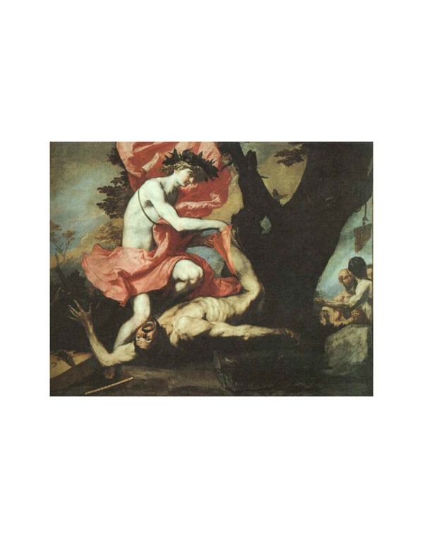
 James Elkins