© 2004 Nature Publishing Group http://www.nature.com/naturegenetics
B R I E F C O M M U N I C AT I O N S
Gene-culture coevolution between
cattle milk protein genes and
human lactase genes
a
N
E
W
S
Albano Beja-Pereira1,2, Gordon Luikart1, Phillip R England1,
Daniel G Bradley3, Oliver C Jann4, Giorgio Bertorelle5,
Andrew T Chamberlain6, Telmo P Nunes7, Stoitcho Metodiev8,
Nuno Ferrand2,9 & Georg Erhardt4
Milk from domestic cows has been a valuable food source
for over 8,000 years, especially in lactose-tolerant human
societies that exploit dairy breeds. We studied geographic
patterns of variation in genes encoding the six most important
milk proteins in 70 native European cattle breeds. We found
substantial geographic coincidence between high diversity
in cattle milk genes, locations of the European Neolithic
cattle farming sites (>5,000 years ago) and present-day
lactose tolerance in Europeans. This suggests a gene-culture
coevolution between cattle and humans.
Some, but not all, human populations have the genetically determined
ability to digest milk lactose in adulthood, thereby benefiting from the
rich food resources in cow’s milk1. These societies (e.g., Northern
Europe) are lactose-tolerant and highly dependent on milk products.
Lactose tolerance is an example of selection-based evolutionary
change in humans from milk-drinking cultures2. Has there also been a
detectable evolutionary change in the gene pool of domestic cattle
from these cultures?
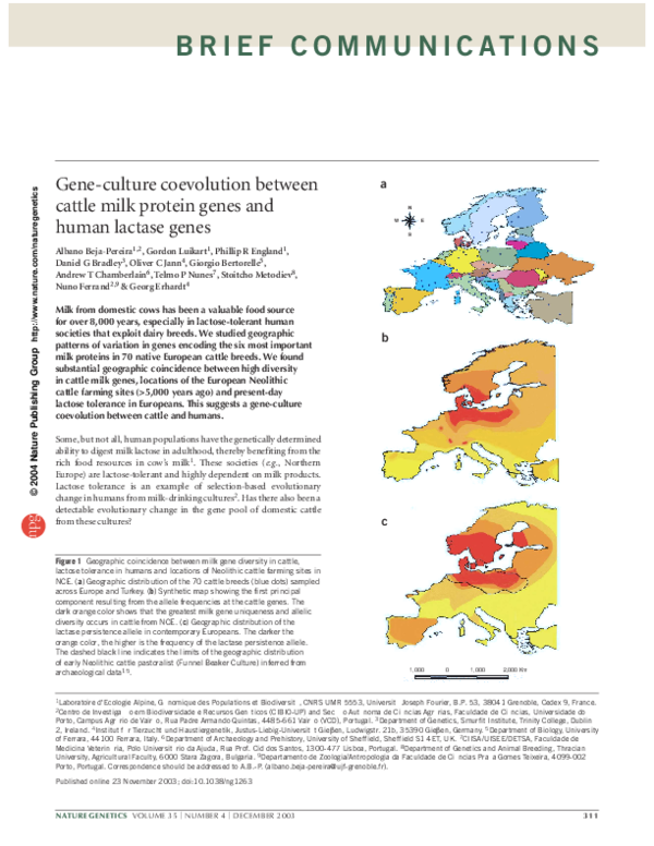
Figure 1 Geographic coincidence between milk gene diversity in cattle,
lactose tolerance in humans and locations of Neolithic cattle farming sites in
NCE. (a) Geographic distribution of the 70 cattle breeds (blue dots) sampled
across Europe and Turkey. (b) Synthetic map showing the first principal
component resulting from the allele frequencies at the cattle genes. The
dark orange color shows that the greatest milk gene uniqueness and allelic
diversity occurs in cattle from NCE. (c) Geographic distribution of the
lactase persistence allele in contemporary Europeans. The darker the
orange color, the higher is the frequency of the lactase persistence allele.
The dashed black line indicates the limits of the geographic distribution
of early Neolithic cattle pastoralist (Funnel Beaker Culture) inferred from
archaeological data15.
b
c
1,000
0
1,000
2,000 Km
1Laboratoire
d'Ecologie Alpine, Génomique des Populations et Biodiversité, CNRS UMR 5553, Université Joseph Fourier, B.P. 53, 38041 Grenoble, Cedex 9, France.
de Investigação em Biodiversidade e Recursos Genéticos (CIBIO-UP) and Secção Autónoma de Ciências Agrárias, Faculdade de Ciências, Universidade do
Porto, Campus Agrário de Vairão, Rua Padre Armando Quintas, 4485-661 Vairão (VCD), Portugal. 3Department of Genetics, Smurfit Institute, Trinity College, Dublin
2, Ireland. 4Institut für Tierzucht und Haustiergenetik, Justus-Liebig-Universität Gieβen, Ludwigstr. 21b, 35390 Gieβen, Germany. 5Department of Biology, University
of Ferrara, 44100 Ferrara, Italy. 6Department of Archaeology and Prehistory, University of Sheffield, Sheffield S1 4ET, UK. 7CIISA/UISEE/DETSA, Faculdade de
Medicina Veterinária, Polo Universitário da Ajuda, Rua Prof. Cid dos Santos, 1300-477 Lisboa, Portugal. 8Department of Genetics and Animal Breeding, Thracian
University, Agricultural Faculty, 6000 Stara Zagora, Bulgaria. 9Departamento de Zoologia/Antropologia da Faculdade de Ciências Praça Gomes Teixeira, 4099-002
Porto, Portugal. Correspondence should be addressed to A.B.-P. (albano.beja-pereira@ujf-grenoble.fr).
2Centro
Published online 23 November 2003; doi:10.1038/ng1263
NATURE GENETICS VOLUME 35 | NUMBER 4 | DECEMBER 2003
311
�B R I E F C O M M U N I C AT I O N S
Table 1 Spearman correlation coefficient values between the first principal component from the milk protein gene frequencies, the
lactase persistence allele frequency and the presence or absence of archeological evidence for Neolithic cattle pastoralists
© 2004 Nature Publishing Group http://www.nature.com/naturegenetics
Spearman correlation
Degrees of freedom
P
First principal component versus neolithic cattle pastoralists
–0.750
21.2–27.3
<0.0005
First principal component versus lactase persistence allele frequency
–0.593
17.7–24.6
<0.01
Neolithic cattle pastoralists versus lactase persistence allele frequency
0.730
19.3–24.8
<0.0005
Our study of nonsynonymous mutations in six milk protein genes
in ∼20,000 cattle from 70 breeds across Europe (Fig. 1a) found high
allelic richness and genetic distinctiveness in the native cattle from
North Central Europe (NCE), as illustrated by the synthetic map of
cattle milk protein genes (Fig. 1b and Supplementary Tables 1 and 2,
Supplementary Fig. 1 and Supplementary Methods online).
Notably, this synthetic map (inner contour) closely matches the
European distribution of the allele for human lactase persistence that
is most frequent in NCE (P < 0.0005; Table 1). This is in stark contrast to the lower levels of lactose tolerance found in people of
Southern Europe and the Near East. There was also strong concordance (P < 0.001) of the geographic distribution of cattle milk gene
diversity with the early Neolithic distribution of a European cattle
pastoralist society3 (Fig. 1c).
How can we explain the strong geographic concordance between
cattle milk gene diversity, human lactose tolerance and the distribution of the earliest European cattle pastoralists? We propose that
since Neolithic times, there has been gene-culture coevolution
between the domestic cattle and human culture driven by the advantages conferred by milk consumption. This led to the maintenance of
larger herds and selection for increased milk yield and altered milk
protein composition. This coevolution seemingly influenced the frequencies of the important milk protein genes in cattle and the gene
encoding lactase in humans. In fact, a recent study suggested that the
relatively old variant for lactose tolerance was only recently driven to
high frequencies in North Central Europeans after the introduction
of dairy culture in this region4.
This scenario is also supported by evidence for selection at milk
protein loci in bovids5. For example, directional selection can explain
high intraspecific divergence and low intraspecific polymorphism in
k-casein sequences across bovids5. Our data also show patterns consistent with selection: 19 NCE breeds deviated significantly from
neutrality (Ewens-Watterson test, 32% of 114 tests with P < 0.01 versus 2% of 306 tests with P < 0.01 in the 51 non-NCE breeds; Fu test
and Tajima test, all NCE breeds showed P < 0.05 versus 4 of 51 nonNCE cattle).
Our genetic data corroborate recent archaeological evidence suggesting that the early European cattle pastoralists in NCE were dependent on milk6,7, as early Neolithic sites in NCE are rich in cattle
remains4. Based on the analysis of intratooth change in nitrogen isotope ratios from archaeological cattle teeth, it seems that cattle herds
were managed for early weaning of calves, making cow’s milk more
available for human consumption. Meat production, practiced outside
NCE, necessitates later weaning to optimize weight gain7.
Among several phenomena that might have shaped our data, selection seems the most probable explanation. Recent studies have shown
that high diversity in human genes can evolve rapidly due to selection8.
In addition, analysis of bovine myostatin alleles showed signals of balancing selection in a number of independently occurring mutations
that cause double-muscling in beef breeds9.
Given that population surveys of mtDNA sequence, microsatellite
markers and protein polymorphisms in European cattle breeds show
no evidence of elevated diversity in NCE10-12, it is likely that selection
pressure imposed by early pastoralists and their successors in different
regions of NCE has left the legacy of high allelic diversity at these specific milk genes. It is also possible that some of the diversity represents
relatively recent mutation (<10,000 years), although, under a neutral
model, mutation rates are too low (10–6–10–9; ref. 5) for this to be a
primary factor. Selection may have maintained many favorable new
mutations by protecting them from the normal process of attrition
due to drift.
Another possible source of the unique diversity found in cattle in
NCE is historical gene flow from an as yet unidentified origin. Two
candidates for this source are local wild aurochs (Bos primigenius),
which persisted in NCE until the sixteenth century, and domestic cattle other than those that gave rise to present day European cattle (outside NCE). Extensive wild auroch introgression seems unlikely, and no
mtDNA sequences have been detected in European cattle which match
aurochs sequences identified using ancient DNA sequencing13.
Notably, our findings contradict the results of previous surveys
of genetic variation in European cattle10–12, which suggested that
diversity declines with distance from the Fertile Crescent region.
This discrepancy could be explained by selection on the milk
genes, and it may also reflect different sampling strategies. Our
analysis is based on a sample set that is unprecedented in size, geographic coverage and breed diversity. Furthermore, unlike previous studies, we analyzed only nonsynonymous polymorphisms in
strong candidate genes most likely to yield unusual geographic
patterns in milk gene diversity.
Our study provides evolutionary insights and identifies high diversity in cattle genes that are economically important, suggesting that
cattle in NCE are a potentially precious genetic resource for future
agricultural productivity. Farming practices since the Neolithic seem
to have left reciprocal genetic signatures in cattle and human populations from NCE. This may represent a rare example of cultural-genetic
coevolution between humans and another species. Other examples of
coevolution have been documented for human genes and genes of parasites, such as Plasmodium14. But our study represents the first nondisease-related example of genetic coevolution between humans and
domestic animals, reflecting the extent to which domestication has
shaped human societies and the genomes of both humans and cattle.
312
Note: Supplementary information is available on the Nature Genetics website.
ACKNOWLEDGMENTS
We thank M. Zvelebil for ideas and discussion. A.B.-P. is supported by a grant from
Fundação para a Ciência e Tecnologia through the Graduate Programme in Areas of
Basic and Applied Biology, and the work was partially supported by a Praxis project
grant. G.L. and P.R.E. were funded by the European Union (Econogene). D.G.B. is a
Science Foundation Ireland Investigator.
COMPETING INTERESTS STATEMENT
The authors declare that they have no competing financial interests.
Received 19 June; accepted 29 October 2003
Published online at http://www.nature.com/naturegenetics/
VOLUME 35 | NUMBER 4 | DECEMBER 2003 NATURE GENETICS
�B R I E F C O M M U N I C AT I O N S
© 2004 Nature Publishing Group http://www.nature.com/naturegenetics
1. Schrimshaw, N.S. & Murray, E.B. Am. J. Clin. Nutr. 48, 1059–1179 (1988).
2. Feldman, M.W. & Cavalli-Sforza, L.L. in Mathematical evolutionary theory (ed.
Feldman, M.W.) 145–173 (Princeton University Press, Princeton, New Jersey,
1989).
3. Midgley, M.S. TRB Culture: The First Farmers of the North European Plain
(Edinburgh University Press, Edinburgh, 1992).
4. Enattah, N.S. et al. Nat. Genet. 30, 233–237 (2002).
5. Ward, R., Honeycutt, L. & Derr, J.N. Genetics 147, 1863–1872 (1997).
6. Dudd, S. & Evershed, P. Science 282, 1478–1480 (1998).
7. Balasse, M. & Tresset, A. J. Archaeol. Sci. 29, 853–859 (2002).
8. Tishkoff, S.A. et al. Science 293, 455–462 (2001).
Mutations in the polyglutamine
binding protein 1 gene cause Xlinked mental retardation
Vera M Kalscheuer1, Kristine Freude1, Luciana Musante1,9,
Lars R Jensen1,9, Helger G Yntema2, Jozef Gécz3, Abdelaziz Sefiani4,
Kirsten Hoffmann1, Bettina Moser1, Stefan Haas1, Ulf Gurok1,
Sebastian Haesler1, Beatriz Aranda1, Arpik Nshedjan1,
Andreas Tzschach1, Nils Hartmann1, Tim-Christoph Roloff1,
Sarah Shoichet1, Olivier Hagens1, Jiong Tao1, Hans van Bokhoven2,
Gillian Turner5, Jamel Chelly6, Claude Moraine7,
Jean-Pierre Fryns8, Ulrike Nuber1, Maria Hoeltzenbein1,
Constance Scharff1, Harry Scherthan1, Steffen Lenzner1,
Ben C J Hamel2, Susann Schweiger1 & Hans-Hilger Ropers1
We found mutations in the gene PQBP1 in 5 of 29 families
with nonsyndromic (MRX) and syndromic (MRXS) forms of Xlinked mental retardation (XLMR). Clinical features in affected
males include mental retardation, microcephaly, short stature,
spastic paraplegia and midline defects. PQBP1 has previously
been implicated in the pathogenesis of polyglutamine
expansion diseases. Our findings link this gene to XLMR and
shed more light on the pathogenesis of this common disorder.
XLMR is a prominent unsolved problem in clinical genetics. Based on
the distribution of linkage intervals in 125 unrelated families, we
recently showed that roughly one-third of all mutations underlying
MRX are clustered on proximal Xp1. This observation prompted us to
search for mutations in families with linkage intervals overlapping
this region.
In 5 of 29 families studied, we detected mutations in PQBP1 that
cause frameshifts in the fourth coding exon (Supplementary
Methods online), which contains a stretch of six AG dinucleotides
in the DR/ER repeat (Fig. 1 and Supplementary Fig. 1 online). In
two families (family N9 (not previously reported) and family SHS
with Sutherland-Haan syndrome (MRXS3; ref. 2)), all affected
males carry an extra AG dinucleotide (3898_3899dupAG), whereas
in two others (family N45 (not previously reported) and family
MRX55; ref. 3), two AG dinucleotides are deleted
(3896_3899delAGAG). A single AG unit (3898_3899delAG) is
9. Dunner, S. et al. Genet. Sel. Evol. 35, 103–118 (2003).
10. Loftus, R.T. et al. Mol. Ecol. 8, 2015–2022 (1999).
11. MacHugh, D.E., Loftus, R.T., Cunningham, P. & Bradley, D.G. Anim. Genet. 29,
333–340 (1998).
12. Medjugorac, I., Kustermann, W., Lazar, P., Russ, I. & Pirchner, F. Anim. Genet. 25,
19–27 (1994).
13. Troy, C. et al. Nature 410, 1088–1091 (2001).
14. Hill, A.V., Jepson, A., Plebanski, M. & Gilbert, S.C. Philos. Trans. R. Soc. Lond. B
Biol. Sci. 352, 1317–1325 (1997).
15. Zvelebil, M. in Archaeogenetics: DNA and the Population History of Europe (ed.
Boyle, K.) 57–79 (MacDonald Institute Cambridge, Cambridge, 2000).
deleted in affected males of family N40 (ref. 4). In all families, these
mutations segregated with the disease and were present in all obligate heterozygotes that we tested. Except for one, all obligate heterozygotes that we examined have random X-chromosome
inactivation (data not shown) and have IQs in the normal range.
Apart from a single-nucleotide polymorphism (IVS2–3C→T), we
found no sequence variation in control X chromosomes.
The duplication observed in families N9 and SHS and the deletion found in families N45 and MRX55 give rise to almost the
same frameshift (Fig. 1b). Still, there is considerable inter- and
intrafamilial phenotypic variation (Supplementary Table 1
online). For example, males with SHS show mental retardation,
short stature, microcephaly, brachycephaly, spastic diplegia, small
testes and anal stenosis or atresia, whereas there is no spastic diplegia or small testes in family N9, with an identical mutation. In
both families the disease is not progressive. In family MRX55, in
whom the predicted mutant protein differs by only two amino
acids, affected individuals are moderately retarded but have no
other clinical signs, except for a somewhat smaller body size in one
individual (height was 159 cm, ≥2 s.d. below normal at the age of
20 years). In contrast, in addition to mental retardation, all
affected individuals in family N45 have microcephaly, one has anal
atresia and another has complete situs inversus. Some of this clinical variability may be due to differences in the genetic background.
Family MRX55 is from Morocco, families N9 and N45 are from the
Netherlands and family SHS has English ancestry.
In family N40, all affected males have congenital heart defects in
addition to severe mental retardation, microcephaly, spasticity, short
stature, cleft or highly arched palate and other craniofacial abnormalities4. The mother of two of the affected individuals has a corrected
atrial septal defect. Facial features coarsened with age.
Several PQBP1 splice variants have been described5. All but one
very rare variant contain exon 4, which is mutated in the five families.
The three different types of mutations cause frameshifts that lead to
premature stop codons, resulting in truncated PQBP1 proteins that
lack several important domains.
The particularly severe clinical phenotype seen in family N40 may
be due to the fact that the C-terminal end of the predicted truncated
protein is entirely different from that of the mutant proteins in the
other families (Fig. 1b) and may give rise to aberrant protein-protein
interactions.
1Max-Planck-Institute
for Molecular Genetics, Ihnestrasse 73, D-14195 Berlin, Germany. 2Department of Human Genetics, University Medical Centre, Nijmegen,
The Netherlands. 3Women’s and Children’s Hospital and The University of Adelaide, Adelaide, Australia. 4Département de Génétique et de Biologie Moléculaire INH,
Rabat, Morocco. 5Hunter Genetics and University of Newcastle, P.O. Box 84, Waratah, New South Wales 2298, Australia. 6Institut Cochin de Génétique Moleculaire,
CNRS/INSERM, CHU Cochin 75014 Paris, France. 7Services de Génétique-INSERM U316, CHU Bretonneau, Tours, France. 8Center for Human Genetics, Clinical
Genetics Unit, Leuven, Belgium. 9These authors contributed equally to this work. Correspondence should be addressed to V.M.K. (kalscheu@molgen.mpg.de).
Published online 23 November 2003; doi:10.1038/ng1264
NATURE GENETICS VOLUME 35 | NUMBER 4 | DECEMBER 2003
313
�E R R ATA
Erratum: Chipping away at the chip bias: RNA degradation
in microarray analysis
H Auer, S Lyianarachchi, D Newsom, M I Klisovic, G Marcucci & K Kornacker
Nat. Genet. 35, 292–293 (2003).
© 2004 Nature Publishing Group http://www.nature.com/naturegenetics
The name of the fifth author was spelled incorrectly. The correct spelling is “Guido Marcucci”.
Erratum: Gene-culture coevolution between cattle milk protein
genes and human lactase genes
A Beja-Pereira, G Luikart, P R England, D G Bradley, O C Jann, G Bertorelle, A T Chamberlain, T P Nunes, S Metodiev,
N Ferrand & G Erhardt
Nat. Genet. 35, 311–313 (2003).
The paper mistakenly contained a reference to Supplementary Figure 1 online; there is no such figure.
106
VOLUME 35 | NUMBER 1 | SEPTEMBER 2003 NATURE GENETICS
�
 Phillip England
 Albano Beja-Pereira