Arthropod Structure & Development 52 (2019) 100877
Contents lists available at ScienceDirect
Arthropod Structure & Development
journal homepage: www.elsevier.com/locate/asd
Using controlled vocabularies in anatomical terminology: A case study
with Strumigenys (Hymenoptera: Formicidae)
Thiago S.R. Silva*, Rodrigo M. Feitosa
�, Francisco Hera
�clito dos Santos Ave., Curitiba, PR, Brazil
Department of Zoology, Universidade Federal do Parana
a r t i c l e i n f o
a b s t r a c t
Article history:
Received 11 March 2019
Received in revised form
23 July 2019
Accepted 24 July 2019
Available online xxx
Morphological studies of insects can help us to understand the concomitant or sequential functionality of
complex structures and may be used to hypothetize distinct levels of phylogenetic relationship among
groups. Traditional morphological works, generally, have encompassed a set of elements, including descriptions of structures and their respective conditions, literature references and images, all combined in
a single document. Fast forward to the digital era, it is now possible to release this information simultaneously but also independently as data sets linked to the original publication in an external environment. In order to link data from various fields of knowledge, disseminating morphological
information in an open environment, it is important to use tools that enhance interoperability. For
example, semantic annotations facilitate the dissemination and retrieval of phenotypic data in digital
environments. The integration of semantic (i.e. web-based) components with anatomic treatments can
be used to generate a traditional description in natural language along with a set of semantic
annotations.
The ant genus Strumigenys currently comprises about 840 described species distributed worldwide. In
the Neotropical region, almost 200 species are currently known, but it is possible that much of the
species' diversity there remains unexplored and undescribed. The morphological diversity in the genus is
high, reflecting an extreme generic reclassification that occurred in the late 20th and early 21st centuries.
Here we define the anatomical concepts in this highly diverse group of ants using semantic annotations
to enrich the anatomical ontologies available online, focussing on the definition of terms through subjacent conceptualization.
© 2019 Elsevier Ltd. All rights reserved.
Keywords:
Morphology
Ants
Ontology
Semantic annotation
Terminology
1. Introduction
In biology, morphology is used to describe or redescribe taxa
�a and Branda
~o,
(Fernandes et al., 2014; Pinheiro et al., 2016; Ulysse
2012); to determine phenotypic traits linked to certain pathologies
(Ortiz et al., 2017; Fiaz et al., 2018); to infer kinship among lineages
in phylogenetic analyses (Fusari et al., 2014; Ramos and Melo, 2010;
Zacca et al., 2016); to trace phenotypic variation during ontogenetic
development (Thompson, 1999; Toyama et al., 2018); to investigate
functional community organization (Gibb and Parr, 2013; Gibb
~o, 2014); to explore evolutionary hyet al., 2015; Silva and Branda
potheses within anatomical groups (Kawada et al., 2015; Boudinot,
2013, 2018). The morphological terminology used by different
* Corresponding author.
E-mail addresses: tsranzanidasilva@gmail.com (T.S.R. Silva), rsmfeitosa@gmail.
com (R.M. Feitosa).
https://doi.org/10.1016/j.asd.2019.100877
1467-8039/© 2019 Elsevier Ltd. All rights reserved.
research groups for related organisms, however, may differ (Silva,
2017), making analyses of broader groups based on comparative
morphology very difficult (Deans et al., 2012).
Morphology is the product of multiple factors. Some are
intrinsic to individuals, such as the expression of differential traits
through gene modularity and hormone variation throughout the
ontogenetic development (Corona et al., 2016; Molet et al., 2012),
while others are extrinsic, for instance climatic oscillations and
environmental heterogeneity (Oms et al., 2017; Purcell et al., 2016).
For this reason, morphological studies result in complex data sets
that have been historically challenging in terms of referencing
(Vogt et al., 2010).
Morphological works generally include descriptions of traits
and their conditions, literature references, images and hypotheses
about trait evolution. Morphologists establish connections between
specimens and their anatomic features, using images and text to
document what they see. They also make interpretations and
advance hypotheses. In traditional morphological publications, all
�2
T.S.R. Silva, R.M. Feitosa / Arthropod Structure & Development 52 (2019) 100877
those elements are combined in a single document. In modern
times, however, there is the possibility of releasing data sets in an
external environment that is linked to the original publication and
making them available to a broader scientific community (Deans
et al., 2012; Godfray, 2002; Miller et al., 2012; Padial et al., 2010).
In order to link data from various fields of knowledge in an open
environment, tools such as semantic annotations can be used to
enhance interoperability and to facilitate the dissemination and
retrieval of phenotypic data in digital environments (Silva, 2017).
Semantic annotation is the process of attaching additional information to various concepts (e.g., terms, organisms etc) in a given
text or any other content. These annotations link multiple concepts
across different domains inside a digital environment and can be
easily retrieved and used by machines. For example, they can be
used in ontology-based information retrieval queries for efficient
data mining, thus facilitating the spread of the current understanding of a specific concept to multiple domains (Silva, 2017).
These annotations are possible because there are multi-species
anatomic ontologies (i.e. data models which represent a set of
concepts belonging to a specific domain, along with their relations),
which contain definitions of anatomic concepts that are logically
related (Dahdul et al., 2010). Annotations are logically composed
using references to these anatomic concepts along with descriptive
concepts from other ontologies. They make it possible, for example,
to understand which processes are involved in the evolution of
anatomic traits (Mabee et al., 2007).
The taxonomic and morphological diversity in Hymenoptera
(ants, bees, and wasps), combined with various researchers focussing on projects in multiple domains, has led to the creation of
several anatomical glossaries within the order (Yoder et al., 2010).
The morphological terminology used for the group is, for the most
part, family-specific (Seltmann et al., 2012), and it is not at all uncommon for a structure to be named differently across publications
(e.g., “paramere” Yoder et al., 2010; see Boudinot, 2018 for a
throughout discussion of genital terminology in Hexapoda)
depending on the hymenopterists' community the publication
originated from (Yoder et al., 2010).
Despite the fact that anatomic studies in myrmecology have
become less common with the advent of molecular endeavours and
the development of robust analytic tools to understand variation at
the molecular level (Keller, 2011), new-generation tools have
become more popular in anatomical studies and have enabled the
generation of fine-grained morphological data on the external and
internal morphology of ants (Agavekar et al., 2017; Sarnat et al.,
2016; Staab et al., 2018) in ways that are useful to other disciplines, such as biomechanics, ecology, behavioural and developmental biology (e.g., Gibb and Parr, 2013; Keller et al., 2014; Larabee
and Suarez, 2014; Larabee et al., 2017; Molet et al., 2012; Silva and
~o, 2014).
Branda
The ant genus Strumigenys, with 840 described species distributed worldwide, is most diverse in the tropics (Bolton, 2000, 2019).
Their great morphological diversity (Baroni Urbani and de Andrade,
1994, 2007) reflects the fact that a number of genera that were
historically kept separate have been synonymized with it in the late
20th and early 21st centuries (cf. Bolton, 2000 for a historical
overview of the taxonomic history for the genus). New publications
in the last fifteen years (Baroni Urbani and De Andrade, 2007;
Bharti and Akbar, 2013; Lattke and Aguirre, 2015; Longino, 2006;
Rigato and Scupola, 2008; Sosa-Calvo et al., 2010; Xu and Zhou,
2004; Zhou and Xu, 2003) have added approximately 30 valid
names to the genus, indicating a crescent e although, seemingly
slow e rate of new species discovery for this group, and the possibility that more species will be described in the future.
The present work defines anatomical concepts in Strumigenys
using semantic annotations to enrich the anatomical ontologies
available online, focussing on the definition of terms through
subjacent conceptualization. For this, we perform a morphological
investigation to substantiate previous definitions, and to establish
structural correspondence between the studied group and other
groups of ants and hymenopteran insects.
2. Material and methods
2.1. Sample information
The specimens used in this study were obtained from the
following institutions:
CASC California Academy of Sciences, San Francisco, CA, USA.
~o Entomolo
�gica Padre Jesus Santiago Moure, UniDZUP Coleça
�, Curitiba, PR, Brazil.
versidade Federal do Parana
~o Paulo, Sa
~o
MZSP Museu de Zoologia da Universidade de Sa
Paulo, SP, Brazil.
� ria, ES, Brazil.
UFES Universidade Federal do Espírito Santo, Vito
For the study of comparative morphology, 11 Formicidae subfamilies were studied, namely Agroecomyrmecinae, Amblyoponinae,
Dolichoderinae, Dorylinae, Ectatomminae, Formicinae, Myrmicinae,
Paraponerinae, Ponerinae, Proceratiinae, and Pseudomyrmecinae. At
least one specimen of each subfamily, apart from Myrmicinae, was
thoroughly studied in search of morphological characteristics (Table 1).
In Myrmicinae, at least one specimen for three tribes (Crematogastrini,
Pogonomyrmecini and Solenopsidini), apart from Attini, were studied
in search of morphological characteristics (Table 1). In Attini, at least
one specimen of the following genera, apart from Strumigenys, were
studied: Acromyrmex Mayr, 1865, Acanthognathus Mayr 1887, Basiceros
Schulz, 1906, Octostruma Forel, 1912, Phalacromyrmex Kempf, 1960,
Pheidole Westwood 1839, and Procryptocerus Emery, 1887.
In Strumigenys, at least one specimen of each of the 74 species was
observed (Table 1). Species belonging to this genus were arbitrarily
chosen as to tentatively explore most of the morphological diversity
present in the genus. Bolton (2000) recognizes several groups of
species, using morphological traits to diagnose them. Since some
representatives of those groups are not usually present in samples,
and we opted to dissect and disarticulate most of the examined
specimens, we used those species which were considered abundant
in samples and were readily available for study. Other non-formicide
Hymenoptera were also examined, reflecting the most recent hypothesis of relationship among hymenopteran families, to encompass a wide range of morphological diversity within the order
(Branstetter et al., 2017a; Peters et al., 2017) (Table 1).
2.2. Equipment
Morphological observations were made with a Leica S8APO
stereomicroscope at 80� magnification. Scanning electron microscope images were obtained using an electronic microscope Tescan
Vega3 LMU, under both low and high pressure. The method provided by Boudinot (2015) was used for mouthpart dissection and
body disarticulation. The methods of Gibson (1985) and Vilhelmsen
et al. (2010) were used in the study of the skeleton and musculature. The specimens were immersed in 100% alcohol one day prior
to dissection for complete dehydration. After the dehydration
process, the specimens were transferred to ethanol-immersed BluTack, which allowed better handling. All the dissections were performed using a razor blade and size zero entomological pins.
�3
T.S.R. Silva, R.M. Feitosa / Arthropod Structure & Development 52 (2019) 100877
Table 1
List containing all species studied (Family, Subfamily, Tribe, Species), sex of specimens studied, morphological form (apterous, brachypterous or macropterous), their collection
of origin, their conservation medium (dry-mounted or ethanol) and their locality information (COUNTRY: Department/Province, Municipality).
Species
Apidae
Apinae
Meliponini
Melipona sp.
Bethylidae
Epyrinae
Epyrinae sp.
Pristocerinae
Pristocerinae sp.
Ceraphronidae
Ceraphronidae sp.1
Ceraphronidae sp.2
Diapriidae
Diapriinae
Diapriinae sp.
Diapriini
Acanthopria sp.
Eucharitidae
Eucharitinae
Eucharitini
Kapala sp.
Evaniidae
Semaeomyia sp.
Ichneumonidae
Ophioninae
Ophioninae sp.
Mutillidae
Sphaerophtalminae
Sphaerophtalminae sp.
Pompilidae
Pepsinae
Pepsinae sp.
Pteromalidae
Cleonyminae
Cleonyminae sp.
Vespidae
Polistinae
Epiponini
Polybia sp.
Formicidae
Agroecomyrmecinae
Agroecomyrmecini
Tatuidris tatusia
Amblyoponinae
Amblyoponini
Fulakora armigera
Dolichoderinae
Tapinomini
Tapinoma melanocephalum
Dorylinae
Labidus coecus
Ectatomminae
Ectatommini
Gnamptogenys sp.
Formicinae
Camponotini
Camponotus atriceps
Paraponerinae
Paraponerini
Paraponera clavata
Ponerinae
Ponerini
Pachycondyla striata
Proceratiinae
Proceratiini
Discothyrea sexarticulata
Pseudomyrmecinae
Pseudomyrmecini
Pseudomyrmex sp.
Myrmicinae
Crematogastrini
Crematogaster sp.
Sex
Form
Collection of origin
Conservation medium
Locality information
Female
Macropterous
DZUP
Ethanol
�, Curitiba
BRAZIL: Parana
Male
Macropterous
DZUP
Ethanol
FRENCH GUIANA: Saint-Laurent-du-Maroni, Saül
Female
Apterous
DZUP
Ethanol
�, Antonina
BRAZIL: Parana
Female
Female
Macropterous
Brachypterous
DZUP
DZUP
Ethanol
Ethanol
�, Piraquara
BRAZIL: Parana
�, Piraquara
BRAZIL: Parana
Female
Macropterous
DZUP
Ethanol
�, Piraquara
BRAZIL: Parana
Female
Apterous
DZUP
Ethanol
�, Piraquara
BRAZIL: Parana
Male
Macropterous
DZUP
Ethanol
�, Antonina
BRAZIL: Parana
?
Macropterous
DZUP
Ethanol
BRAZIL: Santa Catarina, Blumenau
Male
Macropterous
DZUP
Ethanol
BRAZIL: Santa Catarina, Blumenau
Female
Apterous
DZUP
Ethanol
�, Curitiba
BRAZIL: Parana
Female
Macropterous
DZUP
Ethanol
BRAZIL: Rio Grande do Sul, Gramado
Male
Macropterous
DZUP
Ethanol
BRAZIL: Santa Catarina, Blumenau
Female
Macropterous
DZUP
Ethanol
�, Curitiba
BRAZIL: Parana
Female
Apterous
DZUP
Dry-mounted
PERU: Madre de Dios, Puerto Maldonado
Female
Apterous
DZUP
Dry-mounted
BRAZIL: Rio de Janeiro, Itatiaia
Female
Apterous
DZUP
Ethanol
�, Curitiba
BRAZIL: Parana
Female
Apterous
DZUP
Ethanol
�, Antonina
BRAZIL: Parana
Female
Apterous
DZUP
Ethanol
BRAZIL: Rio Grande do Sul, Gramado
Female
Apterous
DZUP
Ethanol
BRAZIL: Santa Catarina, Blumenau
Female
Apterous
DZUP
Dry-mounted
~es
BRAZIL: Mato Grosso, Chapada dos Guimara
Female
Apterous
DZUP
Ethanol
�, Curitiba
BRAZIL: Parana
Female
Apterous
DZUP
Dry-mounted
�, Tibagi
BRAZIL: Parana
Female
Apterous
DZUP
Ethanol
BRAZIL: Rio Grande do Sul, Gramado
Female
Apterous
DZUP
Ethanol
BRAZIL: Santa Catarina, Blumenau
(continued on next page)
�4
T.S.R. Silva, R.M. Feitosa / Arthropod Structure & Development 52 (2019) 100877
Table 1 (continued )
Species
Sex
Form
Collection of origin
Conservation medium
Locality information
Female
Apterous
DZUP
Ethanol
�, Antonina
BRAZIL: Parana
Female
Apterous
DZUP
Ethanol
�, Curitiba
BRAZIL: Parana
Female
Female
Female
Female
Female
Female
Female
Female
Apterous
Apterous
Apterous
Apterous
Apterous
Apterous
Apterous
Apterous
DZUP
DZUP
DZUP
DZUP
DZUP
DZUP
DZUP
DZUP and MZSP
Ethanol
Dry-mounted and ethanol
Dry-mounted
Ethanol
Dry-mounted
Ethanol
Ethanol
Dry-mounted
Strumigenys alberti
Strumigenys appretiata
Female
Female
DZUP
DZUP
Dry-mounted
Dry-mounted and ethanol
Strumigenys beebei
Female
DZUP
Dry-mounted and ethanol
�, Curitiba
Parana
^ nia, Porto Velho
Rondo
Santa Catarina, Indaial
�, Tunas
Parana
Santa Catarina, Painel
�, Curitiba
Parana
Rio Grande do Sul, Gramado
Santa Catarina, Indaial
�, Bocaiúva do Sul
Parana
~o, Açaila
^ndia
Maranha
~o Grande
S~
ao Paulo, Ribeira
Rio de Janeiro, Itatiaia
^ nia, Porto Velho
Rondo
Strumigenys borgmeieri
Female
DZUP and MZSP
Dry-mounted
Strumigenys carinithorax
Strumigenys cincinatta
Strumigenys comis
Female
Female
Female
MZSP
DZUP
DZUP and MZSP
Dry-mounted
Dry-mounted
Dry-mounted
Strumigenys conspersa
Strumigenys cordovensis
Female
Female
Apterous
Apterous and
macropterous
Apterous and
macropterous
Apterous and
macropterous
Apterous
Apterous
Apterous and
macropterous
Apterous
Apterous
BRAZIL:
BRAZIL:
BRAZIL:
BRAZIL:
BRAZIL:
BRAZIL:
BRAZIL:
BRAZIL:
BRAZIL:
BRAZIL:
BRAZIL:
BRAZIL:
BRAZIL:
DZUP
DZUP and MZSP
Dry-mounted
Dry-mounted
Strumigenys cosmostela
Strumigenys crassicornis
Female
Female
MZSP
DZUP
Dry-mounted
Dry-mounted and ethanol
^ nia, Porto Velho
Rondo
Sergipe, Areia Branca
~o Pessoa
Paraíba, Joa
^ nia, Porto Velho
Rondo
Rio de Janeiro, Itatiaia
Santa Catarina, Canoinhas
^ nia, Porto Velho
Rondo
^ nia, Porto Velho
Rondo
Pernambuco, Recife
� polis
S~
ao Paulo, Saleso
Rio de Janeiro, Itatiaia
Strumigenys cultrigera
Strumigenys dapsilis
Strumigenys denticulata
Female
Female
Female
MZSP
MZSP
DZUP
Dry-mounted
Dry-mounted
Dry-mounted and ethanol
BRAZIL: Rio Grande do Sul, Bom Jesus
~o Bento do Sul
BRAZIL: Santa Catarina, Sa
�, Piraquara
BRAZIL: Parana
Strumigenys dentinasis
Female
Apterous
Apterous and
macropterous
Apterous
Apterous
Apterous and
macropterous
Apterous
BRAZIL:
BRAZIL:
BRAZIL:
BRAZIL:
BRAZIL:
BRAZIL:
BRAZIL:
BRAZIL:
BRAZIL:
BRAZIL:
BRAZIL:
DZUP and MZSP
Dry-mounted
Strumigenys depressiceps
Strumigenys eggersi
Female
Female
Macropterous
Apterous
DZUP
DZUP and MZSP
Dry-mounted
Dry-mounted and ethanol
Strumigenys elongata
Female
Apterous
DZUP
Dry-mounted and ethanol
Strumigenys
Strumigenys
Strumigenys
Strumigenys
Strumigenys
Strumigenys
Strumigenys
Strumigenys
Strumigenys
emiliae
emmae
epelys
epinotalis
fridericimuelleri
glenognatha
godmani
grytava
gytha
Female
Female
Female
Female
Female
Female
Female
Female
Female
Apterous
Macropterous
Apterous
Apterous
Apterous
Apterous
Macropterous
Apterous
Apterous
DZUP
UFES
MZSP
MZSP
MZSP
DZUP
DZUP
DZUP
DZUP and MZSP
Dry-mounted
Ethanol
Dry-mounted
Dry-mounted
Dry-mounted
Dry-mounted
Dry-mounted
Dry-mounted
Dry-mounted
Strumigenys
Strumigenys
Strumigenys
Strumigenys
Strumigenys
Strumigenys
hindenburgi
hyphata
infidelis
inusitata
kompsomala
lanuginosa
Female
Female
Female
Female
Female
Female
Apterous
Apterous
Apterous
Apterous
Apterous
Apterous and
macropterous
Apterous
Apterous
Apterous
Apterous
Apterous and
macropterous
MZSP
DZUP
DZUP
DZUP
DZUP
DZUP
Dry-mounted
Dry-mounted
Dry-mounted
Dry-mounted and ethanol
Dry-mounted
Dry-mounted
MZSP
DZUP
DZUP
DZUP
DZUP
Dry-mounted and ethanol
Dry-mounted
Dry-mounted and ethanol
Dry-mounted
Dry-mounted
~o Grande
ao Paulo, Ribeira
BRAZIL: S~
�, Bocaiúva do Sul
BRAZIL: Parana
FRENCH GUIANA: Saint-Laurent-du-Maroni, Saül
BRAZIL: Minas Gerais, Boa Esperança
�us
BRAZIL: Bahia, Ilhe
BRAZIL: Acre, Senador Guiomard
~o, Estreito
BRAZIL: Maranha
BRAZIL: Santa Catarina, Indaial
�ria
BRAZIL: Espírito Santo, Vito
~o
ao Joa
BRAZIL: Bahia, Mata S~
BRAZIL: Minas Gerais, Pedra Azul
�ia
BRAZIL: S~
ao Paulo, Canane
^ nia, Porto Velho
BRAZIL: Rondo
FRENCH GUIANA: Saint-Laurent-du-Maroni, Saül
�va
~o
BRAZIL: Sergipe, S~
ao Cristo
BRAZIL: Sergipe, Malhador
~o Pessoa
BRAZIL: Paraíba, Joa
BRAZIL: Rio Grande do Sul, Morro Reuter
PERU: Madre de Dios, Puerto Maldonado
�s, Caldas Novas
BRAZIL: Goia
^ nia, Porto Velho
BRAZIL: Rondo
^ nia, Porto Velho
BRAZIL: Rondo
�, Piraquara
BRAZIL: Parana
BRAZIL: Rio de Janeiro, Itatiaia
BRAZIL: Bahia, Milagres
PERU: Madre de Dios, Puerto Maldonado
�, Curitiba
BRAZIL: Parana
BRAZIL: Rio de Janeiro, Ilha Grande
BRAZIL: Santa Catarina, Indaial
MZSP
DZUP
DZUP
MZSP
MZSP
DZUP
DZUP
MZSP
DZUP
Dry-mounted
Dry-mounted
Dry-mounted
Dry-mounted
Dry-mounted
Dry-mounted
Dry-mounted
Dry-mounted
Dry-mounted
BRAZIL: Santa Catarina, Blumenau
~o, Açaila
^ndia
BRAZIL: Maranha
^ nia, Porto Velho
BRAZIL: Rondo
�
BRAZIL: Santa Catarina, Chapeco
BRAZIL: S~
ao Paulo, Mirassol
^ nia, Porto Velho
BRAZIL: Rondo
PERU: Madre de Dios, Puerto Maldonado
BRAZIL: S~
ao Paulo, Tapiraí
FRENCH GUIANA: Saint-Laurent-du-Maroni, Saül
Pogonomyrmecini
Hylomyrma sp.
Solenopsidini
Solenopsis sp.
Attini
Acromyrmex crassipisnus
Acanthognathus sp.
Basiceros disciger
Octostruma rugifera
Phalacromyrmex fugax
Pheidole sp.
Procryptocerus sp.
Strumigenys abditivata
Strumigenys lilloana
Strumigenys longimala
Strumigenys louisianae
Strumigenys pr. louisianae
Strumigenys
pr. louisianae
“bruchi complex”
Strumigenys lygatrix
Strumigenys metopia
Strumigenys monstra
Strumigenys minuscula
Strumigenys ogloblini
Strumigenys perparva
Strumigenys planeti
Strumigenys precava
Strumigenys prospiciens
Female
Female
Female
Female
Female
Female
Female
Female
Female
Female
Female
Female
Female
Female
Apterous
Apterous
Apterous
Apterous
Apterous
Apterous
Apterous
Apterous
Macropterous
�5
T.S.R. Silva, R.M. Feitosa / Arthropod Structure & Development 52 (2019) 100877
Table 1 (continued )
Species
Sex
Form
Collection of origin
Conservation medium
Locality information
Strumigenys reticeps
Female
Apterous
DZUP and MZSP
Dry-mounted
Strumigenys rotogenys
Strumigenys rugithorax
Strumigenys saliens
Female
Female
Female
Apterous
Apterous
Apterous
DZUP
MZSP
DZUP and MZSP
Dry-mounted
Dry-mounted
Dry-mounted and ethanol
Strumigenys sanctipauli
Strumigenys schmalzi
Female
Female
Apterous
Apterous
MZSP
DZUP and MZSP
Dry-mounted
Dry-mounted and ethanol
Strumigenys
Strumigenys
Strumigenys
Strumigenys
Strumigenys
Strumigenys
Strumigenys
Strumigenys
Strumigenys
Strumigenys
Strumigenys
Strumigenys
Strumigenys
Strumigenys
Strumigenys
schulzi
signeae
smilax
smithii
subedentata
pr. sublonga
teratrix
trinidadensis
trudifera
urrhobia
wheeleriana
villiersi
xenochelyna
zeteki
sp.n.1 (fe)
Female
Female
Female
Female
Female
Female
Female
Female
Female
Female
Female
Female
Female
Female
Female
DZUP
DZUP
MZSP
DZUP
DZUP
DZUP
DZUP
DZUP
DZUP
DZUP
DZUP
DZUP
DZUP
DZUP
MZSP
Dry-mounted
Dry-mounted
Dry-mounted
Dry-mounted and ethanol
Dry-mounted and ethanol
Dry-mounted
Dry-mounted
Dry-mounted
Dry-mounted and ethanol
Dry-mounted
Dry-mounted
Dry-mounted
Dry-mounted
Dry-mounted
Dry-mounted
Strumigenys
Strumigenys
Strumigenys
Strumigenys
Strumigenys
sp.n.2
sp.n.3
sp.n.4
sp.n.5
sp.n.6
Female
Female
Female
Female
Female
Apterous
Apterous
Apterous
Apterous
Apterous
Apterous
Apterous
Apterous
Apterous
Apterous
Apterous
Apterous
Apterous
Apterous
Apterous and m
acropterous
Apterous
Apterous
Apterous
Apterous
Apterous
~o Paulo, Ribeira
~o Grande
BRAZIL: Sa
~o Paulo, Mogi das Cruzes
BRAZIL: Sa
MALAYSIA: Sabah, Maliau Basin
BRAZIL: Santa Catarina, Blumenau
BRAZIL: Santa Catarina, Blumenau
�, S~
� dos Pinhais
BRAZIL: Parana
ao Jose
~o Paulo, Saleso
� polis
BRAZIL: Sa
�, Jari
BRAZIL: Para
~o Pessoa
BRAZIL: Paraíba, Joa
~o Paulo, Sete Barras
BRAZIL: Sa
MALAYSIA: Sabah, Maliau Basin
^ nia, Porto Velho
BRAZIL: Rondo
^ nia, Porto Velho
BRAZIL: Rondo
BRAZIL: Rio de Janeiro, Itatiaia
PERU: Madre de Dios, Puerto Maldonado
BRAZIL: Sergipe, Nossa Senhora das Dores
^ nia, Porto Velho
BRAZIL: Rondo
^ nia, Porto Velho
BRAZIL: Rondo
BRAZIL: Santa Catarina, Painel
� n, Cerro Los Caracoles
VENEZUELA: Falco
^ nia, Porto Velho
BRAZIL: Rondo
BRAZIL, Tocantins, Porto Nacional
^ nia, Porto Velho
BRAZIL: Rondo
�, Tunas
BRAZIL: Parana
MZSP
MZSP
MZSP and CASC
MZSP
MZSP
Dry-mounted
Dry-mounted
Dry-mounted
Dry-mounted
Dry-mounted
BRAZIL:
BRAZIL:
BRAZIL:
BRAZIL:
BRAZIL:
(ele)
(fast)
(gib)
(ro)
(se)
2.3. Morphological data
All morphological information presented in this work refer to
morphemes (Richter and Wirkner, 2014) and are without functional
or evolutionary implications. Morphemes are characterized as
representations of the smallest morphological unit at a particular
level of description and are useful to reference morphological
characteristics as secondary data.
Each anatomical entity is represented by a class (i.e. concept), a
definition, and class relationships (i.e. the record that links two
concepts via a class relationship). Anatomical classes for Hymenoptera were recovered from the Hymenoptera Anatomy Ontology
(Yoder et al., 2010; HAO version 26.iv.2018), while phenotypic quality
classes are based on the Phenotypic Quality Ontology (PATO version
07.ii.2018). Class relationship types are based on the OBO Relations
Ontology (OBOREL 2018; version 11.vii.2018) (Table 2). All ontologies
are made available through the Open Biomedical Ontologies
Foundry. The list of terms used in the current work and their definitions can be found in the Table 3.
Morphological definitions are constructed as genus-differentia,
which are definitions structured to first describe a more inclusive
class of concepts (genus) and then the characteristics differentiating
(differentia) it from another subordinate of that same concept
(Seltmann et al., 2012). Definitions in this format usually follow the
pattern “The x that is y” (cf. Boudinot, 2018, Appendix A).
To refer to the distinct morphological forms commonly found in
female ants we opted to use the terms apterous and macropterous
rather than workers and queens/gynes, respectively. Since the last
Minas Gerais, Viçosa
Santa Catarina, Palhoça
~o Paulo, Piedade
Sa
�, Tunas
Parana
~o
Bahia, Mata S~
ao Joa
two concepts are intrinsically related to colonial and reproductive
€ lldobler and Wilson, 1990), we refrain from using them,
function (Ho
based on the premise that the external morphology alone is not
sufficient to infer reproductive aptitude (Silva and Feitosa, 2018). The
recognition criterion applied in this study to determine the biological
sex of the specimens is purely anatomical, through the observation of
external elements of the genitalia; in our case, we observed the distal
section of the modified ovipositor (i.e. tip of the sting apparatus).
2.4. Recovering classes from natural language statements
Each examined structure was described through natural language statements and variations found were discussed when
pertinent. The description process was performed simultaneously
by both authors in order to increase agreement during coding
procedures. The descriptions were automatically parsed and concepts were recovered from each ontology using two tools: the
Analyze tool from the Hymenoptera Anatomy Ontology portal
(http://portal.hymao.org/projects/32/public/ontology/analyze)
(Seltmann et al., 2013) for recognition of anatomical classes; and
Bioportal's Annotator (https://bioportal.bioontology.org/annotator)
(Shah et al., 2009) for recognition of phenotypic quality classes.
Both tools recognize input natural language statements, parsing
down textual data and recovering specific classes based on a
defined set of dictionaries (in our case, ontologies). The output data
are normally exported as CSV, JSON, or XML formats, and contains
the classes recognized from the input text, along with their definitions and/or unique resource identifiers (URIs). Classes not
Table 2
Object (class relationship types) and annotation properties used in the annotation of anatomical classes.
Property
Definition
is_a
part_of
has_related_synonym
A subsumption object property. Represents a transitive, reflexive and anti-symmetric relation between two or more classes.
A composition object property. Represents a transitive, reflexive and anti-symmetric relation between two or more classes.
An aggregation annotation property. Represents a predicate between a class and a literal.
�6
T.S.R. Silva, R.M. Feitosa / Arthropod Structure & Development 52 (2019) 100877
Table 3
List of terms, their definitions, and their respective ontology identifiers. To recover definitions of classes and annotations through the OBO Foundry, include http://purl.
obolibrary.org/obo/before the identifier (e.g., http://purl.obolibrary.org/obo/PATO_0000402).
Term
Definition
Ontology identifier
acetabulum
anatomical cluster
anatomical entity
The area that is concave and accommodates the base of a segment.
The anatomical group that has its parts adjacent to one another.
Biological entity that is either an individual member of a biological species or constitutes the
structural organization of an individual member of a biological species.
Anatomical structure consisting of at least two non-overlapping organs, multi-tissue aggregates
or portion of tissues or cells of different types that does not constitute an organism, organ, multitissue aggregate, or portion of tissue.
Material anatomical entity which has inherent 3D shape and is generated by coordinated
expression of the organism's own genome.
A shape quality inhering in a bearer by virtue of the bearer's having at least one salient angle on
the margin.
The fossa that is located on the anterior coxal articular process of the mesotrochanter and
accommodates the anterior trochanteral condyle of the mesocoxa.
The fossa that is located anteriorly on the metatrochanter and accommodates the anterior
trochanteral condyle of the metacoxa.
The fossa that is located anteriorly on the proximal margin of the protrochanter accommodating
the anterior trochanteral condyle of the procoxa.
The foramen that is located on the head in which the radicle is positioned.
The scrobe that is located dorsally of the antennal foramen and is for the reception of the
antenna.
The anatomical structure of the cuticle that is delimited by material or immaterial anatomical
entities.
A surface feature shape inhering in a surface by virtue of the bearer's being divided by ridge-like
structures into a number of small, irregular spaces.
The projection that bears the articular surface.
The lobe that is connected proximodorsally with the manubrium and proximoventrally with the
planta.
The sulcus that corresponds to the basicosta.
The area that is located on the coxa proximal to the basicostal suture.
The tarsomere that is the proximal most annulus of the tarsus, connected proximally with the
tibia and distally with the second tarsomere via membranous conjunctivae.
The condyle that is located on the pectus and inserts into the pectal fossa of the coxa.
A size quality inhering in a bearer by virtue of the bearer's being made wider or larger in all
dimensions.
The dicondylic joint that is composed of the femur and the tibia.
The anatomical space that is surrounded by sclerites and allows for the passage of haemolymph,
nerves and tracheae.
The carina that extends along the lateral margin of the intertorular area towards the vertex.
The anatomical cluster that is composed of two sclerites connected by at least one articulation
and the articular membrane located connecting the two sclerite.
The fossa that is located laterally on the proximal margin of the coxa and accommodates the
lateral coxal condyle of the pectus.
The area that extends between the anterior eye margin and the anterolateral margin of the
cranium, and its width is delimited by the width of the mandible.
The fossa that is located anteriorly on the proximolateral edge of the mandible and
accommodates the pleurostomal coondyle.
The sclerite that is located proximodorsally on the pretarsus and connects the distodorsal
margin of the telotarsus with the dorsal part of the arolium.
The articular process that is located on the ventral margin of the mesopectus medially of the
mesocoxal foramen and bears the medial coxal condyle of the mesopectus.
The articular process that is located on the ventral margin of the metapectus medially of the
metacoxal foramen and bears the medial coxal condyle of the metapectus.
The coxal condyle of the pectus that inserts into the medial pectal fossa of the coxa.
The pectal fossa of the coxa that accommodates the median coxal condyle of the pectus.
The discrimen that is located in the mesothorax and corresponds with the mesodiscrimenal
lamella.
The sclerite that is U-shaped in cross section, connected anteriorly with the pronotum and the
propectus, dorsally with the basalare, the mesonotum, the second axillary sclerite and the
subalare, posteriorly with the metapectus and bears the mesodiscrimenal lamella and the
mesofurca.
The area that is located anteriorly of the metapleural sulcus and ridge.
A shape quality inhering in a bearer by virtue of the bearer's being perfectly circular.
The sclerite that is located proximoventrally on the pretarsus and is connected proximally with
the unguitractor plate and distally with the arolium.
The condyle that is located on the anterior (dorsal) margin of the pleurostoma and inserts into
the mandibular acetabulum.
The mandibular muscle that arises posterodorsally from the cranium and inserts on the tendon
attached anteroproximally on the mandible.
The anatomical cluster that is apical to the telotarsus and composed of the empodium, auxilia,
planta, pulvillum, unguis, unguitractor plate, auxiliar sclerite and manubrium.
The basisternum that is located in the propectus.
The carina that delimits posteriorly the pronotal neck.
HAO_0000082
HAO_0000041
HAO_0000000
anatomical group
anatomical structure
angular
anterior coxal fossa of the mesotrochanter
anterior coxal fossa of the metatrochanter
anterior coxal fossa of the protrochanter
antennal insertion
antennal scrobe
area
areolate
articular process
arolium
basicostal suture
basicoxite
basitarsus
coxal condyle of the pectus
dilated
femoro-tibial joint
foramen
frontal carina
joint
lateral pectal fossa of the coxa
malar area
mandibular acetabulum
manubrium
medial coxal articular process of the mesopectus
medial coxal articular process of the metapectus
medial coxal condyle of the pectus
medial pectal fossa of the coxa
mesodiscrimen
mesopectus
metapectus
orbicular
planta
pleurostomal condyle
posterior cranio-mandibular muscle
pretarsus
probasisternum
pronotal carina
HAO_0000054
HAO_0000003
PATO_0001977
HAO_0001433
HAO_0001501
HAO_0001500
HAO_0001022
HAO_0001432
HAO_0000146
PATO_0002295
HAO_0000150
HAO_0000148
HAO_0000174
HAO_0000175
HAO_0000178
HAO_0001917
PATO_0001571
HAO_0001517
HAO_0000345
HAO_0001533
HAO_0001146
HAO_0001913
HAO_0001393
/HAO_0001391
HAO_0000671
HAO_0001389
HAO_0001388
HAO_0001918
HAO_0001916
HAO_0000545
HAO_0000557
HAO_0000605
PATO_0001934
HAO_0000719
HAO_0000731
HAO_0000745
HAO_0000820
HAO_0001317
HAO_0001031
�7
T.S.R. Silva, R.M. Feitosa / Arthropod Structure & Development 52 (2019) 100877
Table 3 (continued )
Term
Definition
Ontology identifier
pronotal neck
The area of the pronotum that is delimited posterodorsally by an edge and that accommodates
the posterior surface of the head.
The tibial spur that is located on the fore leg, is curved and together with the probasitarsus forms
the strigil.
The fossa that is located posteriorly on the proximal margin of the mesotrochanter
accommodating the posterior trochanteral condyle of the mesocoxa.
The fossa that is located posteriorly on the proximal margin of the metatrochanter and
accommodates the posterior trochanteral condyle of the metacoxa.
The fossa that is located posteriorly on the proximal margin of the protrochanter
accommodating the posterior trochanteral condyle of the procoxa.
The area that is located on the sclerite and that is composed of repetitive anatomical structures.
The patch that differs from the surrounding region by having denser setae.
A shape quality inhering in a bearer by virtue of the bearer's being oblong, with the lower end
very much attenuated.
The anatomical cluster that is composed of the probasitarsus and calcar.
A shape quality inhering in a bearer by virtue of the bearer's being linear, very narrow, tapering
to a very fine point from a narrow base.
The spur that is curved and projects from the apex of the last tarsal segment on either side of the
arolium of the pretarsus.
The area that is located proximally on the femur and is delimited by a groove.
The leg segment that is located proximal to the femur and distal to the coxa.
The sclerite that corresponds to the site of insertion of the flexor of the pretarsus.
HAO_0000837
protibial spur
posterior coxal fossa of the mesotrochanter
posterior coxal fossa of the metatrochanter
posterior coxal fossa of the protrochanter
sculpture
setiferous patch
spatulate
strigil
subulate
tarsal claw
trochantellus
trochanter
unguitractor plate
recognized by those tools were manually annotated to the Hymenoptera Anatomy Ontology (Table 4).
2.5. Annotation development
Annotations were made following a four-step process: 1)
Phenotypic and relational classes available in OWL (Web Ontology
Language, http://www.w3.org/TR/owl2-overview/) were loaded
�ge
� 4.1 (http://protege.stanford.edu/), an open-source
into Prote
ontology editor and a knowledge management system; 2) Anno�ge
� as
tations were manually added to anatomic entities within Prote
OWL class expressions using the built-in Manchester syntax (www.
w3.org/TR/owl2-manchester-syntax/) editor; 3) After annotation,
we used the FaCTþþ reasoner to determine if new annotations did
not violate descriptions and axioms across the ontology; 4) Annotated entities were exported as OWL class expressions and individually added to mx (Mx., 2019), a collaborative open-source
application that facilitates the construction of ontologies through a
multi-feature environment. All phenotype statements in Manchester syntax are available in Table 4.
2.6. Images
Illustrations were made using Adobe Illustrator (version CS6),
based on images available at AntWeb (2019) and personal observations of the studied material. Antweb images used to prepare the
plates have their respective identifier explicitly indicated at the
legend of the figure (in the form of CASENT0000000).
2.7. Data repository
All data obtained are available through an online open-access
repository (https://doi.org/10.6084/m9.figshare.7645400) as .csv
and .owl files. Annotations in Manchester syntax can be easily obtained through the ‘annotations.csv’ file. Novel anatomical classes
were added to the most recent version of the ‘hao.owl’ file and are
deposited as a ‘hao-merged-strumigenys.owl’ file.
3. Results and discussion
Terms and definitions for structures, along with their identifiers
(i.e. persistent uniform resource locators; purl), which were used
HAO_0000875
HAO_0001348
HAO_0001295
HAO_0001297
HAO_0000913
HAO_0000936
PATO_0001937
HAO_0000102
PATO_0001954
HAO_0000989
HAO_0001033
HAO_0001034
HAO_0001043
throughout the text and figures, can be found in Table 3. Annotations for morphological terms discussed in the following sections,
along with their definitions, can be found in Table 4.
We annotated 35 new anatomical classes based on the study of
Strumigenys. From those, 12 were from structures found on the
head and its appendages (i.e. mouthparts and antennae), nine were
from the mesosoma and its appendages (i.e. legs), 13 were from the
metasoma, and one refers to a structure that can occur in more than
one body region (i.e. mesosoma and metasoma). Overall, six classes
presented one synonym, with one class having two synonyms,
totalling seven synonyms.
3.1. Head
Ants have the head oriented horizontally towards the longitudinal axis and mouthparts anteriorly-oriented. Hence, the spatial
orientation terminology follows those of prognathous insects. The
insect head has been divided into different areas (Snodgrass, 1993;
Chapman, 1998): frons, vertex, temple, gena, postgena, malar space
and occiput. However, these areas are difficult to establish, since
their presence and dimensions depend on the presence and position of other structures, such as the compound eyes and ocelli. Most
of those areas were named by Kirby and Spencer (1828), but their
delimitations, for the most part, are elusive.
3.1.1. Fronto-vertexal area and complex
In Hymenoptera, the frons is the area between the epistomal line
and the anterior ocellus and limited laterally by the inner margin of
� , 2009/2019). Macropterous female and male
compound eye (Miko
ants generally retain all three ocelli, with the posterior limits of
the frons and the anterior limits of the vertex being promptly
determined. In apterous ant females, however, the posterior limits
of the frons and anterior limits of the vertex cannot be externally
defined in most groups, due to the loss of ocelli in this form.
Another complex situation found in apterous females is the
reduction or loss of the eyes in some groups, making the lateral
delimitation of the frons and the dorsal delimitation of the gena
somewhat ambiguous (discussed thoroughly in the section
“Gena”).
In Hymenoptera, the vertex is the area delimited by the intersection of the margin of the compound eyes, the interorbital plane, and
the anatomical line tangential to the point on the margin of the
�8
T.S.R. Silva, R.M. Feitosa / Arthropod Structure & Development 52 (2019) 100877
Table 4
List of terms, their definitions, and annotations expressed in Manchester syntax.
Term
Definition
Annotation
Antero-proximal process of the scape
Antero-sternite
The process that is located near the base on the anterior
margin of the scape and is wider or larger in all dimensions
The area that is located on an abdominal sclerite and is
limited anteriorly by the antecostal sulcus and posteriorly
by the transverse impression of the abdominal sclerite.
The antero-sclerite that is located ventrally.
Antero-tergite
The antero-sclerite that is located dorsally.
Antero-ventral notch of the mesopectus
The notch that is located anteriorly on the ventral margin of
the mesopectus.
Apical fork
The anatomical cluster that is consisted by the apicoventral
tooth and the apicodorsal tooth.
Apico-dorsal tooth
The projection that is located distally on the mandible and
limited ventrally by the intercalar tooth or by the apicoventral tooth, forming the apical fork.
The projection that is located distally on the mandible and
limited dorsally by the intercalar tooth or by the apicodorsal tooth, forming the apical fork.
The area of the sclerite that is raised and is divided by ridgelike structures into a number of small, irregular spaces.
is_a some process and part_of some sclerite and
‘bearer of’ some dilated
is_a some area and part_of some metasoma and
part_of some cuticle and has_related_synonym
presclerite
is_a some antero-sclerite and part_of some
metasoma and part_of some cuticle and part_of
some sternum
is_a some antero-sclerite and part_of some
metasoma and part_of some cuticle and part_of
some tergum
is_a some notch and part_of some mesopectus
and part_of some mesosoma and part_of some
margin and part_of some sclerite
is_a some anatomical cluster and part_of some
mandible and part_of some sclerite and part_of
some cuticle
is_a some tooth and part_of some mandible and
part_of some sclerite and part_of some ‘apical
fork’
is_a some tooth and part_of some mandible and
part_of some sclerite and part_of some ‘apical
fork’
is_a some process and part_of some cuticle and
part_of some sclerite and ‘bearer of’ some
areolate and has_related_synonym spongiform
tissue
is_a some foramen and part_of some basicoxite
is_a some foramen and part_of some disticoxite
is_a some area and part_of some coxa
Antero-sclerite
Apico-ventral tooth
Areolate process
Basicoxal foramen
Disticoxal foramen
Disticoxite
Fronto-vertexal area
Fronto-vertexal complex
Intercalar tooth
Lateral areolate process of the petiole
The foramen that is located proximally on the basicoxite.
The foramen that is located distally on the disticoxite.
The area that is located on the coxa distal to the basicostal
suture.
The area of the sclerite that is located between the
epistomal line and the occipital carina and limited laterally
by the inner margin of the compound eye.
The sclerite that is between the epistomal line and the
occipital carina and limited laterally by the inner margin of
the compound eye.
The tooth that is located on the apical fork, limited ventrally
by the apico-ventral tooth and dorsally by the apico-dorsal
tooth.
The areolate process that is located at the lateral margin of
the petiole.
Lateral areolate process of the abdominal
tergum 3
The areolate process that is located at the lateral margin of
the abdominal tergum 3.
Lateral coxal condyle of the pectus
The coxal condyle of the pectus that inserts into the lateral
pectal fossa of the coxa.
Lateral mandibular articular process
The articular process that is located laterally on the
proximal margin of the mandible.
The scrobe that is located proximally on the mandible and
accommodates the anterolateral process of the head.
The gland that is located on the metapleuron and opens
posteriorly near the propodeal foramen, on the orifice of the
metapleural gland.
The anatomical space that is situated posteriorly on the
metapecto-propodeal complex, lateral to the propodeal
foramen.
The impression that is located posteriorly to the oral
foramen.
Lateral mandibular scrobe
Metapleural gland
Orifice of the metapleural gland
Postbucal impression
Postero-sclerite
Postero-sternite
The area of an abdominal sclerite limited anteriorly by the
transverse impression of the abdominal sclerite and
posteriorly by the posterior margin of the abdominal
sclerite.
The postero-sclerite that is located ventrally.
Postero-tergite
The postero-sclerite that is located dorsally.
is_a some area and part_of some fronto-vertexal
complex has_related_synonym fronto-vertex
is_a some sclerite and part_of some frons and
part_of some vertex and part_of some cuticle
is_a some tooth and part_of some mandible and
part_of some sclerite and part_of some cuticle
and part_of some ‘apical fork’
is_a some ‘areolate process’ and part_of some
metasoma and part_of some ‘abdominal
segment 2’ and part_of some cuticle and part_of
some sclerite
is_a some ‘areolate process’ and part_of some
metasoma and part_of some ‘abdominal
segment 3’ and part_of some cuticle and part_of
some sclerite
is_a some ‘coxal condyle of the pectus’ and
part_of some pectus and part_of some sclerite
and part_of some articulation
is_a some ‘articular process’ and part_of some
mandible and part_of some sclerite
is_a some scrobe and part_of some mandible
and part_of some sclerite
is_a some gland and part_of and part_of some
metapleuron and part_of some body
is_a some ‘anatomical space’ and part_of some
‘metapectal-propodeal complex’ and part_of
some metapleuron
is_a some impression and part_of some sclerite
and part_of some integument and part_of some
‘postgenal bridge’
is_a some area and part_of some metasoma and
part_of some cuticle and has_related_synonym
postsclerite
is_a some postero-sclerite and part_of some
metasoma and part_of some cuticle and part_of
some sternum
is_a some postero-sclerite and part_of some
metasoma and part_of some cuticle and part_of
some tergum
�T.S.R. Silva, R.M. Feitosa / Arthropod Structure & Development 52 (2019) 100877
9
Table 4 (continued )
Term
Definition
Annotation
Pronoto-mesonotal complex
Translucent patch of the frontal carina
The sclerite that is located dorsally to the propectus and
mesopectus and is composed of the pronotum and
mesonotum.
The scrobe that is located ventrally on the femur and
accommodates the tibia.
The translucent patch that is located at the frontal carina.
Translucent patch of the metapleuron
The translucent patch that is located at the metapleuron.
Translucent patch of the ventral antennal scape
The translucent patch that is located at the ventral margin of
the antennal scape.
Transverse carina of the fourth postero-tergite
The carina that is located posteriorly to the impression of
the fourth abdominal tergite, belonging to the fourth
postero-tergite
The impression that is located on the external surface of an
abdominal sclerite and does not correspond to a ridge.
is_a some sclerite and part_of some cuticle and
part_of some pronotum and part_of some
mesonotum
is_a some scrobe and part_of some sclerite and
part_of some femur
is_a some ‘translucent patch’ and part_of some
‘frontal carina’ and part_of some integument
and part_of some cuticle
is_a some ‘translucent patch’ and part_of some
metapleuron and part_of some integument
is_a some ‘translucent patch’ and part_of some
scape and part_of some integument and part_of
some cuticle
is_a some carina and part_of some sclerite and
part_of some postero-tergite and
has_related_synonym limbus
is_a some impression and part_of some
metasoma and part_of some sclerite and part_of
some integument and has_related_synonym
cinctus and has_related_synonym ‘girdling
constriction’
is_a some patch and part_of some ‘abdominal
sternum 4’ and part_of some metasoma and
part_of some integument and part_of some
cuticle
is_a some ‘areolate process’ and part_of some
‘abdominal segment 2’ and part_of some
metasoma and part_of some cuticle and part_of
some sclerite
is_a some ‘areolate process’ and part_of some
‘abdominal sternum 3’ and part_of some
metasoma and part_of some cuticle and part_of
some sclerite
is_a some notch and part_of some ‘propodeal
foramen’ and part_of some ‘metapectalpropodeal complex’ and part_of some margin
and part_of some sclerite
Tibial scrobe
Transverse impression of the abdominal sclerite
Transverse patch of the abdominal sternum 4
The patch that is elongated and extends transversely along
the anterior portion of the abdominal sternum 4.
Ventral areolate process of the petiole
The areolate process that is located at the ventral margin of
the petiole.
Ventral areolate process of the abdominal
sternum 3
The areolate process that is located at the ventral margin of
the abdominal sternum 3.
Ventral notch of the propodeal foramen
The notch that is located medially on the ventral margin of
the propodeal foraminal margin.
anterior ocellus which defines the minimum distance between the
anterior ocellus and the oral foramen (Yoder, 2009). Similarly to the
frons, the anterior margin of the vertex cannot be externally
delimited in apterous females of ants, due to the lack of ocelli in
most groups.
Muscular organization has been useful to establish structural
equivalence in other groups of Hymenoptera (Kawada et al., 2015;
Popovici et al., 2014; Zimmermann and Vilhelmsen, 2016) and in
male ants (Boudinot, 2013). There have been several studies
exploring the internal anatomy of the head of ants, especially in
groups with drastic modifications in the cephalic appendages
(Gronenberg, 1995, 1996; Gronenberg and Ehmer, 1996;
Gronenberg et al., 1997, 1998a,b; Larabee et al., 2017, 2018; Paul
and Gronenberg, 1999), high intraspecific morphological variation
(Lilico-Ouachour et al., 2018) or novel appendage functionality
(Khalife et al., 2018). Since these investigations are mainly focused
on the mechanics and physiology of the muscular groups related to
the mandibular motion, the cephalic muscular organization in
different lineages of ants has remained relatively unexplored,
hampering our understanding of the regionalization and sclerite
fusion in this tagma.
Since we are not confident about the current understanding of
the muscular arrangement in ants in a comparative context, and
due to our inability to properly define the frons and vertex of
apterous females both internally and externally, we opted to refer
to them collectively as the fronto-vertexal complex when referring to
the structure as an anatomical cluster, and fronto-vertexal area (or
fronto-vertex) when referring to the externally observable area of
the structure (Table 4).
The main distinction between both classes is how they are
recognized. An anatomical complex is understood as being an
anatomical cluster, which is defined as an anatomical group that has
its parts adjacent to one another (Haendel et al., 2008). An
anatomical group is understood as an anatomical structure consisting
of at least two non-overlapping organs, multi-tissue aggregates or
portion of tissues or cells of different types that does not constitute an
organism, organ, multi-tissue aggregate, or portion of tissue (Haendel
et al., 2008). On the other hand, an anatomical area can be understood as the anatomical structure of the cuticle that is delimited by
� , 2009/2019).
material or immaterial anatomical entities (Miko
Therefore, an anatomical complex can be understood as an assembly of anatomical entities (including cuticle, muscles and apodemes), while an anatomical area can be understood as a twodimensional anatomical entity (e.g., the cuticle) delimited by
other anatomical entities (such as anatomical lines, anatomical
spaces, or anatomical structures).
In Strumigenys the fronto-vertexal area can be easily delimited
laterally even in species with reduced eyes (Fig. 1). Although variable in size and antero-posteriorly positioned, the compound eyes
are rarely displaced in a dorso-ventral axis.
3.1.2. Gena
The gena in hymenopterans is the area delimited by the intersection of the interorbital plane, the margin of the compound eye, the
margin of the oral foramen, the occipital carina and the malar sulcus
(Yoder, 2009). It is divided into three other areas, namely the malar
area, the gena s.s. and the temple (Fig. 1; ma, gn and tm, respectively) (Boudinot et al., 2013). Internally, it appears to not
�10
T.S.R. Silva, R.M. Feitosa / Arthropod Structure & Development 52 (2019) 100877
oc
oc
frvx
frvx
tm
ce
fc
fc
ma
fl
clp
tpfc
clp
posterior
emd
apdt
fl
md
md
imd
emd
anterior
intt
apf
apvt
ocf
pgs
pbi
tm
asc
ai
dorsal
asc
tm
gn
ai
ma
pbi
ventral
gn
poc
ma
pbi
Fig. 1. Schematic illustration of the head and mandibles of apterous females of Strumigenys. Dotted lines indicate the limits of structures. Dashed lines indicate the limits of areas.
Abbreviations: ai: antennal insertion; apdt: apicodorsal tooth; apf: apical fork; apvt: apicoventral tooth; asc: antennal scrobe; ce: compound eye; clp: clypeus; emd: external
margin of the mandible; fc: frontal carina; frvx: fronto-vertexal area; gn: gena; imd: internal margin of the mandible; intt: intercalar teeth; ma: malar area; md: mandible; oc:
occipital carina; ocf: occipital foramen; pbi: postbucal impression; pgs: postgenal suture; poc: preocular carina; tm: temple; tpfc: translucent patch of frontal carina.
�T.S.R. Silva, R.M. Feitosa / Arthropod Structure & Development 52 (2019) 100877
correspond precisely to any muscular group, although most of the
cranio-mandibular muscle group has its origins in this region. In
� et al., 2007) and in Bethylidae (Lanes, 2013), this
Scelionidae (Miko
area corresponds to the origin sites of the median band of the
anterior cranio-mandibular muscle and of the posterior craniomandibular muscle. This condition is similarly found in Formicidae, although there is major variation in muscle size, area of
origin, apodeme length, and area of insertion in those muscles
depending on the group (personal observation), age of individuals
and task repertoire (Muscedere et al., 2011). Mandibular shape
appears to be intrinsically related to the characteristics of those
muscles, possibly affecting the characteristics of the gena, such as
size and shape.
In Formicidae, there is a developed median longitudinal ridge
that extends from the hypostomal margin to the occipital foramen,
referred to by Khalife et al. (2018) as the ventromedial phragma,
and appears to be the site of origin of a ventral band of the posterior
cranio-mandibular muscle (as in Melissotarsus Emery, 1877 and
Myrmoteras Forel, 1893; Khalife et al., 2018 and Larabee et al., 2017,
respectively). It varies greatly in height and degree of sclerotization
and could probably represent an internal folding of the lateral
margins of the postgenal bridge (cf. “Postgenal bridge and postgenal suture section”). In some observed specimens belonging to
long mandibulate species of Strumigenys (e.g., Strumigenys saliens
Mayr, 1887, Strumigenys elongata Roger, 1863, Strumigenys pr. louisianae), this ridge is reduced but stills represents the site of origin of
part of the posterior cranio-mandibular muscle. However, this
structure may vary in other species that possess distinct mandibular morphologies.
3.1.3. Postgenal bridge and postgenal suture
According to Burks and Heraty (2015), a lot of variation can be
found on the subforaminal bridges in Aculeata, and the modifications in cephalic ventral elements in this group have not been
properly studied. Internally, the putative lateral margins of the
postgenal bridge are greatly expanded in some ants, acting as an
attachment site for mandibular muscles (cf. discussion at the
Mandible section). The modifications of the postgenal bridge and
the reduction of the postgenal suture in various groups of ants are
still poorly understood. In view of the high sexual polymorphism
found in the family, comparative studies are needed to investigate
the modifications in this region.
We observed, in Strumigenys, that the postgenal bridge is fused
anteriorly. In some species, the postgenal suture (Fig. 1; pgs) is
visible only from the occipital carina to the medial region of the
ventral surface of the head. Internally, there is a reduced longitudinal ridge spanning from near the occipital foramen to the postgenal inflection anteriorly (in which the labiomaxillary complex
rests). In some dissected specimens, part of the medial band of the
posterior cranio-mandibular muscle has its origins in this ridge.
Compared with other groups that rely on mandibular strength to
process resources (such as major females of Pheidole), the longitudinal ridge of Strumigenys is greatly reduced.
3.2. Antenna
3.2.1. Scape
Among the female ants observed, the basal antennal segment is
usually extremely long when compared with other groups of Hymenoptera (Fig. 2). In most genera of ants, the scape may be as long
as the flagellum (Fig. 2; scp and F1-4, respectively). Much of the
variation in the scape of different ant groups has remained undocumented. For example, the muscular and glandular aspects of
this particular structure in Strumigenys have been broadly overlooked by researchers.
11
One example of variation in the scape that might be informative
in Strumigenys can be found on its ventral surface. While some
species have an unsculptured patch there (e.g., Strumigenys reticeps
(Kempf, 1969) and Strumigenys thaxteri (Wheeler, 1916); Lattke
et al., 2018)), others (e.g., Strumigenys alberti Forel, 1893, Strumigenys appretiata (Borgmeier, 1954), Strumigenys borgmeieri Brown,
1954) have this area mostly occupied by a translucent patch. The
scape also varies in overall shape (from cylindrical to dorsoventrally flattened) and presence of a dilated process near its
base on the anterior margin (Fig. 2; scp) (such as in Strumigenys
crassicornis Mayr, 1887). When the dilated process is present, it can
be defined as “The process that is located near the base on the anterior
margin of the scape and is wider or larger in all dimensions” and
named as antero-proximal process of the scape. Rows of erect and
decumbent setae on the anterior margin of the scape occur in
several species and they can vary from subulate to orbicular.
3.2.2. Flagellum
In Hymenoptera, variation in the number of segments of the
flagellum is significant, with various conditions of fusion or
reduction of segments (Polaszek et al., 1992; Heraty, 2002). In
Formicidae, the antennal segments of many groups seem to be
fused. Some individuals of some species seem to have asymmetrical
numbers of segments (Fischer et al., 2015). There is a slight variation in the number of segments of the flagellum in Strumigenys
(Fig. 3AeE), ranging from two to four, possibly representing the
more reduced number of flagellar segments in the entire family. In
other genera of the tribe, the number varies from five (e.g., Eurhopalothrix) to ten segments (e.g., Basiceros) (Fig. 9). Since we cannot
easily trace fusion or reduction of segments in the flagellum based
on structural equivalence, previous definitions based on this
method are questionable.
3.3. Mouthparts
3.3.1. Labrum
In Hymenoptera, the labrum is defined as the sclerite that is
situated along the distal margin of the clypeus and is connected along
its proximal margin with the distal margin of the epipharyngeal wall
(Vilhelmsen and Miko, 2010). In non-formicid hymenopterans it is
connected posterodorsally to the clypeus by the clypeolabral
articulation, contrasting with the anterior connection found in
many other insects. In Formicidae, the position of the clypeolabral
articulation varies among groups, but most often it is articulated
lateroventrally with the clypeus.
In Strumigenys, the labrum is located posteroventrally in relation
to the clypeus, as in most groups of ants. Most commonly, the
apodeme of the posterior fronto-labral muscle is well-developed
and sclerotized, bearing the site of attachment of the posterior
fronto-labral muscle, which acts as the retractor of the labrum
(Gronenberg, 1996). Since the shape of the labrum is extremely
variable within the genus, the apodeme of the posterior frontolabral muscle also appears extremely variable, especially in position, which, although appearing laterally in the sclerite, differs in
distance from the clypeolabral articulation (Fig. 2; cllba). The setae
located in the labral lobes (Fig. 2; lbl) vary among species, ranging
from orbiculate to subulate. Most long-mandibulate species
possess long subulate setae that are as long as the mandibles.
According to Bolton (1999) the labrum can be modified in two
distinct conditions: (i) the labrum lacks lateral processes and the
anterior labral lobes are large; and (ii) the labrum is T-shaped (sic),
with lateral processes, and the anterior labral lobes are reduced or
vestigial. However, J.C.M. Chaul and coworkers (pers. comm.)
observed that there is immense variation in shape of the labrum
within the genus, even within distinct types of Bolton's
�12
T.S.R. Silva, R.M. Feitosa / Arthropod Structure & Development 52 (2019) 100877
Fig. 2. Illustration of the labrum in dorsal (top left) and ventral (top right) views and of the antenna in ventral view (bottom) of apterous females of Strumigenys. Abbreviations:
afrlb: apodeme of the posterior fronto-labral muscle; amscp: anterior margin of the scape; cllba: clypeo-labral articulation; F1: first segment of the flagellum; F2: second segment
of the flagellum; F3: third segment of the flagellum; F4: fourth segment of the flagellum; lbl: labral lobe; lbst: labral seta; lplb: lateral process of the labrum; pdc: pedicel; scp:
antennal scape; tpscp: translucent patch of the antennal scape.
�T.S.R. Silva, R.M. Feitosa / Arthropod Structure & Development 52 (2019) 100877
13
Fig. 3. Variation in the number of antennal segments in specimens of Strumigenys (AeE), Eurhopalothrix (F), Pilotrochus (G) and Basiceros (H). A: Strumigenys anchis
CASENT0900916; B: Strumigenys minuscula CASENT0281948; C: Strumigenys clypeata CASENT0103000; D: Strumigenys chroa CASENT0436719; E: Strumigenys ornata
CASENT0104478; F: Eurhopalothrix floridana CASENT0003195; G: Pilotrochus besmerus CASENT0047617; H: Basiceros disciger CASENT0914887.
�14
T.S.R. Silva, R.M. Feitosa / Arthropod Structure & Development 52 (2019) 100877
Fig. 4. Illustration of the mandible in dorsal (top left, top right, and bottom left) and ventral (bottom right) views of apterous females of Strumigenys. Abbreviations: bpm: basal
process of the mandible; cmd: carina of the mandible; dia: diastema; dmap: dorsal articular process of the mandible; tpmd: translucent patch of the mandible; vmap: ventral
articular process of the mandible.
�T.S.R. Silva, R.M. Feitosa / Arthropod Structure & Development 52 (2019) 100877
15
Fig. 5. Basal section of the external margin of the mandible of two apterous females of Strumigenys. A: Disarticulated mandible of Strumigenys saliens; B: Mandible inserted in the
head of Strumigenys planeti, showing the points were the lateral scrobe of the mandible (marked by asterisks) would approximately contact the head capsule (marked by circles)
when the mandible is open. The star indicates the surface of the lateral articular process of the mandible, which is concealed within the oral foramen when the mandible is open.
Abbreviations: bpm: basal process of the mandible; dmap: dorsal articular process of the mandible; lmap: lateral articular process of the mandible; lms: lateral scrobe of the
mandible; vmap: ventral articular process of the mandible.
�16
T.S.R. Silva, R.M. Feitosa / Arthropod Structure & Development 52 (2019) 100877
dorsal
posterior
anterior
bcf
pc
bcs
ventral
pn
bc
mti
pmsa
dc
pr
fmtp
pdsp
pmss
pd
anep
pdsr
tc
fm
pdc
pdl
mtp
avnmsp
ktep
tbs
tb
tbtp
msepc
btrtp
mspmtps
mtppds
ptr
btr
trcl
pr
pn
anterior
ppl
pmsa
pbst
avnmsp
posterior
msp
msdm
mti
mcapms
mtp
mtdm
pd
mcapmt
pdsp
vnpdfr
Fig. 6. Illustration of the mesosoma and fore leg of apterous females of Strumigenys. From top to bottom: lateral view, dorsal view and ventral view. Abbreviations: anep: anepisternum; avnmsp: antero-ventral notch of the mesopectus; bc: basicoxite; bcf: basicoxal foramen; bcs: basicostal suture; btr: basitarsus; btrtp: translucent patch of the
basitarsus; dc: disticoxite; fm: femur; fmtp: translucent patch of the femur; ktep: katepisternum; mcapms: medial coxal articular process of the mesopectus; mcapmt: medial
coxal articular process of the metapectus; msdm: mesodiscrimen; msepc: mesepisternal carina; msp: mesopectus; mspmtps: mesopecto-metapectal suture; mtdm: metadiscrimen; mti: metanotal impression; mtp: metapectus; mtppds: metapecto-propodeal suture; pbst: probasisternum; pc: pronotal carina; pd: propodeum; pdc: propodeal
carina; pdl: propodeal lobe; pdsp: propodeal spine; pdsr: propodeal spiracle; pms: pronoto-mesonotal complex; pmsa: pronoto-mesonotal area; pmss: pronoto-mesonotal suture;
pn: pronotal neck; ppl: propleura; pr: pronotal rim; ptr: pretarsus; tb: tibia; tbs: tibial spur; tbtp: translucent patch of the tibia; tc: trochanter; trcl: tarsal claw; vnpdfr: ventral
notch of the propodeal foramen.
�T.S.R. Silva, R.M. Feitosa / Arthropod Structure & Development 52 (2019) 100877
17
Fig. 7. Variation in the size of the anteroventral notch of the mesopectus in specimens of Strumigenys (AeC, G and H), Phalacromyrmex (D), Rhopalothrix (E) and Octostruma (H). A:
Strumigenys alperti CASENT0003239; B: Strumigenys actis CASENT0005467; C: Strumigenys denticulata CASENT0178117; D: Phalacromyrmex fugax CASENT0103116; E: Rhopalothrix
ishtmica CASENT0235905; F: Octostruma stenognatha CASENT0280761; G: Strumigenys inusitata; H: Strumigenys saliens. From A to F the white arrow indicates the position of the
anteroventral notch of the mesopectus. In H, the white arrow indicates the position of a small circular impression adjacent to the anteroventral notch of the mesopectus.
�18
T.S.R. Silva, R.M. Feitosa / Arthropod Structure & Development 52 (2019) 100877
Fig. 8. Image representing the transverse impression of the third abdominal segment in a specimen of Gnamptogenys striatula. Blue: transverse impression of the third abdominal
tergite; Green: anterior area of the third abdominal tergite; Red: antecosta of the third abdominal tergite. (For interpretation of the references to colour in this figure legend, the
reader is referred to the Web version of this article.)
“mandibular mode of action” in Strumigenys (cf. “Mandible” section). A more in-depth exploration of labral variation has to be
addressed as to properly establish structural correspondence of its
instances in a broader scenario within the family, so as to enable
accurate annotation within multi-species ontologies.
The labrum of Strumigenys commonly has setae on the anterior
section of the sclerite (Fig. 2; lbst), which in some species appears
to play a major role in prey recognition (Gronenberg, 1996). The
setae can be orbicular, spatulate or linear, each type occurring
alone or in combination with other types. In species in which the
labral setae are not linear, several setae are disposed over the
anterior half of the labrum, most of them concentrated over the
labral lobes. In these cases, we do not know which role they
perform and if it is related to prey recognition. The lateral processes (Fig. 2; lplb) can be present or absent; when present, the
lateral labral margin appears angular. The lateral processes in
Strumigenys have been extensively studied and appear to function
as part of the locking mechanism that holds the mandibles wideopen during hunting, interlocking with the proximal processes of
the mandible (Brown and Wilson, 1959; Gronenberg, 1996).
Similar to other groups of ants (Gotwald, 1969), the anterior
fronto-labral muscle is absent, while the paired posterior fronto-
�19
T.S.R. Silva, R.M. Feitosa / Arthropod Structure & Development 52 (2019) 100877
at3
ptn
pt
pt3
ptp
ps3
dorsal
tppt
anterior
posterior
ventral
cpt4
pt4
tpps4
ps4
Fig. 9. Illustration of the metasoma in lateral view of apterous females of Strumigenys. Abbreviations: at3: third antero-tergite; cpt4: carina of the fourth postero-tergite; ps3: third
postero-sternite; ps4: fourth postero-sternite; pt3: third postero-tergite; pt4: fourth postero-tergite; pt: petiole; ptn: petiolar node; ptp: petiolar peduncle; tpps4: transverse patch
of the fourth postero-sternite; tppt: translucent patch of the petiole.
�20
T.S.R. Silva, R.M. Feitosa / Arthropod Structure & Development 52 (2019) 100877
labral muscles are present and are solely responsible for the
retraction of the sclerite.
3.3.2. Mandible
Overall, the mandible in Formicidae has three well-defined
margins (Bolton, 1994): the masticatory margin, the basal margin
and the external margin. The masticatory margin corresponds to
the longitudinal margin of each mandibular blade near the anterior
extension of the median line of the head. This margin, generally
bearing one or more teeth, can be also called apical margin. The
teeth can be well-developed or extremely reduced and both types
can be found in the same specimen. Normally, the dentition occurs
along all the masticatory margin (Bolton, 1994). When there is a
space in the teeth row, it is called diastema (Bolton, 1994) (Fig. 4;
dia).
The base of the masticatory margin finds a basal angle and turns
into a transverse or oblique basal margin (Bolton, 1994). The
external margin of each mandible can be straight, concave or
convex (Bolton, 1994). In most species of ants, the external margins
converge anteriorly and become triangular in dorsal view. In some
cases, they project anteriorly, in an elongate-triangular shape
(Bolton, 1994).
When the mandibles are slender and linear, the distinction between the masticatory and basal margins is absent, due to the loss
of the basal angle (Bolton, 1999). In some lineages of Formicidae,
the mandible is linear, with a long and slender blade and the
masticatory and external margins approximately parallel or gradually converging anteriorly (Bolton, 1999). In groups with linear and
elongate mandibles, the section with teeth is positioned at the apex
of the mandible and is called apical fork (Fig. 1; apf) (Brown, 1948).
In their 1994 contribution, Baroni Urbani and de Andrade (1994)
asserted that previous authors had never properly defined the
apical fork. According to Bolton (2000), in most species with
elongate and slender mandibles, the teeth at the apical section
commonly form a fork of two or three vertically disposed teeth. Still
according to the author, an apical fork is normally composed of two
teeth, one dorsal and one ventral, called apicodorsal and apicoventral tooth, respectively (Fig. 1; apdt and apvt, respectively). The
tooth that is located between those teeth is called intercalar tooth
(Fig. 1; intt). According to Baroni Urbani and de Andrade (1994), one
of the important characteristics of Strumigenys is that a pair of acute
mandibular blades, dented or pointy, are opposed, but do not
overlap. Still according to Baroni Urbani and de Andrade (op. cit.), a
putative reduction occurred in the number and/or size of the median and basal teeth until their total loss, normally accompanied by
the atrophy of one, or the formation of an apical fork with two or
more distal teeth.
According to Bolton (op. cit.) the section that receives all teeth
positioned apically would correspond to the masticatory margin
(e.g. Strumigenys fridericimuelleri Forel, 1886) in species previously
classified in Pyramica (a current synonym of Strumigenys), Schmidt
and Shattuck (2014) mentioned that in the Ponerinae genera Anochetus Mayr, 1861 and Odontomachus Latreille, 1804, the masticatory margin corresponds to the internal margin of the mandible, in
dorsal view. However, in Strumigenys the accurate delimitation of
the distinct regions of the mandible can be difficult. The delimitation between the basal and masticatory margins is clear in some
species (e.g., Strumigenys alberti, Strumigenys schulzi Emery, 1894,
Strumigenys urrhobia (Bolton, 2000)) but not in others (e.g., Strumigenys crassicornis, Strumigenys denticulata Mayr, 1887, Strumigenys elongata Roger, 1863), especially in the case of the masticatory
margin and the apical fork. Although this last can be easily
observed, according to Bolton (2000), the variation in shape and
presence sometimes leads to confusing interpretations. Hence, the
term masticatory margin should be avoided, since the structural
equivalence in distinct groups of ants cannot be correctly
established.
On the basal section of the internal margin of the mandible a
cuticular process with variable shape and size is normally present,
the basal process of the mandible (basimandibular process according
to Bolton (2000)) (Fig. 4; bpmd). In some species this structure
connects with the dorsolateral margin of the labrum when the
mandibles are open, thus locking them and preventing the torsion
or displacement of the mandibles while they are open.
In some species, there is a translucent patch near the base of the
mandible where a putative gland reservoir can be seen through
transparency (Fig. 4; tpmd).
Internally, the basal section of the mandible has two angled
processes, one near the dorsoproximal margin and the other near
the ventroproximal margin of each mandible, corresponding,
respectively, to the dorsal articular process of the mandible and the
ventral articular process of the mandible (Fig. 4; dmap and vmap,
respectively). Both processes are responsible for maintaining the
mandible in place during mandibular movement. In all observed
formicide specimens, there is a lateral dilated process at the
external margin of the basal section of the mandible; in Wasmannia
affinis, it is understood as the process that bears the posterior
cranio-mandibular muscle apodeme (Richter et al., 2019; see discussion below). In Strumigenys, this lateral articular process
(abductor swelling in Richter et al., 2019; Fig. 5A, lms) can greatly
vary in size (as in some long-mandibulate species, such as Strumigenys godmani, Strumigenys saliens and Strumigenys planeti
Brown, 1953), and is limited anteriorly by a lateral mandibular
scrobe (Fig. 5A; lms). It is uncertain if this scrobe is structurally
equivalent to the mandibular acetabulum found in other hymenopteran groups (e.g., in Opius dissitus and in Biosteres carbonarius;
Karlsson and Ronquist, 2012). In species, in which the lateral process is hypertrophied, its dorsal surface (Fig. 5B; star) possibly
connects with an internal fossa at the head near the mandibular
insertion during mandibular opening. The anterior margin of the
mandibular scrobe (Fig. 5B; asterisks) would connect to an anterolateral process at the head of the specimen (Fig. 5B; circles), near
the mandibular insertion, holding the mandible into place, preventing its displacement from excessive opening. This lateral process of the head could be structurally equivalent to the pleurostomal
condyle in other hymenopteran groups, although overall modification of Strumigenys' head hampers more acurate interpretations
of structural equivalence. A similar mechanism has been observed
in Acanthognathus rudis (Gronenberg et al., 1998b), although its
morphology differs drastically.
Mandibular movement occurs by two muscles attached to the
external articular margins of the mandibles via two apodemes: the
anterior cranio-mandibular muscle apodeme and the posterior craniomandibular muscle apodeme. The anterior cranio-mandibular muscle
apodeme receives the anterior cranio-mandibular muscle, which
originates at the internal surface of the temple, in the posterodorsal
section of the head and inserts on its respective short apodeme. The
posterior cranio-mandibular muscle apodeme receives the posterior
cranio-mandibular muscle, which originates at the ventromedian
region of the head and on the postgenal ridge and inserts on its
respective long apodeme. This apodeme is inserted in the lateral
mandibular articular process through a membrane, similar to other
genera (W. affinis; Richter et al., 2019). According to Gronenberg
(1996), the first muscle is responsible for the adduction movement
of the mandible, whilst the latter is responsible for the abduction
movement of the same structure.
3.3.3. Labiomaxillary complex
In Hymenoptera, the labium and maxilla are united posteriorly
by a membranous cuticle, forming the labiomaxillary complex
�T.S.R. Silva, R.M. Feitosa / Arthropod Structure & Development 52 (2019) 100877
(Deans, 2009). In Formicidae, the maxillary palp is composed of
1e6 sclerites, being rarely absent, while the labial palp is composed
of 1e4 sclerites (Fisher and Bolton, 2016).
According to Gotwald (1969), the galea and the lacinia are broadly
joined in members of the family, and although the former is larger
than the latter, the lacinia is still conspicuous in ants, compared to the
reduced condition found in other families of Hymenoptera. According
to Popovici et al. (2014), the anatomical complex comprised by the
fusion of the galea and lacinia is termed as galeo-lacinial complex and
can be defined as the area of the stipes that is delimited proximomedially
by the stipito-premental conjunctiva, proximolaterally by the stipitomandibular conjunctiva and posteroproximally by the margin of the
� 2014).
posterior stipital sclerite (Miko
Compared to other Hymenoptera and particularly to Formicidae,
the labiomaxillary complex of Strumigenys presents reduction of
several sclerites, especially the lacinia, which is poorly sclerotized
and broadly joined to the proximolateral galeal sclerite. The lacinial
lever, lacinial bar, lacinial lobe and lacinial setiferous patch could
not be observed. Regarding the galeal sclerite, this structure is
easily distinguished in Strumigenys, with both distolateral and
proximolateral galeal setiferous patches well-developed. Although
most sclerites of the labium appear fused, they are more sclerotized
if compared to the maxilla, especially the prementum. Both the
glossa and the prementum are well-developed. The number of
palpi, both the maxillary and labial, are reduced, with most species
having one segment each.
3.4. Mesosoma
3.4.1. Pronoto-mesonotal complex
In Hymenoptera and in most groups of Formicidae the pronotum is articulated posteriorly with the mesonotum. In Myrmicinae
ants, however, the pronotum and mesonotum are externally fused
�ndez, 2003) and represent an anatomical
in apterous females (Ferna
cluster. In some groups within the subfamily, an arched, transverse
impression marks the section in which the putative areas representing the pronotum and the mesonotum might be located.
Internally, the pronotal surface in apterous females of ants correspond to the site of origin of multiple muscles, such as the pronotolaterocervical, pronoto-postoccipital, pronoto-profurcal, pronotopropleural, and pronoto-procoxal muscles. Other muscles normally
observed in macropterous specimens have not been observed in
apterous females belonging to the subfamily, possibly as a result of
loss or fusion of several axillary sclerites. The same pattern of
pronotal muscle organization was observed for apterous females of
Strumigenys.
As in all specimens of Myrmicinae observed, the prophragma of
apterous females of Strumigenys could not be observed. In fact, in
other non-myrmicine ants (such as the formicine Camponotus
Mayr, 1861 and the ponerine Pachycondyla Smith, 1858), a small
anterior ridge can be observed on the mesonotum. However, there
seems to be no muscles arising from it, suggesting the absence or
reduction of the prophragma in apterous female ants. Although the
reduction or absence of this structure can be intuitive when
considering the loss of flight sclerites in apterous insects (since it is
one of the main points of insertion of the indirect flight muscles;
Snodgrass, 1993), a reduced prophragma can be observed in other
species of apterous insects (Leubner et al., 2016) and may be related
to the process of head movement. However, since the present study
did not extensively focus on the muscular characteristics of the
pronoto-mesonotal complex in Myrmicinae (and hence, in Strumigenys) and other subfamilies of ants, the skeletomuscular features of this anatomical cluster remains poorly understood.
Externally, the shape and surface features of the dorsal area of
the pronoto-mesonotal complex of Strumigenys (Fig. 6; pms)
21
greatly varies among species. However, all species of the genus
have a distinct pronotal neck, delimited anteriorly by the pronotal
rim and posteriorly by the transverse pronotal carina (Fig. 6; pn, pr
and pc respectively). In some species (e.g., Strumigenys nitens
Sanstchi, 1932 and Strumigenys villiersi (Perrault, 1986)), the
transverse pronotal carina is absent.
3.4.2. Mesopectus
In Hymenoptera, the mesopectus can be defined as the sclerite
that is U-shaped in cross section, connected anteriorly to the pronotum
and the propectus, dorsally with the basalare, the mesonotum and the
second axillary sclerite and the subalare, posteriorly with the metapectus and bears the mesodiscrimenal lamella and the mesofurca
� 2014). Since this definition considers the presence of fully(Miko
developed flight sclerites, much of the structures in the definition
cannot be observed in apterous specimens. Hence, in apterous females, the mesopectus can be roughly defined as the anatomical
cluster composed of the mesopleura and mesosternum, connected
anteriorly to the pronotum and the propectus, dorsally with the
mesonotum, posteriorly with the metapecto-propodeal complex
and bears the mesodiscrimenal lamella and the mesofurca. However, since there is a wide range of variation in this complex in ants,
especially in Myrmicinae, throughout studies of skeletomusculature of this specific complex have to be conducted,
encompassing females and males, apterous and macropterous
specimens, to properly define the limits of the mesopectus.
The anteroventral region of the mesopectus in females of Strumigenys has a transverse notch (Fig. 6; avnmsp) that is variously
developed (Fig. 7AeC). In some species, there is a setiferous patch
occurring throughout the ventral margin of the structure, with the
shape of the setae varying among species. Bolton (1999) considered
that the lateral margin of this structure would bear a glandular
apparatus, although this could not be observed in the current work.
Externally, a circular minute impression was observed in several
specimens, positioned laterally to the notch (Fig. 7H). However, it
was not possible to determine if the circular structure was an
external indicative of the pleural apophyses, or the opening connected to a glandular duct. Although most common in Strumigenys,
the anteroventral notch occurs in other groups of myrmicine ants,
such as Phalacromyrmex, Octostruma and Eurhopalothrix
(Fig. 7DeF).
3.4.3. Metapecto-propodeal complex
In Hymenoptera, the fusion of the first abdominal tergite with
the third thoracic tergite is considered a diagnostic trait for the
order. This anatomical complex has been studied in several hymenopteran lineages (Vilhelmsen et al., 2010; Kawada et al., 2015).
It can be defined as the sclerite that is connected anteriorly with the
mesopectus, dorsally with the metanotum, the metabasalare, the
second axillary sclerite of the hind wing and the metasubalare and
� 2014). In
articulates with the metacoxae and the metasoma (Miko
apterous female ants, it can be roughly defined as the sclerite that is
connected anteriorly with the mesopectus and dorsally with the
metanotal-propodeal sulcus and articulates with the metacoxae
and the metasoma.
This anatomical complex varies greatly among different groups
of ants, reflecting their variety of habits. This region accommodates
the metapleural gland, metacoxae, and petiolar articulation. In
Strumigenys, the propodeal foramen invaginates ventrally between
the metacoxae, forming a medial notch (Fig. 6; vnpdfr). Some
distinct pilosity can be found near the metacoxal foramina, and its
shape varies from filiform to spatulate. Distinct types of processes
can be found in the propodeal lobe, and some species lack any
processes (e.g. Strumigenys denticulata, Strumigenys elongata, Strumigenys eggersi Emery, 1890). The same can be said regarding the
�22
T.S.R. Silva, R.M. Feitosa / Arthropod Structure & Development 52 (2019) 100877
propodeal spine. Although this structure is present in most species,
showing various degrees of development, it can be translucent in
some species.
One of the main characteristics of ants is the presence of a gland
located dorso-laterally to the coxal foramen and named as metapleural gland. It is composed of a reservoir (commonly known in
myrmecology as bulla) and a duct that connects the reservoir to an
orifice near the propodeal foramen. In most species of ants, the
orifice is easily observed and varies greatly in shape and size. In
some species, the reservoir can be observed externally through a
translucent patch of the metapleuron, while in others the putative
location of the reservoir can be inferred based on the presence of a
dilated area of the cuticle, anterior to the orifice of the metapleural
gland.
3.5. Legs
3.5.1. Coxa
Similar to the general coxal conformation of Formicidae, the
coxa of Strumigenys is divided in a small basicoxite, separated from
the larger disticoxite by a basicostal suture (Boudinot, 2015) (Fig. 6,
bc, dc and bcs, respectively). In most female ants (both apterous and
macropterous), the disticoxa of the fore leg is evidently enlarged if
compared to the disticoxite of the other pairs of legs (such as in
Myrmecia nigrocincta; Liu et al., 2019), and, according to Liu et al.
(2019), it is possible that this could be considered as an apomorphic condition for the family, despite a similar condition occurring
in other non-related hymenopteran families (Branstetter et al.,
2017b). The disticoxite of the fore legs in Strumigenys follows the
general elongated pattern occurring in ants.
According to Boudinot (2015), the basicoxite is limited proximally by the coxal articulation with the mesosoma consisting of a
basicoxal foramen (Fig. 6; bcf), and lateral and medial pectal fossae of
the coxa, which, in turn, articulate with the lateral and medial coxal
condyle of the pectus, respectively. In Strumigenys, several muscles
arise from distinct regions of the pronoto-mesonotal complex,
propleuron and profurca and connect directly to the procoxal rim
(anteriorly, mesally and laterally), similar to the condition observed
in other species (M. nigrocincta; Liu et al., 2019). The disticoxa articulates distally with the anterior and posterior trochanteral condyles through the anterior and posterior coxal fossae of the
trochanter, respectively (Boudinot, 2015). The disticoxal foramen of
the fore leg is directed laterally, similar to other groups of ants
(Boudinot, 2015; Liu et al., 2019).
Externally, the coxae of Strumigenys can be variously sculptured,
ranging from areolate to totally smooth.
studied (e.g., Octostruma and Pheidole). The protrochanteral base is
completely enclosed by the disticoxal foramen, providing additional support to the hypothesis of the apomorphic condition of this
character for Formicidae (Boudinot, 2015; char. 6).
3.5.3. Femur
Trochantero-femoral joint is partly enclosed by the distal
trochanteral foramen. The femur (Fig. 6; fm) in Strumigenys does
not vary much apart from size and sculpture. As in all members of
the family (Bolton, 2003), the trochantellus is absent. Translucent
patches (Fig. 6; fmtp) are present on the dorsal area of the femur,
mainly on the middle and distal section of the structure. Some
species have a tibial scrobe on the ventral margin of the femur,
which receives the tibia when it is pressed towards the femur.
3.5.4. Tibia
Femoro-tibial joint is enclosed by the distal femoral foramen.
Although its sculpture pattern appears to be mainly smooth, in
some species this region is completely areolate (e.g. Strumigenys
lilloana), differing significantly from the remainder of the tibia
(Fig. 6; tb). Protibial spur (Fig. 6; tbs) is similar to other ant groups,
forming the proximal section of the strigil. Translucent patches are
present on the dorsal area of the tibia, mainly on its dorsodistal
margin, varying among species, being most common on the
protibia.
3.5.5. Tarsus
The basitarsus (Fig. 6; btr) of Strumigenys varies considerably
both in size and shape. The proximoventral margin of the probasitarsus is concave and bears a row of setae, forming the distal
section of the strigil. In some species, the distodorsal margin of the
basitarsus bears a translucent patch.
The pretarsus (Fig. 6; ptr) of Strumigenys is simplified, if
compared to other hymenopteran and formicide groups. The tarsal
claws (Fig. 6; trcl) are structurally simple, varying in size, distance
and curvature. The arolium is extremely reduced, which is a common condition within the family; the manubrium and the unguitractor plate are extremely reduced and the planta was not observed
due to reduction, fusion or possible loss. The pretarsal anatomy is
poorly studied in ants as a whole, despite their putative importance
during exploration of structurally complex landscapes and their
variation across different lineages in the family. Lattke et al. (2018)
discussed the putative importance of pretarsal morphology (mainly
size and shape of tarsal claws) during foraging in two species of
Strumigenys.
3.6. Metasoma
3.5.2. Trochanter
Studies on external and internal trochanteral anatomy in Hymenoptera are extremely scarce. Some studies superficially address
this structure, when exploring adjacent structures (Boudinot, 2015;
Johnson, 1988; Vilhelmsen et al., 2010); muscular organization and
overall variation of said structure, however, are still poorly understood, especially in ants. Boudinot (2015) provided some commentaries on the trochanter for the family, whilst briefly discussing
coxal variation in ants and Liu et al. (2019) briefly described and
discussed trochanteral variations in M. nigrocincta.
The trochanter of Strumigenys is reduced in size (Fig. 6; tc), if
compared to other hymenopteran specimens observed in this study
(e.g. Diapriidae, Evaniidae), but has a size similar to some formicide
groups (e.g. Camponotus, Pheidole, Octostruma). The size and shape
of this structure is somewhat variable in the genus and its variation
deserves a more throughout exploration. However, all specimens of
Strumigenys observed have the meso- and metatrochanters proximally slender and distally dilated, similarly to other ant groups
The metasoma of ants is unique among other extant Hymenoptera families in having a transverse impression from the first
metasomal segment (¼ petiole) to the second-third metasomal
segment in female ants, varying in presence and size depending on
the group. Internally, this impression does not correspond to the
site of attachment of any muscular group. The metasomal acrosclerites are located laterally on their respective sclerites (Fig. 8 in
red), followed posteriorly by a reduced antecostal sulcus. Posteriorly, a distinct plate follows (Fig. 8 in green) and, posterior to that,
an arched impression occurs in all the sclerites (Fig. 8 in blue).
Bolton (1994) refers to this impression as a girdling constriction,
while Serna and Mackay (2010) refer to the same condition as
cinctus. Nevertheless, Bolton (1990) mentioned that the region
anterior to the impression, and posterior to the antecostal sulcus,
should be referred to as presclerites and the region posterior to it
should be referred to as postsclerites. However, according to
Snodgrass (1993), the postsclerites (i.e. posttergites and
�T.S.R. Silva, R.M. Feitosa / Arthropod Structure & Development 52 (2019) 100877
poststernites) correspond to the postcostal lip of a definitive plate
that includes the intersegmental sclerotization following, with the
anterior counterpart being the acrosclerites, both separated from
each other by the antecostal sulcus. Since the transverse impression
of the metasomal segment does not appear to be structurally
equivalent to an antecostal sulcus, the pre- and postsclerites sensu
Bolton (1990) are not structurally equivalent to the “pre”- and
postsclerites of Snodgrass (1993). Hence, the most adequate term to
refer to the region of the sclerites anterior and posterior to the
transverse impression of the abdominal segment would be the
antero- and postero-sclerites of the said segment. For the anterosclerites, the definition would be the area of the sclerite delimited
anteriorly by the antecostal sulcus and posteriorly by the transverse
impression of the abdominal segment for the antero-sclerite (Fig. 9;
aat3) and the area of the sclerite delimited anteriorly by the transverse
impression of the abdominal segment and posteriorly by the posterior
margin of said sclerite for the postero-sclerite (Fig. 9; pt3, ps3, pt4
and ps4).
3.6.1. Areolate processes (¼spongiform tissue)
Several groups of ants possess a set of intricate shaped processes
located in various regions of their metasoma, such as Tetheamyrma
(Bolton, 1991), Dacetinops (Brown and Wilson, 1957) and, most
commonly, Strumigenys (Bolton, 2000). These processes possess an
overall areolate shape that superficially resembles a sponge and,
historically, have been collectively referred as spongiform tissues (or
spongiform processes) (Bolton, 2000). It is not clear when the term
was first proposed and if an explicit definition was provided. For the
best of our knowledge, a similar term (spongy substance; p. 149
under Strumigenys lewisi Cameron, 1886) was first used by Bingham
(1903), while Forel (1886; p.5) described Strumigenys fridericimuelleri Forel, 1886 as having ‘a small foaming growth’ in the
ventral margin of the petiole. In both cases, neither of the authors
explicitly defines the concepts adopted. Since then, subsequent
publications that dealt with the description of the Strumigenys
fauna used some modifications of the term - membranous appendices (Emery, 1924), spongiform processes (Smith, 1931), spongiform
tissue (Brown, 1953), spongiform appendages (Brown, 1954; Kempf,
1959; Bolton, 1972) - while not providing explicit definitions for
the structure. Based on an etymological analysis, the possible
definition of the concept could be ‘a structure that resembles a
sponge’. The concept itself could also be understood as a “structural
quality (..) resembling a sponge in elasticity, absorbency, or porousness” similar to the spongy class from PATO (http://purl.obolibrary.
org/obo/PATO_0001480). Since we do not explicitly know which
condition (i.e. shape, elasticity or permeability) a “spongiform”
structure needs to possess to attain such quality, and, so far, we do
not have documented observations that objectively describe other
structural qualities of this structure (i.e. we do not know if it is
elastic or permeable in live specimens), we opted to use a surface
feature shape class to describe and define it. Hence, we also opted
to use the term areolate process, in opposition to the traditional use
of spongiform tissue to refer to this concept.
3.6.2. Second abdominal segment (¼petiole)
The tergosternal fusion in the second abdominal segment
(Fig. 9; pt) was observed in all Myrmicinae in the present work,
although this is not the case in other subfamilies of ants. The tergosternal fusion of the second abdominal segment in Strumigenys is
evident, and the lateral limits of the tergite and the sternite cannot
be observed. In some species of Strumigenys there is a wide range of
variation in the development of the areolate processes in the
ventral margin of the petiole, when they are present. Apart from
that, all studied specimens present a transverse carina in the
23
anteriormost region of the ventral margin of the petiole, near the
articulation with the propodeal foramen.
3.6.3. Third abdominal segment (¼postpetiole)
The tergosternal fusion of the third abdominal segment is universal in the Dorylinae females and almost universal among
poneroid ants (Ward, 1994; Bolton, 2003; Keller, 2011). In Myrmicinae, however, it has only been documented for Cataulacus Smith,
1853, Myrmicaria Saunders, 1842, and Cephalotes Latreille, 1802
(Bolton, 2003). We did not observe the tergosternal fusion in any of
the specimens of Myrmicinae available to us. On the other hand, we
observed the tergosternal fusion of the third abdominal segment in
all examined specimens of female Strumigenys. The segment is
fused along its length and, although the putative tergosternal limits
are present, the impression defining it is often hard to see as
areolate processes covers it in many species.
3.6.4. Transverse carina on the fourth abdominal tergite (¼limbus)
According to Bolton (2000), the limbus is an elevated cuticular
rim that prolongs transversely in the entire anterior region of the
fourth abdominal tergite, near its articulation with the third
abdominal tergite. According to Bolton (op. cit.), this structure
would be diagnostic for the genus, and, hence, exclusive to Strumigenys. However, Baroni Urbani and De Andrade (2007)
mentioned that in some species belonging to other genera (e.g.
Octostruma stenognatha Brown and Kempf 1980) there is a similar
structure. Similar structures are also present in species of other
attine genera, namely Basiceros scambognathus (Brown, 1964),
Colobostruma sp. and Ishakidris ascitaspis Bolton, 1984.
In the description of B. scambognathus, Brown (1949) defined
this structure as an “anterior border semicircularly excised to
receive the postpetiole”. Later, Feitosa et al. (2007) described the
structure similarly.
Besides the abovementioned works, there are no citations to
similar structures in the groups mentioned. Although differing in
size and shape, the position of the structure remains constant in the
studied specimens. In Strumigenys, the structure has been found in
the examined specimens, presenting drastic variation in shape and
size. It can be described as a transverse carina that is located posteriorly to the impression of the fourth abdominal tergite, belonging to
the fourth postero-tergite (Fig. 9; cpt4).
4. Conclusions
Previous studies on the morphology of Strumigenys were
insufficient, in part due the great specific and morphological diversity of the genus. Also, in the past, it was difficult to exploit the
minute and fragile structures or complexes of structures (such as
the labio-maxillary complex). These structures can now be more
easily addressed with the advent of new-generation tools. Specifically, for Strumigenys, some aspects related to skeleto-musculature
of the head are currently being studied (D. Booher, pers. comm.)
and collaborative efforts are being made to extensively explore the
variation in the morphology of both males and females of Strumigenys J.C.M. Chaul, pers. comm. The use of ontologies to reference
and to organize anatomical knowledge is extremely important to
align anatomical terminology (and thus, anatomical data) with
other groups of ants and families of Hymenoptera, aiming for an
unambiguous referencing of morphological classes. However, one
of the trade-offs for annotating anatomical classes is the need of indepth exploration of anatomical parts and their instances. Therefore, the coarser the level of anatomical study, the more difficult it is
to generate a fine-tuned class annotation. The ideal scenario would
be to individually explore specific body regions (such as subforaminal bridges, or the legs and associated structures) and
�24
T.S.R. Silva, R.M. Feitosa / Arthropod Structure & Development 52 (2019) 100877
provide the recognition criteria and conceptualization for those
parts and their instances using an ontology framework. Under
these circumstances, definitions construed on a genus differentia
strategy promote a basis for unambiguous referencing and could
possibly enhance annotation capability in ontologies, although
lacking the qualities to define epistemological recognition criteria
(Vogt and Bartholomeus, 2019).
Author contributions
TSRS and RMF designed the study and conducted the morphological documentation. TSRS recovered anatomical classes from
natural language statements, developed the anatomical class annotations, performed the scanning electron microscopy, and prepared the figure plates. TSRS and RMF wrote the first version of the
manuscript. All authors revised the manuscript and read and
approved the final version.
Acknowledgements
^nica
The authors are deeply indebted to Ricardo Kawada, Mo
�a, John Lattke and Cla
�udio Carvalho for providing important
Ulysse
observations in previous versions of this work. We are equally
indebted to three anonymous reviewers who pointed out several
issues throughout the text, while providing insights on the discussion of data accessibility and methodological consistency. We
�n Miko
� for patiently
would like to thank Matthew Yoder and Istva
explaining important aspects of the Hymenoptera Anatomy
Ontology and for providing access to HAO through mx. We are also
indebted to Brian Fisher and all the Antweb team for providing
open-access high-quality images of several Strumigenys species.
Also, Alexandre Casadei Ferreira, Mila Martins, Weslly Franco,
Natalia Ladino, Aline Oliveira, Gabriela Camacho, Júlio Chaul,
Douglas Booher and Brendon Boudinot provided important
knowledge on ant morphology as a whole, greatly improving the
discussion of this work. The results presented were supported by
~o de Aperfeiçoamento de
the grant 40001016005P5 (Coordenaça
Pessoal do Nível Superior - CAPES) to TSRS and the grant 02462/
2016-3 (Brazilian Council of Research and Scientific Development CNPq) and project 3-188 from the Partnerships for Enhanced
Engagement in Research (PEER) Science Program (NAS/USAID) to
RMF. This work was supported in part by Finance Code 001 (CAPES).
Appendix A. Supplementary data
Supplementary data to this article can be found online at
https://doi.org/10.1016/j.asd.2019.100877.
References
Agavekar, G., Hita Garcia, F., Economo, E.P., 2017. Taxonomic overview of the
hyperdiverse ant genus Tetramorium Mayr (Hymenoptera, Formicidae) in India
with descriptions and X-ray microtomography of two new species from the
Andaman Islands. PeerJ 5, e3800.
AntWeb, 2019. Available from: https://www.antweb.org. Accessed 03 August 2019.
Baroni Urbani, C., de Andrade, M.L., 1994. First description of fossil Dacetini ants
with a critical analysis of the current classification of the tribe (amber collection
€ge Zur NaturStuttgart: Hymenoptera, Formicidae. VI: Dacetini). Stuttg. Beitra
kunde Ser. B 198, 1e65.
Baroni Urbani, C., De Andrade, M.L., 2007. The ant tribe Dacetini: limits and constituent genera, with descriptions of new species. Ann. Mus. Civ. Storia Nat.
Giacomo Doria 99, 1e191.
Bharti, H., Akbar, S.A., 2013. Taxonomic studies on the ant genus Strumigenys Smith,
1860 (Hymenoptera, Formicidae) with report of two new species and five new
records including a tramp species from India. Sociobiology 60, 387e396.
Bingham, C.T., 1903. The fauna of British India, including Ceylon and Burma. Hymenoptera, Vol. II. Ants and Cuckoo-Wasps. Taylor and Francis, London.
Bolton, B., 1972. Two new species of the ant genus Epitritus from Ghana, with a key
to the world species (Hym., Formicidae). Entomol. Mon. Mag. 107, 205e208.
Bolton, B., 1990. Abdominal characters and status of the cerapachyine ants (Hymenoptera, Formicidae). J. Nat. Hist. 24, 53e68.
Bolton, B., 1991. New myrmicine genera from the oriental region (Hymenoptera:
Formicidae). Syst. Entomol. 16, 1e13.
Bolton, B., 1994. Identification Guide to the Ant Genera of the World. Harvard
University Press, Cambridge, Mass.
Bolton, B., 1999. Ant genera of the tribe Dacetonini (Hymenoptera: Formicidae).
J. Nat. Hist. 1639e1689.
Bolton, B., 2000. The ant tribe Dacetini. Mem. Am. Entomol. Inst. 65, 1e1028.
Bolton, B., 2003. Synopsis and classification of Formicidae. Mem. Am. Entomol. Inst.
71, 1e370.
Bolton, B., 2019. An Online Catalog of the Ants of the World. Available from: http://
antcat.org [Accessed 10.iii.2019].
Boudinot, B., 2013. The male genitalia of ants: musculature, homology, and functional morphology (Hymenoptera, Aculeata, Formicidae). J. Hymenopt. Res. 30,
29e49.
Boudinot, B.E., 2015. Contributions to the knowledge of Formicidae (Hymenoptera,
Aculeata): a new diagnosis of the family, the first global male-based key to subfamilies, and a treatment of early branching lineages. Eur. J. Taxon. 120, 1e62.
Boudinot, B.E., 2018. A general theory of genital homologies for the Hexapoda
(Pancrustacea) derived from skeletomuscular correspondences, with emphasis
on the Endopterygota. Arthropod Struct. Dev. 47, 563e613.
Boudinot, B.E., Sumnicht, T.P., Adams, R.M.M., 2013. Central American ants of the
genus Megalomyrmex Forel (Hymenoptera: Formicidae): six new species and
keys to workers and males. Zootaxa 3732, 1e82.
Branstetter, M.G., Longino, J.T., Ward, P.S., Faircloth, B.C., 2017a. Enriching the ant
tree of life: enhanced UCE bait set for genome-scale phylogenetics of ants and
other Hymenoptera. Methods Ecol. Evol. 8, 768e776.
Branstetter, M.G., Danforth, B.N., Pitts, J.P., Faircloth, B.C., Ward, P.S., Buffington, M.L.,
Gates, M.W., Kula, R.R., Brady, S.G., 2017b. Phylogenomic insights into the
evolution of stinging wasps and the origins of ants and bees. Curr. Biol. 27 (7),
1019e1025.
Brown, W.L.,J., 1948. A preliminary generic revision of the higher Dacetini. Trans.
Am. Entomol. Soc. 74, 101e129.
Brown, W.L., 1949. Revision of the ant tribe Dacetini: IV. Some genera properly
excluded from the Dacetini, with the establishment of the Basicerotini new
tribe. Trans. Am. Entomol. Soc. 75, 83e96.
Brown, W.L., 1953. Revisionary studies in the ant tribe Dacetini. Am. Midl. Nat. 50,
1e137.
Brown, W.L., 1954. The ant genus Strumigenys Fred. Smith in the Ethiopian and
Malagasy regions. Bull. Mus. Comp. Zool. 112, 1e34.
Brown, W.L., Wilson, E.O., 1957. Dacetinops, a new ant genus from New Guinea.
Breviora 77, 1e7.
Brown, W.L., Wilson, E.O., 1959. The evolution of the dacetine ants. Q. Rev. Biol. 34,
278e294.
Burks, R.A., Heraty, J.M., 2015. Subforaminal bridges in Hymenoptera (Insecta), with
a focus on Chalcidoidea. Arthropod Struct. Dev. 44, 173e194.
Chapman, R.F., 1998. The Insects: Structure and Function, fifth ed. Camb. Univ. Press.
Corona, M., Libbrecht, R., Wheeler, D.E., 2016. Molecular mechanisms of phenotypic
plasticity in social insects. Curr. Opin. Insect Sci. 13, 55e60.
Dahdul, W.M., Balhoff, J.P., Engeman, J., Grande, T., Hilton, E.J., Kothari, C., Lapp, H.,
Lundberg, J.G., Midford, P.E., Vision, T.J., Westerfield, M., Mabee, P.M., 2010.
Evolutionary characters, phenotypes and ontologies: Curating data from the
systematic biology literature. PLoS One 5, e10708.
Deans, A.R., 2009. Curator. Hymenoptera Anatomy Ontology.
Deans, A.R., Yoder, M.J., Balhoff, J.P., 2012. Time to change how we describe biodiversity. Trends Ecol. Evol. 27, 78e84.
Emery, C., 1924. Hymenoptera. Fam. Formicidae. Subfam. Myrmicinae Concl. Gen.
Ins. 174C, 207e397.
Feitosa, R.M., Brand~
ao, C.R.F., Dietz, B.H., 2007. Basiceros scambognathus (Brown,
1949) n. comb., with the first worker and male descriptions, and a revised
�is Avulsos
generic diagnosis (Hymenoptera: Formicidae: Myrmicinae). Pape
~o Paulo 47, 31e42.
Zool. Sa
Fernandes, I.O., De Oliveira, M.L., Delabie, J.H.C., 2014. Description of two new
species in the Neotropical Pachycondyla foetida complex (Hymenoptera: Formicidae: Ponerinae) and taxonomic notes on the genus. Myrmecol. News 19,
133e163.
�n a
Fern�
andez, F., 2003. Subfamilia Myrmicinae. In: Fern�
andez, F. (Ed.), Introduccio
� n Neotropical. Instituto de Investigacio
� n de Recursos
Las Hormigas de La Regio
� gicos Alexander von Humboldt, Bogota
�, pp. 307e330.
Biolo
Fiaz, M., Martínez, L.C., Plata-Rueda, A., Gonçalves, W.G., Shareef, M., Zanuncio, J.C.,
~o, J.E., 2018. Toxicological and morphological effects of tebufenozide on
Serra
Anticarsia gemmatalis (Lepidoptera: Noctuidae) larvae. Chemosphere 212,
337e345.
Fischer, G., Azorsa, F., Hita Garcia, F., Mikheyev, A.S., Economo, E.P., 2015. Two new
phragmotic ant species from Africa: morphology and next-generation
sequencing solve a caste association problem in the genus Carebara Westwood. ZooKeys 525, 77e105.
Fisher, B.L., Bolton, B., 2016. Ants of Africa and Madagascar: A Guide to the Genera.
University of California Press, Berkeley.
Forel, A., 1886. Einige Ameisen aus Itajahy (Brasilien). Mitt. Schweiz. Entomol. Ges.
7, 210e217.
Fusari, L.M., De Oliveira Roque, F., Hamada, N., 2014. Systematics of Oukuriella epler,
1986, including a revision of the species associated with freshwater sponges.
Insect Syst. Evol. 45, 117e157.
�T.S.R. Silva, R.M. Feitosa / Arthropod Structure & Development 52 (2019) 100877
Gibb, H., Parr, C.L., 2013. Does structural complexity determine the morphology of
assemblages? An experimental test on three continents. PLoS One 117, e64005.
Gibb, H., Stoklosa, J., Warton, D.I., Brown, A.M., Andrew, N.R., Cunningham, S.A.,
2015. Does morphology predict trophic position and habitat use of ant species
and assemblages? Oecologia 8, e64005.
Gibson, G.A.P., 1985. Some pro- and mesothoracic structures important for phylogenetic analysis of Hymenoptera, with a review of terms used for the structures.
Can. Entomol. 117, 1395e1443.
Godfray, H.C.J., 2002. Challenges for taxonomy. Nature 417, 17.
Gotwald, W.H.J., 1969. Comparative morphological studies of the ants, with
particular reference to the mouthparts. Mem. Cornell Univ. Agric. Exp. Stn. 408,
1e150.
Gronenberg, W., 1995. The fast mandible strike in the trap-jaw ant Odontomachus.
J. Comp. Physiol. A 176 (3), 399e408.
Gronenberg, 1996. The trap-jaw mechanism in the dacetine ants Daceton armigerum
and Strumigenys sp. J. Exp. Biol. 199, 2021e2033.
Gronenberg, W., Ehmer, B., 1996. The mandible mechanism of the ant genus Anochetus (Hymenoptera, Formicidae) and the possible evolution of trap-jaws.
Zoology 99 (3), 153e162.
€ lldobler, B., Alpert, G.D., 1998a. Jaws that snap: control of
Gronenberg, W., Ho
mandible movements in the ant Mystrium. J. Insect Physiol. 44 (3e4), 241e253.
~o, C.R.F., Dietz, B.H., Just, S., 1998b. Trap-jaws revisited: the
Gronenberg, W., Branda
mandible mechanism of the ant Acanthognathus. Physiol. Entomol. 23 (3),
227e240.
€lldobler, B., 1997. Mandible muscle fibers in ants:
Gronenberg, W., Paul, J., Just, S., Ho
fast or powerful? Cell Tissue Res. 289 (2), 347e361.
Haendel, M.A., Neuhaus, F., Osumi-Sutherland, D., Mabee, P.M., Mejino, J.L.V.,
Mungall, C.J., Smith, B., 2008. CARO - the common anatomy reference ontology.
In: Burger, A., Davidson, D., Baldock, R. (Eds.), Anatomy Ontologies for Bioinformatics: Principles and Practice. Springer, London, pp. 311e333.
Heraty, J.M., 2002. A revision of the genera of Eucharitidae (Hymenoptera, Chalcidoidea) of the world. Mem. Am. Entomol. Inst. 68, 1e359.
€lldobler, B., Wilson, E.O., 1990. The Ants. Harvard University Press.
Ho
Johnson, N.R., 1988. Midcoxal articulations and phylogeny of the order Hymenoptera. Ann. Entomol. Soc. Am. 81, 870e881.
Karlsson, D., Ronquist, F., 2012. Skeletal morphology of Opius dissitus and Biosteres
carbonarius (Hymenoptera: Braconidae), with a discussion of terminology. PLoS
One 7 (4), e32573.
Kawada, R., Lanes, G.O., Azevedo, C.O., 2015. Evolution of metapostnotum in flat
wasps (Hymenoptera, Bethylidae): implications for homology assessments in
Chrysidoidea. PLoS One 10, e0140051.
Keller, R.A., 2011. A phylogenetic analysis of ant morphology (Hymenoptera: Formicidae) with special reference to the poneromorph subfamilies. Bull. Am. Mus.
Nat. Hist. 355, 1e90.
Keller, R.A., Peeters, C., Beldade, P., 2014. Evolution of thorax architecture in ant
castes highlights trade-off between flight and ground behaviors. eLife 3,
e01539.
Kempf, W.W., 1959. Two new species of Gymnomyrmex Borgmeier, 1954 from
southern Brazil, with remarks on the genus (Hymenoptera, Formicidae). Braz. J.
Biol. 19, 337e344.
Khalife, A., Keller, R.A., Billen, J., Hita Garcia, F., Economo, E.P., Peeters, C., 2018.
Skeletomuscular adaptations of head and legs of Melissotarsus ants for
tunnelling through living wood. Front. Zool. 15, 30.
Kirby, W., Spencer, W., 1828. An Introduction to Entomology: or Elements of the
Natural History of Insects, fifth ed. Longman et al., London.
^mico de Bethylidae (Hymenopetera, Chrysidoidea):
Lanes, G.O., 2013. Atlas anato
Diversidade de estruturas e termos dos caracteres fenotípicos (PhD Thesis).
Universidade Federal do Espírito Santo.
Larabee, F.J., Gronenberg, W., Suarez, A.V., 2017. Performance, morphology and
control of power-amplified mandibles in the trap-jaw ant Myrmoteras (Hymenoptera: Formicidae). J. Exp. Biol. 220, 3062e3071.
Larabee, F.J., Smith, A.A., Suarez, A.V., 2018. Snap-jaw morphology is specialized for
high-speed power amplification in the Dracula ant, Mystrium camillae. R. Soc.
Open Sci. 5, 181447.
Larabee, F.J., Suarez, A.V., 2014. The evolution and functional morphology of trapjaw ants (Hymenoptera: Formicidae). Myrmecol. News 20, 25e36.
Lattke, J.E., Aguirre, N., 2015. Two new Strumigenys F. Smith (Hymenoptera: Formicidae: Myrmicinae) from Montane forests of Ecuador. Sociobiology 62,
175e180.
Lattke, J.E., Silva, T.S.R., Delsinne, T., 2018. Taxonomy and natural history of Strumigenys thaxteri Wheeler and Strumigenys reticeps (Kempf) (Hymenoptera:
Formicidae). Zootaxa 4438, 137e147.
€ rnschemeyer, T., Bradler, S., 2016. The thorax of the cave cricket
Leubner, F., Ho
Troglophilus neglectus: anatomical adaptations in an ancient wingless insect
lineage (Orthoptera: Rhaphidophoridae). BMC Evol. Biol. 16, 39.
Lillico-Ouachour, A., Metscher, B., Kaji, T., Abouheif, E., 2018. Internal head
morphology of minor workers and soldiers in the hyperdiverse ant genus
Pheidole. Can. J. Zool. 96, 383e392.
Liu, S.P., Richter, A., Stoessel, A., Beutel, R.G., 2019. The mesosomal anatomy of
Myrmecia nigrocincta workers and evolutionary transformations in Formicidae
(Hymenoptera). Arthropod Syst. Phylo. 77 (1), 1e19.
Longino, J.T., 2006. New species and nomenclatural changes for the Costa Rican ant
fauna (Hymenoptera: Formicidae). Myrmecol. Nachrichten 8, 131e143.
Mabee, P.M., Arratia, G., Coburn, M., Haendel, M., Hilton, E.J., Lundberg, J.G.,
Mayden, R.L., Rios, N., Westerfield, M., 2007. Connecting evolutionary
25
morphology to genomics using ontologies: a case study from cypriniformes
including zebrafish. J. Exp. Zoolog. B Mol. Dev. Evol. 308, 655e668.
� , I., 2009/2019. Curator. Hymenoptera Anatomy Ontology.
Miko
� , I., Vilhelmsen, L., Johnson, N.F., Masner, L., Pe
�nzes, Z., 2007. SkelMiko
etomusculature of Scelionidae (Hymenoptera: Platygastroidea): head and
mesosoma. Zootaxa 1571, 1e78.
Miller, J., Dikow, T., Agosti, D., Sautter, G., Catapano, T., Penev, L., Zhang, Z.Q.,
Pentcheff, D., Pyle, R., Blum, S., Parr, C., Freeland, C., Garnett, T., Ford, L.S.,
�nichou, L., 2012. From taxonomic
Muller, B., Smith, L., Strader, G., Georgiev, T., Be
literature to cybertaxonomic content. BMC Biol. 10, 87.
Molet, M., Wheeler, D.E., Peeters, C., 2012. Evolution of novel mosaic castes in ants:
modularity, phenotypic plasticity, and colonial Buffering. Am. Nat. 180, 328e341.
Muscedere, M.L., Traniello, J.F.A., Gronenberg, W., 2011. Coming of age in an ant
colony: cephalic muscle maturation accompanies behavioral development in
Pheidole dentata. Naturwissenschaften 98, 783. https://doi.org/10.1007/s00114011-0828-6.
Mx, 2019. Available from: http://mx.phenomix.org. Accessed 03 August 2019.
Oms, C.S., Cerd�
a, X., Boulay, R., 2017. Is phenotypic plasticity a key mechanism for
responding to thermal stress in ants? Sci. Nat. 104, 42.
Ortiz, G., Vieira, A.S., Bueno, O.C., 2017. Toxicological and morphological comparative studies of insecticides action in leaf-cutting ants. Int. J. Agric. Innov. Res. 6,
516e522.
Padial, J.M., Miralles, A., De la Riva, I., Vences, M., 2010. The integrative future of
taxonomy. Front. Zool. 7, 16.
Paul, J., Gronenberg, W., 1999. Optimizing force and velocity: mandible muscle fibre
attachments in ants. J. Exp. Biol. 202 (7), 797e808.
Peters, R.S., Krogmann, L., Mayer, C., Donath, A., Gunkel, S., Meusemann, K.,
Kozlov, A., Podsiadlowski, L., Petersen, M., Lanfear, R., Diez, P.A., Heraty, J.,
Kjer, K.M., Klopfstein, S., Meier, R., Polidori, C., Schmitt, T., Liu, S., Zhou, X.,
Wappler, T., Rust, J., Misof, B., Niehuis, O., 2017. Evolutionary history of the
Hymenoptera. Curr. Biol. 27, 1013e1018.
Pinheiro, L.R., Cerda, J.A., Duarte, M., 2016. Description of five new species of Heliura
Butler (Lepidoptera: Erebidae: Arctiinae: Arctiini: Ctenuchina), with discussion
on H. pierus (Cramer) and the H. stolli (Rothschild) species group. Can. Entomol.
148, 396e415.
Polaszek, A., Evans, G.A., Bennett, F.D., 1992. Encarsia parasitoids of Bemisia tabaci
(Hymenoptera: Aphelinidae, Homoptera: Aleyrodidae): a preliminary guide to
identification. Bull. Entomol. Res. 82, 375e392.
Popovici, O., Miko, I., Seltmann, K., Deans, A., 2014. The maxillo-labial complex of
Sparasion (Hymenoptera, Platygastroidea). J. Hymenopt. Res. 37, 77e111.
Purcell, J., Pirogan, D., Avril, A., Bouyarden, F., Chapuisat, M., 2016. Environmental
influence on the phenotype of ant workers revealed by common garden
experiment. Behav. Ecol. Sociobiol. 70, 357e367.
Ramos, K.S., Melo, G.A.R., 2010. Taxonomic revision and phylogenetic relationships
of the bee genus Parapsaenythia Friese (Hymenoptera, Apidae, Protandrenini),
with biogeographic inferences for the South American Chacoan Subregion. Syst.
Entomol. 35, 449e474.
Richter, A., Keller, R.A., Rosumek, F.B., Economo, E.P., Garcia, F.H., Beutel, R.G., 2019.
The cephalic anatomy of workers of the ant species Wasmannia affinis (Formicidae, Hymenoptera, Insecta) and its evolutionary implications. Arthropod
Struct. Dev. 49, 26e49.
Richter, S., Wirkner, C.S., 2014. A research program for evolutionary morphology.
J. Zool. Syst. Evol. Res. 52, 338e350.
Rigato, F., Scupola, A., 2008. Two new species of the Pyramica gundlachi-group from
Ecuador (Hymenoptera Formicidae). Biodivers S. Am. Mem Biodiver Memoirs
on Biodiversity 477e481.
Sarnat, E.M., Fischer, G., Economo, E.P., 2016. Inordinate Spinescence: taxonomic
revision and microtomography of the Pheidole cervicornis species group (Hymenoptera, Formicidae). PLoS One 11, e0156709.
Schmidt, C.A., Shattuck, S.O., 2014. The higher classification of the ant subfamily
Ponerinae (Hymenoptera: Formicidae), with a review of ponerine ecology and
behavior. Zootaxa 3817, 1e242.
Seltmann, K., Yoder, M., Miko, I., Forshage, M., Bertone, M.A., Agosti, D., Austin, A.D.,
Balhoff, J., Borowiec, M., Brady, S.G., Broad, G., Brothers, D.J., Burks, R.,
Buffington, M.L., Campbell, H., Dew, K., Ernst, A., Fernandez-Triana, J., Gates, M.,
Gibson, G., Jennings, J., Johnson, N., Karlsson, D., Kawada, R., Krogmann, L., Kula, R.,
Mullins, P., Ohl, M., Rasmussen, C., Ronquist, F., Schulmeister, S., Sharkey, M.,
Talamas, E., Tucker, E., Vilhelmsen, L., Ward, P.S., Wharton, R., Deans, A.R., 2012.
A hymenopterists' guide to the Hymenoptera Anatomy Ontology: utility, clarification, and future directions. J. Hymenopt. Res. 27, 67e88.
�nzes, Z., Yoder, M., Bertone, M.A., Deans, A.R., 2013. Utilizing
Seltmann, K., Pe
descriptive statements from the biodiversity Heritage library to expand the
Hymenoptera anatomy ontology. PLoS One 8 (2), e55674.
Serna, F., Mackay, W., 2010. A descriptive morphology of the ant genus Procryptocerus (Hymenoptera: Formicidae). J. Insect Sci. 10, 137.
Shah, N.H., Bhatia, N., Jonquet, C., Rubin, D., Chiang, A.P., Musen, M.A., 2009.
Comparison of concept recognizers for building the open biomedical annotator.
BMC Bioinf. 10, S14.
~o, C.R.F., 2014. Ecosystem-wide morphological structure of leafSilva, R.R., Branda
litter ant communities along a tropical latitudinal gradient. PLoS One 9,
e93049.
Silva, T.S.R., 2017. Species descriptions and digital environments: alternatives for
accessibility of morphological data. Rev. Bras. Entomol. 61, 277e281.
Silva, T.S.R., Feitosa, R.M., 2018. On titles and royalty: a terminological discussion
over castes in myrmecology. Insectes Sociaux 66, 25e35.
�26
T.S.R. Silva, R.M. Feitosa / Arthropod Structure & Development 52 (2019) 100877
Smith, M.R.,1931. A revision of the genus Strumigenys of America, north of Mexico, based
on a study of the workers (Hymn.: Formicidae). Ann. Entomol. Soc. Am. 24, 686e710.
Snodgrass, R.E., 1993. Principles of Insect Morphology. Cornell University Press,
Ithaca.
Sosa-Calvo, J., Schultz, T.R., LaPolla, J.S., 2010. A review of the dacetine ants of
Guyana (Formicidae: Myrmicinae). J. Hymenopt. Res. 19, 12e43.
Staab, M., Hita Garcia, F., Liu, C., Xu, Z.H., Economo, E.P., 2018. Systematics of the ant
genus Proceratium Roger (Hymenoptera, Formicidae, Proceratiinae) in China e
with descriptions of three new species based on micro-CT enhanced nextgeneration-morphology. ZooKeys 770, 137e192.
Thompson, D.B., 1999. Genotype-environment interaction and the ontogeny of dietinduced phenotypic plasticity in size and shape of Melanoplus femurrubrum
(Orthoptera: Acrididae). J. Evol. Biol. 12, 38e48.
�rez, J.M., 2018. Ontogenetic changes
Toyama, K.S., Junes, K., Ruiz, J., Mendoza, A., Pe
in the diet and head morphology of an omnivorous Tropidurid lizard (Microlophus thoracicus). Zoology 129, 45e53.
�a, M.A., Branda
~o, C.R.F., 2012. A new ant species of Oxyepoecus (HymenopUlysse
tera: Formicidae: Myrmicinae), with the description of Oxyepoecus browni gyne
�is Avulsos Zool. 52, 167e173.
and new records for the genus. Pape
Vilhelmsen, L., Miko, I., 2010. Curator. Hymenoptera Anatomy Ontology.
� , I., Krogmann, L., 2010. Beyond the wasp-waist: structural diVilhelmsen, L., Miko
versity and phylogenetic significance of the mesosoma in apocritan wasps
(Insecta: Hymenoptera). Zool. J. Linn. Soc. 159, 22e194.
Vogt, L., Bartolomaeus, T., 2019. Adapting Ontologies to the Diagnostic Needs of
AnatomydRevisiting the Type Concept in Biology. https://doi.org/10.13140/
RG.2.2.14892.51844.
Vogt, L., Bartolomaeus, T., Giribet, G., 2010. The linguistic problem of morphology:
structure versus homology and the standardization of morphological data.
Cladistics 26, 301e325.
Ward, P.S., 1994. Adetomyrma, an enigmatic new ant genus from Madagascar (Hymenoptera: Formicidae), and its implications for ant phylogeny. Syst. Entomol.
19, 159e175.
Xu, Z., Zhou, X.-G., 2004. Systematic study on the ant genus Pyramica Roger (Hymenoptera: Formicidae) of China. Acta Zootaxonomica Sin. 29, 440e450.
Yoder, M.J., 2009. Curator. Hymenoptera Anatomy Ontology.
� , I., Seltmann, K.C., Bertone, M.A., Deans, A.R., 2010. A gross anatYoder, M.J., Miko
omy ontology for Hymenoptera. PLoS One 5, e15991.
Zacca, T., Casagrande, M.M., Dias, F.M.S., Pyrcz, T.W., Boyer, P., Mielke, O.H.H., 2016.
Systematics of the Neotropical genus Pampasatyrus (Lepidoptera: Satyrinae:
Pronophilina), with description of three new taxa. Insect Syst. Evol. 48, 201e255.
Zhou, S.Y., Xu, Z., 2003. Taxonomic study on Chinese members of the ant genus
Strumigenys F. Smith (Hymenoptera, Formicidae) from the mainland of China.
Acta Zootaxonomica Sin. 28, 737e740.
Zimmermann, D., Vilhelmsen, L., 2016. The sister group of Aculeata (Hymenoptera)
- evidence from internal head anatomy, with emphasis on the tentorium.
Arthropod Syst. Phylogeny 74, 195e218.
�
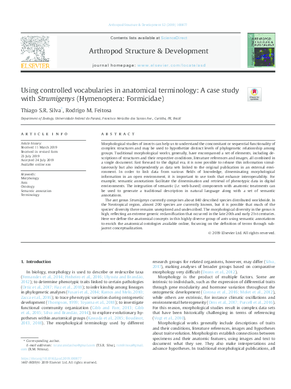
 Rodrigo Feitosa