Social Cognitive and Affective Neuroscience, 2017, 197–211
doi: 10.1093/scan/nsw108
Advance Access Publication Date: 16 September 2016
Original article
The neural component-process architecture of
endogenously generated emotion
Haakon G. Engen, Philipp Kanske, and Tania Singer
Department of Social Neuroscience, Max Planck Institute for Human Cognitive and Brain Sciences, Leipzig,
Germany
Correspondence should be addressed to Haakon G. Engen, Department of Social Neuroscience, Max Planck Institute of Human Cognitive and Brain
Sciences, Stephanstraße 1a, D-04103 Leipzig, Germany. E-mail: engen@cbs.mpg.de.
Abstract
Despite the ubiquity of endogenous emotions and their role in both resilience and pathology, the processes supporting their
generation are largely unknown. We propose a neural component process model of endogenous generation of emotion (EGE)
and test it in two functional magnetic resonance imaging (fMRI) experiments (N ¼ 32/293) where participants generated and
regulated positive and negative emotions based on internal representations, usin self-chosen generation methods. EGE activated nodes of salience (SN), default mode (DMN) and frontoparietal control (FPCN) networks. Component processes implemented by these networks were established by investigating their functional associations, activation dynamics and integration. SN activation correlated with subjective affect, with midbrain nodes exclusively distinguishing between positive and
negative affect intensity, showing dynamics consistent generation of core affect. Dorsomedial DMN, together with ventral anterior insula, formed a pathway supporting multiple generation methods, with activation dynamics suggesting it is involved
in the generation of elaborated experiential representations. SN and DMN both coupled to left frontal FPCN which in turn was
associated with both subjective affect and representation formation, consistent with FPCN supporting the executive coordination of the generation process. These results provide a foundation for research into endogenous emotion in normal, pathological and optimal function.
Key words: emotion generation; endogenous emotion; emotion regulation; positive emotion; negative emotion
Introduction
From melancholic reminiscence to joyful anticipation, we frequently experience emotions caused by internal mental processes, such as thoughts and memories (Killingsworth and
Gilbert, 2010). Such endogenous emotion is described as richer
and more intense than emotion elicited by external events
(Salas et al., 2012) and is known to play an important role in affective psychopathology, such as depression (Nolen-Hoeksema
et al., 2008) and anxiety (Freeston et al., 1996). There is also evidence that the endogenous generation of positive emotional
states can used as an effective means to regulate emotional reactions to external events (Engen and Singer, 2015), and the trait
tendency to do this is a predictor of psychological resilience
(Tugade and Fredrickson, 2004). Thus, understanding the psychological and neural mechanisms of endogenous generation of
emotion (EGE) can yield important insight into normal, pathological and even optimal emotional function.
Despite this, research into EGE has been limited, stemming
mainly from behavioral studies using EGE as a method to induce
emotional states. This research shows that EGE can be occasioned by a range of information-processing modalities, including mental imagery and semantic analysis of emotional
information (Vrana et al., 1986), interoception of bodily signals
(Philippot et al., 2002) or recall of episodic autobiographical memories (Mayberg et al., 1999). It has also been shown that EGE can
effectively occur when individuals immerse themselves in
Received: 27 January 2016; Revised: 27 June 2016; Accepted: 3 August 2016
C The Author (2016). Published by Oxford University Press.
V
This is an Open Access article distributed under the terms of the Creative Commons Attribution Non-Commercial License (http://creativecommons.org/
licenses/by-nc/4.0/), which permits non-commercial re-use, distribution, and reproduction in any medium, provided the original work is properly cited.
For commercial re-use, please contact journals.permissions@oup.com
197
Downloaded from https://academic.oup.com/scan/article-abstract/12/2/197/2544451
by Sachsische Landesbibliothek - Staats- u. Universitaetsbibliothek Dresden user
on 03 July 2018
�198
|
Social Cognitive and Affective Neuroscience, 2017, Vol. 12, No. 2
hypothetical scenarios (Wilson-Mendenhall et al., 2013a). This
latter finding demonstrates the theoretically important point
that EGE is not limited to reinstantiation of previously experienced emotional situations but can also simulate states appropriate for novel contexts. Indeed, emotions are frequently
elicited by spontaneous cognition about future events (Ruby
et al., 2013), suggesting that an important use of EGE is predicting
the affective relevance of hypothetical future scenarios
(Baumgartner et al., 2008). Although these studies were not
focused at exploring EGE as a process in its own right, they show
that multiple means (e.g. different strategies or different information modalities) can be utilized to generate emotional states,
dependent on the representational content of the target emotional experience. Mirroring recent constructivist theories of
emotion (Barrett and Wilson-Mendenhall, 2014; Russell, 2014),
this suggests that a comprehensive account of EGE needs to distinguish between (i) processes supporting the generation of the
hedonic or core affective quality (Wilson-Mendenhall et al.,
2013b) of an endogenous emotional experience from (ii) processes supporting the formation of representations of the context
to which this affective state applies or stems from. Importantly,
this opens for the possibility that the two are mechanistically
distinct, with different neural systems supporting core affect
generation varying as a function of hedonic qualities, while systems supporting representation formation should vary as a
function of the specific implementation of the generation
process.
Presently neuroimaging studies of EGE using comparable
protocols are limited, making evaluation of this hypothesis difficult. One exception is a series of early positron emission tomography (PET) experiments in which participants generated
emotional states by volitionally recalling significant emotional
experiences (Pardo and Raichle, 1993; Gemar et al., 1996; George
et al., 1996; Reiman et al., 1997; Kimbrell et al., 1999; Mayberg et al.,
1999; Damasio et al., 2000; Liotti et al., 2000). Considered in aggregate (Supplementary Figure S1), these studies suggest the involvement of three large-scale functional networks in EGE: (i)
The default mode network (DMN; Raichle and Snyder, 2007),
including ventromedial prefrontal cortex (vmPFC), posterior cingulate cortex (PCC), left temporoparietal junction (TPJ) and hippocampus (HC), (ii) the extended Salience Network (SN; Seeley et al.,
2007), including anterior insula (AI), dorsomedial PFC (dmPFC)
and structures in basal ganglia and midbrain and (iii) The frontoparietal control network (Spreng et al., 2010; FPCN; Laird et al.,
2011; Spreng et al., 2013) centered on lateral and dorsomedial prefrontal and inferior parietal cortices. There is a notable overlap
between this putative neural architecture and that known to
support the construction of mental representations in general:
DMN is associated with numerous forms of psychological processes involving simulation based on endogenous information
(Spreng et al., 2009) and appears to be involved in the integration
of information about a given topic into detailed episodic representations. Interestingly, DMN does not appear to support the
initial generation of the representational core that these details
pertain to (Addis et al., 2007). Rather, this initial generation is
thought to involve the direct activation of domain-specific and
task-relevant networks (Hassabis and Maguire, 2007). In the context of emotion, the SN is a likely candidate such a network.
Composed of cortical (AI, dmPFC) limbic [amygdala (AMY), ventral striatum (VS)] and midbrain structures [periaqueductal gray
(PAG), substantia nigra/ventral tegmentum (SN/VTA)], the SN is
closely associated with the representation and generation of
core affect and homeostatic regulation (Seeley et al., 2007;
Lindquist et al., 2012). Interestingly, DMN and SN appears to be
Downloaded from https://academic.oup.com/scan/article-abstract/12/2/197/2544451
by Sachsische Landesbibliothek - Staats- u. Universitaetsbibliothek Dresden user
on 03 July 2018
intrinsically anticorrelated (Buckner et al., 2013; Spreng et al.,
2013), strengthening the claim that they support dissociable
component processes in EGE. This anticorrelation also suggests
that an intermediary network coordinates and maintains activation of the SN and DMN, pointing to the need for executive processes to coordinate and maintain the generation process.
Possibly, the FPCN supports this role as it is known to support
adaptive cognitive control, as well as interfacing with
SN (Dosenbach et al., 2008), affording a pathway by which core
affective states can be generated in a goal-directed fashion.
Similarly, FPCN and DMN are known to couple during goaldirected internal mentation (Spreng et al., 2010) and to be implicated in the domain-general control of retrieval processes important for representation formation (Badre and Wagner, 2007).
To the degree that this functional-process architecture holds,
an interesting question is how these processes interact over the
course of a given EGE event. Addis et al. (2007) have shown that
the construction of endogenous simulations of events involves
distinct generation and elaboration phases, with the initial
phase involving retrieval of the core semantic features of the
representation and the subsequent elaboration phase involving
the elaboration of the core information in question with details
about the specific event. If this model holds for EGE, one would
expect involvement of SN primarily in the early stages of process, corresponding to core affect serving as a semantic anchor
for later elaboration efforts. Plausibly, however, the opposite
could be true, such that generation involves setting up representations of emotional situations, which in turn elicit core affective
states (Kross et al., 2009). A major objectives of the current investigations was to establish this relationship.
The objective of this study was to investigate this and to establish a comprehensive neural component-process architecture
for EGE. Based on the earlier considerations, we expected EGE to
be neurally implemented by DMN, SN and FPCN. Each of these
networks were hypothesized to support dissociable functional
component processes. Specifically, we suggest that SN supports
the generation of core affective states that serve as a guide for
the formation of detailed representations via processes instantiated in the DMN, resulting in an emotional experience. Finally,
we propose that FPCN supports the executive maintenance and
coordination of the generation process, coupling with both SN
and DMN. Importantly, as we propose they form the functional
core of EGE, we expect that these networks should partake in
EGE irrespective of the hedonic quality of the emotional state or
the precise means or modality used to generate it. We tested
this model in two experiments (N ¼ 32 and N ¼ 293) with a newly
developed paradigm aimed at maximizing ecological validity
and generalizability of EGE. To ensure that participants generated comparable emotional states, they were anchored using a
multimodal emotion induction procedure prior to scanning.
This procedure elicited multiple markers of emotional states (semantic, visual, auditory and bodily) prior to scanning, avoiding
artificially biasing participants’ implementation toward particular information modalities. To maximize ecological validity and
task compliance, participants were instructed to implement EGE
as they experienced most efficacious. Thus, in Experiment 1 participants were given complete freedom in how they generated
emotions, while in Experiment 2 they were allowed to combine
four generation modalities (Semantic Analysis, Episodic and
Auditory Imagery and Bodily Interoception; i.e. the endogenous
analogs to the modalities used in the induction procedure), in
whichever way they found most effective.
Participants then completed a cue-based fMRI paradigm
(Figure 1A). Trials consisted of a Generation phase and a
�H. G. Engen et al.
|
199
Fig. 1. Experimental task, behavior and psychophysiological validation. (A) Schematic of a single trial. (B) Subjective ratings of affect in Experiment 1. (C) Subjective ratings of affect for Experiment 2. (D) Main effect of Condition on SCL in a subset (N ¼ 244) of participants in Experiment 2. Error-bars ¼ within-subject SEs (Loftus and
Masson, 1994). * ¼ P< .05, ** ¼ P< .01, *** ¼ P< .001.
Modulation phase. In the Generation phase, participants used
their self-selected techniques to generate positive and negative
emotional states or actively attempted to remain neutral. Thus,
we could distinguish the neural correlates of general emotion
generation, from those supporting generation of particular implementations of generation. In the subsequent Modulation
phase, participants maintained this state (Maintain condition),
actively suppressed it (Regulate condition) or simply ceased
their generation efforts (Cease condition; Experiment 1 only).
This approach enabled us to dissociate neural systems supporting different component processes based on their activation
profiles. Finally, participants provided ratings of their affective
states following each trial, allowing identification of the neural
correlates of generation success.
Methods
Participants
For Experiment 1, 32 participants were recruited from an inhouse participant database (15 female, mean age ¼ 30.3, range
21–51, SD ¼ 9). For Experiment 2, participants were recruited in
the context of the large-scale longitudinal ReSource Project (see
Supplementary Materials for screening procedure). Baseline data
from this study were used. Three hundred and thirty-two participants were recruited for the ReSource Project, with 305 participants completing the current paradigm. Of these, five
participants were excluded on account of missing auxiliary data
(post-scan questionnaire, structural MRI) and technical difficulties. Four participants reported difficulties (e.g. nausea or sleepiness) during the scanning session and were dropped from
analysis. From the sample of 296 with complete data, a further
three participants were removed due to aberrant behavioral report and/or unacceptable data quality after preprocessing (>1
Downloaded from https://academic.oup.com/scan/article-abstract/12/2/197/2544451
by Sachsische Landesbibliothek - Staats- u. Universitaetsbibliothek Dresden user
on 03 July 2018
voxel movement, >5% corrupted time points, design VIF > 2),
leaving a final sample of 293 (170 female, mean age ¼ 40.4,
range: 20–55, SD ¼ 9.3). All participants had normal or corrected
to normal vision. The study was approved by the Ethics
Committee of the University of Leipzig and Humboldt
University, Berlin and was carried out in compliance with the
Declaration of Helsinki. All participants gave written informed
consent, were paid for their participation and were debriefed
after the study was completed.
fMRI experimental procedure
Before scanning, participants underwent an automated training
procedure (see Supplementary Materials for details), including a
multimodal emotion induction aimed at minimizing betweenparticipant variance in implemented emotional states. In
Experiment 2, participants were also instructed in the use of
four generation modalities (Semantic, Episodic, Auditory and
Bodily) and instructed to select to which degree they to use
each in the following experiment according to their own preferences. Additionally, participants were shown a number of neutral stimuli (e.g. pictures of scenery) and instructed to actively
attain the sort of neutral emotional state these stimuli evoked
(see Supplementary Materials for details). Participants were instructed to attempt to attain such states during the Neutral
condition, and also when requested to downregulate their erstwhile generated emotional states. After the scanning session,
participants were debriefed. In Experiment 1, verbal debriefing
was done with an experimenter. In Experiment 2, participants
reported the degree to which they used each of the generation
modalities using a nine-point Likert.
Each trial (Figure 1A) started with a 4–6 s white fixation cross
indicating the start of trial. Then a 10 s Generation phase was
entered, in which subjects were shown a colored symbol
�200
|
Social Cognitive and Affective Neuroscience, 2017, Vol. 12, No. 2
indicating which emotional state to generate (Red
minus ¼ Negative, Green plus ¼ Positive, Blue 0 ¼ Neutral). This
was followed by a 5 s Modulation phase where participants either maintained the generation of the emotional state or downregulated it so as to attain a neutral emotional state. In the
Maintain condition, the instruction symbol remained the same
as in the Generation phase. In the Regulation condition, the
symbol changed to a Blue 0. Finally, in Experiment 1 we
included a partial-trial condition where the instruction cue
changed to a fixation cross (Cease condition; Experiment 1
only). For the Neutral condition the symbol did not change but
remained a Blue 0. Thus, Experiment 1 consisted of a total of
seven different conditions (Maintain Positive/Negative, Regulate
Positive/Negative, Cease Positive/Negative and Neutral).
Experiment 2 omitted the Cease condition and thus had a total
of five conditions. Experiment 1 had two runs of five trials per
condition (35 per run), while Experiment 2 had a single run of 10
trials per condition (50 total). Condition sequence was pseudorandomized, ensuring no direct repetitions of conditions
occurred. Finally, a 5 s fixation cross was presented followed by
a 5 s presentation of a continuous Visual Analog rating Scale
ranging from ‘Extremely negative’ via ‘Neutral’ to ‘Extremely
positive’ [range 6 251 from the neutral point (0)]. Initial cursor
position was jittered randomly around the Neutral point.
Participants responded using a button box and the right hand
index and middle finger. Participants were instructed to report
their affective state as it was at the moment of report. Stimuli
were back-projected using a mirror setup. Task setup was identical in both experiments, except for the omission of the Cease
condition in Experiment 2 due to time constraints.
MRI acquisition
For both experiments, MRI data were acquired on a 3T Siemens
Verio Scanner (Siemens Medical Systems, Erlangen, Germany)
using a 32-channel head-coil. High-resolution structural images
were acquired using a T1-weighted 3D-MPRAGE sequence
(TR ¼ 2300 ms, TE ¼ 2.98 ms, TI ¼ 900 ms, flip angle ¼ 7� , iPat ¼ 2;
176 sagittal slices, FOV ¼ 256 mm, matrix size ¼ 240 � 256,
1̂3 mm voxels; total acquisition time ¼ 5.10 min). For the functional imaging, we employed a T2*-weighted gradient EPI sequence that minimized distortions in medial orbital and
anterior temporal regions (TR ¼ 2000 ms, TE ¼ 27 ms, flip
angle ¼ 90� , iPat ¼ 2; 37 slices tilted �30� from the AC/PC axial
plane, FOV ¼ 210 mm, matrix size ¼ 70 � 70, 33 mm voxels, 1 mm
gap). For Experiment 2, we acquired B0 field maps using a
double-echo gradient-recalled sequence with matching dimensions to the EPI images (TR ¼ 517 ms, TE ¼ 4.92 and 7.38 ms).
fMRI preprocessing
Preprocessing was performed using a combination of SPM12
(r6225) functions and the ArtRepair toolbox (Mazaika et al., 2005)
running on Matlab 2013b. Functional images were realigned
(Experiment 1) or realigned and unwarped to additionally correct for distortion using B0 field maps (Experiment 2). ArtRepair
procedures were then employed, including slice wise artifact
detection and repair using interpolation (art_slice; 5% cutoff),
time series diagnostics (art_global) identifying and repairing via
interpolation volumes showing large global intensity fluctuation (>1.3%), volume-by-volume movement exceeding 0.5 mm
and overall movement (>3 mm) and despiking with a 5% signal
change cutoff (art_despike). T1 structural images were registered to the mean realigned volume and segmented. Using
Downloaded from https://academic.oup.com/scan/article-abstract/12/2/197/2544451
by Sachsische Landesbibliothek - Staats- u. Universitaetsbibliothek Dresden user
on 03 July 2018
DARTEL (Ashburner, 2007) procedures, functional images were
normalized and smoothed with an isotropic kernel of 8 mm
FWHM.
First level fMRI analyses
Individual-level models included separate sets of regressors for
the Generation and Modulation phase. For the Generation
phase, three regressors were specified corresponding to the
emotional target (Positive, Negative and Neutral) of the trial. For
the Modulation phase, separate regressors were specified for
each condition. Thus, the model in Experiment 1 included seven
regressors [Valence (Positive and Negative) * Modulation
(Maintain, Cease and Regulate) þ Neutral] for the Modulation
phase, for a total of 10 regressors The model in Experiment 2,
where the Cease condition was omitted, included five regressors for the Modulation phase, for a total of eight regressors.
Regressors were convolved with canonical hemodynamic
response functions (HRFs) with a 10 s (Generation) or 5 s
(Modulation) duration, as well as regressors specifying parametric modulations by trial-wise subjective affect ratings. An additional regressor was specified for the Rating period. Movement
parameters derived from the realignment step (six regressors),
their derivatives and squared values were added (24 regressors).
Potential physiological confounds were controlled for by adding
four additional regressors reflecting volume-wise mean signal
from white matter and cerebrospinal fluid, global signal and
highest-variance voxel time course.
Second level fMRI analyses
All second analyses were conducted using robust regression
(Wager et al., 2005), with covariates of no interest coding elected
arousal level, age and gender. Second level models for
Experiment 2 additionally included regressors coding selfreported generation modality usage (four regressors) as continuous covariates.
All results were corrected for multiple comparisons using
cluster extent family-wise error rate (FWEc) correction at an
alpha of P < 0.05, unless otherwise indicated. Cluster extents
were estimated using Monte Carlo simulation and estimated intrinsic smoothness [3DClustSim and 3DFWHMx from the AFNI
package (Forman et al., 1995)], as implemented in NeuroElf. Note
that peak-forming thresholds were adapted for Experiments 1
(P < 0.001) and 2 (P < 0.00005) to account for differences in sample size. Correlational and mediation results also used a less
strict peak threshold of P < 0.0005.
All analyses were masked with a gray matter template
derived from the DARTEL created template, thresholded at 95%
gray matter probability, supplemented by a hand-drawn masks
of brainstem nuclei due to poor differentiation of white from
gray matter in these regions.
Constrained principal component analysis. In Experiment 2, we
adopted a data-driven approach using constrained principal
components analysis (CPCA; see Woodward et al., 2013 for details) of fMRI time series using the CPCA-fMRI package (www.
nitrc.org/projects/fmricpca). CPCA analysis of fMRI data is a
multivariate method that involves a singular value decomposition of BOLD time series to identify functional networks followed by an estimation of BOLD change in each network over
peristimulus time as a function of experimental condition.
Here, we used finite impulse response (FIR) modeling to identify
task-specific functional connectivity networks based on the 15
�H. G. Engen et al.
Table 1. Comparison of self-reported experienced affect
Comparison of emotion ratings
Comparison
Maintain
Cease
Cease
Regulate
Cease
Experiment 1
vs
vs
vs
vs
vs
Experiment 2
Comparison
Maintain
vs
Regulate
vs
Positive
Negative
Neutral
Neutral
Maintain
Maintain
Regulate
t-value (df ¼ 31)
11.25***
�10.50***
10.94***
�9.50***
�3.27**
2.43*
�7.79***
6.88***
6.96***
�6.08***
Neutral
Maintain
t-value (df ¼ 292)
Positive
Negative
27.24***
�27.94***
�19.44***
20.69***
Descriptives are reported in Figure 1. * ¼ P < .05, ** ¼ P < .01, *** ¼ P < .001.
bins (i.e. 30 s, allowing for hemodynamic lag) following the
onset of the generation cue. Importantly, using a FIR model
allows hemodynamic response (HDR) profiles to be identified
for each component separately, allowing the identification of
task-relevant functional connectivity networks with dissociable
temporal profiles. Finally, CPCA provides HDR estimates at the
individual level, allowing the resultant predictor weights to be
used to explore the correlates of individual differences in component activation.
Mediation analyses. To differentiate components of the generation network involved in generation using a specific modality
from components involved in generation in general, we followed previous work aimed at identifying the large-scale networks supporting emotion regulation performance via
mediation modeling (Denny et al., 2014). First, regions whose activation during generation of emotion (relative to neutral) were
identified using robust regression. Mediation effect parametric
mapping as implemented in the M3 mediation toolbox (Wager
et al., 2008) was used to investigate modality-specific and modality general pathways of emotion generation. We performed a
whole-brain search for voxels whose activity during emotion
generation (relative to the neutral baseline) showing a relationship with reported use of a given modality that was mediated
by the activity in regions independently correlated with usage
of that modality in a robust regression model. Statistics were assessed using the bootstrapping approach implemented in the
M3 toolbox (10 000 samples).
Analysis approach
The first objective our analyses was to establish the overall neural architecture of EGE. To achieve this, we first sought establish
the validity of our experiment by investigating subjective and
physiological indices of emotional states. Next, we contrasted
combined positive and negative EGE with the neutral baseline,
thereby identifying the overall neural basis of EGE. We next
sought to test the component process mapping proposed in the
introduction in two ways: first, based on the data from
Experiment 1, we enacted a contrast-based decomposition,
based on a model of the activation dynamics expected for each
of the component processes. To complement this, we next performed a data-driven decomposition of the data from
Experiment 2 using CPCA, to identify the functional networks
central in EGE. Together, the results from these three analyses
Downloaded from https://academic.oup.com/scan/article-abstract/12/2/197/2544451
by Sachsische Landesbibliothek - Staats- u. Universitaetsbibliothek Dresden user
on 03 July 2018
|
201
allowed a description of the overall network and functional subcomponents supporting EGE in general. Following on this, the
second objective of the analyses was to differentiate general
EGE networks from those supporting specific implementations
of EGE, such as the generation of a particular valence, or using a
specific modality. By investigating how subjective ratings
for positive and negative generation parametrically modulated
signal, we could differentiate regions activated in a valencespecific manner from those supporting specifically the generation of positive and negative emotional states. Finally, by
investigating the correlation of activation with reported usage
of different modalities, we could identify specific regions supporting modality-specific implementation, and, using mediation analysis, identify the networks supporting EGE modality
usage. Moreover, by comparing these networks we could differentiate parts of these networks supporting specific modalities
from those supporting EGE in general.
Results
Behavioral and psychophysiological validation
Our first objective was to validate our experimental design,
using a combination of behavioral and psychophysiological
measures to ascertain that participants were able to generate
and regulate emotional states as measured by subjective and
objective markers of emotional arousal.
Post-trial ratings were analyzed using paired t-tests, reported
in Table 1. Figure 1B shows subjective ratings in each condition
for Experiment 1. Relative to the Neutral baseline condition,
increased reports of corresponding affect were observed for both
Maintain and Cease conditions. The Cease condition also
showed significantly higher ratings for both positive and negative affect compared with their respective Maintain conditions.
Finally, regulation resulted in decreased ratings for both positive
and negative emotion relative to their respective Maintain conditions. Figure 1C shows subjective ratings as function of condition for Experiment 2. Relative to the neutral baseline condition,
increased reports of corresponding affect were observed for both
positive and negative Maintain conditions. Regulation conditions also showed decreased ratings for both positive and negative affect, relative to their respective Maintain conditions.
These results demonstrate that participants were subjectively
able to generate and regulate endogenous emotional states of
both positive and negative valence in both experiments.
Importantly, they also show that, while a generated emotional
states decay without active maintenance, they remain subjectively significant for at least a short time following generation,
consistent with the representation of the emotional state persisting even without active generation efforts.
We next sought to establish whether participants’ generation efforts also elicited objective emotional arousal responses.
To this end, we concurrently assessed elicited skin conductance
levels (SCL) in Experiment 2 (see Supplementary Materials for
details on data acquisition and preprocessing). Two hundred
and twenty-five recordings had acceptable data quality and
were used to investigate the impact of generation instructions
on objective measures of emotional arousal, as well as their
interaction with subjective ratings. As SCL is the most frequently reported measure in investigations of exogenously
induced emotional states (Kreibig, 2010), an interaction would
suggest that the elicited states can be construed of as bona fide
emotional states and that behavioral ratings can be taken as
proxy for emotional arousal. Using linear mixed modeling
�202
|
Social Cognitive and Affective Neuroscience, 2017, Vol. 12, No. 2
of trial-wise SCL responses during the Generation period, we
predicted the trial-wise log-transformed estimates of SCL measured in microsiemens (lS) using a subject-level random intercept model. The model further included a factorial fixed effect
for condition (Generate Positive, Generate Negative, Neutral)
and a continuous fixed covariate for scaled trial-wise ratings of
subjective affect. To control for potential learning/fatigue effects, trial number was entered as a nuisance covariate (for
more detail on the effect of fatigue in the current experiment,
please see Supplementary Analyses). This analysis revealed a
main effect of Condition [F(2, 11012.639) ¼ 3.155, P < 0.05], Rating
[F(1, 11013.700) ¼ 4.625, P < 0.05], as well as a Condition * Rating
interaction [F(2, 11014.119) ¼ 17.815, P < 0.001]. Bonferroni corrected t-tests (Figure 1D) were performed to clarify the main effect of Condition, showing that, relative to the neutral
(mean ¼ 1.205, SE ¼ 0.32) baseline condition, higher SCL levels
were observed for both negative [mean ¼ 1.267, SD ¼ 0.032;
paired t-test: t(224) ¼ 4.44, P < 0.001] and positive [mean ¼ 1.268,
SD¼ 0.032; paired t-test: t(224) ¼ 5, P < 0.001] emotion generation
conditions. Closer investigation of the Condition * Rating interaction showed that it consisted of a significant difference in the
slopes of the rating effect between negative and positive generation [t(8770.515) ¼ 5.63, P < 0.0001]. Specifically, SCL had a negative relationship with ratings [t(4278.64) ¼ �3.71, P < 0.001]
during negative generation and a positive relationship
[t(4278.64) ¼ 4.16, P < 0.001] during positive generation.
Corresponding to the bipolar scale used (Figure 1A), this shows
that SCL levels increased with stronger affect ratings for both
positive and negative emotion (Figure 1E). These results show
that participants were capable of generating both positive and
negative emotional states, as measured by both subjective and
objective indices of emotional arousal, and these indices were
correlated, such that behavioral report corresponded to objective physiological arousal.
Finally, we sought to explore what kind of emotional states
participants elected to generate. During debriefing, participants
in Experiment 2 were asked whether they generated high or low
arousal exemplars of positive and negative emotional states
after the experiment. Thirty-nine percent of participants reported generating high arousal positive emotional states, like joy
or happiness, with the complementary 61% generating low
arousal positive emotion like calmness or caring. Similarly, 29%
of participants reported generating high arousal negative emotions like fear or anger, while 71% reported generating low
arousal states like sadness or melancholia. All subsequent analyses in Experiment 2 control for this between-subject variance.
Exploring the neural architecture of EGE
Our next objective was to establish whether our hypothesized
three-network architecture of EGE was in evidence. To identify
the neural correlates of emotion generation, we contrasted the
combined Generation and Maintenance periods for both positive and negative affect generation with the Neutral baseline
condition, with one sample t-tests performed using robust regression (Wager et al., 2005). For Experiment 1, a primary
cluster-forming threshold of P < 0.001, T > 3.38 was used. In
Experiment 2, a more stringent threshold of P < 0.00005, T > 3.95
was used for the primary contrasts to balance increased power.
Using Monte-Carlo simulation (Forman et al., 1995), cluster
thresholds were determined to be k > 40 and k > 10, respectively,
for FWEc a < 0.05.
In Experiment 1 (Figure 2A and Supplementary Table S1),
we observed activation in core nodes of the DMN [vmPFC, PCC,
Downloaded from https://academic.oup.com/scan/article-abstract/12/2/197/2544451
by Sachsische Landesbibliothek - Staats- u. Universitaetsbibliothek Dresden user
on 03 July 2018
left TPJ, left middle temporal gyrus (MTG) and HC] and SN [AI,
dmPFC, including pre-supplemental motor area (pre-SMA) and
dorsal anterior cingulate cortex (dACC)]. Activation was also
observed in nodes of the SN most closely associated with hedonic processing (VS, SN/VTA), as well as cerebellar regions.
Deactivations were observed in right FPCN, in addition to inferior temporal gyrus (ITG) and superior occipital gyrus. In
Experiment 2 (Figure 2B and Supplementary Table S1), we
observed activation and deactivation patterns substantially
similar to Experiment 1, albeit markedly stronger, consistent
with the increased power in Experiment 2 (N ¼ 293). Additional
activation was also observable in the frontal portions of the left
FPCN [bilateral inferior (IFG) and middle (MFG) frontal gyrii].
Stronger activations were observed in midbrain, including both
SN/VTA and PAG, as well as hypothalamus, thalamus, basal
ganglia and ventral AI. Again, deactivations centered on right
FPCN and occipital regions.
These results replicate previous work and support our contention that the DMN, SN and FPCN key are components in the
neural architecture supporting EGE. They also expand on them
demonstrating that this relationship holds for EGE as it is freely
implemented in the population. Finally, they suggest that EGE
additionally involves the active suppression of right frontoparietal and occipital regions, explainable by the known deactivation
of these regions in internally focused processing (AndrewsHanna et al., 2014).
Model-based component process mapping
Differentiating the initial generation of endogenous states from
their subsequent elaboration into experiential representations
is commonly done by having participants report the moment
they subjectively experience to have completed the generation
process (e.g. retrieval of core semantic information about an
event) and begin the process of elaborated mental simulation
by adding details about the context (Addis et al., 2007, 2009).
In the context of EGE, achieving a similar subjective differentiation is difficult, since the emotional experiences are inherently
situated in a given context (Wilson-Mendenhall et al., 2013a).
We therefore took a model-based decomposition approach to
test our proposed component process structure. We reasoned
that the 10 second Generation period of each trial should include activation of all constituent component processes (i.e.
generation, elaboration and maintenance), and that regions
supporting these processes should be distinguishable by their
activation dynamics in different conditions during the
Modulation phase. By masking out activation attributable to either maintenance or elaboration from the 10 second Generation
period, one should therefore be left with regions involved exclusively in the initial generation of emotional experiences.
Figure 3A schematically illustrates the hypothesized activation dynamics for each component as a function of condition.
Specifically, we hypothesized that regions supporting the initial
generation of the affective state should show early and phasic
activation corresponding to their involvement in the generation
of the affective core of the experiences. Importantly, they
should also be largely unaffected by modulation efforts, as
these should target the neural substrates of representation of
the emotional experience rather than those involved in generation (Gross et al., 2011). Conversely, regions supporting the elaborated representation of emotional experiences should be
affected by modulation efforts and be mainly in evidence in the
later part of the trial. Moreover, given that emotional experiences tend to persist over time (Buchanan, 2007; Verduyn et al.,
�H. G. Engen et al.
|
203
Fig. 2. Core networks of endogenous emotion generation. (A) Regions activated in Experiment 1 for the Generation conditions (Positive and Negative Generate and
Maintain) relative to the Neutral condition. (B) Equivalent contrast for Experiment 2.
2009), it should be possible to dissociate the neural substrates of
the elaborated mental simulation from those supporting the active maintenance of the generation process in contrasting activation in the Cease condition with the Maintenance condition
(in the Modulation phase only). For these analyses alone, we
used a more lenient threshold of P < 0.005 (uncorrected), k > 10,
due to the lower power of the component contrasts.
To begin, component contrasts were calculated according
to our process dynamic logic (Supplementary Figure S2 and
Table S4). The Generate > Neutral contrast for the Generation
period defined the neural reference space for EGE overall
(Supplementary Figure S2A). Regions involved in the elaborated
representation of emotional experiences were identified in contrasting the average of the Cease and Maintain conditions with
the Regulate condition (Supplementary Figure S2B). This was
done because both Cease and Maintain conditions were associated with elevated subjective emotional experiences relative to
both Neutral and Regulate conditions (Figure 1B). We opted to
use the Regulate rather than the Neutral condition since the
Regulate condition actively suppress emotional representations.
Finally, regions involved in effortful maintenance of EGE was
identified by the Maintain > Cease contrast (Supplementary
Figure S2C). Next, the maintenance and representation contrasts
were inclusively masked with the Generate > Neutral contrast to
ensure that all activations were associated with EGE. To ensure
orthogonality maintenance and representation processes, these
were mutually masked (overlapping regions are reported in
Supplementary Figure S2D). Finally, the Generate > Neutral contrast was exclusively masked with both maintenance and representation process contrasts, leaving exclusively activation not
attributable to either of the two.
The results from this masking approach are reported in
Figure 3B and Supplementary Table S3. Generation was primarily associated with activation of the extended SN, including left
dorsal AI, dACC, basal ganglia and brainstem regions, in addition
to known mnemonic structures such as temporal pole and HC.
Maintenance, conversely, uniquely activated nodes of FPCN,
including left IFG, PMC and pre-SMA, in addition to occipital regions and subgenual ACC. Finally, elaborated representation
uniquely activated large portions of the DMN (PCC, vmPFC, TPJ
and MTG) and thalamus, in addition to the ventral AI, nucleus
accumbens (NACC) and AMY—all regions traditionally thought
to subserve core affect processing (Lindquist et al., 2012).
Downloaded from https://academic.oup.com/scan/article-abstract/12/2/197/2544451
by Sachsische Landesbibliothek - Staats- u. Universitaetsbibliothek Dresden user
on 03 July 2018
As a final step, we verified that our approach indeed identified regions with appropriate temporal dynamics by extracting
FIR-fitted time courses using MarsBar for select regions in each
contrast. This revealed strong correspondence between
observed and hypothesized dynamics (Figure 3C).
These results conform to our hypotheses, showing that EGE
is supported by at least three separable component processes,
and that these roughly overlap with each of the three core intrinsic networks observed in our main contrasts. Specifically,
FPCN appears primarily to support the active maintenance of
generation efforts, while DMN primarily supports the representation of the generated states as evidenced by it being the primary target for downregulation, as well as it remaining active
even in the absence of generation efforts. Finally, the cortical
and midbrain aspects of SN selectively responded in manner
consistent with being involved initial generation of emotional
states. Interestingly, we found that several regions in the limbic
subcomponent of the SN (vAI, AMY and NACC) responded in a
manner consistent with them supporting elaborated representations, suggesting that midbrain and limbic components of SN
differ in their functional contribution to EGE.
Functional significance and dynamic integration of EGE
networks
Having found evidence consistent with our hypothesized component process mapping, our next step was to (i) establish the
functional significance of these networks and (ii) investigate
their dynamic interaction during EGE. To address this, we used
CPCA (Woodward et al., 2013; Lavigne et al., 2015) in our larger
sample in Experiment 2. Briefly, CPCA is a data-driven multivariate method that combines multiple regression with PCA
analysis to identify component of mutually correlating voxels,
i.e. functional networks, involved in a task based on their specific task-related activation dynamics (see Experimental Procedure
for detail). As CPCA provides individual-level estimates of activation of each network component, we could identify networks
specifically predicting individual differences in EGE efficacy,
thereby establishing both their involvement and their functional significance. Finally, as CPCA does not enforce spatial orthogonality on components it allows the identification of
regions partaking in multiple network components with
�204
|
Social Cognitive and Affective Neuroscience, 2017, Vol. 12, No. 2
Fig. 3. Model-based mapping of component processes of EGE. (A) Activation profiles of three hypothesized component processes of EGE. Briefly, processes involved in
the Generation of endogenous emotion experiences should show early activation and be unmodulated by later regulatory efforts, while processes involved in their
Maintenance should show extended activation but only when active efforts are expended to sustain the emotional state. Finally, processes involved in the Representation
of the emotional experience should show a graded response, such that highest activation occurs during active maintenance and lowest activation occurs during active
regulation. (B) Regions showing activation patterns consistent with partaking in the three hypothesized component processes in Experiment 1. (C) Time courses from
select regions (white rings in B) of the components showing correspondence with hypothesized activation profiles. Error-bars ¼ SEM.
differing temporal dynamics, as would be expected if, as
hypothesized, FPCN coupled to both SN and DMN.
Eigenvalue plots indicated that six components should be
extracted using the scree criterion. To differentiate components
supporting general task processes (e.g. sensory processing,
motor responses) from those specifically supporting EGE, we
calculated the component-specific AUC of loadings in the task
window (4–22 s post-stimulus; allowing for 4–6 s hemodynamic
lag) for the Maintain condition, subtracting the Neutral baseline.
This yielded individual-level estimates of overall component
activation during EGE, which were orthogonalized and entered
into a multiple regression model predicting individual differences in self-reported generation success (i.e. average affect ratings in the Maintain condition only). Individual differences in
component activation explained a significant amount of variance in generation success [F(6286) ¼ 3.124, P < 0.01, R2 ¼ 0.062],
with two components directly predicting generation success. To
interpret these, loading maps were thresholded at the dominant
10% of component loadings with k > 30 (Lavigne et al., 2015).
The first component [b ¼ 0.153, t(292) ¼ 2.675, P < 0.01; Figure
4A] included central nodes of the DMN (vmPFC, PCC, left TPJ)
and FPCN (bilateral BA47, BA45, MFG and PMC), as well as VS
Downloaded from https://academic.oup.com/scan/article-abstract/12/2/197/2544451
by Sachsische Landesbibliothek - Staats- u. Universitaetsbibliothek Dresden user
on 03 July 2018
(NACC, caudate) dACC, SMA/pre-SMA, mid cingulate cortex
(MCC), bilateral superior temporal gyrus/transversal gyrus (STG/
TRANS), left MTG and right somatosensory cortex, similar to the
Representation network identified earlier. This similarity extended to activation dynamics, showing both early and sustained activation in the Modulation phase for the Maintain
condition and evidence of suppression in the Regulate condition. The second component [b ¼ 0.146, t(292) ¼ 2.574, P ¼ 0.01;
Figure 4B] included cortical nodes of SN (bilateral AI, pre-SMA)
as well as portions of the FPCN (bilateral BA47, left BA45, angular gyrus and MFG), plus thalamus, occipital cortex and superior
cerebellum. Dynamics closely resembled the Generation network
identified in earlier, with no observed difference between
Maintain and Regulate conditions. Conjoining the individually
thresholded component maps (Figure 4C and Supplementary
Table S5), we found that left lateral FPCN regions (IFG, BA45/45,
MFG), as well as pre-SMA and dACC were part of both components, in addition to thalamus and retrosplenial cortex.
Notably, this closely overlaps with the Maintenance network
identified in our model-based analyses. These results expand
on our model-based approach earlier, establishing the unique
functional significance of our three candidate networks in EGE.
�H. G. Engen et al.
Further, they demonstrate that FPCN coupled with both SN and
DMN, while these did not show evidence of coupling, supporting the hypothesis that FPCN coordinates activation of these
two networks during EGE.
Identifying the neural basis of core affect generation
Having found evidence for both the functional significance and
dissociability of our putative EGE networks, we next sought to establish neural implementation of core affect generation and representation formation. Overall success at generating emotions
was evaluated in the debriefing session of Experiment 2 on a 0–9
scale ranging from completely unsuccessful to completely successful. Participants reported significantly higher success at generating positive (mean ¼ 5.99) than negative (mean ¼ 5.54)
emotions [paired t-test: t(292) ¼ 4.11, P < 0.001]. To avoid biasing
results by potential effort or success effects, we therefore combined positive and negative valence conditions, and instead
focused on trial-wise parametric modulation of activation as a
function of ratings. Using robust regression, we performed one
sample t-tests on parametric modulation maps separately for
positive and negative trials, averaging across all conditions
involving emotion generation for both Generation and
Modulation periods (Supplementary Table S5). To differentiate regions supporting emotion generation success in general from
valence-specific regions, we conjoined the resulting FWEc thresholded maps (Figure 5). In Experiment 1, this revealed valencegeneral modulation in the basal ganglia, including putamen and
caudate body, while positive ratings uniquely modulated caudate
head/NACC and negative ratings uniquely modulated left dorsal
AI and pre-SMA. In Experiment 2, valence-general modulation
was more extensive, including left frontal portions of the FPCN,
particularly IFG and MFG, as well as key nodes of the SN (dorsal
AI, dACC and pre-SMA) in addition to thalamus. Valence-specific
modulation of activation was observed in caudate head/NACC
and SN/VTA for positive affect ratings, while modulation of deactivation was observed in occipital and right lateralized frontal
regions overlapping with the deactivated regions reported in the
main contrast. Negative affect ratings modulated activation in
right dorsal AI and PAG. These results show that activation levels
of FPCN and SN support successful emotion generation.
Moreover, midbrain portions of the SN were shown to be recruited in a valence-specific fashion, consistent with the known
association of these regions with domain general hedonic processing (Kringelbach and Berridge, 2009; Buhle et al., 2013). This
supports our hypothesis that SN is particularly important for the
generation of the affective core of EGE. Further, in line with our
CPCA analysis, we find that frontal FPCN supports a general role
in the generation of core affective states, possibly associated with
the initiation of the generation process.
Pathways of representation formation
Finally, we sought to identify the neural bases of representation formation. To this end, participants in Experiment 2 were
constrained to use four specific generation modalities: (i)
Semantic Analysis, involving the use of verbalized thoughts affective thoughts (ii) Episodic Imagery, involving the generation
of visual emotional imagery, (iii) Auditory Imagery, involving
the generation of affective soundscapes and (iv) Bodily
Interoception, involving focus on and interpretation of bodily
signatures of emotional states (for precise instructions, see
Supplementary Materials). These modalities corresponded to our
multimodal induction procedure, ensuring participants were
Downloaded from https://academic.oup.com/scan/article-abstract/12/2/197/2544451
by Sachsische Landesbibliothek - Staats- u. Universitaetsbibliothek Dresden user
on 03 July 2018
|
205
equally primed to using each of them, and have clear analogs
in daily life [e.g. thinking self-deprecating thoughts (semantic),
remembering or anticipating an emotional event (episodic),
humming a sad song (auditory) or noticing a dry mouth and
racing heart when making a presentation (bodily)]. Finally, participants were allowed to freely combine these modalities in
whichever they found best enabled them to generate emotional
states. These four modalities can be combined to different degrees in an intuitive fashion (e.g. internal affective monolog
combined with a concrete emotional episode where a specific
song was playing) ensuring variance in the combinations utilized by participants.
Post-scan self-reports of the degree to which participants
used each of these modalities (Figure 6B) showed that the
Episodic modality was the most used (40%), followed by
Semantic (24%), Bodily (21%) and Auditory (15%). Entering the
degree to which each participant reported using each modality
as covariates in our main robust regression analysis (Generate
and Maintain > Neutral), we could identify the neural correlates
of modality usage (Figure 6C). Due to the noisy nature of self-report, we used a more lenient cluster-forming threshold of P <
0.0005, T > 3.32 (k > 42, FWEc P < 0.05). This revealed that use of
the Semantic modality was correlated with activation of the left
MTG, corresponding to the inferior border of Wernicke’s area, as
well as a region in the left dorsal frontal cortex. Use of the
Episodic modality was correlated with signal in the anterior superior PCC, a region known to be a part of the mnemonic subsystem of the DMN (Andrews-Hanna et al., 2014). Use of Bodily
modality was correlated with signal in bilateral dorsal and
mid-AI, a region known as interoceptive cortex representing
bodily signals (Craig, 2011). As no significant correlations
were found for the Auditory modality, we did not explore this
further.
We next identified the extended neural pathways by which
these regions influenced the generation network as a whole,
using mediation analysis (Wager et al., 2008; Denny et al., 2014).
Specifically, we implemented a mediation model (Figure 6A) consisting of a whole-brain search for voxels where the relationship
between their Generate > Neutral contrast value and reported
modality usage was mediated by the contrast values in the
modality-specific regions identified earlier. Thus, these analyses
identify voxels whose relationship with modality usage is mediated by activation of the modality-specific regions, suggesting
that they are part of the functional pathway by which that modality is implemented. For each analysis, reported usage of all
other modalities were entered as covariates. To identify unique
pathways for each modality mediation maps were thresholded
at Z > 3.25, P < 0.005, k > 30 and masked exclusively with the
maps for the remaining two modalities, revealing exclusive
modality-specific pathways (Figure 6D and Supplementary Table
S3). The Semantic pathway included left BA45 and BA22/35,
approximating Broca’s and Wernicke’s areas, respectively, left
temporal pole and premotor regions and dACC and anterior PCC,
closely corresponding to the extended semantic system
described in a recent meta-analysis (Binder et al., 2009). The
Episodic pathway included the majority of the DMN, including
vmPFC, PCC, left MTG and HC, bilateral angular gyrus, ventral AI
and left IFG, as well as subgenual ACC extending into VS, caudate
and pallidum. Finally, the Bodily pathway included regions
involved in body representation, including right posterior insula
(Craig, 2011), bilateral fusiform body area and left extrastriate
body area (Taylor et al., 2007), in addition to MCC and PC, perigenual ACC and bilateral dorsolateral PFC, including premotor
cortices.
�206
|
Social Cognitive and Affective Neuroscience, 2017, Vol. 12, No. 2
Fig. 4. Dynamic differentiation and integration of EGE networks. Results from the CPCA analysis showing the two components that predicted endogenous emotion generation ability, their dynamics and correlation with generation efficacy. (A) The primary component, composed mainly of FPCN and DMN regions. (B) The secondary
component, composed mainly of FPCN and SN nodes. (C) Conjunction of A and B, showing regions partaking in both task-relevant components.
Downloaded from https://academic.oup.com/scan/article-abstract/12/2/197/2544451
by Sachsische Landesbibliothek - Staats- u. Universitaetsbibliothek Dresden user
on 03 July 2018
�H. G. Engen et al.
|
207
Fig. 5. Neural correlates of core affect. Regions parametrically modulated by subjective ratings of affect in Experiment 2. Green and red regions, respectively, show
modulation in positive and negative conditions exclusively. Blue colored showed parametric modulation in both positive and negative conditions. Results are averaged
across all emotional conditions for both Generation and Modulation phases.
To identify modality-independent pathways, we conjoined
the thresholded maps, revealing a shared pathway (Figure 6E
and Supplementary Table S3) overlapping with the Representation
network identified earlier including the ventral AI and portions
of the dorsomedial subsystem of the DMN (Andrews-Hanna et al.
, 2010), as well as a substantial portion of the FPCN (bilateral IFG
and MTG, left angular gyrus, MCC), as well as posterior insula.
Thus, our findings support our hypothesis that DMN, together
with vAI, support representation formation in cooperation with
FPCN. Importantly, the Generation network was not involved in
either general nor modality-specific pathways, supporting the
hypothesized distinction between representation formation and
core affect generation.
Discussion
We hypothesized that the EGE involves the cooperation of three
core functional networks: the saliency network (SN), the DMN
and the FPCN, respectively, supporting core affect generation,
episodic representation and executive maintenance of emotional states. In two independent samples, we found that EGE
activations centered on our three candidate networks.
Decomposing these networks based on hypothesized activation
profiles of component processes, we found support for our
process-network mapping, showing that cortical and midbrain
SN primarily contributed in the initial stages of the generation
process and were unaffected by subsequent modulation efforts.
Activation of cortical (dorsal AI, pre-SMA) and limbic (basal ganglia) nodes of SN were modulated by subjective experience of
both positive and negative emotion, while midbrain nodes of
SN showed valence-specific modulation, with PAG tracking
with negative affect and SN/VTA tracking with positive affect.
Overall, this is strongly supportive of the SN primarily supporting the initial generation of core affect in EGE.
Conversely, activation of DMN was observed in all conditions
where participants reported elevated affect, also after active
generation had ceased, and was deactivated when participants
suppressed their emotional states. Notably, this pattern was not
exclusive to DMN, but was also observed for limbic regions
(AMY, NACC) heavily implicated in affective processing
(Lindquist et al., 2012), and ventral AI, known to be associated
with the intensity of emotional experience (Touroutoglou et al.,
2012). Furthermore, dorsomedial DMN together with ventral AI
was found to be part of a general network supporting representation formation. Overall, this supports our hypothesis that the
DMN plays a central role in the representational component of
EGE, expanding on it by showing that key affective regions partake in this process, which is a likely signature of the emotional
nature of the representations in question.
We also found that left lateral and dorsomedial portions of
FPCN, together with ITG and posterior MTG uniquely activated
Downloaded from https://academic.oup.com/scan/article-abstract/12/2/197/2544451
by Sachsische Landesbibliothek - Staats- u. Universitaetsbibliothek Dresden user
on 03 July 2018
during extended generation efforts. The FPCN was also found to
be unique being part of both components found to predict generation success in our data-driven decomposition analysis, coupling with both DMN and SN, consistent with it coordinating
activation of these networks. Supporting this, left lateral frontal
FPCN activation was found to predict trial-wise generation success and also to partake in the core, modality-independent
pathway supporting representation formation. Thus, left lateral
FPCN appears important for both the initiation and maintenance of EGE, is coherent with the known role of this region in
the cognitive control of other internal processes, like memory
retrieval (Badre and Wagner, 2007) and working memory. In
summary, our findings suggest that EGE is a dynamic process in
which left FPCN engages cortical and midbrain portions of the
SN to establish a hedonic core affective state. Concurrently,
FPCN couples to DMN, key limbic and insular regions, and regions supporting specific representational content, enabling the
elaboration of the core affective state into an emotional
experience.
It is notable that our findings show a large degree of overlap
of with recent meta-analytical models of the neural bases of
emotion derived from experiments using mainly exogenous,
typically pictorial, stimuli (Kober et al., 2008; Lindquist et al.,
2012). Based on clustering of coactivation patterns, Kober et al.
(2008) described six functional networks supporting emotion
processing. Interestingly, though the precise functional clustering differs somewhat, the current data suggest the involvement
of at least five of these clusters, suggesting a large degree of
overlap between the neural bases of exogenous and endogenous emotion. Indeed, the one component distinguishing the
Kober results from the current primarily include occipital regions known to be involved primarily in visual processing—as
one would expect given the lack of emotionally relevant stimuli
in the current experiment. As such, our findings are largely consistent with constructivist models of emotion (Lindquist and
Barrett, 2008; Barrett and Wilson-Mendenhall, 2014) and suggest
that the neural architecture of emotional processing is largely
similar across induction modalities.
However, despite the similarity in overall architecture, the
current results do suggest that EGE might involve different
functional roles for specific structures. This is most notable in
the case of AMY and basal ganglia. It is commonly assumed
that these support generation of core affect, i.e. the qualities of
valence and arousal that form the emotional foundation of experiences (Lindquist and Barrett, 2012). Although the observation in the current data that basal ganglia activity is correlated
with the intensity of both positive and negative subjective affect
could be taken in support of such a relationship for emotional
arousal, valence generation appears to be centered in midbrain
regions, consistent with their known role in reward (SN/VTA;
Berridge and Kringelbach, 2013) and aversion (PAG; Buhle et al.,
�208
|
Social Cognitive and Affective Neuroscience, 2017, Vol. 12, No. 2
Fig. 6. Pathways of modality-specific and general representation usage. Results from robust regression and mediation analyses of Experiment 2, differentiating general
neural networks supporting emotion generation from modality-specific instances. (A) The mediation model used to perform a whole-brain search for predictors (X) of
reported modality usage (Y) that were mediated by activation in regions correlated with reported modality usage (M; reported in panel C and Supplementary Table S6).
(B) Reported usage of the four modalities in post-scan questionnaires. (C) Regions correlated with modality usage. (D) Mutually exclusively masked mediation maps,
showing pathways uniquely supporting usage of each emotion generation modality. (E) Conjunction of the three modality-specific pathways, showing the regions
common to all three modalities.
2013). In our data, limbic structures like AMY and NACC appear
instead to support the extended representation of emotional
states, and thus are more closely linked to the experience than
the generation of emotion. Although this could be specific to endogenous emotions, we note that a recent meta-analysis of
mainly exogenous emotion generation experiments did not find
evidence for valence-specific processing in limbic regions
(Lindquist et al., 2016).
A natural question our findings raise is whether they are applicable to endogenous emotions that are not actively generated
but occur spontaneously. Although ultimately an empirical
question, extant evidence from the study of spontaneous mental activity in general suggests that this also is supported by the
coupling of FPCN, DMN and mnemonic regions (Christoff et al.,
2009), in a manner similar to what we observe. However, while
the general architecture is similar, one could expect that
Downloaded from https://academic.oup.com/scan/article-abstract/12/2/197/2544451
by Sachsische Landesbibliothek - Staats- u. Universitaetsbibliothek Dresden user
on 03 July 2018
spontaneous EGE might show different dynamics. For one,
while we find evidence that core affective states are elicited
concurrently with representations when EGE is volitionally initiated, in spontaneous EGE the representation is likely to be a
dynamic process in which occasionally affectively salient constellations appear. One possibility is that SN is then triggered
via its dmPFC node, known to be associated with monitoring
ongoing cognitive processes (Dosenbach et al., 2008).
An interesting aspect of our findings is the similarity of the
neural networks supporting EGE to those supporting emotion
regulation. Reappraisal, one of the most closely investigated
and efficacious regulation strategies in the current literature,
consistently shows activation in FPCN and, in particular, the
same regions of the left lateral frontal cortex we found to be at
the functional core of EGE (Buhle et al., 2014). This region has
also been implicated in a prefrontal-subcortical pathway
�H. G. Engen et al.
|
209
predicting the capacity to regulate emotion using reappraisal
(Wager et al., 2008) and is thought to play a key role in the cognitive control of memory (Badre and Wagner, 2007). Interestingly,
a study comparing a variety of cognitive emotion regulation
strategies showed that Reappraisal uniquely activated the left
FPCN (Dörfel et al., 2014). This could relate to Reappraisal requiring the active generation of new emotional meaning, and thus
indicate that Reappraisal partly depends on the capacity to endogenously generate emotion. Dörfel et al. (2014)also showed
that right FPCN appears to form a core regulation network utilized across strategies, thought to implement inhibitory processes. We found that these regions were strongly deactivated
during EGE. This could point to a neural basis for distinguishing
between the regulation and generation of emotion, an important issue in current emotion theory (Gross and Barrett, 2011).
A corollary to this is whether emotion generation can itself be
used as an emotion regulation technique. Recent work in our
lab (Engen and Singer, 2015) suggests so, showing that
meditation-based generation of compassion can be used to actively regulate emotional responses to external stimuli, and is
associated with activation of largely the same network we describe here. Importantly, no activation was observable in the
right FPCN in that study, suggesting that this constituted a noninhibitory type of regulation. Though it is unknown whether
this generalizes to other generation-techniques, this suggests
that regulation based on counter-generation of emotion should
be considered distinct from inhibitory strategies. The current
findings appear to support this, showing that EGE in general appears to, if anything, involve the deactivation of right FPCN.
This is of potential practical importance, as it points to the possibility of developing interventions aimed at enhancing emotion
generation ability, which could facilitate coping in individuals
who are unable to utilize inhibitory strategies due to either circumstance or pathology.
minimizing potential confounds stemming from fatigue/habituation like we observed here. Moreover, acquiring more details
on the specific scenarios used during EGE (i.e. whether they are
past or future related or whether they involved specific emotional states) would allow for more nuanced modeling of how
information about emotion is stored, retrieved and combined in
the construction of emotional experiences.
Limitations
References
The large scale of the current data required the use of a relatively
compressed paradigm, which could have skewed the results. For
one, as we used a fixed length for the Generation phase, it is possible that some of the effects seen are attributable to anticipation
of the next phase of the experiment. Future studies could avoid
this by including a variable length generation phase akin to that
used in previous studies of constructive memory (Addis et al.,
2009). A potential limitation of our design is that we only used a
single symbol (a Blue 0) to denote that participants should aim to
achieve a neutral emotional state. Although piloting indicated
that using a single cue was less confusing for participants than
having separate regulation cues, it is possible that this could
have led to participants conflating the neutral and regulation
conditions.
Another limitation to the paradigm was that we had no clear
indications of the precise discrete emotions the participants
generated or how these fluctuated over the course of taskimplementation, as a function of for example fatigue or habituation. Future studies could investigate this by performing more
detailed analysis of trial-wise contents of generated states,
which could provide more information about the differences between emotional states in terms of their neural underpinnings.
Similarly, a more fine-grained analysis of trail-wise variation
could provide valuable insight into potential fluctuations of
generation strategies during repeated emotion generation,
which could provide valuable insight into the relative efficacies
of generation modalities. This would also be a possible way of
Addis, D.R., Pan, L., Vu, M.-A., Laiser, N., Schacter, D.L. (2009).
Constructive episodic simulation of the future and the past:
distinct subsystems of a core brain network mediate imagining and remembering. Neuropsychologia, 47, 2222–38.
Addis, D.R., Wong, A.T., Schacter, D.L. (2007). Remembering the
past and imagining the future: common and distinct neural
substrates during event construction and elaboration.
Neuropsychologia, 45, 1363–77.
Andrews-Hanna, J.R., Reidler, J.S., Sepulcre, J., Poulin, R.,
Buckner, R.L. (2010). Functional-anatomic fractionation of the
brain’s default network. Neuron, 65, 550–62.
Andrews-Hanna, J.R., Smallwood, J., Spreng, R.N. (2014). The default network and self-generated thought: component processes, dynamic control, and clinical relevance. Annals of the
New York Academy of Sciences, 1316, 29–52.
Ashburner, J. (2007). A fast diffeomorphic image registration algorithm. NeuroImage, 38, 95–113.
Badre, D., Wagner, A.D. (2007). Left ventrolateral prefrontal cortex and the cognitive control of memory. Neuropsychologia, 45,
2883–901.
Barrett, L.F., Wilson-Mendenhall, C.D. (2014). The conceptual act
theory: a road map. In: Barrett L.F., Russell J.A., editors. The
Psychological Construction of Emotion. New York: Guilford
Publications, 83–110.
Baumgartner, H., Pieters, R., Bagozzi, R.P. (2008). Future-oriented
emotions: conceptualization and behavioral effects. European
Journal of Social Psychology, 38, 685–96.
Downloaded from https://academic.oup.com/scan/article-abstract/12/2/197/2544451
by Sachsische Landesbibliothek - Staats- u. Universitaetsbibliothek Dresden user
on 03 July 2018
Funding
Tania Singer, as principal investigator, received funding for
the ReSource Project from a) the European Research Council
under the European Community s Seventh Framework
Program (FP7/2007-2013/ ERC Grant Agreement Number
205557 to T.S.), and b) from the Max Planck Society.
Acknowledgements
We are thankful to all the members of the Department of Social
Neuroscience involved in the ReSource study over many years, to
Astrid Ackermann, Christina Bochow, Matthias On the endogenous control of affect Bolz, and Sandra Zurborg for managing the
large-scale longitudinal study, to Hannes Niederhausen, Henrik
Grunert and Torsten Kðstner for their technical support, to Sylvia
Tydeks, Elisabeth Murzick, Manuela Hofmann, Sylvie Neubert,
and Nicole Pampus for their help with recruitment and data
collection.
Supplementary data
Supplementary data are available at SCAN online.
Conflict of interest. None declared.
�210
|
Social Cognitive and Affective Neuroscience, 2017, Vol. 12, No. 2
Berridge, K.C., Kringelbach, M.L. (2013). Neuroscience of affect:
brain mechanisms of pleasure and displeasure. Current Opinion
in Neurobiology, 23, 294–303.
Binder, J.R., Desai, R.H., Graves, W.W., Conant, L.L. (2009). Where is
the semantic system? A critical review and meta-analysis of 120
functional neuroimaging studies. Cerebral Cortex, 19, 2767–96.
Buchanan, T.W. (2007). Retrieval of emotional memories.
Psychological Bulletin, 133, 761–79.
Buckner, R.L., Krienen, F.M., Yeo, B.T.T. (2013). Opportunities and
limitations of intrinsic functional connectivity MRI. Nature
Neuroscience, 16, 832–7.
Buhle, J.T., Kober, H., Ochsner, K.N., et al. (2013). Common representation of pain and negative emotion in the midbrain periaqueductal gray. Social Cognitive and Affective Neuroscience, 8, 609–16.
Buhle, J.T., Silvers, J.A., Wager, T.D., et al. (2014). Cognitive reappraisal of emotion: a meta-analysis of human neuroimaging
studies. Cerebral Cortex, 24, 2981–90.
Christoff, K., Gordon, A.M., Smallwood, J., Smith, R., Schooler,
J.W. (2009). Experience sampling during fMRI reveals default
network and executive system contributions to mind wandering. Proceedings of the National Academy of Science of the United
States of America, 106, 8719–24.
Craig, A.D.B. (2011). Significance of the insula for the evolution of
human awareness of feelings from the body. Annals of the New
York Academy of Sciences, 1225, 72–82.
Damasio, A.R., Grabowski, T.J., Bechara, A., et al. (2000).
Subcortical and cortical brain activity during the feeling of
self-generated emotions. Nature Neuroscience, 3, 1049–56.
Denny, B.T., Ochsner, K.N., Weber, J., Wager, T.D. (2014).
Anticipatory brain activity predicts the success or failure of
subsequent emotion regulation. Social Cognitive and Affective
Neuroscience, 9, 403–11.
Dosenbach, N.U.F., Fair, D.A., Cohen, A.L., Schlaggar, B.L.,
Petersen, S.E. (2008). A dual-networks architecture of topdown control. Trends in Cognitive Sciences, 12, 99–105.
Dörfel, D., Lamke, J.-P., Hummel, F., Wagner, U., Erk, S., Walter,
H. (2014). Common and differential neural networks of emotion regulation by detachment, reinterpretation, distraction,
and expressive suppression: a comparative fMRI investigation.
NeuroImage, 101, 298–309.
Engen, H.G., Singer, T. (2015). Compassion-based emotion regulation up-regulates experienced positive affect and associated
neural networks. Social Cognitive and Affective Neuroscience, 10,
1291–301.
Forman, S.D., Cohen, J.D., Fitzgerald, M., Eddy, W.F., Mintun, M.A.,
Noll, D.C. (1995). Improved assessment of significant activation
in functional magnetic resonance imaging (fMRI): use of a
cluster-size threshold. Magnetic Resonance in Medicine, 33, 636–47.
Freeston, M.H., Dugas, M.J., Ladouceur, R. (1996). Thoughts,
images, worry, and anxiety. Cognitive Therapy and Research, 20,
265–73.
Gemar, M.C., Kapur, S., Segal, Z.V., Brown, G.M., Houle, S. (1996).
Effects of self-generated sad mood on regional cerebral activity: a PET study in normal subjects. Depression, 4, 81–8.
George, M.S., Ketter, T.A., Parekh, P.I., Herscovitch, P. (1996).
Gender differences in regional cerebral blood flow during transient self-induced sadness or happiness. Biological Psychiatry,
40, 859–71.
Gross, J.J., Barrett, L.F. (2011). Emotion generation and emotion
regulation: one or two depends on your point of view. Emotion
Review, 3, 8–16.
Gross, J.J., Sheppes, G., Urry, H.L. (2011). Emotion generation and
emotion regulation: a distinction we should make (carefully).
Cognition and Emotion, 25, 765–81.
Downloaded from https://academic.oup.com/scan/article-abstract/12/2/197/2544451
by Sachsische Landesbibliothek - Staats- u. Universitaetsbibliothek Dresden user
on 03 July 2018
Hassabis, D., Maguire, E.A. (2007). Deconstructing episodic memory with construction. Trends in Cognitive Sciences, 11, 299–306.
Killingsworth, M.A., Gilbert, D.T. (2010). A wandering mind is an
unhappy mind. Science, 330, 932–932.
Kimbrell, T.A., George, M.S., Parekh, P.I., et al. (1999). Regional
brain activity during transient self-induced anxiety and anger
in healthy adults. Biological Psychiatry, 46, 454–65.
Kreibig, S.D. (2010). Autonomic nervous system activity in emotion: a review. Biological Psychology, 84, 394–421.
Kringelbach, M.L., Berridge, K.C. (2009). Towards a functional
neuroanatomy of pleasure and happiness. Trends in Cognitive
Sciences, 13, 479–87.
Kober, H., Barrett, L.F., Joseph, J. et al, (2008). Functional grouping
and corticalsubcortical interactions in emotion: A meta-analysis of neuroimaging studies. NeuroImage, 42, 998–1031.
Kross, E., Davidson, M., Weber, J., Ochsner, K. (2009). Coping with
emotions past: the neural bases of regulating affect associated
with negative autobiographical memories. Biological Psychiatry,
65, 361–6.
Laird, A.R., Fox, P.M., Eickhoff, S.B., et al. (2011). Behavioral interpretations of intrinsic connectivity networks. Journal of
Cognitive Neuroscience, 23, 4022–37.
Lavigne, K.M., Metzak, P.D., Woodward, T.S. (2015). Functional
brain networks underlying detection and integration of disconfirmatory evidence. NeuroImage, 112, 138–51.
Lindquist, K.A., Wager, T.D., Kober, H., Bliss-Moreau, E., Barrett,
L.F. (2012). The brain basis of emotion: a meta-analytic review.
Behavioral and Brain Sciences, 35, 121–43.
Lindquist, K.A., Barrett, L.F. (2008). Constructing Emotion The
Experience of Fear as a Conceptual Act. Psychological Science, 19,
898–903.
Lindquist, K.A., Barrett, L.F. (2012). A functional architecture of
the human brain: emerging insights from the science of emotion. Trends in Cognitive Sciences, 16, 533–40.
Lindquist, K.A., Satpute, A.B., Wager, T.D., Weber, J., Barrett, L.F.
(2016). The Brain Basis of Positive and Negative Affect:
Evidence from a Meta-Analysis of the Human Neuroimaging
Literature. Cerebral Cortex, 26, 1910–22.
Liotti, M., Mayberg, H.S., Brannan, S.K., McGinnis, S., Jerabek, P.,
Fox, P.T. (2000). Differential limbic–cortical correlates of sadness and anxiety in healthy subjects: implications for affective
disorders. Biological Psychiatry, 48, 30–42.
Loftus, G.R., Masson, M.E.J. (1994). Using confidence intervals
in within-subject designs. Psychonomic Bulletin & Review, 1, 476–90.
Mayberg, H.S., Liotti, M., Brannan, S.K., et al. (1999). Reciprocal
limbic-cortical function and negative mood: converging PET
findings in depression and normal sadness. American Journal of
Psychiatry, 156, 675–82.
Mazaika, P.K., Whitfield, S., Cooper, J.C. (2005). Detection and
repair of transient artifacts in fMRI data. NeuroImage, 27, 624–34.
Nolen-Hoeksema, S., Wisco, B.E., Lyubomirsky, S. (2008).
Rethinking rumination. Perspectives on Psychological Science, 3,
400–24.
Pardo, J.V., Raichle, M.E. (1993). Neural correlates of self-induced
dysphoria. American Journal of Psychiatry, 150, 713–9.
Philippot, P., Chapelle, G., Blairy, S. (2002). Respiratory feedback
in the generation of emotion. Cognition and Emotion, 16, 605–27.
Raichle, M.E., Snyder, A.Z. (2007). A default mode of brain function: a brief history of an evolving idea. NeuroImage, 37,
1083–90. discussion1097–9.
Reiman, E.M., Lane, R.D., Ahern, G.L., et al. (1997).
Neuroanatomical correlates of externally and internally generated human emotion. American Journal of Psychiatry, 154,
918–25.
�H. G. Engen et al.
Ruby, F.J.M., Smallwood, J., Engen, H., Singer, T. (2013). How selfgenerated thought shapes mood–the relation between mindwandering and mood depends on the socio-temporal content
of thoughts. PLoS One, 8, e77554.
Russell, J.A. (2014). My psychological constructionist perspective,
with a focus on conscious affective experience. In: Barrett L.F.,
Russell J.A., editors. The Psychological Construction of Emotion.
New York: Guilford Publications, 183–208.
Salas, C.E., Radovic, D., Turnbull, O.H. (2012). Inside-out: comparing internally generated and externally generated basic emotions. Emotion, 12, 568–78.
Seeley, W.W., Menon, V., Schatzberg, A.F., et al. (2007).
Dissociable intrinsic connectivity networks for salience processing and executive control. The Journal of Neuroscience, 27,
2349–56.
Spreng, R.N., Mar, R.A., Kim, A.S.N. (2009). The common neural
basis of autobiographical memory, prospection, navigation,
theory of mind, and the default mode: a quantitative metaanalysis. Journal of Cognitive Neuroscience, 21, 489–510.
Spreng, R.N., Sepulcre, J., Turner, G.R., Stevens, W.D., Schacter, D.L.
(2013). Intrinsic architecture underlying the relations among the
default, dorsal attention, and frontoparietal control networks of
the human brain. Journal of Cognitive Neuroscience, 25, 74–86.
Spreng, R.N., Stevens, W.D., Chamberlain, J.P., Gilmore, A.W.,
Schacter, D.L. (2010). Default network activity, coupled with
the frontoparietal control network, supports goal-directed cognition. NeuroImage, 53, 303–17.
Taylor, J.C., Wiggett, A.J., Downing, P.E. (2007). Functional MRI analysis of body and body part representations in the extrastriate
and fusiform body areas. Journal of Neurophysiology, 98, 1626–33.
Downloaded from https://academic.oup.com/scan/article-abstract/12/2/197/2544451
by Sachsische Landesbibliothek - Staats- u. Universitaetsbibliothek Dresden user
on 03 July 2018
|
211
Touroutoglou, A., Hollenbeck, M., Dickerson, B.C., Barrett, L.F.
(2012). Dissociable large-scale networks anchored in the right
anterior insula subserve affective experience and attention.
NeuroImage, 60, 1947–58.
Tugade, M.M., Fredrickson, B.L. (2004). Resilient individuals
use positive emotions to bounce back from negative emotional
experiences. Journal of Personality and Social Psychology, 86,
320–33.
Verduyn, P., Delvaux, E., Van Coillie, H., Tuerlinckx, F., Van
Mechelen, I. (2009). Predicting the duration of emotional experience: two experience sampling studies. Emotion, 9, 83–91.
Vrana, S.R., Cuthbert, B.N., Lang, P.J. (1986). Fear imagery and
text processing. Psychophysiology, 23, 247–53.
Wager, T.D., Davidson, M.L., Hughes, B.L., Lindquist, M.A.,
Ochsner, K.N. (2008). Prefrontal-subcortical pathways mediating successful emotion regulation. Neuron, 59, 1037–50.
Wager, T.D., Keller, M.C., Lacey, S.C., Jonides, J. (2005). Increased
sensitivity in neuroimaging analyses using robust regression.
NeuroImage, 26, 99–113.
Wilson-Mendenhall, C.D., Barrett, L.F., Barsalou, L.W. (2013a).
Situating emotional experience. Frontiers in Human
Neuroscience, 7, 764.
Wilson-Mendenhall, C.D., Barrett, L.F., Barsalou, L.W. (2013b).
Neural evidence that human emotions share core affective
properties. Psychological Science, 24, 947–56.
Woodward, T.S., Feredoes, E., Metzak, P.D., Takane, Y., Manoach,
D.S. (2013). Epoch-specific functional networks involved in
working memory. NeuroImage, 65, 529–39.
�
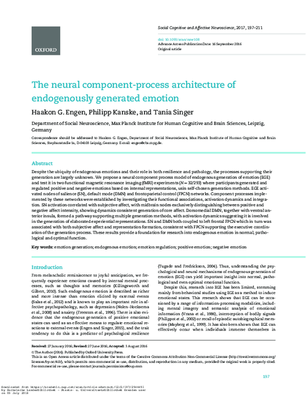
 Philipp Kanske