49
Egypt. J. Bot., Vol. 56, No. 3, pp. 817-835 (2016)
Bacterial Detoxification of Copper and Its
Impacts on Germination Indices of Barley and
Mung Bean
D. E. M. Radwan 1,3, A. M. M. Essa 2,3, M. N. Awad 3
1
Botany Department, Faculty of Science, Sohag University,
Sohag, ,2 Botany Department, Faculty of Science, Fayoum
University, Fayoum, Egypt and 3 Biology Department, Faculty
of Science, Jazan University, Kingdom of Saudi Arabia
O
N ACCOUNT of incessant human activities, copper is
……...accumulated in the environment at elevated concentrations that
induce harmful influences on all kinds of living organisms. An oxic
bioreactor was employed to transform copper ions into copper
particles using volatile metabolites of Escherichia coli culture. SEM
and EDX analysis of the transformed copper showed the formation of
elongated particles with 1-5 µm in length comprising of copper,
sulfur, carbon, oxygen and nitrogen elements. Mung bean seeds and
barley grains exposed to ionic copper demonstrated low germination
and apparent decline of seedlings growth parameters while higher
germination and growth rates were recorded with those treated with
copper particles. At the same time, an enhanced POD activity was
noticed with all Cu treatments, CAT activity seemed to be induced in
response to ionic Cu only meanwhile APX activity was markedly
affected with both types of Cu. Furthermore, seedlings subjected to
Cu particles showed higher protein contents. Toxicity reduction of
copper treated with E. coli volatiles was ascribed to the decrease of
the mobile copper concentration as a result of interaction with
vaporized chelators that reduce bioavailability of copper.
Keywords: Copper - Detoxification - E. coli - Antioxidant enzymes Hordeum vulgare - Vigna radiata.
At the moment, there is an extensive raise in the discharge of industrial effluents
into the environment, primarily soil and water that generally leads to the
accumulation of heavy metals. Heavy metals pollution is a major health concern
since they are non-degradable and have long-lasting effects on the ecosystem.
Heavy metals discharge from industrial activities is considered common source
of heavy metal pollution such as fertilizer production, electroplating and plastics
manufacturing in addition to mining and metallurgical processes (Zouboulis
et al., 2004). Most heavy metals such as arsenic, cadmium, chromium, copper,
lead, mercury, nickel, silver, zinc are toxic to all living organisms even at very
low concentrations. Toxic consequences of excess levels of heavy metals in
1
Corresponding author:Tel.: +2-093-4611042; Fax: +2-093-4601159 E-mail address:
deya90@yahoo.com (D. Radwan).
�818
D. E. M. Radwan et al.
plants include cellular damage and production of elevated amounts of reactive
oxygen species (ROS)-led to oxidative stress, and cellular metabolic arrest (Gill
and Tuteja, 2010). At relatively low concentrations, a number of heavy metal
such as Co2+, Cu2+, Fe2+, Mn2+, Mo2+, Ni2+, and Zn2+ are recognized as essential
trace elements for living cells as they act as cofactors of metalloproteins and
some enzymes (Jansen et al., 1994). These metals can also modulate plant ROSmetabolizing/scavenging system that comprising of enzymatic such as catalase,
guaiacol peroxidase, glutathione sulfotransferase, ascorbate peroxidase,
monodehydroascorbate reductase, glutathione reductase , dehydroascorbate
reductase and non-enzymatic such as ascorbate, glutathione, carotenoids,
tocopherols, and phenolics (Anjum et al., 2012).
Although copper is required for normal cell growth, it is considered as a
common soil contaminant. It is growing environmental problem due to the
continuity of human activities leading to copper pollution. These activities
include mining, sewage sludge application to soils and copper containing
fungicides (Mackie et al., 2012 and Ruyters et al., 2013).
The ionic form of Cu2+ caused root growth alterations in durum wheat at low
concentration; 1.0 μM and high Cu concentrations (200 and 500 μM) may reduce
the growth of both root and shoots (Michaud et al., 2008 and Thounaojam et al.,
2014). Toxicity of Cu is affected by bioavailability of metal in the soil and by the
concentration of the metal as well as pH of the soil (Wang et al., 2009). High
concentration of Cu2+ are toxic and leads to inhibition of plant growth,
disturbance of the mitosis, inhibition of root elongation, damage to root cell
membranes (Ouzounidou et al., 1995). Cu tends to be accumulated in the root
tissue with little translocate to the shoots (Marschner, 1995).
The capability of microbial cells to stay alive in the occurrence of elevated
doses of heavy metals could be assigned to the occurrence of various strategies
to accommodate with these fatal pollutants. Of these mechanisms is the aptitude
of the microbial cell to discharge non-specific extracellular molecules that are
engaged in the attenuation of heavy metals toxicity through altering the
physicochemical conditions around microbial biomass. Such these modifications
induce the bioprecipitation of these pollutants in the microbial surroundings
(Fomina et al., 2008; Dupraz et al., 2009 and Jang et al., 2015).
It is well known that plants produce root exudates containing an assortment
of organic substances which have considerable consequences on the
development of microbial communities and their activities. Therefore, the
interaction between plant roots and soil microbes can influence on the
mobilization and immobilization of metals in the soil (Seshadri et al., 2015).
Protection of plants from the toxicity of heavy metals such as copper, zinc and
lead occurs through microbial changing metal speciation into biologically
unavailable species (He and Yang, 2007; Huang et al., 2005 and Dixit et al.,
2015). Thus the aim of the present study was to evaluate the impact of ionic
Egypt. J. Bot., Vol. 56, No. 3 (2016)
�BACTERIAL DETOXIFICATION OF COPPER…
819
copper and copper treated with the volatile metabolites of E. coli on seed
germination and metabolism of barley and mung bean seedlings.
Materials and Methods
Treatment of CuSO4 by bacterial volatiles
Two concentrations of copper sulfate (CuSO4.5H2O) solutions (0.2 mM and
0.4 mM) were prepared from stock solution (1.0 M CuSO 4). The bacterial strain,
Escherichia coli, (Essa, 2012) was used in this study. It was grown aerobically in
Luria Broth medium on shaking incubator (200 rpm) for 24 hr at 30 oC. Bacterial
growth was monitored by measuring the optical density at 600 nm. Two sets of
the CuSO4 solutions, (200 ml of each) were exposed to the E. coli biogases (OD
≈ 0.8) for 24 hr in an oxic bioreactor according to Essa et al. (2012). Then one
set of the treated copper solutions were used to study their effect on seed
germination and seedlings growth criteria while the other set was subjected for
centrifugation at 10000 rpm for 15 min. The supernatant was discarded and
precipitates were washed with 20 ml deionized water followed by centrifugation
as before. The last step was repeated three times and Cu-precipitate were
collected and dried at 30 oC. Cu-precipitates were examined with a JEOL JSM
5900 scanning electron microscope with the elemental composition determined
by energy dispersive X-ray microanalysis (EDX) using an Oxford Link ISIS
System according to Essa and Khallaf (2014).
Plant materials and treatments
Mung bean (Vigna radiata; Fabaceae) seeds and barley (Hordeum vulgare;
Poaceae) grains were surface sterilized by immersion in ethyl alcohol (70%) for
2 min followed by rinsing three times with sterilized deionized water, 5 minutes
each. Surface sterilized seeds or grains were germinated in 10 cm plastic Petri
dishes containing sterilised filter papers witted with 20 ml of treatment solutions,
10 seeds or grains in per dish where control dishes contained 20 ml of sterilized
water. Triplicates of each treatment were done and all dishes were kept in the
dark at 18 ± 20 °C.
The treatments were carried out as following:
1) Control: sterilized deionized water.
2) Cu-IL: Ionic Cu solution (CuSO4.5H2O) at low concentration (8 µM).
3) Cu-NL: Cu-particles solution prepared via E. coli volatile metabolites at low
concentration (8 µM).
4) Cu-IH: Ionic Cu solution (CuSO4.5H2O) at high concentration (16 µM).
5) Cu-NH: Cu-particles solution prepared via E. coli volatile metabolites at high
concentration (16 µM).
Analysis of growth parameters
The percentage of germinated seeds or grains, length of seedlings (in
centimetres), fresh weight and dry weight of seedlings were determined to
express metal toxicity in both ionic and non-ionic Cu forms.
Egypt. J. Bot., Vol. 56, No. 3 (2016)
�820
D. E. M. Radwan et al.
Antioxidant enzymes
All extraction procedures were carried out at 4 ºC, where one gram of fresh
seedlings was grinded in 5 ml of phosphate buffer pH 7.0 followed by
centrifugation at 14000 g at 10 °C for 15 min. The supernatants were used for
determination of enzyme activity.
Peroxidase activity (POD)
For determination of POD (EC 1.11.1.7) activity, supernatants (0.1 ml)
were mixed with assay mixture (3 ml) and the development of the brown color
was monitored. The assay mixture for POD activity contained 40 mM potassium
phosphate pH 7.2, 0.1 mM EDTA, 5 mM guaiacol, 0.3 mM H2O2. The increase
in the absorbance due to oxidation of guaiacol (Extinction factor = 26.2 mM cm 1
) was measured spectrophotometrically at 470 nm. POD activity was calculated
in terms of µmol of guaiacol oxidized min-1 g-1 Fresh weight at 25 ± 2 °C
(MacAdam et al., 1992; Zhang, 1992).
Catalase activity (CAT)
By monitoring the disappearance of H2O2, CAT (EC 1.11.1.6) activity was
measured according to the method of Chandlee and Scandalios (1984). The
disappearance of H2O2 was measured by the decrease in absorbance at 240 nm
(E= 0.036 mM-1 cm-1) of a reaction mixture (3 ml) consisting of 25 mM
potassium phosphate buffer (pH 7.0), 10 mM H2O2 and enzyme extract (0.1ml).
One CAT unit is the amount of enzyme necessary to decompose 1 µmol min -1
H2O2 under the above-mentioned assay conditions.
Ascorbate peroxidase activity (APX)
APX (EC 1.11.1.11) activity was determined according to Nakano and Asada
(1981). The supernatants were mixed with the assay medium for testing the
activity of APX. The assay medium consists of 3 ml containing 50 mM
phosphate buffer (pH 7.0), 0.1 mM EDTA, 0.3 mM ascorbate, 0.06 mM H 2O2
and 0.1 ml enzyme extract. The decrease in ascorbate concentration was
followed by decline in absorbance at 290 nm and activity was calculated using
the extinction coefficient (E =2.8 mM-1 cm-1) for ascorbate.
Protein content
Soluble, insoluble and total protein contents of seedlings were determined
according to Lowry et al. (1951). Dry tissues were extracted in 10 ml distilled
water for 2 hr at 90 °C for analysis of soluble protein. Moreover, the total
proteins were extracted in 10 ml NaOH (0.1 N) for 2 hr at 90 °C. The extracts
were centrifuged and the supernatants were collected. One ml of extract was
added to 5 ml of alkaline reagent (50 ml of 2% Na2CO3 prepared in 0.1 N NaOH
and 1 ml 0.5% of CuSO4.5H2O prepared in 1% sodium potassium tartrate),
mixed thoroughly and then allowed to stand for 10 min. Folin reagent diluted 1:1
(v/v) was then added and mixed immediately. After 30 min, the extinction
against appropriate blank was measured at 700 nm. Results were expressed as
milligrams per gram dry weight. Insoluble proteins were calculated as the
Egypt. J. Bot., Vol. 56, No. 3 (2016)
�BACTERIAL DETOXIFICATION OF COPPER…
821
difference between the amounts of total and water-soluble proteins. Bovine
serum albumin was used for calibration curve.
Total free amino acids content
According to Moore and Stein (1948), total free amino acids were extracted
and determined. Dry tissue samples were extracted in distilled water by heating
in water bath at 90 oC for 2 hrs. The extracts were then centrifuged and the
supernatants were collected. Supernatant (0.1 ml) was added to 1 ml of ninhydrin
solution with stannous chloride. The tubes were heated in boiling water bath for
20 min till a purple color was developed. Five milliliter of the diluents were
added and mixed well. After 15 min, the intensity of the color against a reagent
blank was measured in a colorimeter at 570 nm. The free amino acids
concentrations were calculated as mg/g dry matter.
Statistical analyses
The resulted data were tested by using the ANOVA test for significance.
Means were compared by least significant differences (LSD) test at levels P
<0.05 and P <0.01. All statistical tests were carried out using SPSS (v. 16.0)
software (Garth, 2008).
Results
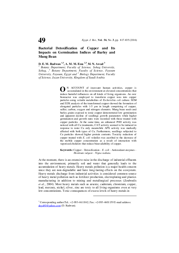
Effect of E. coli biogas on CuSO4 solution
Data in Table 1 showed that changes in the optical density (OD), pH and
color of CuSO4 solutions in the precipitation chamber of the bioreactor as a result
of exposing to the volatiles produced aerobically by E. coli. There was a gradual
increase in the OD and pH values of copper solution by increasing the exposure
time where maximum OD was recorded after 12 hr with pH value 8.2. After 24
hr exposure time, the pH value reached the highest value (8.8) and a pale blue
precipitate was formed. At the same time, there was a marked change in the
color of CuSO4 solution by increasing the exposure time until 24 hr where pale
blue precipitate was formed. Scanning electron microscope analysis of the Cuprecipitate (Fig. 1) showed the formation of elongated particles with 1 - 5 µm in
length. At the same time, electron dispersive X-ray analysis of these particles
elucidated their elemental composition. They comprised of 61.71% copper,
8.44% sulfur, 4.03% carbon, 22.19% oxygen and 1.98% nitrogen (Fig. 1).
Analysis of growth parameters
The germination of both mung bean seeds and barley grains under the effect
of Cu treatment was presented in Fig. 2. Compared to control, all treatments
could reduce the growth of seedlings at variable levels. The effect of Cu toxicity
was more obvious in radicals than plumules. In case of mung bean seedlings,
radicals become reduced and totally inhibited in barley seedlings. Non-ionic Cu
form seemed to be less toxic than the ionic form of Cu in both low and high Cu
concentrations.
Egypt. J. Bot., Vol. 56, No. 3 (2016)
�822
D. E. M. Radwan et al.
TABLE 1. Influence of exposure time of E. coli volatiles on the physical characters of
copper sulfate solution in the precipitation chamber of the oxic bioreactor.
Exposure time (hrs)
0
2
O.D600
0.000
0.069
pH
6.3
6.5
Color
Blue
Blue
4
8
0.113
0.437
7.4
7.8
Turbid blue
Cloudy blue
12
24
0.792
0.547
8.2
8.8
Light blue precipitate
Pale blue precipitate
Fig. 1. Scanning electron microscope (SEM) and energy dispersive X-ray (EDX) analysis
of copper particles produced in the bioreactor through the interaction of volatile
metabolites of E. coli culture with copper sulfate solution.
The percentage of seed or grain germination was affected by Cu application
in either ionic or non-ionic form (Fig. 3). Cu could lower the percentage of
germination and the reduction depended on Cu concentration, the higher Cu
concentration the lower the percentage of germination. Different percentages of
germination were obtained in different forms of Cu. In mung beans, the
percentage of germination at low Cu were 53% and 73% in case of ionic and
non-ionic Cu forms, respectively. Almost similar responses were detected in
barely grains germinated in ionic and non-ionic Cu forms. Results concerning
the percentage of germination revealed that, the toxicity of Cu was lower in case
of non-ionic Cu for the same concentration in both plants.
The length of seedlings produced in all Cu treatments was reduced (Fig 3).
The severity of reduction was concentration and Cu-form dependent. In all
concentrations, the non-ionic Cu form was less toxic to both mung bean and
barely seedlings. Compared to the control, low concentration caused reduction of
57 % and 90% in cases of mung bean treated with low and high concentration of
ionic Cu, respectively. Barley seedlings grown on high non-ionic Cu were 65 %
Egypt. J. Bot., Vol. 56, No. 3 (2016)
�BACTERIAL DETOXIFICATION OF COPPER…
823
longer than those germinated on high ionic Cu. Moreover, in case of mung bean
seedlings, high ionic and non-ionic Cu concentrations reduced seedling lengths
to 92% and 47% less than control, respectively.
Fig. 2. Impact of ionic and non-ionic copper on the germination of mung bean seeds
(Vigna radiate; A) and barley grains (Hordeum vulgare; B).
Egypt. J. Bot., Vol. 56, No. 3 (2016)
�824
D. E. M. Radwan et al.
Fig. 3. Growth parameters of mung bean seeds (Vigna radiata) and barley grains
(Hordeum vulgare) germinated on ionic and non-ionic Cu-solutions where
(A) is germination percentage, (B) is seedling length, (C) is fresh weight and
(D) is dry weight.
Egypt. J. Bot., Vol. 56, No. 3 (2016)
�BACTERIAL DETOXIFICATION OF COPPER…
825
Comparing the weights (fresh and dry) of the produced seedlings under
stressful conditions with the corresponding controls, the obtained results
demonstrated lower weights of seedlings due to Cu treatments. Similarly, Cu
toxicity leads to reduced fresh and dry weights in case of ionic Cu in low and
high concentrations in both plants. On the other hand, non-ionic Cu showed less
of no toxicity in mung bean where the fresh and dry weights were almost similar
to controls.
Antioxidant enzymes analyses
The analyzed antioxidant enzymes peroxidase (POD), Catalase (CAT) and
Ascorbate peroxidase (APX) extracted from both mung bean and barely seedling
grown for 10 days on different Cu treatments were tested for their activities.
Alterations in enzyme activities were detected in response to different forms of
Cu as well as different plants (Table 2). Comparing the controls, POD extracted
from barely was 10 fold more active than mung bean POD. Peroxidase activity
was enhanced in seedlings grown on all Cu treatments. High concentration of
non-ionic Cu could induce the activity of POD to be 390 % and 102% more than
control for mung bean and barely, respectively. Peroxidase activity was induced
with lesser amounts in non-ionic Cu treatments compared with the corresponding
ionic forms. For high concentration of non-ionic Cu, POD activity increased with
percentages of 50 % and 89% for mung bean and barley, respectively. Cu in nonionic form could lower the POD activities compared with the corresponding
ionic forms but still the values higher than those of the controls.
Catalase activity was lowered by subjection to Cu in most treatments. In
comparison to control, low concentration of ionic Cu could lower the activity of
CAT to be 98% and 49% for mung bean and barely, respectively. In case of nonionic Cu, CAT activity increased by 9% and 7% for low and high concentrations
in mung bean. On the other plant, the increase reached 6% and 15% above
control in case of low and high concentration of non-ionic Cu applied to barley
seedlings. Compared to the other analyzed enzymes, the effect of Cu treatments
on CAT activity was less in both plants.
Ascorbate peroxidase (APX) activity showed similar behavior as peroxidase
in its response towards Cu in its different forms. In details, high concentration of
non-ionic Cu induced the activity of APX to be 176% and 332% above controls
in case of mung bean and barely, respectively. Non-ionic Cu form reduced the
activity of APX to be 9% in mung bean and double folded the activity in barely
compared with their controls. Generally, non-ionic Cu had less effect on the
activity of APX than ionic Cu.
Proteins and amino acids
Soluble, insoluble and total proteins were determined for mung bean and
barley seedlings (Table 3). The obtained data revealed that soluble, insoluble and
total proteins were lowered with all Cu treatments in both plants. On exception,
low non-ionic Cu could increase the soluble, insoluble and total proteins content
Egypt. J. Bot., Vol. 56, No. 3 (2016)
�D. E. M. Radwan et al.
826
in barely seedlings while high non-ionic Cu form increased the soluble protein
only. It seemed that non-ionic Cu could increase the protein contents compared
with ionic Cu. Meanwhile, soluble and total proteins were induced by non-ionic
Cu in barely seedlings where the values reached 130% and 121% for soluble and
total proteins, respectively when seedlings were germinated in low concentration
of non-ionic Cu. There are slight differences between the effect of low and high
concentrations of ionic Cu on total protein contents in mung bean whereas in
barely it was found that the higher the concentration of ionic Cu the lower the
total proteins content.
TABLE 2. Effect of ionic and non-ionic Cu treatments on antioxidant enzymes (Unit g -1 FW) of
Vigna radiata and Hordeum vulgare seedlings. Values are means (M) of four
replicates ± standard deviation (SD).
Plants
Treatments
H. vulgare
V. radiata
Control
POD
SD
M
CAT
SD
%
M
25.80
±
8.40
APX
SD
%
M
100.00
19.97
±
2.05
100.00
%
60.87
19.34
±
4.58
100.00
Cu-IL
35.37**
±
7.29
182.89
25.20
±
7.85
97.67
12.15*
±
0.82
Cu-NL
29.77**
±
1.82
153.95
28.20*
±
9.00
109.30
13.76**
±
0.41
68.90
Cu-IH
94.85**
±
2.62
490.46
26.20
±
1.79
101.55
55.27**
±
6.14
276.81
Cu-NH
28.94**
±
7.61
149.67
27.60
±
2.16
106.98
16.08**
±
2.05
80.43
Control
188.10
±
3.13
100.00
19.50
±
4.95
100.00
9.84
±
4.09
100.00
Cu-IL
320.55**
±
9.56
170.41
19.30
±
0.71
98.97
19.97**
±
0.41
202.94
Cu-NL
300.64**
±
6.72
159.82
20.56
±
4.24
105.44
16.94*
±
4.09
70.59
Cu-IH
379.58**
±
3.76
201.79
25.73**
±
5.66
131.95
42.53**
±
4.50
432.35
Cu-NH
353.88**
±
3.97
188.13
22.38*
±
1.41
114.77
20.25**
±
6.55
205.88
Statistical significance of differences compared to control: *, significant at P<0.05; **, significant at
P<0.01.
TABLE 3. Effect of ionic and non-ionic Cu treatments on soluble, insoluble and total proteins (mg
g-1 FW) of Vigna radiata and Hordeum vulgare seedlings. Values are means (M) of four
replicates ± standard deviation (SD).
H. vulgare
V. radiata
Plants
Soluble proteins
Treatments
Control
Cu-IL
Cu-NL
Cu-IH
M
127.73
94.53**
107.73*
94.27**
Cu-NH
Insoluble proteins
±
±
±
±
SD
7.02
6.74
9.98
3.14
%
100.00
74.01
84.34
73.80
M
171.93
108.80**
107.27**
118.40**
112.93*
±
4.80
88.41
Control
58.67
±
9.26
Cu-IL
54.27
±
Cu-NL
75.73**
Cu-IH
Cu-NH
Total proteins
±
±
±
±
SD
3.73
2.32
8.41
4.74
%
100.00
63.28
62.39
68.86
M
299.67
203.33**
215.00**
212.67**
±
±
±
±
SD
5.70
8.04
3.61
3.32
%
100.00
67.85
71.75
70.97
89.73**
±
2.59
52.19
202.67**
±
4.39
67.63
100.00
146.00
±
3.07
100.00
204.67
±
2.82
100.00
6.65
92.50
137.40
±
6.36
94.11
191.67*
±
7.32
93.65
±
1.52
129.09
171.27**
±
5.72
117.31
207.00
±
8.54
120.68
96.53**
±
3.89
164.55
62.13**
±
4.66
42.56
158.67*
±
7.50
77.52
65.87**
±
1.59
112.27
107.47*
±
4.69
73.61
173.33*
±
3.50
84.69
Statistical significance of differences compared to control: *, significant at P<0.05; **, significant at
P<0.01.
Egypt. J. Bot., Vol. 56, No. 3 (2016)
�BACTERIAL DETOXIFICATION OF COPPER…
827
Total free amino acids of control and Cu treated seedlings of mung bean and
barley after 10 days of germination were shown in Table 4. Free amino acids
were increased in most of Cu treatments by 5 - 40% above the corresponding
controls. The ionic forms of Cu could increase the total free amino acids in both
mung bean and barley. In case of mung bean, the increase caused by low and
high ionic Cu was 10 and 40%. Similarly in barley seedlings, low and high
concentrations of ionic Cu increased the total free amino acids by 15 and 19%
more than controls. Non-ionic Cu could keep the content of total free amino
acids more or less than controls in both mung bean and barley seedlings. The
values were 89 and 103% in case of mung bean and barley at high concentration
of non-ionic Cu. It can be concluded that ionic forms could induce more contents
of free amino acids while non-ionic Cu form could keep the values almost
similar to those of the corresponding controls.
TABLE 4. Effect of ionic and non-ionic Cu treatments on total free amino acids (mg g-1
FW) of Vigna radiata and Hordeum vulgare seedlings. Values are means (M)
of four replicates ± standard deviation (SD).
H. vulgare
V. radiata
Plants
Treatments
Control
Cu-IL
Cu-NL
Cu-IH
Cu-NH
Control
Cu-IL
Cu-NL
Cu-IH
Cu-NH
Total free amino acids
M
29.96
33.1*
31.74
41.82**
26.92*
8.60
9.90*
8.44
10.24*
8.90
±
±
±
±
±
±
±
±
±
±
SD
4.10
2.76
3.63
7.76
1.25
0.47
0.21
1.12
1.18
1.03
%
100.00
110.48
105.94
139.59
89.85
100.00
115.12
98.14
119.07
103.49
Statistical significance of differences compared to control: *, significant at P<0.05; **,
significant at P<0.01.
Discussion
Although Copper is considered essential for physiological processes within
plant cells, it persuades an obvious toxicity depending on its concentration and
availability in soil. The present study showed the influence of ionic Cu and nonionic Cu on seed germination and antioxidant enzymes’ activities of mung bean
and barely. Non-ionic Cu particles were prepared by a bioprocess using E. coli
volatile gases in a specific bioreactor. EDX analysis revealed that Cu-particles
comprised copper, sulfur, nitrogen, oxygen and carbon but with no measurable
phosphorus that confirms the nonexistence of contamination by bacterial cells.
According to our preceding studies (Essa et al., 2005 and Macaskie et al., 2007),
the bacterial biogas included sulfur-based and nitrogen-based gaseous metabolites
such as amines and dimethyl disulfide that are discharged during the excessive
metabolic activity of the bacterial culture. Bacterial volatile sulfur molecules hold
immense affinity to complex metal ions into insoluble metallo-sulfur compoundsd
Egypt. J. Bot., Vol. 56, No. 3 (2016)
�828
D. E. M. Radwan et al.
(Brummett et al., 2015; Essa and Khallaf, 2016). At the same time, the incidence
of ammonia in E. coli biogas (confirmed using Nessler's solution) is in charge for
altering pH value of copper solution in the direction of alkalinity (Table 1).
Biogenic ammonia beside other amines liberated from E. coli culture have vital
task in the complexation of copper ions into less soluble Cu-particles. The
complete chemical identification of Cu-particles was not available in this
investigation but EDX-analysis verified the incidence of nitrogen and sulfur in
addition to carbon elements in the transformed Cu-particles. These results are in
agreement with our previous study (Essa and Mostafa, 2011; Essa et al., 2012)
where some organo-sulfur and amine groups such as thiocarbonyl, disulfide and
amine functional groups were spotted by Fourier-Transform Infrared Spectroscopy
in the metal complexes resulting from the treatment of heavy metals with gaseous
metabolites of oxic bacterial culture.
The obtained results revealed less toxicity of non-ionic Cu particles
compared with the ionic Cu form in both low and high concentrations. The
percentage of seed germination as well as all growth parameters analyzed;
seedling lengths, fresh and dry weights, confirmed less toxicity of Cu particles.
Inhibition of root elongation caused by heavy metals may be due to metal
interference with cell division, chromosomal aberrations and abnormal mitosis
(Jain et al., 2010 and Liu et al., 2003). Moreover, reduced seedling growth in
metal treatments could be as a result of the reduction in meristematic cells (Kabir
et al., 2008). The obtained results in this investigation revealed acceptable
growth patterns with non-ionic Cu treatments indicating less toxicity of Cu in its
non-ionic Cu form and hence Cu detoxification. Furthermore, in the present
work, the antioxidant enzyme activities were significantly altered in response to
Cu ions and non-ionic Cu. For example, all antioxidant enzymes had higher
activities in seedlings treated with ionic forms of Cu. Moreover, non-ionic Cu
forms could lower the POD and APX activities compared with the corresponding
ionic forms but still the values higher than those of the controls. Similar to the
present results, Nekrasova et al. (2011) reported decrease in CAT activity in E.
densa at higher concentrations of copper ions. This can be explained by enzyme
inactivation, might be, due to substitution of Fe2+ ion in its active center with
Cu2+ ion (Hou et al., 2007; Mallick, 2004). Clearly, Cu ions inhibited the
antioxidant enzymes more strongly, compared to non-ionic Cu particles
(Nekrasova et al., 2011). In the present, a reason behind the APX inhibition in
some cases of Cu treatments might be the lack of ascorbate due to presence of
high amounts of Cu ions. It was reported previously that, copper ions promote
rapid loss of ascorbate (Packer, 2001).
Several mechanisms were suggested by which the plant cells resist the toxic
effects of heavy metals in particles form. One of those mechanisms is that plant
cell walls present in natural settings, are primarily composed of carbon hydrate
polymers, and are semi permeable. Thus, exotics need to penetrate through the
cell wall prior to the membrane invagination. Limited size of pores in plant cell
walls prevents larger molecules from free passing through cell wall (Carpita
Egypt. J. Bot., Vol. 56, No. 3 (2016)
�BACTERIAL DETOXIFICATION OF COPPER…
829
et al., 1979). Moreover, creating an apoplastic pool of Cu as a way for Cu
detoxification affects chemical and structural changes in cell wall under the
influence of Cu excess. Increasing the permeability of plant cell walls by metal
exposure lead to create “holes”, and then enter into the cells by penetrating
through the “holes”. After entering the cells, the non-ionic Cu particles are able
to transport between cells via plasmodesmata which are microscopic channels of
plants traversing the cell walls and enabling transport and communication
between cells. Plasmodesmata or intercellular bridges were reported to be
cylindrical channels with 40nm in diameter (Tilney et al., 1991). These
plasmodesmata allow particles to pass from cell to another depending on their
size causing less toxic effects than ionic Cu. Furthermore, several investigations
excluded dissolution from the main mechanisms regulating the toxicity of metalbased non-ionic particles (Nel et al., 2006). Even though the dissolution of nonionic Cu to cupric ions has a negligible effect in plant agar media, some amount
of non-ionic Cu may dissolve to become cupric ions within the cell. Most toxic
action results from the non-ionic Cu because of the presence of particles or
aggregated nanoparticles within the cell (Lee et al., 2008). Another mechanism
is changing the permeability of plasma membrane. Previously reported,
damaging the plasma membrane through formation of OH· radicles which can be
achieved by traces of transition metal ions such as iron and copper (Packer,
2001). Reduction in seed germination can also be attributed to alterations of
selection permeability properties of cell membrane. Alteration in plasma
membrane permeability might be due to changes in membrane protein channels
leading to metal ions passage and hence metal toxicity. It is well known that,
cells become active produce the hydrolytic enzymes they begin to digest the
stored food which is converted into soluble form and transported to the primary
root and shoot tips for enzyme amylase which converts starch into sugar and
proteases act on proteins. When the activities of hydrolytic enzymes are affected,
the food does not reach to the primary root and shoot, thereby affecting the
seedling growth (Kabir et al., 2008).
Another mechanism of Cu toxicity is through the generation of reactive
oxygen intermediates (H2O2, O2-, OH, superoxide etc.) and then oxidative stress.
Previously reported, exposure to excessive Cu ions leads to generation of
oxidative stress in plant system (Gaetke and Chow, 2003). A suggested strategy
to avoid toxicity of heavy metals is through the induction of antioxidant enzyme
system and increase in the non-enzymatic antioxidants to reduce the oxidative
damage related to excessive ROS formation caused by heavy metals (Verma and
Dubey, 2003). In the present work, induced antioxidant enzyme activities were
noticed in both ionic and non-ionic Cu treatments with different rations.This
supports the theory of detoxification through management of oxidative stress.
Antioxidant enzyme activities were highly induced in case of ionic Cu indicating
more stressful conditions. Presence of ROS causes oxidative damage to
biomolecules such as lipids, proteins, nucleic acids, etc. (Radwan et al., 2010
and Radwan, 2012). The transition metals such as Cu can act as specific
cofactors for numerous metalloproteins because of their physical and chemical
Egypt. J. Bot., Vol. 56, No. 3 (2016)
�830
D. E. M. Radwan et al.
properties. It is involved in the maintenance of the functional and structural
integrity of plant cells. Heavy metals exercise a detrimental influence on cells by
binding to vital proteins and manage protein/enzyme functioning. In this
experiment, reduction of soluble and total proteins contents was noticed in both
ionic and non-ionic Cu treatments except for barley soluble protein contents.
Comparing the contents of proteins, plants treated with non-ionic Cu had higher
contents than those exposed to ionic Cu. Obviously, Cu could reduce the total
protein contents in mung bean and barley. Packer (2001) reported that copper
cations are bound to proteins leading to their degradation. Cu toxicity is
determined by binding to SH-groups in proteins, thereby inhibiting enzyme
activity or protein function (Cohu and Pilon, 2010).
In the present work, a noticeable decrease in the amino acids content with Cu
stress that might be involved in detoxification of copper (ionic or non- ionic).
Organic acids, amino acids, or peptides are potential ligands for phytochelation
of metals (Clemens, 2001). Moreover, amino acids such as citric, malic, and
histidine are potential ligands for heavy metals and so could play a role in
tolerance and detoxification (Clemens, 2001). In the cytoplasm, metals can either
bind to free amino acids, the non proteinogenic nicotianamine, protein ligands
that are rich in Cys residues, such as metallothioneins, metallochaperones,
phytochelatins, and low molecular weight thiols (Callahan et al., 2006;
Trampczynska et al., 2010).
Conclusions
This work highlights the effect of ionic and non-ionic Cu on germination and
metabolism of barley grains and mung bean seeds. The non-ionic Cu was
prepared using a bioprocess through bacterial biogases in a specific bioreactor.
Barley and mung bean subjected to non-ionic Cu particles showed significantly
lower toxicity than those treated with ionic Cu. The reduced toxicity of non-ionic
copper was attributed to the decline of copper capability to penetrate plant cells.
Further studies are required to investigate the impact of this bioprocess on the
toxicity of other metals in order to understand the collaborative impact of
bacteria in diminishing heavy metals toxicity against plants in heavily polluted
environment.
References
Anjum, N. A., Iqbal,A., Iram M., Mário, P., Armando,D. C., Eduarda,P., Shahid, U.,
Altaf, A. and Nafees K. A. (2012) Modulation of glutathione and its related enzymes
in plants’ responses to toxic metals and metalloids - a review. Environ. Exp. Bot., 75,
307-324.
Brummett, A.E., Schnicker, N.J., Crider, A., Todd, J.D. and Dey, M. (2015)
Biochemical,
kinetic,
and
spectroscopic
characterization
of Ruegeria
pomeroyi DddW- a mononuclear iron-dependent DMSP lyase. PLoS ONE,10 (5),
e0127288. http://Dol.org/10.1371/journal.pone.0127288.
Egypt. J. Bot., Vol. 56, No. 3 (2016)
�BACTERIAL DETOXIFICATION OF COPPER…
831
Callahan, D. L., Baker, A. J., Kolev, S. D. and Wedd, A. G. (2006) Metal ion ligands in
hyperaccumulating plants. J. Bio. Inorg. Chem., 11(1), 2-12.
Carpita, N., Sabularse, D., Montezinos, D. and Delmer, D. P. (1979) Determination of
the pore size of cell walls of living plant cells. Science, 205(4411), 1144-1147.
Chandlee, J. and Scandalios, J. (1984) Analysis of variants affecting the catalase
developmental program in maize scutellum. Theor. Appl. Genet., 69 (1), 71-77.
Clemens, S. (2001) Molecular mechanisms of plant metal tolerance and homeostasis.
Planta, 212 (4), 475-486.
Cohu, C.M. and Pilon, M. (2010) Cell biology of copper. In: “Cell Biology of Metals
and Nutrients. Ed. by Ru¨diger H. and Mendel R.R. pp. 55–74. Springer, Berlin.
Dixit R., Malaviya D., Pandiyan K., Singh U. B., Sahu A., Shukla R., et al.
(2015) Bioremediation of heavy metals from soil and aquatic environment: an
overview of principles and criteria of fundamental processes. Sustainability 7, 2189–
2212. 10.3390/su7022189
Dupraz, C., Reid, R. P., Braissant, O., Decho, A. W., Norman, R. S. and Visscher, P.
T. (2009) Processes of carbonate precipitation in modern microbial mats. Earth-Sci.
Rev., 96 (3), 141-162.
Essa A. M. M. (2012) Effect of a continuous mercury stress on mercury reducing
community of some characterized bacterial strains. Afr. J. Microbiol. Res. 6: (6)
1255-1261.
Essa, A.M., Abd-Alsalam, E.S. and Ali, R.M. (2012) Biogenic volatile compounds of
activated sludge and their application for metal bioremediation. Afr. J. Biotechnol.,
11(42), 9993-10001.
Essa, A. M., Macaskie, L. E. and Brown, N. L. (2005) A new method for mercury
removal. Biotechnol. Lett., 27(21), 1649-1655.
Essa A. M. and Khallaf M. K. (2014) Biological nanosilver structures for the
preservation of archaeological stones against microbial colonization. Int. Biodeter.
Bioremed., 94, 31-37.
Essa A. M. and Khallaf M. K. (2016) Antimicrobial potentiality of consolidation
polymers impregnated with copper nanoparticles. BMC Microbiol. 2016, 16:144 DOI:
10.1186/s12866-016-0766-8.
Essa, A. M. M. and Mostafa, S. M. M. (2011) Biomineralization of some heavy metals
by cyanobacterial biogas. Egyp. J. Bot. 31, 112-121.
Fomina, M., Charnock, J. M., Hillier, S., Alvarez, R., Livens, F. and Gadd, G. M.
(2008) Role of fungi in the biogeochemical fate of depleted uranium. Curr. Biol., 18
(9), R375-R377.
Gaetke, L.M. and Chow, C.K. (2003) Copper toxicity, oxidative stress, and antioxidant
nutrients. Toxicol. 189 (1), 147-163.
Egypt. J. Bot., Vol. 56, No. 3 (2016)
�832
D. E. M. Radwan et al.
Garth, A. (2008) Analysing Data Using SPSS: A Practical Guide for Those Unfortunate
Enough To Have to Actually do it. Sheffield Hallam University, 94.
Gill, S. S. and Tuteja, N. (2010) Reactive oxygen species and antioxidant machinery in
abiotic stress tolerance in crop plants. Plant Physiol. Biochem., 48 (12), 909-930.
He, Z. and Yang, X. (2007) Role of soil rhizobacteria in phytoremediation of heavy metal
contaminated soils. J. Zhejiang Univ. Sci. B., 8 (3), 192-207.
Hou, W., Chen, X., Song, G., Wang, Q. and Chang, C. C. (2007) Effects of copper and
cadmium on heavy metal polluted waterbody restoration by duckweed (Lemna
minor). Plant Physiol. Biochem., 45(1), 62-69.
Huang, Y., Tao, S. and Chen, Y. (2005) The role of arbuscular mycorrhiza on change of
heavy metal speciation in rhizosphere of maize in wastewater irrigated agriculture
soil. J. Environ. Sci. (China), 17(2), 276-280.
Jain, R., Srivastava, S., Solomon, S., Shrivastava, A. and Chandra, A. (2010) Impact
of excess zinc on growth parameters, cell division, nutrient accumulation,
photosynthetic pigments and oxidative stress of sugarcane (Saccharum spp.). Acta
physiol. plant., 32(5), 979-986.
Jang, G. G., Jacobs, C. B., Gresback, R. G., Ivanov, I. N., Meyer, H. M., Kidder, M.
and Moon, J. W. (2015) Size tunable elemental copper nanoparticles: extracellular
synthesis by thermoanaerobic bacteria and capping molecules. J. Mater. Chem.
C, 3(3), 644-650.
Jansen, E., Michels, M., Van Til, M. and Doelman, P. (1994) Effects of heavy metals in
soil on microbial diversity and activity as shown by the sensitivity-resistance index,
an ecologically relevant parameter. Biol. Fertility Soils, 17(3), 177-184.
Kabir, M., Iqbal, M. Z., Shafiq, M. and Farooqi, Z. (2008) Reduction in germination
and seedling growth of Thespesia populnea L., caused by lead and cadmium
treatments. Pak. J. Bot, 40(6), 2419-2426.
Lee, W. M., An, Y. J., Yoon, H. and Kweon, H. S. (2008) Toxicity and bioavailability of
copper nanoparticles to the terrestrial plants mung bean (Phaseolus radiatus) and
wheat (Triticum aestivum): Plant agar test for water‐insoluble nanoparticles.
Environ. Toxicol. Chem., 27(9), 1915-1921.
Liu, D., Jiang, W. and Gao, X. (2003) Effects of cadmium on root growth, cell division
and nucleoli in root tip cells of garlic. Biol. Plant., 47 (1), 79-83.
Lowry, O.H., Rosebrough, N.J., Farr, A.L. and Randall, R.J. (1951) Protein
measurement with the Folin phenol reagent. J. Biol. Chem., 193(1), 265-275.
MacAdam, J.W., Nelson, C. J. and Sharp, R. E. (1992) Peroxidase activity in the leaf
elongation zone of tall fescue I. Spatial distribution of ionically bound peroxidase
activity in genotypes differing in length of the elongation zone. Plant Physiol., 99 (3),
872-878.
Egypt. J. Bot., Vol. 56, No. 3 (2016)
�BACTERIAL DETOXIFICATION OF COPPER…
833
Macaskie, L., Creamer, N., Essa, A. and Brown, N. (2007) A new approach for the
recovery of precious metals from solution and from leachates derived from electronic
scrap. Biotechnol. Bioeng., 96(4), 631-639.
Mackie, K., Müller, T. and Kandeler, E. (2012) Remediation of copper in vineyards–a
mini review. Environ. Pollut., 167, 16-26.
Mallick, N. (2004) Copper-induced oxidative stress in the chlorophycean microalga
Chlorella vulgaris: response of the antioxidant system. J. Plant Physiol., 161(5), 591597.
Marschner, H. (1995) Mineral Nutrition of Higher Plants. Academic Press, London.
Michaud, A.M., Chappellaz, C. and Hinsinger, P. (2008) Copper phytotoxicity affects
root elongation and iron nutrition in durum wheat (Triticum turgidum durum L.).
Plant Soil, 310 (1-2), 151-165.
Moore, S. and Stein, W.H. (1948) Photometric ninhydrin method for use in the
chromatography of amino acids. J. Biol. Chem., 176 (1), 367-388.
Nakano, Y. and Asada, K. (1981) Hydrogen peroxide is scavenged by ascorbate-specific
peroxidase in spinach chloroplasts. Plant and cell physiol., 22 (5), 867-880.
Nekrasova, G., Ushakova, O., Ermakov, A., Uimin, M. and Byzov, I. (2011) Effects of copper
(II) ions and copper oxide nanoparticles on Elodea densa. Russ. J. Ecol., 42(6), 458-463.
Nel, A., Xia, T., Mädler, L. and Li, N. (2006) Toxic potential of materials at the
nanolevel. Science, 311(5761), 622-627.
Ouzounidou, G., Čiamporová, M., Moustakas, M. and Karataglis, S. (1995)
Responses of maize (Zea mays L.) plants to copper stress—I. Growth, mineral content
and ultrastructure of roots. Environ. Exp. Bot., 35 (2), 167-176.
Packer, L. (2001)"Handbook of Antioxidants" CRC Press.
Radwan, D.E.M. (2012) Salicylic acid induced alleviation of oxidative stress caused by
clethodim in maize (Zea mays L.) leaves. Pestic. Biochem. Physiol., 102 (2), 182-188.
Radwan, D.E.M., Fayez, K.A., Mahmoud, S.Y. and Lu, G. (2010) Modifications of
antioxidant activity and protein composition of bean leaf due to Bean yellow mosaic
virus infection and salicylic acid treatments. Acta physiol. plant., 32 (5), 891-904.
Ruyters, S., Salaets, P., Oorts, K. and Smolders, E. (2013) Copper toxicity in soils
under established vineyards in Europe. Sci. Total Environ., 443, 470-477.
Seshadri, B., Bolan, N. and Naidu, R. (2015) Rhizosphere-induced heavy metal (loid)
transformation in relation to bioavailability and remediation. Journal of soil science
and plant nutrition(AHEAD), 0-0.
Thounaojam, T. C., Panda, P., Choudhury, S., Patra, H. K. and Panda, S. K. (2014)
Zinc ameliorates copper-induced oxidative stress in developing rice (Oryza sativa L.)
Egypt. J. Bot., Vol. 56, No. 3 (2016)
�834
D. E. M. Radwan et al.
seedlings. Protopl., 251(1), 61-69.
Tilney, L.G., Cooke, T.J., Connelly, P.S. and Tilney, M.S. (1991) The structure of
plasmodesmata as revealed by plasmolysis, detergent extraction, and protease
digestion. J. cell biol., 112 (4), 739-747.
Trampczynska, A., Küpper, H., Meyer-Klaucke, W., Schmidt, H. and Clemens, S.
(2010) Nicotianamine forms complexes with Zn (II) in vivo. Metallomics, 2(1), 57-66.
Verma, S. and Dubey, R. (2003) Lead toxicity induces lipid peroxidation and alters
activities of antioxidant enzymes in growing rice plants. Plant Sci., 164(4), 645-655.
Wang, X., Ma, Y., Hua, L. and McLaughlin, M. J. (2009) Identification of hydroxyl
copper toxicity to barley (Hordeum vulgare) root elongation in solution culture.
Environ. Toxicol. Chem., 28 (3), 662-667.
Zhang, X. (1992) Research methodology of crop physiology. Agri. Press, Beijing, 208211.
Zouboulis, A., Loukidou, M. and Matis, K. (2004) Biosorption of toxic metals from
aqueous solutions by bacteria strains isolated from metal-polluted soils. Process
Biochem., 39 (8), 909-916.
Received 9 /6 / 2016;
accepted 1 /8 / 2016)
Egypt. J. Bot., Vol. 56, No. 3 (2016)
�835
…BACTERIAL DETOXIFICATION OF COPPER
إستخدام البكتيريا فى التخلص من سمية النحاس و أثر ذلك على مؤشرات إنبات
الشعير و فول المانج
ضياء الدين رضوان - 1,3أشرف عيسى -2,3محمد عوض
1قسم النبات – كلیة العلوم – جامعة سوهاج 2قسم النبات – كلیة العلوم – جامعة
الفیوم – مصر و 3قسم األحیاء – كلیة العلوم – جامعة جازان -المملكة العربیة
السعودية
3
يحدث تراكم أيونات النحاس في البیئة بتركیزات مرتفعة كنتیجة لألنشطة البشرية
المختلفة والتي ينجم عنها الكثیر من التأثیرات الضارة على جمیع أنواع الكائنات
الحیة .إن الهدف من هذه الدراسة هو تقییم تأثیر أيونات النحاس على إنبات ونمو
بادرات الشعیر و فول المانج مقارنة بجزيئات النحاس التى تم تحضیرها بإستخدام
النواتج المتطايرة المصاحبة لنمو بكتیريا .Escherichia coliلقد أظهرت نتائج
تحلیل المیكروسكوب االلكتروني الماسح SEMو تحلیل تشتت األشعة السینیة
الطیفي EDXأن أيونات النحاس قد ترسبت على شكل جزيئات مستطیلة يتراوح
طولها من 5-1میكرون و التي تتركب من عناصر النحاس والكبريت والكربون
واألكسجین والنیتروجین .و عند إختبار تأثیر النحاس بالصورة األيونیة والغیر
أيونیة علي إ نبات ونمو بادرات الشعیر وفول المانج أوضحت النتائج أن البذور
التي تعرضت للنحاس بصورته األيونیة أظهرت إنخفاض واضح في نسبة اإلنبات
وكافة معايیر النمو المختلفة للبادرات في حین سجلت معدالت أعلى لإلنبات والنمو
مع المعالجة بجزيئات النحاس الغیر أيونیة .وفي الوقت نفسه لوحظ زيادة في نشاط
إنزيمات البیروكسیديز واالسكوربات بیروكسیديز مع جمیع معامالت النحاس
بینما كانت المعاملة بالنحاس في الصورة األيونیة فقط محفزة لنشاط إنزيم
الكاتالیز .عالوةعلى ذلك فإن تعرض البادرات للنحاس في الصورة الغیر أيونیة
سجلت محتويات أعلى من البروتینات بالمقارنة مع تلك المعالجة بالنحاس في
الصورة األيونیة .وقد أرجعت الدراسة إنخفاض سمیة جزيئات النحاس المعالج الى
تراجع معدل ذوبان النحاس نتیجة لتفاعل أيونات النحاس مع بعض المركبات
المخلبیة المتواجدة فى األبخرة المصاحبة لنمو بكتیريا E. coliوالتي بدورها تقلل
من إتاحیة أيونات النحاس فى بیئة نمو النباتات.
)Egypt. J. Bot., Vol. 56, No. 3 (2016
�
 Ashraf Essa