SYSTEMATICS
Three Remarkable New Fungus-Growing Ant Species of the Genus
Myrmicocrypta (Hymenoptera: Formicidae), With a Reassessment of
the Characters That Define the Genus and
Its Position Within the Attini
J. SOSA-CALVO1,2
AND
T. R. SCHULTZ2,3
Ann. Entomol. Soc. Am. 103(2): 181Ð195 (2010); DOI: 10.1603/AN09108
ABSTRACT Three new species of the fungus-growing ant genus Myrmicocrypta Fr. Smith are
described from Brazil and Peru, all unique within the genus due to their shared character state of erect
pilosity. Myrmicocrypta erectapilosa sp. nov. and Myrmicocrypta bucki sp. nov. are otherwise typical for
the genus in their small size and effaced, tuberculate sculpture, whereas Myrmicocrypta camargoi sp.
nov. is also unique in its large size and pronounced sculpture. M. erectapilosa and M. bucki are closely
related but can be distinguished by differences in the frontoclypeal and hypostomal teeth, frontal
lobes, mesonotal sculpture, and propodeal spines. All castes (workers, gynes, and males) are described
for M. camargoi, workers and gynes are described for M. erectapilosa, and only workers are described
for M. bucki. Because the erect pilosity encountered in these species contradicts the state previously
considered diagnostic for the genus, that of appressed, spatulate or squamiform pilosity found in all
other Myrmicocrypta species, we necessarily discuss the characters that deÞne the genus Myrmicocrypta and review its phylogenetic position within the tribe Attini.
KEY WORDS Brazil, Myrmicocrypta erectapilosa sp nov., Myrmicocrypta camargoi sp nov., Myrmicocrypta bucki sp. nov., Myrmicinae
The ant genus Myrmicocrypta (Formicidae: Myrmicinae:
Attini) was established by Smith (1860) based on an alate
gyne collected in São Paulo, Brazil. The genus has never
been revised; genus-level taxonomic actions consist
solely of a junior synonym (Glyptomyrmex, Emery
1894), the transfer of nine species to the attine genera
Mycetophylax Emery, Kalathomyrmex Klingenberg &
Brandão, Paramycetophylax Kusnezov, and Trachymyrmex Forel (Emery 1913, 1922; Santschi 1922, 1929;
Weber 1958; Bolton 1995), and the transfer from the
genus Apterostigma of the species Myrmicocrypta uncinatum (Emery 1894). Currently, the genus comprises 28
described species and subspecies (Bolton 1995, Bolton et
al. 2007) distributed in the Neotropics from southern
Mexico through Northern Argentina (Kempf 1972, Fernandez and Sendoya 2004). Except for Trinidad and
Tobago, which are biotic extensions of the mainland, the
genus is unknown in the Caribbean (Wheeler 1922a;
Weber 1958, 1968; Wilson 1988; see Kempf 1972 for
distributional information).
The genus Myrmicocrypta is one of 15 genera within
the monophyletic ant tribe Attini (Schultz and Meier
1 Maryland Center for Systematic Entomology, Department of Entomology, University of Maryland, 4112 Plant Sciences Bldg., College
Park, MD 20742.
2 Department of Entomology, National Museum of Natural History,
Smithsonian Institution, P.O. Box 37012, MRC 188 CE516, Washington, DC 20013-7012.
3 Corresponding author, e-mail: schultzt@si.edu.
1995, Wetterer et al. 1998, Price et al. 2003, Schultz and
Brady 2008). Like all other attine ants, Myrmicocrypta
species, so far as their biology is known, cultivate
fungus gardens upon which they depend for food
(Wilson 1971, Garling 1979, Hölldobler and Wilson
1990, Mueller et al. 2005, Schultz et al. 2005). Chief
among the putative synapomorphies uniting Myrmicocrypta species are the recurved, appressed, squamiform or spatulate setae present in workers and
gynes (but not in males). Indeed, the Þrst species
described was named M. squamosa, of which the
author, Smith (1860) (p. 74), wrote, “. . . covered on
every part with separate and not very distant scales,
which are of a glittering transparent white,Ðthose on
the scape of the antennae and legs most dense, the
ßagellum alone being naked; . . .” (Smith 1860). This
character state has been cited consistently in the
subsequent taxonomic history of the genus and in
identiÞcation keys (Smith 1860, Mayr 1865, 1887;
Emery 1913, 1922; Mann 1916, 1922; Wheeler 1922b,
1925; Santschi 1936, Weber 1937, 1938, 1947, 1958,
1972; Borgmeier 1948, Hölldobler and Wilson 1990,
Bolton 1994, Palacio and Fernandez 2003). Here, as
part of a larger taxonomic revision and phylogenetic
analysis of the genus currently in progress, we describe three new species of Myrmicocrypta in which
this character state is contradicted, requiring a redeÞnition of the genus based on previously described as
well as newly discovered synapomorphies.
�182
ANNALS OF THE ENTOMOLOGICAL SOCIETY OF AMERICA
Materials and Methods
Morphology and Standard Measurements and Indices. Specimens were examined at various magniÞcations using an MZ125 light stereomicroscope
(Leica, Wetzlar, Germany). All measurements were
taken to the nearest 0.001 mm and unless otherwise
noted are in millimeters. Measurements of paratypes
are listed within parentheses. Specimens were photographed using a JVC KY-F70B video camera
mounted on an M420 stereomicroscope (Leica) attached to an IBM Intellistation M Pro computer, on
which composite images were assembled using AutoMontage Pro version 5.03.0018 BETA software (Synoptics Ltd., Frederick, MD). Images were cropped and
edited using Photoshop CS2 version 9) (Adobe Systems, Mountain View, CA). The measurements, indices, and morphological terminology used throughout
follow Gauld and Bolton (1988), Hölldobler and Wilson (1990), Huber and Sharkey (1993), Bolton (1994),
and Kugler (1994), with modiÞcations where noted.
Anatomical abbreviations are as follows: eye length
(EL): in proÞle, the maximum diameter of the eye
measured from the dorsal margin to the ventral margin; frontal lobes distance (FLD): in full-face view,
the maximum horizontal distance between the outer
borders of the frontal lobes; gaster length (GL), in
proÞle, the length of the gaster from the anteriormost
point of Þrst gastral segment (fourth abdominal segment) to the posteriormost point of the last segment;
head length (HL), in full-face view, the maximum
vertical distance from the posteriormost margin of the
head to the midpoint of the anterior clypeal margin,
excluding the mandibles; head width (HW), in fullface view, the maximum horizontal width of the cephalic capsule excluding the eyes; mandible length
(ML), in full-face view, the maximum diagonal-line
distance from the base of the external mandibular
insertion to the apical tooth; petiole length (PL), in
lateral proÞle, the straight-line distance from the posteriormost margin of the petiole to the posteriormost
margin of the metapleural lobe; postpetiole length
(PPL), in lateral proÞle, the maximum length of the
postpetiole; scape length (SL), in full-face view, the
maximum length of the scape excluding the basal condyle; total length (TL), HL!ML!WL!PL!PPL!GL;
WeberÕs length (WL), in lateral proÞle, the diagonal
length of the alitrunk as measured from the anteriormost
dorsal extent of the pronotum to the posteriormost ventral angle of the propodeum; cephalic index (CI), (HW/
HL) "100; frontal lobes index (FLI), FLD/HW " 100;
mandibular index (MI), (ML/HL) " 100; and scape
index (SI), (SL/HW) " 100.
Depositories of Material. The specimens examined
were borrowed from and/or have been deposited in
the following institutions: CPDC Jacques Delabie Collection, CEPEC/CEPLAC, Itabuna, Bahia, Brazil;
INPA, Instituto Nacional de Pesquisas da Amazônia,
Manaus, Brazil; LACM, Natural History Museum of
Los Angeles County, Los Angeles, CA; MCZC, Museum of Comparative Zoology, Harvard University,
Cambridge, MA; MUSM, Museo de Historia Natural
Vol. 103, no. 2
“Javier Prado,” Universidad Nacional Mayor de San
Marcos, Lima, Perú; MZSP, Museu de Zoologia, Universidade de São Paulo, São Paulo, Brazil; SMNK,
Staatliches Museum fuer Naturkunde Karlsruhe, Germany; and USNM, National Museum of Natural History, Washington, DC.
Results and Discussion
Systematic Treatment
Genus Myrmicocrypta Mayr
Myrmicocrypta is currently regarded as relatively
“primitive” for the tribe Attini, i.e., as retaining many
character states considered plesiomorphic for the
tribe, including characters of wing venation (Kusnezov 1963); male antennae (Kusnezov 1961); degree
of queen/worker polymorphism (Wheeler 1910);
monomorphism of the worker caste (Wheeler 1910,
Emery 1912); larval morphology, including the form of
the galea in some species and straight (rather than
curved) body proÞle (Schultz and Meier 1995); position on the integument of mutualistic Pseudonocardia Henssen (Actinomycetes) bacterial symbionts
(Currie et al. 1999); and the use, by the nest-founding
gyne, of her shed forewing as a platform for the incipient garden (Fernandez-Marin et al. 2004).
Colonies of most Myrmicocrypta species are small,
consisting of #200 individuals (Weber 1945, Murakami and Higashi 1997, Price et al. 2003). Nest form
varies across species, usually consisting of either a
single, spherical, shallow chamber in the soil or of a
single, irregular chamber within rotting wood (Mann
1916, Weber 1941, 1945, 1947, 1968, 1969; Hölldobler
and Wilson 1990, Murakami and Higashi 1997; unpublished data). Workers are cryptic foragers in the leaf
litter and thus rarely hand-collected in the Þeld. Myrmicocrypta species reportedly use a wide variety of
organic matter as substrates for their fungus gardens,
including arthropod frass, wood pellets, insect
corpses, seeds, ßower parts, dry leaves, and other plant
debris (Weber 1941, 1945, 1947, 1966, 1968, 1969; Hölldobler and Wilson 1990, Murakami and Higashi 1997,
Mueller et al. 2005). The only thorough study of Myrmicocrypta biology (Murakami and Higashi 1997) reports that M. ednaella Mann garden substrate consists
mainly of wood chips and occasional insect corpses
and that adult workers feed primarily upon plant nectar and sap, which they share with other workers via
trophallaxis.
Smith (1860) created the genus Myrmicocrypta
based on an alate gyne collected in São Paulo, Brazil.
Mayr (1865) (p. 24) brießy deÞned the genus, citing
the characters: wings of gynes with short hairs, with
submarginal cell enclosed, lacking stigma and lacking
discal cell; and very reduced frontal lobes in workers
and gynes. Forel (1885) created the genus Glyptomyrmex, based on a single male collected in Orizaba,
Mexico, noting its resemblance to males in the genera
Apterostigma Mayr and Cyphomyrmex Mayr. Mayr
(1887) described the species Apterostigma uncinatum,
�March 2010
SOSA-CALVO AND SCHULTZ: THREE NEW SPECIES OF Myrmicocrypta
based on a worker collected in St. Catharina, Brazil.
After examining the type specimen and additional
specimens (worker, gyne, and male) collected in
Asuncion, Paraguay, Emery (1890) (p. 70) transferred
this species to Glyptomyrmex. Referring to the description by Smith (1860) and reexamining the gyne,
Emery (1894) (p. 224) synonomized the genus Glyptomyrmex with Myrmicocrypta and synonomized G.
uncinatus with M. squamosa. Subsequently, Forel
(1911) (p. 295) revived uncinata as a variety of M.
squamosa.
Interestingly, in his description Smith (1860) (p. 74)
points out a possible relationship between Myrmicocrypta and Oecodoma Latreille, the latter now regarded
as a junior synonym of the attine leaf-cutting genus
Atta F. (Roger (1863) p. 35). Because, contrary to
other authors of the day, SmithÕs (1858) concept of
Oecodoma seemed to comprise our modern concept of
the leaf-cutting attine genera Atta and Acromyrmex
Mayr, his suggestion of a relationship between
Oecodoma and Myrmicocrypta was unusually prescient. Forel (1885) was the Þrst researcher to propose
the monophyly of the Attini, grouping the seven genera known at the time (including Myrmicocrypta),
three of which were regarded as “subgenera” of Atta.
Six years later, however, he expanded his tribal deÞnition to include additional genera (“the former Cryptocerides excluding Cryptocerus Latreille and Procryptocerus Emery”), none of which are fungus growers
nor any longer considered to be attines. It was only
after the fungus-growing behavior became known for
multiple attine genera (Möller 1893, Forel 1893) that
the tribal composition became relatively stable.
The Attini are morphologically heterogeneous, with
few unreversed synapomorphies. The tribe is characterized by 1) 11 antennal segments in workers and
gynes, 13 in males (reduced to 12 in some Cyphomyrmex Mayr and Trachymyrmex Forel species, in
Pseudoatta argentina Gallardo, and in all Sericomyrmex
Mayr species); 2) palpal formula of 4,2 (reduced to 3,2
in all Apterostigma species and in Pseudoatta argentina); 3) anterior tarsus dilated, with the distal tarsomere long (reversed in some Cyphomyrmex species); 4) larvae with short, narrow labrum; 5) larval
mandibles ßeshy, straight, and subconical; 6) larvae
with leg vestiges present as open integumental slits
(Schultz and Meier 1995); 7) obligate cultivation of
fungi for food (Leucoprineaceae or, in some derived
Apterostigma species, Pterulaceae) (Schultz et al.
2005); 8) presence of a long unpaired median clypeal
seta that arises from the border of the clypeus and
clypeal apron, secondarily lost in all Apterostigma except A. megacephala Lattke and in Kalathomyrmex
emeryi (Forel) (Lattke 1997, 1999; Brandão and
Mayhé-Nunes 2001, Klingenberg and Brandão 2009).
Some, but not all, previous researchers have suggested that members of Myrmicocrypta possess the
most plesiomorphic characters within the Attini, i.e.,
that Myrmicocrypta species may be morphologically
little diverged from the ancestral attine and that the
genus may occupy a phylogenetic position near the
root of the attine tree. Wheeler (1910) was the Þrst to
183
propose that Myrmicocrypta is the “most primitive”
attine genus, based on low degree of worker/queen
polymorphism and on the monomorphic worker caste.
Emery (1912) produced the Þrst phylogenetic diagram for the tribe, dividing it into two clades, one
containing (Apterostigma ! Myrmicocrypta) and the
other containing (Cyphomyrmex ! the rest of the
attines). A year later he added Mycocepurus Forel to
the clade containing Myrmicocrypta and Apterostigma,
based on the relative size of the male antennal pedicel
(Emery 1913), and subsequently reinforced this
grouping (Emery 1922). Kusnezov (1955) (p. 23)
also hypothesized that the genera Myrmicocrypta !
Apterostigma ! Mycocepurus were “primitive” based
on nest architecture, number of individuals per colony, fungal substrate, worker monomorphism, and defense behavior. He subsequently grouped the three
genera together under the name Paleoatiini (Kusnezov 1963). The phylogeny of Schultz and Meier
(1995), based solely on morphological characters of
larvae, reconstructs Myrmicocrypta as paraphyletic
with regard to both the remaining Paleoattini (Mycocepurus ! Apterostigma) and the Neoattini.
The Paleoattini share a number of plausible synapomorphies including 1) the presence of a fenestra
(clear spot) on the forewings of gynes, to our knowledge unique among ants (Emery 1913, 1922; Fernandez-Marin et al. 2005, T.R.S., unpublished data); 2)
inferior corner of the pronotum rounded, entirely
lacking a spine, tooth, or angle; 3) male antennal funicular segment I (pedicel) much shorter (almost as
twice as short) than funicular segment II; 4) the presence of the Pseudonocardia actinomycete symbiont on
basisternum II under the forelegs (Currie et al. 1999);
and 5) hypostoma of workers and gynes bearing a pair
of lateral teeth, secondarily lost in some Apterostigma
and some Mycocepurus. The monophyly of the Paleoattini is also supported by molecular phylogenetic
analyses (Schultz and Brady 2008). Myrmicocrypta can
be separated from the other two Paleoattine genera by
the characters listed in the following diagnosis.
Genus Myrmicocrypta Fr. Smith, 1860
Myrmicocrypta, Smith, J. Entomol. i. p. 73, t. 4. Figures
14 Ð17 (1860). Type-species: Myrmicocrypta squamosa by monotypy.
Junior synonymy of Myrmicocrypta.
Glyptomyrmex, Forel, Bull. Soc. Vaud. Sci. Nat. (2) xx. p.
50 (1885). Type-species: Glyptomyrmex dilaceratum
by monotypy.
Glyptomyrmex as junior synonym of Myrmicocrypta
Emery, Bull. Soc. Ent. Ital. 26: 224 (1894).
The monophyly of Myrmicocrypta is well supported
in molecular phylogenetic analyses of DNA sequences
from four nuclear protein-coding genes for six species
(Schultz and Brady 2008) and one nuclear proteincoding gene and one mitochondrial protein-coding
gene for fourteen species (J.S.-C., unpublished data).
Putative morphological synapomorphies for the genus
include the following.
�184
ANNALS OF THE ENTOMOLOGICAL SOCIETY OF AMERICA
Worker:
1. Antennal scapes bilobed at the base at the junction
of the antennal condyle.
2. Posterior lateral margins of the clypeus, anterior to
the frontal lobes, produced into a pair of blunt to
acuminate frontoclypeal teeth.
3. Area of propleuron adjacent to the inferior pronotal angle bearing a tooth, tubercle, or carina.
4. Postpetiole with lateral margins usually conßuent
with the anterior lateral margins of the gaster.
5. Body of most species typically covered with appressed to suberect squamate or spatulate hairs,
reversed to erect or simple hairs in M. camargoi sp.
nov., M. erectapilosa sp. nov., and M. bucki sp. nov.
Male: Propodeal spines extremely long and thin.
Diagnosis (Worker). Monomorphic. Posterior border of head in full-face view convex, interrupted by a
median concavity and sometimes by blunt tubercles
but never by teeth or spines. Eyes of variable size,
strongly convex, hemispherical, or globose. Lacking
ventral subocular prominence. Antennal scapes long
usually surpassing occipital corners and bilobed at
base at junction of antennal condyle. Clypeal apron
(“anteclypeus” of Brandão and Mayhé-Nunes 2001)
always present as smooth to weakly sculptured shining
strip. Posterior lateral margins of clypeus, anterior to
frontal lobes, produced into a pair of blunt to acuminate frontoclypeal teeth. Frontal lobes narrow, in
some species incompletely covering antennal sockets,
and always separated by Þngerlike posterior projection of clypeus. Lateral corners of hypostoma with
acute hypostomal teeth (hypostomal teeth rounded or
absent in Apterostigma and Mycocepurus). Area of propleuron adjacent to inferior pronotal angle bearing a
tooth, tubercle, carina, or otherwise sculptured and
bearing erect hairs (sculpture and hairs absent in Mycocepurus and sculpture absent in Apterostigma).
Promesonotum usually with spines or tubercles, rarely
reduced to low ridges or carinae (as in Apterostigma),
anteriorly with three pairs of spines or tubercles, but
never with a crown of well-differentiated spines (as in
Mycocepurus). Petiole with long peduncle and welldeÞned petiolar node lacking spines but sometimes
with posterior carina (petiolar node weakly deÞned in
Apterostigma and armed with two pairs of spines in
Mycocepurus). Postpetiole, in dorsal view, usually
trapezoidal with or without posterior margin emarginate, lateral margins usually conßuent with anterior
lateral margins of gaster. First gastral segment somewhat longer than wide; in dorsal view, its anterior and
posterior margins straight, the lateral margins convex
and anteriorly carinate. Sting present, protruding, and
visible; frequently large. Body of most species covered
with appressed to suberect squamate or spatulate
hairs, in rare cases (described below) with erect or
simple hairs (in Apterostigma hairs always long, simple,
and ßexuous [Lattke 1997, 1999], in Mycocepurus simple, short, and either erect, curved, or decumbent
[Kempf 1963]).
Type Species. Myrmicocrypta squamosa Fr. Smith.
Vol. 103, no. 2
Myrmicocrypta camargoi Sosa-Calvo & Schultz,
new species
(Figs. 1Ð15)
Diagnosis (Worker). The largest known species of
Myrmicocrypta (TL $ 4 mm); body covered with erect
hairs; frontal lobes, in full-face view, evenly rounded
and, in proÞle, strongly protruding; antennal scapes
covered with simple, suberect hairs; lateral pronotal
and lateral mesonotal spines long, the latter longer
than the former and blunt at apex.
Type Material. HOLOTYPE. Worker, labeled: “Brazil: São Paulo, Botucatu, 29-XI-2002, 23%15& S 48%15& W,
825 m., nest series, pasture, (RS Camargo).” (MZSP).
USNM No. 00412647. PARATYPES. Nine workers,
same data as holotype. Deposited in MCZC (1) USNM
ENT No. 00537327; MZSP (3) USNM ENT Nos.
00537318, 00537309, 00413572; and USNM (5) USNM
ENT Nos. 00413572, 00413574, 00413577, 00412653,
00412649, 00412651; 1 gyne labeled: “Brasil: Jatai,
Goiás, Faz. Aceiro, 30-X-1962, Exp. Dep. Zool. Cerrado” (MZSP) USNM ENT No. 00537319; 1 gyne labeled: “São Paulo, Botucatu, 5-X-1991, (BH Dietz)”
(MZSP) USNM ENT No. 00537310; 2 males labeled:
“Brasil: GB [refers to state of Guanabara, actually state
of Rio de Janeiro (RJ)], Rio de Janeiro (Floresta da
Tijuca), IV-1966, (M Alvarenga). (MZSP) USNM ENT
Nos. 00537311, 00537311; 2 males labeled: “Brasil:
Espirito Santo, Santa Teresa, XI-1928, (O Conde).”
(MZSP) USNM ENT No. 00537320.
Worker. Measurements: TL ' 4.58 (4.49 Ð 4.89);
WL ' 1.36 (1.33Ð1.46); HL ' 0.97 (0.94 Ð1.02); HW '
0.74 (0.72Ð 0.80); SL ' 1.13 (1.10 Ð1.19); ML ' 0.61
(0.58 Ð 0.69); EL ' 0.12 (0.11Ð 0.12); PL ' 0.39 (0.39 Ð
0.42); PPL ' 0.22 (0.21Ð 0.23); GL ' 1.03 (0.97Ð1.13);
CI ' 76 (76 Ð 81); SI ' 153 (147Ð154); MI ' 63 (60 Ð
71); FLD ' 0.31 (0.28 Ð 0.34) (n ' 10).
Head. Almost 1.3" as long as wide (excluding mandibles); in full-face view head narrowing posterior to
eyes, posterior corners more or less evenly rounded;
head dorsally with erect, spatulate hairs on carinae,
areas between carinae lacking pilosity; sculpture on
head restricted to discrete carinae, areas in between
smooth, minutely punctate; in full-face view, frontal
carinae branching posteriorly into fully developed
median vertexal carinae and continuing laterally to
connect with a carina arising in area of eye, possibly
preocular carina; denticles occur at junctions of carinae forming, in lateral view, projections of almost
similar size and shape; eyes with seven to eight ommatidia in longest row and 37Ð 42 in total; eyes globose,
located posterior to middle of head at level above
frontal lobes; antennal scapes very long and thin,
slightly thickened at apex; antennae with 11 segments;
antennal ßagellum gradually thickened distally rather
than abruptly clubbed, last three segments noticeably
larger than rest, last two very much so, all typical for
Attini; antennal scapes surpassing occipital corners by
$0.4" scape length; anterior border of clypeus hyaline, shining, and imbricate, with a distinct median
angle; clypeal setae arising from posterior margin of
clypeal apron and consisting of six to seven pairs of
�March 2010
SOSA-CALVO AND SCHULTZ: THREE NEW SPECIES OF Myrmicocrypta
185
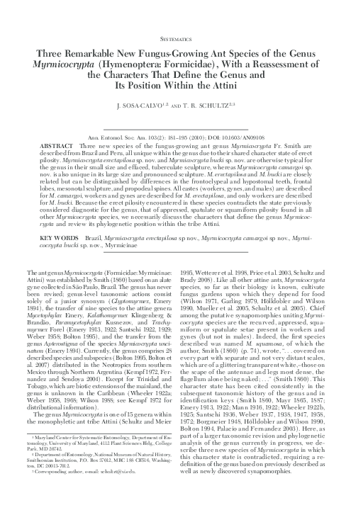
Fig. 1. M. camargoi, new species, life habitus reconstruction by V. Malikul.
simple, appressed hairs slightly overhanging mandibles and single thick, long (0.16 Ð 0.19 mm) unpaired
median seta that originates from posterior of clypeal
apron, slightly anterior to junction of clypeal apron
and body of clypeus (sensu Kugler 1994:22 deÞned as:
“the medial portion of the clypeus anterior to the
frontal lobes and dorsal to the clypeal apron”); frontoclypeal teeth acute, covered with suberect simple
hairs; mandibles with eight to 10 teeth, increasing
uniformly in size from base to apex; sculpture on
dorsal surface of mandibles rugulose-strigulate, frontal
lobes evenly rounded, expanded laterally (0.28 Ð 0.34
mm), covering antennal insertions; in proÞle, frontal
lobes strongly protruding; posterior margin of hypostoma with simple hairs that project over hypostomal
plate. Dorsum of hypostomal plate shining and glabrous; anterior-lateral margin of hypostoma with a pair
of acute teeth; in proÞle, occiput drawn out posterolaterally into an enlarged bilobed “neck” or “collar”
that extends backwards, covering anterior-lateral portions of pronotum. Mesosoma: median pronotal tubercles present, small; humeral tubercles present, (1.5"
length of median pronotals; lateral pronotal tubercles
long and robust, (2" length of median pronotals,
directed forward; lateral mesonotal tubercles longest
on mesosoma, (2" length of lateral pronotals; Þrst
median anterior mesonotal tubercles absent; second
median posterior mesonotal tubercles present, acute,
subequal in length to humeral tubercles; Þrst and second posterior mesonotal tubercles present, small, sub-
equal, joined by carinae, similar in size to median
pronotals; inferior angle of pronotum evenly rounded;
propleuron, adjacent to inferior pronotal edge, lacking
distinct tubercles, site bearing roughenings and erect,
spatulate hairs. mesonotal groove shallow but conspicuous,. Metanotal groove deep and conspicuous,
with median longitudinal carina. Base of propodeum,
in proÞle, ßat and slightly longer than declivity; both
laterally carinate; propodeal spines reduced to tubercles. Base of forecoxa with a conspicuous and lamellate
carina. Metasoma: petiolar peduncle lacking ventral
process; node of petiole, in dorsal view, rounded anteriorly and longer than wide and, in lateral view,
anteriorly rounded and posteriorly straight; postpetiole, in dorsal view, 1.3" wider than long; posterior
border emarginate; postero-lateral postpetiolar processes absent; dorsum of abdominal segment IV
(gaster) Þnely reticulate and covered with short,
erect, spatulate hairs; very subtle longitudinal sculpturing visible anteriorly; anterior margin straight, anterior lateral carinae absent. Individuals yellow to light
ferruginous; pilosity restricted mainly to tubercles and
carinae, spoon-shaped or spatulate; antennal scapes
and legs with simple, erect hairs.
Gyne. Measurements: TL ' 7.42Ð7.51; WL ' 2.13Ð
2.18; HL ' 1.20 Ð1.24; HW ' 1.05Ð1.13; SL ' 1.44;
ML ' 0.42; EL ' 0.22Ð 0.25; PL ' 0.67Ð 0.72; PPL '
0.38 Ð 0.39; GL ' 2.15Ð2.22; CI ' 85Ð94; SI ' 127Ð137;
MI ' 67Ð 69; FLD ' 0.42Ð 0.43 (n ' 2).
�186
ANNALS OF THE ENTOMOLOGICAL SOCIETY OF AMERICA
Vol. 103, no. 2
Figs. 2–7. M. camargoi, new species. Automontage photographs of worker habitus: 2, full-face (dorsal) view; 3, proÞle;
4, dorsal view. Scanning electron micrographs of worker habitus: 5, proÞle; 6, dorsal view; and 7, waist segments and gaster
in lateral view.
Similar to worker except for differences typical of
caste. Head: In full-face view, excluding mandibles,
slightly longer than wide with lateral and posterior
margins straight, posterior corners more angular and
less rounded than in worker; vertexal carinae conspicuous and lamellate, each produced into a series of
three processes: a pair of obtuse, triangular denticles
ßanking anterior ocellus, a pair of rectangular lamellae
overhanging posterior ocelli, and a pair of long, acute,
spinelike denticles mounted on vertex; cephalic mar-
gin with raised carina; posterior ocelli very small, median ocellus small (0.06 mm in diameter); frontal carinae complete to junction with vertexal carinae, but
lateral branches absent (present in worker); supraocular tubercle present; mandibles with 11 teeth;
clypeus as in worker and likewise with 7Ð9 pairs of
hairs and with a long unpaired median seta (length '
0.22Ð 0.23 mm). Mesosoma: Median and lateral pronotal and humeral tubercles present, well developed;
inferior margin of pronotum evenly rounded; in lateral
�March 2010
SOSA-CALVO AND SCHULTZ: THREE NEW SPECIES OF Myrmicocrypta
187
Figs. 8–11. Gyne of M. camargoi. 8, full-face view; 9, proÞle; 10, dorsal view; 11, fore and hind wings.
view, propleuron adjacent to inferior pronotal edge
weakly and obtusely angulate; median pronotal spines
connected by a weak carina, easily visible in frontodorsal view; mesoscutum with notauli (i.e., median
pair of longitudinal carinae) carinate and extending
through entire length of mesoscutum, developed anteriorly into blunt triangular denticles; median sulcus
present as a low raised carina on anterior half of
mesoscutum; parapsidal lines present as carinae in
posterior half of mesoscutum; transscutal articulation
present; parascutal lobes prominent and well developed, in lateral view posteriorly narrowed to a blunt,
acute angle, tip bearing a transverse lamella on its
outer face visible in oblique or dorsal view; axillae
large, in dorsal view tapering posteriorly, dorsal and
lateral margins developed into lamellae that form excavated, hemispherical concavities visible in posterior
view; scutellum without lateral projections and ending
posteriorly as two prominent dorsoventrally extended,
laterally compressed, distally rounded processes; base
and declivity of propodeum laterally carinate, carinae
continuing downward to join propodeal lobes; propodeal teeth short, triangular; pleural suture wide
and deep. Metasoma: Peduncle of petiole with a pair of
dorsolateral longitudinal carinae along nearly entire
length; petiole with a pair of ventral carinae that may
(one individual) or may not (one individual) end
anteriorly in a small tooth-like process, visible laterally; node of petiole dorsolaterally carinate; postpetiole in dorsal view 1.9" wider than long, with
pronounced posterolateral corners, posteriorly
emarginate; tergite of abdominal segment IV Þnely
reticulate-punctate. Wings: Forewing (length 4.80
mm) with very reduced fenestra, which appears as a
rounded spot near apical margin (Fig. 5.9); hind wing
(length 3.57 mm) with one closed cell. Head and body
dark ferrugineous; gaster yellowish to light ferrugineous; wings smoky. Pilosity as in worker.
Male. Measurements: TL ' 5.96 Ð 6.34; WL ' 1.90 Ð
1.98; HL ' 0.90 Ð 0.94; HW ' 0.77Ð 0.83 (including
eyes ' 1.08); SL ' 0.48 Ð 0.54; ML ' 0.48 Ð 0.58;
EL ' 0.37Ð 0.40; PL ' 0.64 Ð 0.73; PPL ' 0.23Ð 0.33;
GL ' 1.64 Ð1.86; CI ' 82Ð92; SI ' 58 Ð70; MI ' 53Ð 62;
FLD ' 0.25Ð 0.29 (n ' 4).
Head. In full-face view, triangular, wider posteriorly and laterally angulate, cephalic margin interrupted laterally by a pair of tubercles and medially
(behind posterior ocelli) by a pair of denticulate tubercles mounted on posterior extensions of vertexal
carinae; ocelli larger than those in gynes, median ocellus 0.12 mm in diameter and with its anterior margin
slightly concave or straight, posterior margin evenly
rounded; frontal carinae extending posterad to intersect with vertexal carinae, not continuing laterad; low
�188
ANNALS OF THE ENTOMOLOGICAL SOCIETY OF AMERICA
Vol. 103, no. 2
Figs. 12–15. Male of M. camargoi. 12, full-face view; 13, proÞle; 14, dorsal view; 15, fore and hind wings.
tubercles present at point of intersection; a median
carina extends from between frontal lobes to anterior
margin of median ocellus, more pronounced anteriorly, weaker posteriorly; dorsum of mandibles punctate; masticatory margin with eight to nine teeth gradually diminishing in size toward base; outer margin
of mandibles slightly convex; frontoclypeal teeth
present, short, blunt; anterior margin of clypeus hyaline, shining, and imbricate; three to four pairs of
lateral clypeal hairs barely exceeding the anterior clypeal margin, a single thick median seta (0.07 mm in
length; body of clypeus with a median carina arising
posterior to hyaline border at socket of median clypeal
seta, extending posterad and dividing into two carinae
that intersect frontoclypeal teeth; posterior border of
clypeus carinate; antennae with 13 segments; antennal
scapes barely surpassing cephalic margin, shorter than
funicular segments I-III combined; funicular segment
II $ 2" longer than funicular segment I (antennal
pedicel); posterior margin of head, in proÞle, concave;
with a median longitudinal carina that originates at
occipital carina and extends to level of vertexal tubercles; occipital collar present laterally, short and, in
lateral view, quadrate; hypostomal teeth large and
rounded at tip. Mesosoma: Pronotum with humeral and
lateral tubercles present, short and angulate; median
pronotal spines absent, replaced by a transverse carina
that connects lateral spines; inferior margin of pronotum evenly rounded; propleuron adjacent to inferior
pronotal edge with a low tubercle bearing simple hairs;
mesoscutum and scutellum similar to those of gyne,
axillary concavities less pronounced; propodeal spines
long and dorsolaterally compressed, in lateral view
appearing somewhat expanded apically, in dorsal view
appearing apically blunt, but in dorsolateral (edge-on)
view appearing linear; base of propodeum with a pair
of lateral carinae (Rio de Janeiro specimens; carinae
absent in Espiritu Santo specimens), declivity with a
pair of lamellate carinae (Rio de Janeiro specimens;
carinae lower in Espiritu Santo specimens). Metasoma:
Petiole pedunculate; in proÞle, node of petiole low
and evenly convex; petiole with dorso-lateral carinae
that extend along entire length of petiole, petiolar
node with single median (Espiritu Santo specimens)
or a few longitudinal carinae (Rio de Janeiro specimens); petiole with a pair of ventral lateral longitudinal carinae extending its entire length; node of petiole 1.6" longer than wide; node of postpetiole 1.7"
wider than long; abdominal tergite IV Þnely reticulate.
Wings: as in female but lacking fenestra. Forewing
length ' 4.56 Ð 4.68 mm; hind wing length ' 3.25Ð
3.47 mm.
�March 2010
SOSA-CALVO AND SCHULTZ: THREE NEW SPECIES OF Myrmicocrypta
189
Fig. 16. M. erectapilosa, new species, life habitus reconstruction by E. Hodges.
Individuals ferruginous in color; antennae and legs
testaceous; pilosity on antennal scape simple and appressed; head and mesosoma with hook-like hairs
mostly on carinae and tubercles; legs with simple appressed or decumbent hairs; abdominal tergite IV with
very short simple appressed hairs.
Etymology. It gives us great pleasure to name this
striking and unusual fungus-growing ant after its discoverer, Roberto S. Camargo.
Natural History. Workers of this species have been
collected in Cerrado habitat in Botucatú (São Paulo)
and Jatai (Goias) and in Mata Atlantica (Atlantic forest) in Floresta da Tijuca (Rio de Janeiro) and Santa
Teresa (Espiritu Santo). The nest series from Botucatú
was collected while digging out a colony of Atta
capiguara Gonçalves in a pasture Þeld composed
mostly of Brachiaria spp. and Paspalum spp. (Poaceae:
Panicoideae). Workers were observed carrying small
pieces of dry grass, but the nest entrance was not
located (R. S. Camargo, personal communication).
Myrmicocrypta erectapilosa Sosa-Calvo & Schultz,
new species
(Figs. 16Ð22)
Diagnosis (Worker). body covered with erect simple hairs; hypostomal teeth short, triangular, and
acute; sculpture on mesosoma reduced; frontal lobes
triangular and partially covering the antennal insertions.
Type Material. HOLOTYPE. Worker, labeled: “Brazil: Amazonas, Manaus, BR. 174 Km 45 EEST 51, 13IX-1991, (AY Harada and AG Bandeira).” (INPA)
USNM ENT No. 00537304.
Paratypes. 2 workers, same locality as holotype; one
dealate gyne, same locality as holotype. (INPA)
USNM ENT No. 00537305; one worker labeled: “Brazil: Amazonas 4832, Manaus, Dimona Camp, RS 2108,
21-X-93, plot C-5, (AB Casimiro) P. Coleção do Laboratório de Myrmecologia # 62 (CEPEC)” USNM ENT
No. 00537329.
Worker. Measurements: TL ' 3.22 (3.23Ð3.33);
WL ' 0.87 (0.84 Ð 0.91); HL ' 0.67 (0.66 Ð 0.71); HW '
0.58 (0.57Ð 0.63); SL ' 0.60 (0.61Ð 0.65); ML ' 0.46
(0.44 Ð 0.47); EL ' 0.05 (0.05Ð 0.06); PL ' 0.36 (0.29 Ð
0.33); PPL ' 0.16 (0.15Ð 0.16); GL ' 0.70 (0.76 Ð 0.78);
CI ' 87 (86 Ð 89); SI ' 103 (103Ð107); MI ' 69 (63Ð
68); FLD ' 0.22 (0.21Ð 0.24) (n ' 4).
Head. In full-face view 1.13"Ð1.15" as long as wide
(excluding mandibles); cephalic margin with shallow
median concavity, posterior corners evenly convex,
lacking sharp angles, spines, or tubercles; frontal carinae vestigial, past level of eyes merging with abundant
scabrous sculpture; eyes very small (six to eight ommatidia total) and in full-face view located approximately midway between posteriormost margin of head
�190
ANNALS OF THE ENTOMOLOGICAL SOCIETY OF AMERICA
Vol. 103, no. 2
Figs. 17–22. M. erectapilosa, new species. 17, 19, and 21 worker habitus (17, full-face view; 19, proÞle; and 21, dorsal view).
18, 20, and 22 gyne habitus (18, full-face view; 20, proÞle; and 22, dorsal view).
and insertion of mandibles; antenna with 11 segments;
antennal scape slightly wider at midpoint; hairs on
antennal scape long and erect or semierect; antennal
scapes surpassing cephalic margin by nearly 1.8" their
apical width; clypeal apron evenly convex, transparent, and shiny with shallow transverse striae and with
Þve pairs of simple, long, slender, appressed hairs that
originate on posterior margin of clypeal apron and
overhang mandibles; medially with unpaired long
(0.14 Ð 0.16 mm), thick seta; frontoclypeal teeth prominent, triangular, dentiform, and covered with simple
erect hairs; mandibles with six to seven teeth increasing in size from base to apex; dorsal surface of mandibles striate and bearing hairs, shorter and more appressed on lateral margins, longer and suberect toward
tips. Frontal lobes, in full face view, triangular and
partially covering antennal insertions; hypostomal
teeth short, triangular, and acute; hypostoma glabrous,
�March 2010
SOSA-CALVO AND SCHULTZ: THREE NEW SPECIES OF Myrmicocrypta
191
Fig. 23. M. bucki, new species, life habitus reconstruction by V. Malikul.
smooth, and shiny; occiput not extended into a “neck”
or “collar.” Head covered mainly with erect and suberect hairs; integument evenly reticulate-rugose. Mesosoma: pronotal humeral and lateral spines reduced
to eroded tubercles occurring at intersections of carinae; anterior pronotal spines absent; dorsum of
pronotum with a median longitudinal carina arising at
anterior pronotal margin and extending posteriorly
at least to level of lateral pronotal spines; dorsum
of pronotum sometimes with low wrinkles; inferior
pronotal margin evenly rounded; propleuron adjacent
to inferior pronotal edge with one or two very small
tubercles, each bearing a simple hair; all mesonotal
spines reduced to carinae; anterior propodeal spines
absent; posterior portion of propodeum with short,
blunt teeth; base of propodeum, in proÞle, ßat and as
long as declivity of propodeum; declivity of propodeum with a very reduced but conspicuous lamella on
each side. Metasoma: petiole pedunculate; ventrally
with a pair of closely approximated longitudinal median carinae that end anteriorly in a slightly raised
process that, when viewed laterally, looks like a single
longitudinal carina ending in a very small tooth; node
of petiole in proÞle rounded, in dorsal view ellipsoidal
and with a longitudinal carina; postpetiole in proÞle
longer than high and dorsally convex; posterior edge
of postpetiole in dorsal view slightly emarginate and
with distinct lateral corners; postpetiole wider than
long (1.6 Ð1.9"). In lateral view, base of abdominal
segment IV at junction with postpetiole ventrally
evenly convex and smooth; in dorsal view, base of
abdominal segment IV at junction with postpetiole
with a transverse carina with lateral corners; laterally
a few short carinae extend posterad from these corners. Dorsum of abdominal segment IV punctulatereticulate.
Individuals uniformly brown ferruginous; antennal scapes, head, and mesosoma covered with simple, erect hairs restricted mainly to carinae or tubercles; hairs on dorsum of abdominal segment IV
hook-like.
Gyne. Measurements: TL ' 4.24; WL ' 1.19; HL '
0.81; HW ' 0.71; SL ' 0.75; ML ' 0.51; EL ' 0.12;
PL ' 0.46; PPL ' 0.22; GL ' 1.05; CI ' 88; SI ' 106;
MI ' 63; FLD ' 0.28. (n ' 1).
Characters similar to those in worker with modiÞcations expected for caste and with following differences: Head: Frontal carina extending posterad to almost level of mid-ocellus and splitting into two low
carinae, vertexal ones being more conspicuous; eyes
large, containing eight ommatidia in largest row, (45
ommatidia in total; ocelli small (maximum length of
middle ocellus ' 0.06); occipital collar (neck) moderately developed laterally, with both upper and lower
extensions small and blunt, lower one slightly larger
than upper. Mesosoma: Dorsum of pronotum conspicuously rugose; humeral and lateral pronotal tubercles
present; propleuron adjacent to inferior pronotal edge
�192
ANNALS OF THE ENTOMOLOGICAL SOCIETY OF AMERICA
Figs. 24–26.
Vol. 103, no. 2
Habitus of worker of M. bucki, new species. 24, full-face view; 25, lateral view; 26, dorsal view.
somewhat angulate and bearing several curved hairs;
mesoscutum overall rugose; mesoscutal sulcus, in dorsal view, conspicuous and short, not extending $one
third length of mesoscutum; notauli absent; parapsidal
lines conspicuous and extending nearly to anterior
margin of mesoscutum; transscutal articulation conspicuous; scutellum posteriorly bidentate, dorsally
rugose; propodeum with a pair of short denticles; declivity of propodeum with a conspicuous lamella on
each side. Metasoma: Petiole as in worker; postpetiole
2.5x wider than long, with projecting latero-posterior
corners; dorsum of abdominal tergite IV densely reticulate, basally with widely spaced, short costulae and
with conspicuous antero-lateral carinae, as in worker.
IndividualÕs body dark yellow or light brown; pilosity
as in worker.
Etymology. The name erectapilosa derives from the
latin erecta ' standing and pilosa ' hairy and refers
to the erect hairs present in this species, the Þrst such
species to come to our attention.
Myrmicocrypta bucki Sosa-Calvo & Schultz,
new species
(Figs. 23Ð26)
Diagnosis. Similar in size and habitus to M. erectapilosa, but differing from it by having frontal lobes ves-
tigial, failing to cover antennal insertions; hypostomal
teeth long; vertexal carina present; humeral and lateral
pronotal spines acute; dorsum of pronotum smooth
and glabrous; propodeal spines long and acute; petiole
subquadrate.
Type Material. HOLOTYPE. One worker, labeled:
“Peru: Madre de Dios, Centro de Investigación y Capacitación Rṍo Los Amigos (CICRA), Otorongo Trail,
12%33& 42.28) S 70%05& 32.64) W, elevation 276 m, 19XI-2005, (J. Sosa-Calvo), nest series, hand collected,
forest (JSC051119 Ð 09)” USNM ENT No. 00537326.
(MUSM).
Paratypes. Fourteen workers, part of the same nest
series as holotype. (USNM). USNM ENT Nos.
00537307, 00537308, 00537317, 00537325, and 00301761
(alcohol); four workers, labeled: “Brazil: Amapa, Serra
do Navio (Silverstone).” (LACM 42302). USNM ENT
Nos. 00537306 and 00537316; 1 worker labeled: “Brazil,
Amazonas, Manaus, 9-v-2003, (C. Rabeling and M. Verhaagh).” (USNM). USNM ENT No. 00537315.
Worker. Measurements: TL ' 3.56 (3.28 Ð3.65);
WL ' 0.97 (0.91Ð1.01); HL ' 0.75 (0.72Ð 0.77); HW '
0.64 (0.62Ð 0.67); SL ' 0.69 (0.66 Ð 0.72); ML ' 0.51
(0.45Ð 0.54); EL ' 0.07 (0.05Ð 0.07); PL ' 0.35 (0.31Ð
0.35); PPL ' 0.17 (0.16 Ð 0.19); GL ' 0.81 (0.72Ð 0.83);
CI ' 85 (86 Ð 88); SI ' 108 (101Ð109); MI ' 68 (63Ð
71); FLD ' 0.13 (0.11Ð 0.14) (n ' 9).
�March 2010
SOSA-CALVO AND SCHULTZ: THREE NEW SPECIES OF Myrmicocrypta
Head. Head almost 1.2" longer than broad; posterior margin convex, convexity interrupted by two carinate tubercles in vertexal area; integument matte and
strongly rugole-reticulate; frontal carinae obsolete or
vestigial; eye very small with seven to nine ommatidia
in total; eyes above middle of head; antennal scape
reticulate and long, surpassing cephalic corners by
twice its apical width; clypeal apron convex and medially slightly angulate, hyaline, and transversely
weakly striate; clypeal pilosity originating near posterior margin of apron and extending over mandibles,
consisting of Þve to six pairs of lateral hairs and of one
median unpaired seta, much thicker and 3x as long
(length holotype ' 0.15, paratypes ' 0.13Ð 0.15 mm)
as lateral hairs; clypeus with a pair of blunt frontoclypeal teeth covered with simple, curved hairs; mandibles with six to seven teeth decreasing in size from
apex to base; dorsal surface of mandibles striolate;
frontal lobes strongly reduced, exposing antennal condyles in full-face view; hypostomal teeth long; occipital “collar” (neck) reduced to two low, blunt tubercles; antennal scape wider near its apex than at rest of
its length; anterior edge of antennal scape minutely
denticulate; in full-face view anterior and posterior
edges of antennal scape bearing suberect to erect
hairs; hairs on ventral portion of head narrowly spatulate. Mesosoma: Integument of mesosoma minutely
puncate. Dorsum of pronotum smooth and glabrous
with low humeral tubercles; lateral spines acute and
dentiform, longer and more discrete than other mesosomal tubercles; lacking anterior pronotal tubercles
or spines; inferior corner of pronotum evenly rounded; propleuron adjacent to inferior pronotal edge with
a broadly obtuse angle and with some subdecumbent
or suberect, simple or extremely narrowly spatulate
hairs; mesonotum with lateral tubercles reduced to
carinae, anterior tubercles low; mesonotal groove
smooth and glabrous; median mesonotal tubercles
low; posterior mesonotal tubercles blunt apically and
slightly longer than or similar in size to median tubercles; metanotal groove with a single median longitudinal carina that extends from posterior portion
of mesonotum through anterior portion of propodeum; propodeum anteriorly carinate, lacking anterior spines or tubercles; posterior propodeal spines
long and acute; dorsum and declivous of propodeum
with lateral carinae extending to propodeal lobes; base
of propodeum ßat, in proÞle subequal to declivity of
propodeum. Metasoma: Petiole pedunculate; node of
petiole, in proÞle, with two small anterior tubercles
and two larger posterior tubercles connected by sometimes incomplete carinae; node in lateral view subquadrate, in dorsal view slightly longer than broad; in
dorsal view, postpetiole $1.5" broader than long;
posteriorly emarginate; latero-posterior corners with
inferior wing-like projections giving it a trapezoidal
shape; shape of postpetiole, in proÞle, longer than
higher (1.2Ð1.3"); tergite of abdominal segment IV
punctulate-reticulate; in dorsal view, base of tergite at
junction with postpetiole slightly carinate (as in all
Myrmicocrypta spp.), but lacking lateral carinae beyond this junction, i.e., antero-lateral gastral carinae
193
absent; pilosity on dorsum of abdominal tergite IV
consisting of widely separated, extremely narrowly
spatulate hairs, all curved at tips and directed backward.
Individuals brown ferruginous; pilosity restricted to
wrinkles, tubercles, spines, carinae, appendages, antennal scapes, and gaster, absent elsewhere. Legs and
antennal scapes strongly reticulate.
Gynes and Males: Unknown.
Etymology. Named in honor of Dr. Peter Buck in
recognition of his support for science at the SmithsonianÕs National Museum of Natural History.
Comments. M. bucki is similar in size and habitus to
M. erectapilosa but differs from it by having simple,
curved hairs (simple but entirely erect in M. erectapilosa); frontoclypeal teeth blunt (large and acute in M.
erectapilosa); hypostomal teeth long (short and triangular in M. erectapilosa); frontal lobes narrow (triangular in M. erectapilosa) exposing part of the antennal
condyles; vertexal carinae forming a pair of tubercles
on cephalic margin in full-face view (these tubercles
absent in M. erectapilosa); humeral and lateral tubercles acute (reduced in M. erectapilosa); node of petiole
subquadrate in proÞle (rounded in M. erectapilosa). M.
bucki can be separated from any other Myrmicocrypta
species by the presence of simple, semierect hairs.
Acknowledgments
We are grateful to R. S. Camargo for collecting the type
specimens of M. camargoi, to C. Rabeling and M. Verhaag for
collecting additional specimens of M. bucki and to A. B.
Casimiro, A. Y. Harada, and A. G. Bandeira for collecting the
specimens of M. erectapilosa. We thank an anonymous reviewer for commenting on the manuscript. Carlos Roberto F.
Brandão (MZSP), Jacques Delabie (CPDC), Ana Harada
(MPEG), Christian Rabeling (University of Texas at Austin),
Roy Snelling (LACM), and M. Verhaag (SMNK) loaned us
specimens. Eugenia Okonski (USNM) photographed the
worker of M. erectapilosa. Elaine Hodges and Vichai Malikul
created the illustrations. Astrid Caldas provided information
about Rio de Janeiro. This research was supported in part by
National Science Foundation grants DEB 0110073 and DEB
0431330 (to T.R.S.). J.S.-C. was supported in part by an Ernst
Mayr Travel Award in Animal Systematics (Harvard University), a Jacob K. Goldhaber Travel Award (University of
Maryland), and an Amazon Conservation Association Graduate Research Grant.
References Cited
Bolton, B. 1994. IdentiÞcation guide to the ant genera of the
world. Harvard University Press, Cambridge, MA.
Bolton, B. 1995. A new general catalogue of the ants of the
world. Harvard University Press, Cambridge, MA.
Bolton, B., G. Alpert, P. S. Ward, and P. Naskrecki. 2007.
BoltonÕs Catalogue of Ants of the World: 1758 Ð2005. Harvard University Press, Cambridge, MA. (CD-ROM).
Borgmeier, T. 1948. Die Geschlechtstiere zweier EcitonArten und einige andere Ameisen aus Mittel-und suedamerika (Hym. Formicidae). Rev. Entomol. 19: 191Ð206.
Brandão, C.R.F., and A. J. Mayhé-Nunes. 2001. A new fungus-growing ant genus, Mycetagroicus gen. nov., with the
description of three new species and comments on the
�194
ANNALS OF THE ENTOMOLOGICAL SOCIETY OF AMERICA
monophyly of the Attini (Hymenoptera: Formicidae).
Sociobiology 38: 639 Ð 665.
Currie, C. R., J. A. Scott, R. C. Summerbell, and D. Malloch.
1999. Fungus-growing ants use antibiotic-producing bacteria to control garden parasites. Nature 398: 701Ð704.
Emery, C. 1890. Studi sulle formiche della fauna neotropica.
Boll. Soc. Entomol. Ital. 22: 38 Ð 80.
Emery, C. 1894. Studi sulle formiche della fauna neotropica.
Boll. Soc. Entomol. Ital. 26: 137Ð241.
Emery, C. 1912. Études sur les Myrmicinae. Ann. Soc. Entomol. Belg. 56: 94 Ð105.
Emery, C. 1913. Études sur les Myrmicinae. Ann. Soc. Entomol. Belg. 57: 250 Ð262.
Emery, C. 1922. Hymenoptera, Fam. Formicidae, subfam.
Myrmicinae. Genera Insectorum 174C: 207Ð397.
Fernandez, F., and S. Sendoya. 2004. Synonymic list of the
Neotropical ant (Hymenoptera: Formicidae). Biota Colombiana 5: 3Ð105.
Fernandez-Marin, H., J. K. Zimmerman, and W. T. Wcislo.
2004. Ecological traits and evolutionary sequence of nest
establishment in fungus-growing ants (Hymenoptera,
Formicidae, Attini). Biol. J. Linn. Soc. 81: 39 Ð 48.
Fernandez-Marin, H., J. K. Zimmerman, W. T. Wcislo, and
S. A. Rehner. 2005. Colony foundation, nest architecture, and demography of a basal fungus-growing ant,
Mycocepurus smithii (Hymenoptera, Formicidae). J. Nat.
Hist. 39: 1735Ð1743.
Forel, A. 1885. Études myrmécologiques en 1884; avec une
description des organes sensoriels des antennes. Bull. Soc.
Vaudoise Sci. Nat. 20: 316 Ð380.
Forel, A. 1893. Sur la classiÞcation de la famille des formicides, avec remarques synonymiques. Ann. Soc. Entomol.
Belg. 37: 161Ð167.
Forel, A. 1911. Ameisen des Herrn Prof. v. Ihering aus Brasilien (Sao Paulo usw.) nebst einigen anderen aus Südamerika und Afrika (Hym.). Dtsch. Entomol. Z. 1911:
285Ð312.
Garling, L. 1979. Origin of ant-fungus mutualism: a new
hypothesis. Biotropica 11: 284 Ð291.
Gauld, I., and B. Bolton. 1988. The Hymenoptera. Oxford
University Press, London, United Kingdom.
Hölldobler, B., and E. O. Wilson. 1990. The ants. Belknap
Press, Cambridge, MA.
Huber, J., and M. J. Sharkey. 1993. Structure, pp. 14 Ð59. In
H. Goulet and J. T. Huber [eds.], Hymenoptera of the
world, an identiÞcation guide to families. Monograph
1894E. Agriculture Canada Research Branch, Ottawa,
ON, Canada.
Kempf, W. W. 1963. A review of the ant genus Mycocepurus
Forel, 1893 (Hymenoptera: Formicidae). Stud. Entomol.
(N.S.). 6: 417Ð 432.
Kempf, W. W. 1972. Catalogo abreviado das formigas da
Regiao Neotropical. Stud. Entomol. (N.S.). 15: 3Ð344.
Klingenberg, C., and C.R.F. Brandão. 2009. Revision of the
fungus-growing ant genera Mycetophylax Emery and
Paramycetophylax Kusnezov rev. stat., and description of
Kalathomyrmex n. gen. (Formicidae: Myrmicinae: Attini). Zootaxa 2052: 1Ð31.
Kugler, C. 1994. Revision of the ant genus Rogeria (Hymenoptera: Formicidae) with descriptions of the sting apparatus J. Hymenopt. Res. 3: 17Ð 89.
Kusnezov, N. 1955. Evolución de las hormigas. Dusenia 6:
1Ð34.
Kusnezov, N. 1961. Evolution der Ameisen. Symp. Genet.
Biol. Ital. 12: 103Ð121.
Kusnezov, N. 1963. Zoogeografṍa de las hormigas en Sudamerica. Acta Zool. Lilloana 19: 25Ð186.
Vol. 103, no. 2
Lattke, J. 1997. Revisión del género Apterostigma Mayr (Hymenoptera: Formicidae). Arq. Zool. 34: 121Ð221.
Lattke, J. E. 1999. A new species of fungus-growing ant and
its implications for attine phylogeny (Hymenoptera: Formicidae). Syst. Entomol. 24: 1Ð 6.
Mann, W. M. 1916. The ants of Brazil. Bull. Mus. Comp.
Zool. 60: 399 Ð 490.
Mann, W. M. 1922. Ants from Honduras and Guatemala.
Proc. U.S. Natl. Mus. 61: 1Ð54.
Mayr, G. L. 1865. Reise der Oesterreichischen Fregatte Novara um die Erde in den Jahren 1857, 1858, 1859, unter den
befehlen des Commodore B. von Wuellerstorf-Urbair.
Zoologischer Theil. Formicidae. Vienna, Austria.
Mayr, G. L. 1887. Südamerikanische Formiciden. Verh.
Zool.-Bot. Ges. Wien 37: 511Ð 632.
Möller, A. 1893. Die Pilzgärten einiger südamerikanischer
Ameisen. Bot. Mitt. Tropen. 1893: 1Ð127.
Mueller, U. G., N. Gerardo, T. R. Schultz, D. Aanen, D. Six.
2005. The evolution of agriculture in insects. Annu. Rev.
Ecol. Syst. 36: 563Ð569.
Murakami, T., and S. Higashi. 1997. Social organization in
two primitive attine ants, Cyphomyrmex rimosus and Myrmicocrypta ednaella, with reference to their fungus substrates and food sources. J. Ethol. 2: 135Ð139.
Palacio, E. E., and F. Fernandez. 2003. Claves para las subfamilias y géneros, pp. 233Ð260. In F. Fernandez [ed.],
Introducción a las hormigas de la región Neotropical.
Instituto Humboldt, Bogotá, Colombia.
Price, S. L., T. Murakami, U. G. Mueller, T. R. Schultz, and
C. R. Currie. 2003. Recent Þnding in fungus-growing
ants: evolution, ecology, and behavior of a complex microbial symbiosis. In T. Kikuchi, N. Azuma, and S. Itagashi
[eds.], Genes, behaviors, and Evolution of social insects.
Hokkaido University Press, Sapporo, Japan.
Roger, J. 1863. Verzeichniss der Formiciden-Gattungen
und Arten. Berl. Entomol. Z. 7(Suppl.): 1Ð 65.
Santschi, F. 1922. Myrmicines, dolichodérines et autres formicides néotropiques. Bull. Soc. Vaudoise Sci. Nat. 54:
345Ð378.
Santschi, F. 1929. Nouvelles fourmis de la République Argentine et du Brésil. An. Soc. Cient. Argent. 107: 273Ð316.
Santschi, F. 1936. Fourmis nouvelles ou intéressantes de la
République Argentine. Rev. Entomol. 6: 402Ð 421.
Schultz, T. R., and S. G. Brady. 2008. Major evolutionary
transitions in ant agriculture. Proc. Nat. Acad. Sci. 105:
5435Ð5440.
Schultz, T. R., and R. Meier. 1995. A phylogenetic analysis
of the fungus-growing ants (Hymenoptera: Formicidae:
Attini) based on morphological characters of the larvae.
Syst. Entomol. 20: 337Ð370.
Schultz, T. R., U. G. Mueller, C. R. Currie, and S. A. Rehner.
2005. Reciprocal illumination: a comparison of agriculture in humans and ants, pp. 149 Ð190. In F. Vega and M.
Blackwell [eds.], Ecological and evolutionary advances
in insect-fungal associations. Oxford University Press,
New York.
Smith, F. 1858. Catalogue of the hymenopterous insects in
the collection of the British Museum. Part VI. Formicidae.
Taylor and Francis, London, United Kingdom.
Smith, F. 1860. Descriptions of new genera and species of
exotic Hymenoptera. J. Entomol. 1: 65Ð 84.
Weber, N. A. 1937. The biology of the fungus-growing ants.
Part I. New forms. Rev. Entomol 7: 378 Ð 409.
Weber, N. A. 1938. The biology of the fungus-growing ants.
Part 4. Additional new forms. Part 5. The Attini of Bolivia.
Rev. Entomol. 9: 154 Ð206.
�March 2010
SOSA-CALVO AND SCHULTZ: THREE NEW SPECIES OF Myrmicocrypta
Weber, N. A. 1941. The biology of the fungus-growing ants.
Part 7. The Barro Colorado Island, Canal Zone, species.
Rev. Entomol. 12: 93Ð130.
Weber, N. A. 1945. The biology of the fungus-growing ants.
Part 8. The Trinidad, B.W.I. species. Rev. Entomol. 16: 1Ð88.
Weber, N. A. 1947. Lower Orinoco River fungus-growing
ants (Hymenoptera: Formicidae, Attini). Bol. Entomol.
Venez. 6: 143Ð161.
Weber, N. A. 1958. Evolution in fungus-growing ants. Proc.
Tenth Int. Congr. Entomol. 2: 459 Ð 473.
Weber, N. A. 1966. Fungus-growing ants. Science (Wash.,
D.C.) 153: 587Ð 604.
Weber, N. A. 1968. Tobago Island fungus-growing ants (Hymenoptera: Formicidae). Entomol. News. 74: 141Ð145.
Weber, N. A. 1969. A comparative study of the nests, gardens and fungi of the fungus-growing ants, Attini, pp.
299 Ð307. In Proceedings of the VI Congress IUSSI
(International Union for the Study of Social Insects).
Organizing Committee of the VI Congress IUSSI, Bern,
Switzerland.
Weber, N. A. 1972. Gardening ants, the attines. American
Philosophical Society, Philadelphia, PA.
195
Wetterer, J. K., T. R. Schultz, and R. Meier. 1998. Phylogeny
of fungus-growing ants (Tribe Attini) based on mtDNA
sequence and morphology. Mol. Phylogenet. Evol. 9: 42Ð
47.
Wheeler, W. M. 1910. Ants, their structure, development
and behaviour. Columbia University Press, New York.
Wheeler, W. M. 1922a. The ants of Trinidad. Am. Mus. Novit. 45: 1Ð16.
Wheeler, W. M. 1922b. Keys to the genera and subgenera of
ants. Bull. Am. Mus. Nat. Hist. 45: 631Ð710.
Wheeler, W. M. 1925. Neotropical ants in the collections of
the Royal Museum of Stockholm. Part I. Ark. Zool. 17:
1Ð55.
Wilson, E. O. 1971. The insect societies. Belknap Press, Harvard University, Cambridge, MA.
Wilson, E. O. 1988. The biogeography of the West Indian
ants (Hymenoptera: Formicidae), pp. 214 Ð230. In J. Liebherr [ed.], Zoogeography of Caribbean Insects. Cornell
University Press, Ithaca, NY.
Received 24 July 2009; accepted 7 December 2009.
�
 Jeffrey Sosa-Calvo