seminars in I M M U N OL OG Y, Vol 11, 1999: pp. 227]237
Article No. smim.1999.0179, available online at http:rrwww.idealibrary.com on
T cell receptor-mediated signs and signals governing T
cell development
Nicolai S. C. van Oers
gd . in a non-covalent association with a group of
invariant proteins ŽCD3 g , d , « and TCR z .. These
invariant subunits are responsible for efficient assembly and surface expression of the various TCR complexes Žreviewed in ref 1.. Additionally, the invariant
chains contain a conserved signaling motif that functions to translate effective ligand binding into intracellular biochemical signals Žreviewed in ref 2.. This
motif, termed the immune tyrosine based activation
motif ŽITAM. wYxxLx Ž6-8.YxxLx, is present as a single
copy in CD3 g , d , and « and as three copies in TCR
z .3 The signals propagated through the ITAMs ultimately converge in the nucleus, resulting in the induction of various biological responses such as differentiation, proliferation, programmed cell death,
cytokine release, andror cytolytic functions.
A considerable wealth of knowledge now exists
about the mechanisms of TCR-mediated signaling
Žreviewed in ref 4.. The central feature of this pathway is the modulation of the tyrosine phosphorylation status of many effector molecules through the
activation of several families of protein tyrosine kinases ŽPTKs. Žreviewed in ref 5.. The Src-family of
PTKs are proposed to initiate TCR-induced signal
transduction by phosphorylating pairs of tyrosine
residues present in the ITAMs ŽFigure 2.. Once phosphorylated, the ITAMs form a consensus binding
motif for the two Src-homology 2 ŽSH2. domains of
the SykrZAP-70 family of PTKs. The recruitment of
SykrZAP-70 results in the combined activation of
both families of kinases, culminating in the phosphorylation of many additional effector molecules such
as LAT, SLP-76, vav, and PLC-g 1.4 These changes
lead to a well-established elevation in intracellular
calcium and the activation of ras and its downstream
MAPK cascades Žreviewed in refs 6 and 7.. The following review will examine the roles of three families
of PTKs in TCR signal transduction, with particular
emphasis on their functions during thymopoeisis.
Several of the more recently identified regulatory
molecules will also be reviewed, again with an em-
The developmental fate of T cells is largely controlled by the
nature and success of signals mediated by the pre-T cell
receptor (TCR) and TCR complexes. These intracellular
signals are regulated by cascades of protein tyrosine phosphorylations initiated following ligand binding to the preTCR or TCR complexes. The phosphorylation cascades are
primarily orchestrated by two distinct families of protein
tyrosine kinases (PTKs), the Src- and the Syk r ZAP-70families. Germline gene targeting experiments, several human immunodeficiencies, and somatic cell mutants have all
contributed to our understanding of how these families of
kinases coordinate their actions to promote signaling. Upon
activation, the PTKs transmit their signals to a number of
newly described adaptor proteins including LAT, SLP-76,
and vav, among others. The following review combines
results derived from different experimental strategies to examine the contributions of the PTKs and the adaptor
molecules to pre-TCR and TCR signaling processes.
Key words: adaptor proteins r protein tyrosine kinases r
signal transduction r T cell receptor
Q1999 Academic Press
Introduction
THE PRE-T CELL RECEPTOR Žpre-TCR. and T cell receptor ŽTCR. complexes are multi-subunit complexes
that mediate the differentiation and expansion of
both ab and gd T lineage cells during T cell development ŽFigure 1.. The complexes themselves comprise the ligand binding subunits Žpre-Tarb ; ab ;
From the Center for Immunology and the Department of Microbiology, UT Southwestern Medical Center, Room NA7.201,
6000 Harry Hines Blvd., Dallas, TX 75235-9093, USA
Q1999 Academic Press
1044-5323r 99 r 040227q 11 $30.00r 0
227
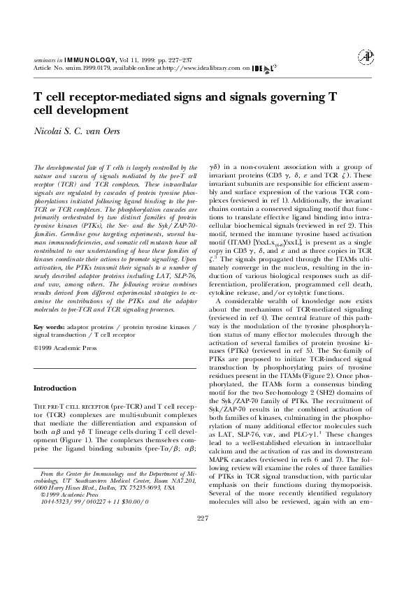
�N. S. C. van Oers
Figure 1. Pre-T Cell Receptor and T cell Receptor Complexes. The immune tyrosine-based
activation motifs are represented as darkened rectangles. The pre-TCR complex appears to have
a very weak association with the TCR z subunit.
to convert the CD25q CD44 lory cells into a
CD25yCD44 lory subpopulation that begins expressing the CD4 and CD8 coreceptor molecules. The
CD4yCD8y to CD4qCD8q transition coincides with
the termination of pre-Ta expression, and initiation
of rearrangements at the TCR a locus. A successful
pairing and surface expression of the ab TCR complex allows the CD4qCD8q thymocytes to undergo
the stringent processes of positive and negative selection.8 The consequences of these TCR-mediated
selection events results in the establishment of a
peripheral T cell repertoire that is restricted by selfMHC molecules.
phasis on TCR-mediated signal transduction processes
necessary for T cell development.
T cell development
The majority of ab T cells, a subset of gd T cells,
and certain NK cell populations develop in the thymus. For ab T cells, this developmental process
follows a highly ordered series of selection events that
are phenotypically defined with the specific expression of certain cell surface molecules such as CD3,
CD4, CD8, CD25, and CD44. A simplified overview of
this process is illustrated in Figure 3. The entire
process of selection will be covered in great detail in
the many accompanying review articles in this volume.
Following the arrival of precursor cells into the thymus, the TCR b locus undergoes a rearrangement
process. A successfully rearranged TCR b gene
product subsequently pairs with the pre-TCR a subunit, and this complex is expressed on the surface of
cells that lack the CD4 and CD8 coreceptor molecules
ŽFigure 3.. Such CD4y CD8y cells can be further
subdivided into four distinct subpopulations on the
basis of CD25 and CD44 expression. Signals generated through the pre-TCR complex are proposed
TCR r CD3 itams
The generation of the peripheral T cell repertoire is
contigent upon the nature and success of the TCR
signaling pathways that are activated during thympoeisis. The main functions of the ITAMs of the TCR
complex are to convey appropriate ligand binding
events into intracellular signals. As previously descibed, the TCR complex comprises upwards of ten
ITAMs, with six contributed by the TCR z subunit
ŽFigure 1.. Given the presence of ten ITAMs, the
228
�Signals governing T cell development
Figure 2. Model of Protein Tyrosine Kinase Activation by the T Cell Receptor ITAMs. Following
TCR interactions with peptidermajor histocompatibility complexes, the Src-kinases are activated,
resulting in the phosphorylation of the tyrosine residues in the ITAMs. This promotes the
recruitment of the SykrZAP-70 family of kinases to the TCR complex, in interactions mediated by
the two SH2 domains of the kinase with the doubly phosphorylated tyrosines in the ITAMs. The
association of SykrZAP-70 with the phosphorylated ITAMs allows for their subsequent phosphorylation and activation. Cross-communication between the two families of kinases promotes the
phosphorylation of multiple downstream effector molecules.
phosphorylation of increasing numbers of ITAMs may
contribute quantitatively to the strength of signaling.
The concept of quantitative signaling with increasing
TCRrCD3 ITAMs was originally described for cell
lines expressing chimeric molecules with the extracellular domain of CD8 and an intracellular region
containing either one, two, or three TCR z ITAMs.9
However, many studies have also described qualitative differences between the various phosphorylated
TCRrCD3 ITAMs and their abilities to interact with
downstream effector molecules Žreviewed in ref 5..
The issues of qualitative vs quantitative signaling differences may have important ramifications for T cell
selection processes in the thymus, and these have yet
to be resolved.
With regards to thymopoeisis, some apparent distinctions between the various invariant chains have
been described. For example, the targeted disruption
of the CD3 « allele Žalso affects CD3 g and d transcription. results in a complete development arrest of
both ab and gd T cells at the CD4yCD8y stage,
similar to mice lacking the RAG genes.10,11 In contrast, the targeted disruption of the TCR z molecule
results in a much less severe development block.12 ] 14
Thus, CD4qCD8q thymocytes lacking surface TCR
expression are still apparent in these mice, although
at numbers that vary from two- to 30-fold less than
age matched, wild-type littermates. These results
would imply that pre-TCR signaling is still functional
in TCR z-null mice, consistent with the concept that
the TCR, and perhaps the pre-TCR, contain two
independent signal transduction modules wTCR z vs
CD3 g , d , and « x.15 Alternatively, TCR z may not be
required for the assembly of the pre-TCR complex. It
229
�N. S. C. van Oers
Figure 3. Overview of T Cell Development. The development of ab T cells in the thymus follows
a well-defined series of development changes phenotypically characterised by the expression of
various cell surface markers, as indicated. The effects of the targeted disruptions of various
effector molecules on this developmental pathway are high-lighted below the pathways. A solid
bar indicates a complete developmental arrest, whereas a dashed bar implies an incomplete
developmental block.
is interesting to note that normal T cell development
can be restored in the TCR z knock-out mice by
introducing TCR z transgenes comprising zero, one,
or all three z ITAMs.16 These studies demonstrated
that the TCR z ITAMs are not essential for thymopoeisis, but z is important for proper TCR assembly and surface expression. In fact, only in a well-defined ab trangenic TCR background ŽH-Y TCR
transgenic mice. does it become obvious that the
number of TCR z ITAMs can contribute in a quantitative capacity to positive and negative selection.17
A second distinction between the various invariant
chains is the presence of a pool of constitutively
tyrosine phosphorylated TCR z subunits in murine
thymocytes and peripheral T cells.18,19 This constitutive phosphorylation, resulting in the formation of a
21-kDa phosphorylated form of TCR z , is unique to z
as none of the other CD3 subunits are constitutively
phosphorylated. The constitutive phosphorylation of
TCR z is, in part, a consequence of ongoing TCR
interactions with peptiderMHC molecules expressed
in the thymus.18,20 As discussed below, both the Lck
and ZAP-70 protein tyrosine kinases ŽPTKs. are required for the maintenance of the constitutively
phosphorylated TCR z subunits.21,22 In addition to
the 21-kDa form of TCR z , a second phosphorylated
form of TCR z Žapparent molecular mass of 23 kDa.
can be induced following receptorrligand interactions.20,23 ] 25 Notably, a strong and provocative correlation has been established between the induction of
particular phosphorylated forms of TCR z Ž21 vs 23
kDa. and the functional responsiveness of T cells
Žreviewed in ref 23.. The presence or absence of
these distinct phosphorylated forms of TCR z may
also have relevance to the processes of positive and
negative selection in the thymus.
Protein tyrosine kinases
Three families of protein tyrosine kinases ŽPTKs., the
230
�Signals governing T cell development
Src-, Syk-, and Tec-families, have important and distinct functional contributions to T cell development.
Lck and Fyn, two members of the Src-family of PTKs,
are primarily restricted in expression to T cells.26
They have unique N-terminal sequences followed by
a Src-homology 3 ŽSH3., a Src-homology 2 ŽSH2., and
a kinase domain. Although these kinases are targeted
to the plasma membrane through myristylation and
palmitylation modifications at their NH 2-terminus, a
proportion of Lck molecules are found associated
with the CD4 and CD8 coreceptor molecules while
Fyn can complex with the TCR, albeit at low stoichiometry.27 The important contributions of the CD4
and CD8 coreceptor molecules to T cell development
are reviewed elsewhere in this issue.
A combination of biochemical and genetic data has
elegantly demonstrated a principle role for Lck in
initiating TCR-signaling cascades by phosphorylating
the ITAMs in the TCR complex 21,28 Žreviewed in ref
27.. This pathway was initially uncovered following
the description of a Lck-deficient leukemic cell that
was unable to initiate TCR-mediated signaling
processes unless reconstituted with Lck.29 With regards to thymopoeisis, Lck is expressed in all thymocyte subsets, and numerous experiments have revealed a role for Lck in pre-TCR and TCR-signaling
Žreviewed in refs 30 and 31.. In brief, mice rendered
deficient in Lck have a 5]20 fold reduction in thymic
cellularity and a severe paucity of both CD4qCD8q
and mature single positive T cells.32 The mature T
cells that can develop in these mice are unable to
respond effectively to TCR stimulations.32 Such problems are primarily related to the fact that Lck is
required for initiating TCR signal transduction by
phosphorylating the ITAMs and for phosphorylating
and activating the SykrZAP-70 family of PTKs following their high affinity binding to the phosphorylated ITAMs ŽFigure 2..33 ] 35 As a consequence, very
little constitutive phosphorylation of the TCR z
molecules can be detected in thymocytes from Lcknull mice.21 Moreover, no inducible phosphorylation
of TCRrCD3 ITAMs or ZAP-70 is noted in these
knock-out animals.
Fyn is a second Src-family member that functions
in the TCR signaling cascade, in part, by phosphorylating the TCR ITAMs. In contrast to Lck, Fyn is
expressed at 10-fold higher levels in mature single
positive thymocytes relative to CD4qCD8q cells.26
Interestingly, Fyn-deficient mice have normal numbers of thymocytes and peripheral T cells.36,37 The
single positive thymocytes and mature T cells in Fyn-
null mice are hypo-responsive following TCR crosslinking, as evidenced by their poor proliferative responses and low mobilisation of intracellular
calcium.36,37 Consistent with the knock-out animals,
mice expressing a dominant negative Fyn transgene
only have a defect in single positive TCR-mediated
signal transduction.38 This defect is partly a consequence of reduced IL-2 production by the stimulated
T cells, suggesting that Fyn couples to the IL-2 pathway.36,37 The inability of Lck to promote signaling in
the absence of Fyn may relate to the unique substrate
specificity of Fyn. Thus, Fyn can selectively phosphorylate both Pyk2, a member of the focal adhesion
family of PTKs, and Fyb ŽSLAP130., a Fyn and SLP-76
associating signaling protein.39 ] 41 This selectivity suggests a bifurcation point in the functions of Fyn and
Lck and may indicate a unique contribution for Fyn
in IL-2 production in mature T cells through a pathway involving Fyb ŽSLAP130.. Mice rendered
deficient in both Lck and Fyn have a complete ab
T cell developmental arrest at the CD4yCD8y
ŽCD25y CD44y . stage of thympoeisis.42,43 Taken
together, the aforementioned data would suggest that
Lck is the primary Src-family PTK responsible for T
cell development although Fyn can partially compensate for Lck in pre-TCR and TCR signaling. The
requirements for Fyn and Lck in gd and NK cell
development are discussed in the accompanying review articles.
Syk and ZAP-70 are members of a second family of
PTKs that function as critical mediators of pre-TCR
and TCR signaling, with ZAP-70 having a predominant role in mature T cells Žreviewed in ref 31..
Defined by the presence of two NH 2 terminal SH2
domains and a COOH-terminal kinase domain, ZAP70 is localised as a diffuse band under the plasma
membrane.27,44 The two SH2 domains form a high
affinity interaction with the doubly phosphorylated
ITAMs. Upon TCR ligation, both kinases can associate with the tyrosine phosphorylated ITAMs of the
pre-TCR and TCR complexes, and, in turn, are tyrosine phosphorylated.45 ] 47 The phosphorylations on
ZAP-70 increase the intrinsic catalytic activity of the
kinase and allow for the binding of additional signaling proteins Žvav, Lck., or subsequent attenuators of
signaling Žc-cbl, SHP-1.. Both ZAP-70 and Syk are
expressed in developing thymocytes, with ZAP-70 upregulated during the CD4qCD8q to the single positive transition, while Syk undergoes a concomitant
down-modulation following pre-TCR signaling.46,48
The functional requirements for ZAP-70 in TCRmediating signaling processes were elucidated in sev231
�N. S. C. van Oers
eral severe combined immunodeficiency ŽSCID.
patients that harbored mutations in the ZAP-70
gene.49 ] 51 Phenotypically, immature CD4qCD8q thymocytes from such patients could only mature into
CD4q T cells, and these cells were functionally incompetent to transduce TCR-mediated signals. Thus,
TCR cross-linking resulted in a very poor induction
of tyrosine phosphoproteins, demonstrating a central
role for ZAP-70 in initiating TCR signal transduction
cascades. Mice lacking ZAP-70 or mice harboring a
catalytically inactive ZAP-70 molecule have normal
numbers of CD4qCD8q thymocytes that are unable
to undergo either positive or negative selection
processes, such that no CD4q or CD8q cells are
generated.22,52,53 The phenotypes described for the
ZAP-70 deficiencies are largely explained by the requirements of ZAP-70 to phosphorylate several key
signalingradaptor substrates including LAT, SLP-76,
and vav.5,54 Moreover, the two SH2 domains of ZAP-70
are required for maintaining the constitutive and
inducible phosphorylations of the TCR z chain.22 It
is interesting to note that the results with the mice
differ somewhat from that described for the human
SCIDs, and this may relate to the differences in Syk
expression and function, as described below.
In contrast to ZAP-70, Syk-deficient mice have a
completely normal pattern of ab T cell development
and a selective impairment of certain gd lineage T
cells.55,56 The targeted disruption of both ZAP-70 and
Syk results in a complete attenuation of thymocyte
development at the CD4yCD8y stage, with the majority of cells in these mice maintaining a
CD25qCD44yrlo phenotype, analogous to the subsets
in LckrFyn- and Rag-null mice.31 Such findings again
demonstrate the existence of functional redundancy
within a given family of PTKs. Thus, Syk can partially
compensate for the absence of ZAP-70 by supporting
pre-TCR signal transduction.31 The failure of Syk to
promote differentiation of thymocytes to the single
positive stage in mice may relate to the marked
down-regulation of Syk expression that occurs in
double positive thymocytes.46,48 This is consistent with
the ability of Syk to support complete T cell development and TCR-mediated signaling when over-expressed as a transgene in ZAP-70-null mice.57 One
important distinction between ZAP-70 and Syk is the
observation that Syk has a much higher intrinsic
enzymatic activity and much less dependence on Srcfamily kinases for its activation.58,59
Itk is a member of the TecrBtk family of PTKs that
is also implicated in thymopoeisis. This family of
kinases has several functional domains including a
NH 2 terminal plextrin homology ŽPH. domain, followed by a SH3, SH2, and a kinase domain. The PH
domain is involved in localising these PTKs to the
cellular membrane by interacting with the phospholipid PIP3 . Mice deficient in Itk have a small reduction in single positive thymocytes, an effect more
obviously noted in a ab TCR transgenic background.60 Moreover, the mature T cells in these mice
have reduced TCR-mediated proliferative responses
largely as a consequence of reduced IL-2 production.
It will be interesting to determine whether Itk connects to the FynrFyb pathway for IL-2 production.
Mutations in the B cell family member, Btk, results in
X-linked agammaglobulinemia in humans and X-linked immunodeficiency in mice.61 These extremely
severe B cell phenotypes relative to the mild T cell
defects in Itk-deficient mice may suggest the presence
of additional Tec family members in T cells that can
facilitate thymopoeisis. The exact role of Itk in TCRsignaling requires further investigation.
Signaling molecules
The preliminary events of pre-TCR- or TCR-mediated
PTK activation must be successfully relayed to appropriate downstream substrates. A theme common
to many different signaling systems is the recruitment
and activation of selected enzymes through the use of
scaffold, anchoring, andror adaptor proteins.62 T
cells are no exception, with an exciting number of
newly identified and essential adaptor molecules having recently been identified Žreviewed in ref 4.. One
key T cell adapter protein Žalso in NK cells, mast
cells, and basophils. is termed LAT Žlinker for activation of T cells..54 LAT is a 36]38-kDa palmitylated
transmembrane glycoprotein that becomes heavily
tyrosine phosphorylated following TCR stimulation.54
At least 6 tyrosine residues are phosphorylated in
LAT, most by ZAP-70, and these phosphorylated sequences form consensus binding motifs for the SH2
domains of phosphopholipase C gamma 1 ŽPLC-g 1.,
the Grb2 adapter protein, and Grap ŽGrb2-like. ŽFigure 4..54 Thus, LAT serves as an adaptor protein by
connecting the PTKs to downstream substrates. The
crucial role of LAT in T cell signaling was established
with an analysis of a LAT-deficient Jurkat T cell
line.63 Cross-linking the TCR on these cells revealed a
normal ITAM and ZAP-70 phosphorylation without
any subsequent mobilisation of intracellular calcium
or ras activation.63 Additionally, overexpression of a
mutant LAT bearing tyrosine to phenylalanine substi232
�Signals governing T cell development
acterised phosphorylated substrates in T cells Žreviewed in ref 7.. Activated PLC-g 1 hydrolyses phosphatidylinositol 4,5 bisphosphate into the second
messengers IP3 and diacylglycerol ŽDAG., which are,
in turn, responsible for the well-characterised intracellular calcium increases and PKC activation, respectively.6 The calcium increase is required to activate
the serinerthreonine phosphatase calcineurin, which,
in turn, activates an important IL-2 transcription factor, nuclear factor of activated T cells ŽNFAT..
A second critical molecule recruited to phosphory-
tutions reduces the magnitude of NF-AT driven transcriptional reporter constructs.54 Given the fact that
the highest expression levels of LAT are in the thymus Žassessed by Northern blotting., LAT-deficient
mice are likely to have a complete block in ab and
gd T cell development. The lack of intracellular
calcium mobilisation in the LAT-deficient cells is
likely a consequence of the failure to recruit and
phosphorylate PLC-g 1. Interestingly PLC-g 1, an enzyme essential for the activation of the phosphatidylinositol pathway, was one of the first well-char-
Figure 4. T Cell Receptor Signaling Cascades. Following the initiation of TCR signaling, a
number of downstream adaptor proteins are phosphorylated. One key adaptor protein phosphorylated by ZAP-70 is LAT. The phosphorlation sites on LAT serve as consensus binding motifs for
the SH2 domains of PLC-g 1 and Grb2. The subsequent phosphorylation and activation of PLC-g 1
results in the release of intracellular calcium stores and the activation of PKC. Since Grb2 forms
complexes with SLP-76 and SOS, the recruitment of Grb2 to LAT provides a mechanism for
relocating SOS to the membrane, facilitating ras activation and the downstream MAPK pathway.
The re-localisation andror phosphorylation of SLP-76 is also necessary for both ras activation and
PLC-g 1 phosphorylation. This may be partly attributed to the association of SLP-76 with vav, a
guanine nucleotide exchanger on the rhorracrCDC42 family of GTPases. The actions of vav may
regulate actin polymerisation and cytoskeletal re-organisation. Vav can also form an independent
complex with phosphorylated ZAP-70, and this may also contribute to vav activation. Finally, Fyn
may couple to the IL-2 pathway through its association with a SLP-76 interacting protein, termed
Fyb, or SLAP-130.
233
�N. S. C. van Oers
lated LAT is Grb2, an adapter protein that itself
complexes to several other key regulatory molecules
including sos-of-sevenless ŽSOS., SLP-76 ŽSH2 domain
leukocyte protein of 76 kDa., and the c-cbl protooncogene ŽFigure 4..6,54,64 Grb2 contains one SH2
domain flanked on either side by a SH3 domain. The
SH3 domains of Grb2 can bind to proline rich peptide sequences in both SOS and SLP-76. SOS is an
important guanine nucleotide exchanger for the ras
family of small GTPases, functioning to activate ras.
Activated ras has a central role in IL-2 gene activation
by regulating the MAPK ŽErk. pathway, and this pathway has been reviewed elsewhere.6 With regards to
thymopoeisis, an activated form of ras, when expressed as a transgene in Rag-deficient embryonic
stem cells, is capable of promoting the differentiation
of ab T cells to the CD4qCD8q stage.65 Since ras
connects to the MAPK pathway, these results are also
consistent with the observations that a dominant negative MAPKK, when expressed in fetal thymic organ
cultures, blocks the transition of thymocytes from
double negative to double position cells.66 Using the
Lck proximal promoter to express a dominant negative ras transgene in thymocytes, a specific block in
positive but not negative selection was revealed.67
Grb2 also associates with SLP-76, a recently identified T and NK cell specific molecule that contains
three tyrosine residues located near the NH 2terminus, a central proline rich stretch, and a
COOH-terminal SH2 domain.64,68,69 Following TCR
cross-linking, ZAP-70 phosphorylates SLP-76 on the
three NH 2-terminal tyrosines, presumably following
the association of SLP-76rGrb2 with phosphorylated
LAT. In mature T cells, overexpression of SLP-76
potentiates TCR-mediated IL-2 gene induction while
a mutant form lacking either the three NH 2-terminal
tyrosines or a functional SH2 domain can attenuate
IL-2 responses.69 Interestingly, T cell lines lacking
SLP-76 have revealed a requirement for SLP-76 in
PLC-g 1 phosphorylation and ras activation, but not
in the TCR-induced phosphorylation of most other
proteins.70 These results would suggest that the
Grb2rSOS pathway is not sufficient to activate ras.
The importance of SLP-76 in thymopoeisis was
recently described with the generation of two independent SLP-76 deficient mice.71,72 The thymocyte
subpopulations in these mice revealed a complete
developmental arrest within the CD4yCD8y subset,
at the transition point from CD44yCD25q to
CD44yCD25y cells. This is the same developmental
arrest point as the LckrFyn and ZAP-70rSyk double
knock-out mice. As TCR b gene rearrangements and
pre-TCR a expression were apparent in the SLP-76
null animals, the absence of SLP-76 most probably
impairs pre-TCR signaling. The ability of the dominant negative MAPKK to block thymocyte development is consistent with a requirement for SLP-76 to
promote ras activation in early thymocyte development.
In mature T cells, the mechanism by which SLP-76
couples to IL-2 production is not clear but certainly
involves another TCR-induced phosphoprotein, the
vav proto-oncogene Žreviewed ref 5.. Vav functions as
a guanine nucleotide exchange factor ŽGEF. for the
rhorracrCDC42 families of GTPases, and also contains a PH domain, and two SH3 domains at its
COOH terminus interspersed by an SH2 domain.
Following TCR ligation, vav becomes tyrosine phosphorylated and also forms two independent complexes with phosphorylated ZAP-70 or SLP-76 through
its SH2 domain. Overexpression of wild-type vav can
significantly increase both the basal and TCR-induced NFAT and IL-2-driven transcriptional activity.73
Notably, the combined overexpression of vav and
SLP-76 can synergistically potentiate the aforementioned transcriptional activity. As described earlier,
PLC-g 1 is not phosphorylated in SLP-76 deficient
cells. This may be a consequence of the absence of
vav association, since vav may be required to promote
cytoskeletal reorganisation through its GEF functions.
With regards to T cell development, vav-deficient
mice have been generated by a RAG-complementation approach, and these mice exhibit a 10]20-fold
reduction in all thymocyte subsets.74 ] 76 Moreover,
these cells have severely reduced proliferative responses to TCR stimuli. It remains to be established
why the phenotype of the SLP-76 deficient mice is
much more severe than that of the vav-null mice, but
may relate to the ability of SLP-76 to associate with
several additional phosphorylated proteins.68 As mentioned earlier, SLP-76-deficient T cells cannot
mobilise intracellular calcium. In contrast, most of
the data with vav-deficient thymocytes indicates that a
normal calcium flux can be elicited following TCR
cross-linking. Taken together, these results would imply that SLP-76 may couple to multiple downstream
effector molecules. Alternatively, a vav homologue
may also be expressed in hematopoeitic cells and this
may partially compensate for the absence of vav.
Protein tyrosine phosphatases
The pre-TCR and TCR-mediated signals are coordi234
�Signals governing T cell development
nately regulated by the actions of PTKs and protein
tyrosine phosphatases ŽPTPases.. Although many PTPases will likely be identified as key regulators of
antigen receptor signal transduction, two well-characterised ones include CD45 and SHP-1. CD45 is a
transmembrane PTPase that comprises two intracellular PTPase domains and is expressed on almost all
hematopoeitic cells.77 Several isoforms have been
identified on T cells as a consequence of regulated
processes of alternative splicing. CD45 primarily
functions as a positive regulator of antigen receptor
signal transduction by activating the Src-kinases Lck
and Fyn through the dephosphorylation of the negative regulatory tyrosine residue in these. The importance of CD45 in T cell development was evident
with the 4]10-fold reduction in the numbers of
CD4qCD8y and CD4yCD8q thymocytes and peripheral T cells in mice lacking either all CD45 isoforms
or CD45-exon 6-null mice.78,79 The CD4qCD8q thymocytes from the CD45-null mice are refractory to
anti-TCR mAb-induced negative selection and those
mature T cells that do develop display reduced TCRmediated proliferative responses.78,79 This is largely
explained by the fact that Fyn and Lck are hyperphosphorylated at their COOH-terminal negative
regulatory tyrosine in CD45-null thymocytes. As a
consequence, these kinases are relatively inactive resulting in hypo-phosphorylation of the TCR ITAMs
and a relatively poor recruitment of ZAP-70 to the
TCR complex.80. What remains unclear at present in
how T cell development can proceed to the
CD4qCD8q stage in the absence of CD45.
SHP-1 is a hematopoeitic specific PTPase that appears to down-modulate antigen receptor signaling
systems.77 Two naturally occurring SHP-1 mutations
were identified in mice Žmotheaten mice. several
years ago. Phenotypically, the thymocytes from these
mice have a normal developmental profile. Functionally, both thymocytes and peripheral T cells from
these SHP-1 mutant mice hyper-proliferate in response to anti-TCR stimulations.1,81 Additionally, the
levels of the constitutively and inducibly phosphorylated proteins in these thymocytes are markedly elevated relative to normal mice.81 With regards to
mechanism, the absence of SHP-1 results in the hyper-activation of the Src-family kinases, leading to
increased basal and TCR-inducible phosphorylation
levels of the ITAMs and ZAP-70. SHP-1 has also been
reported to complex ZAP-70, and this may be an
additional means of regulating antigen responsiveness.82
Concluding remarks
The processes of antigen receptor signal transduction
are emerging as well-defined biochemical pathways
involving a coordinated regulation of PTK and PTPase activities. A new set of exciting signalingradaptor proteins have recently been identified in these
signaling cascades. Thus, a combination of cloning
reports, gene knock-out results, and studies with somatic cell mutants have revealed essential roles for
the LAT, SLP-76, and vav molecules. The exact functional mechanisms of these and other signaling
molecules during pre-TCR and TCR-signaling will
emerge rapidly. Over the next few years, novel activators and attenuators of antigen receptor signals will
likely be identified, many of which will have significant functional contributions to ab , gd , and NK cell
development.
Acknowledgements
I would like to thank Dr T.C. Ayi, Mr. B. Tohlen, and Mrs.
A. Feulner for their reading of this manuscript. This work
was supported in part by a grant from the NIH ŽAI4295301A1. and Fikes support funding from UT Southwestern
Medical Center.
References
1. Ashwell JD, Klausner RD Ž1990. Genetic and mutational
analysis of the T-cell antigen receptor. Ann Rev Immunol
8:139]167
2. Weiss A Ž1993. T cell antigen receptor signal transduction: a
tale of tails and cytoplasmic protein-tyrosine kinases. Cell
73:209]212
3. Cambier JC Ž1995. New nomenclature for the Reth motif Žor
ARH1rTAMrARAMrYXXL.. Immunol Today 16:110
4. Rudd CE Ž1999. Adaptors and molecular scaffolds in immune
cell signaling. Cell 96:5]8
5. Wange RL, Samelson LE Ž1996. Complex complexes: signaling at the TCR. Immunity 5:197]205
6. Cantrell D Ž1996. T cell antigen receptor signal transduction
pathways. Ann Rev Immunol 14:259]274.
7. Weiss A, Littman DR Ž1994. Signal transduction by lymphocyte antigen receptors. Cell 76:263]274
8. Jameson SC, Bevan MJ Ž1998. T-cell selection. Curr Opin
Immunol 10:214]219
9. Irving BA, Chan AC, Weiss A Ž1993. Functional characterization of a signal transducing motif present in the T cell
receptor z chain. J Exp Med 177:1093]1103.
10. Malissen M, Gillet A, Ardoiun L, Bouvier G, Trucy J, Ferrier
P, Viver E, Malissen B Ž1995. Altered T cell development in
mice with a targeted mutation of the CD3-« gene. EMBO J
14:4641]4653
11. Tanaka Y, Ardoiun L, Gillet A, Lin S-Y, Magnan A, Malissen
B, Malissen M Ž1995. Early T-cell development in CD3-deficient mice. Immunol Rev 148:171]199
235
�N. S. C. van Oers
12. Love PE, Shores EW, Johnson MD, Tremblay ML, Lee EJ,
Grinberg A, Huang SP, Singer A, Westphal H Ž1993. T cell
development in mice that lack the z chain of the T cell
antigen receptor compex. Science 261:918]921
13. Liu C-P, Ueda R, She J, Sancho J, Wang B, Weddell G, Loring
J, Kurahara C, Dudley EC, Hayday A, Terhorst C, Huang M
Ž1993. Abnormal T cell development in CD3-zyry mice and
identification of a novel T cell population in the intestine.
EMBO J 12:4863]4875
14. Malissen M, Gillet A, Rocha B, Trucy J, Brun N, Mazza G,
Spanopoulou E, Guy-Grand D, Malissen B Ž1993. T cell
development in mice lacking the CD3-zrh gene. EMBO J
12:4347]4355
15. Wegener A-MK, Letourneur F, Hoeveler A, Brocker T, Luton
F, Malissen B Ž1992. The T cell receptorrCD3 complex is
composed of at least two autonomous transduction modules.
Cell 68:83]95
16. Shores EW, Huang K, Tran T, Lee E, Grinberg A, Love PE
Ž1994. Role of TCR z chain in T cell development and
selection. Science 266:1047]1050
17. Shores EW, Tran T, Grinberg A, Sommers CL, Shen H, Love
PE Ž1997. Role of multiple TCR z chain signaling motifs in
selection of the T cell repertoire. J Exp Med 185:893]900
18. Nakayama T, Singer A, Hsi ED, Samelson LE Ž1989. Intrathymic signalling in immature CD4qCD8q thymocytes results in tyrosine phosphorylation of the T-cell receptor zeta
chain. Nature 341:651]654
19. van Oers NSC, Tao W, Watts JD, Johnson P, Aebersold R, Teh
H-S Ž1993. Constitutive tyrosine phosphorylation of the T cell
receptor ŽTCR. z subunit: Regulation of TCR-associated protein kinase activity by TCR z . Mol Cell Biol 13:5771]5780
20. van Oers NSC, Killeen N, Weiss A Ž1994. ZAP-70 is constitutively associated with tyrosine phosphorylated TCR z in
murine thymocytes and lymph node T cells. Immunity
1:675]685
21. van Oers NSC, Killeen N, Weiss A Ž1996. Lck regulates the
tyrosine phosphorylation of the TCR subunits and ZAP-70 in
murine thymocytes. J Exp Med 183:1053]1062
22. Kadlecek TA, van Oers NSC, Lefrancois L, Olson S, Finlay D,
Chu DH, Connolly K, Killeen N, Weiss A Ž1998. Differential
requirements for ZAP-70 in TCR signaling and T cell development. J Immunol 161:4688]4694
23. Jameson SC, Bevan MJ Ž1995. T cell receptor antagonists and
partial agonists. Immunity 2:1]11
24. Sloan-Lancaster J, Shaw AS, Rothbard JB, Allen PM Ž1994.
Partial T cell signaling: Altered phospho-z and lack of ZAP-70
recruitment in APL-induced T cell anergy. Cell 79:913]922
25. Madrenas J, Wange RL, Wang JL, Isakov N, Samelson LE,
Germain RN Ž1995. z phosphorylation without ZAP-70 activation induced by T cell receptor antagonists or partial agonists. Science 267:515]518
26. Olszowy MW, Leuchtmann PL, Veillette A, Shaw AS Ž1995.
Comparison of p56 l ck and p59 f y n protein expression in thymocyte subsets, peripheral T cells, NK cells, and lymphoid
cell lines. J Immunol 155:4236]4240
27. Chan AC, Desai D, Weiss A Ž1994. Role of protein tyrosine
kinases and protein tyrosine phosphatases in T cell antigen
receptor signaling. Ann Rev Immunol 12:555]592
28. Iwashima M, Irving BA, van Oers NSC, Chan AC, Weiss A
Ž1994. Sequential interactions of the TCR with two distinct
cytoplasmic tyrosine kinases. Science 263:1136]1139
29. Straus D, Weiss A Ž1992. Genetic evidence for the involvement of the Lck tyrosine kinase in signal transduction
throught the T cell antigen receptor. Cell 70:585]593
30. Anderson SJ, Perlmutter RM Ž1995. A signaling pathway
governing early thymocyte maturation. Immunol Today
16:99]105
31. Cheng AM, Cahn AC Ž1997. Protein tyrosine kinases in
thymocyte development. Curr Opin Immunol 9:528]533
32. Molina TJ, Kishihara K, Siderovski DP, van Ewijk W, Narendran A, Timms E, Wakeham A, Paige CJ, Hartmann K-U,
Veillette A, Davidson D, Mak TW Ž1992. Profound block in
thymocyte development in mice lacking p56lck. Nature
357:161]164
33. Chan AC, Dalton M, Johnson R, Kong G-H, Wang T, Thoma
R, Kurosaki T Ž1995. Activation of ZAP-70 kinase activity by
phosphorylation of tyrosine 493 is required for lymphocyte
antigen receptor function. EMBO J 14:2499]2508
34. Watts JD, Affolter M, Krebs DL, Wange RL, Samelson LE,
Aebersold R Ž1994. Identification by electrospray ionization
mass spectromoetry of the sites of tyrosine phosphorylation
induced in activated Jurkat T cells on the protein tyrosine
kinase ZAP-70. J Biol Chem 269:29520]29529
35. Wange RL, Guitian R, Isakov N, Watts JD, Aebersold R,
Samelson LE Ž1995. Activating and inhibitory mutations in
adjacent tyrosines in the kinase domain of ZAP-70. J Biol
Chem 270:18730]18733
36. Stein PL, Lee H-M, Rich S, Soriano P Ž1992. pp59 fyn mutant
mice display differential signaling in thymocytes and peripheral T cells. Cell 70:741]750
37. Appleby MW, Gross JA, Cooke MP, Levin SD, Qian X, Perlmutter RM Ž1992. Defective T cell receptor signaling in mice
lacking the thymic isoform of p59 f y n . Cell 70:751]763
38. Cooke MP, Abraham KM, Forbush KA, Perlmutter RM Ž1991.
Regulation of T cell receptor signaling by a src family protein-tyrosine kinase Žp59 fyn .. Cell 65:281]292
39. Qian D, Lev S, van Oers NSC, Schlessinger J, Weiss A Ž1997.
Tyrosine phosphorylation of Pyk2 is selectively regulated by
Fyn during TCR signaling. J Exp Med 185:1253]1259
40. Musci MA, Hendricks-Taylor LR, Motto DG, Paskind M, Kamens J, Turck CW, Koretzky GA Ž1997. Molecular cloning of
SLAP-130, an SLP-76-associated substrate of the T cell antigen
rceptor stimulated protein tyrosine kinases. J Biol Chem
272:11674]11677
41. daSilva AJ, Li Z, de Vera C, Canto E, Findell P, Rudd CE
Ž1997. Cloning of a novel T-cell protein FYB that binds FYN
and SH2-domain-containing leukocyte protein 76 and modulate interleukin 2 production. Proc Natl Acad Sci USA
94:7493]7498
42. Groves T, Smiley P, Cooke MP, Forbush K, Perlmutter RM,
Guidos CJ Ž1996. Fyn can partially substitute for Lck in T
lymphocyte development. Immunity 5:417]428
43. van Oers NSC, Lowin-Kropf B, Finlay D, Connolly K, Weiss A
Ž1996. ab T cell development is abolished in mice lacking
both Lck and Fyn protein tyrosine kinases. Immunity
5:429]436
44. Huby RDJ, Iwashima M, Weiss A, Ley SC Ž1997. ZAP-70
protein tyrosine kinase is constitutively targeted to the T cell
cortex independently of its SH2 domains. J Cell Biol
137:1639]1649
45. Chan AC, Iwashima M, Turck CW, Weiss A Ž1992. ZAP-70: A
70kD protein tyrosine kinase that associates with the TCR z
chain. Cell 71:649]662
46. Chan AC, van Oers NSC, Tran A, Turka L, Law C-L, Ryan JC,
Clark EA, Weiss A Ž1994. Differential expression of ZAP-70
and Syk protein tyrosine kinases, and the role of this family of
protein tyrosine kinases in T cell antigen receptor signalling.
J Immunol 152:4758]4766
47. van Oers NSC, von Boehmer H, Weiss A Ž1995. The pre-TCR
complex is functionally coupled to the TCR z subunit. J Exp.
Med 182:1585]1590
48. Chu DH, van Oers NSC, Malissen M, Harris J, Elder M, Weiss
A Ž1999. Pre-T cell receptor signals are responsible for the
downregulation of Syk protein tyrosine kinase expression.
Submitted
49. Arpaia E, Shahar M, Dadi H, Cohen A, Roifman CM Ž1994.
Defective T cell receptor signaling and CD8q thymic selection in humans lacking ZAP-70 kinase. Cell 76:947]958
236
�Signals governing T cell development
50. Chan AC, Kadlecek TA, Elder ME, Filipovich AH, Kuo W-L,
Iwashima M, Parslow TG, Weiss A Ž1994. ZAP-70 deficiency in
an autosomal recessive form of severe combined immunodeficiency. Science 264:1599]1601
51. Elder ME, Lin D, Clever J, Chan AC, Hope TJ, Weiss A,
Parslow T Ž1994. Human severe combined immunodeficiency
due to a defect in ZAP-70, a T cell tyrosine kinase. Science
264:1596]1599
52. Negishi I, Motoyama N, Nakayama K-I, Nakayama K, Senju S,
Hatakeyama S, Zhang Q, Chan AC, Loh DY Ž1995. Essential
role for ZAP-70 in both positive and negative selection of
thymocytes. Nature 376:435]438
53. Wiest DL, Ashe JM, Howcroft TK, Lee H-M, Kemper DM,
Negishi I, Singer DS, Singer A, Abe R Ž1997. A spontaneously
arising mutation in the DLAARN motif of murine ZAP-70
abrogates kinase activity and arrests thymocyte development.
Immunity 6:663]671
54. Zhang W, Sloan-Lancaster J, Kitchen J, Trible RP, Samelson
LE Ž1998. LAT: The ZAP-70 tyrosine kinase substrate that
links T cell receptor to cellular activation. Cell 92:83]92
55. Turner M, Mee PJ, Costello PS, Williams O, Price AA, Duddy
LP, Furlong MT, Geahlen RL, Tybulewicz VLJ Ž1995. Perinatal lethality and blocked B cell development in mice lacking
the tyrosine kinase Syk. Nature 378:298]302
56. Cheng AM, Rowley B, Pao W, Hayday A, Bolen JB, Pawson T
Ž1995. Syk tyrosine kinase required for mouse viability and B
cell development. Nature 378:303]306
57. Gong Q, White L, Johnson R, White M, Negishi I, Thomas M,
Chan AC Ž1997. Restoration of thymocyte development and
function in zap-70yry mice by the Syk protein tyrosine
kinase. Immunity 7:369]377
58. Latour S, Chow LML, Veillette A Ž1996. Differential intrinsic
enzymatic activity of Syk and ZAP-70 protein tyrosine kinases.
J Biol Chem 271:22782]22790
59. Chu D, Spits H, Peyron J-F, Rowley RB, Bolen JB, Weiss A
Ž1996. The Syk protein tyrosine kinase can function independently of CD45 or Lck in T cell antigen receptor signaling.
EMBO J 15:6251]6261
60. Liao XC, Littman DR Ž1995. Altered T cell development in
mice lacking Itk. Immunity 3:757]769
61. Rawlings DJ, Witte ON Ž1994. Bruton’s tyrosine kinase is a
key regulator in B cell development. Immunol Rev
138:105]119
62. Pawson T, Scott JD Ž1997. Signaling though scaffold, anchoring, and adaptor proteins. Science 278:2075]2080
63. Finco TS, Kadlecek T, Zhang W, Samelson LE, Weiss A Ž1998.
LAT is required for TCR-edmiated activation of PLC-g1 and
the ras pathway. Immunity 9:617]626
64. Jackman JK, Motto DG, Sun Q, Tanemoto M, Turck CW,
Peltz GA, Koretzky GA, Findell PR Ž1995. Molecular cloning
of SLP-76, a 76 kDa tyrosine phosphoprotein associated with
Grb2 in T cells. J Biol Chem 269:7029]7032
65. Swat W, Shinkai Y, Cheng H-L, Davidson L, Alt FW Ž1996.
Activated ras signals differentiation and expansion of
CD4 q CD8 q thymocytes. Proc Natl Acad Sci USA
93:4683]4687
66. Crompton T, Gilmour KC, Owen MJ Ž1996. The MAP kinase
pathway controls differentiaton from double-negative to double positive thymocyte. Cell 86:243]251
67. Swan KA, Alberola-IIa J, Gross JA, Appleby MW, Forbush KA,
Thomas JF, Perlmutter RM Ž1995. Involvement of p21ras
distinguishes positive and negative selection in thymocytes.
EMBO J 14:276]285
68. Motto DG, Ross SE, Wu J, Hendricks-Taylor LR, Koretzky GA
Ž1996. Implication of the GRB2-associated phosphoprotein
SLP-76 in T cell receptor interleukin 2 production. J Exp
Med 183:1937]1943
69. Wardenburg JB, Fu C, Jackman JK, Flotow H, Wilkinson SE,
Williams DH, Johnson R, Kong G, Chan AC, Findell P Ž1996.
Phosphorylation of SLP76 by the ZAP-70 protein tyrosine
kinase is required for T cell receptor function. J Biol Chem
271:19641]19644
70. Yablonski D, Kuhne MR, Kadlecek T, Weiss A Ž1998. Uncoupling of nonreceptor tyrosine kinases from PLC-g1 in an
SLP-76-deficient T cell. Science 281:413]416
71. Clements JL, Yang B, Ross-Barta SE, Eliason SL, Hrstka RF,
Williamson RA, Koretzky GA Ž1998. Requirment for the
leukocyte-specific adapter protein SLP-76 for normal T cell
development. Science 281:416]419
72. Pivniouk V, Tsitsikov E, Swinton P, Rathbun G, Alt FW, Geha
RS Ž1998. Impaired viability and profound block in thymocyte development in mice lacking the adaptor protein SLP-76.
Cell 94:229]238
73. Wu J, Katzav S, Weiss A Ž1995. A functional T-cell receptor
signaling pathway is required for p95 vav activity. Mol Cell
Biol 15:4337]4346
74. Tarakhovsky A, Turner M, Schaal S, Mee PJ, Duddy LP,
Rajewsky K, Tybulewicz VLJ Ž1995. Defective antigen receptor-mediated proliferation of B and T cells in the absence of
vav. Nature 374:467]470
75. Zhang R, Alt FW, Davidson L, Orkin SH, Swat W Ž1995.
Defective signaling through the B and T cell antigen receptors in lymphoid cells lacking the vav proto-oncogene. Nature
374:470]473
76. Fischer KD, Zmuldzinas A, Gardner S, Barbacid M, Bernstein
A, Guidos C Ž1995. Defective T cell receptor signaling and
positive selection of Vav-deficient CD4qCD8q thymocytes.
Nature 374:474]477
77. Neel BG Ž1997. Role of phosphatases in lymphocyte activation. Curr Opin Immunol 9:405]420
78. Blyth KF, Conroy LA, Howlett S, Smith AJH, May J, Alexander DR, Holmes N Ž1996. CD45-null transgenic mice reveal a
positive regulatory role for CD45 in early thymocyte development, in the selection of CD4qCD8q thymocytes, and in B
cell maturation. J Exp Med 183:1707]1718
79. Kishihara K, Penninger J, Wallace VA, Kundig TM, Kawai K,
Wakeham A, Timms E, Pfeffer K, Ohashi PS, Thomas ML,
Furlonger C, Paige CJ, Mak TW Ž1993. Normal B cell development but impaired T cell maturation in CD45-exon 6
protein tyrosine phosphatase deficient mice. Cell 74:143]156
80. Stone JD, Conroy LA, Byth KF, Hederer RA, Howlett S,
Takemoto Y, Holmes N, Alexander DR Ž1997. Aberrant
TCR-mediated signaling in CD45-null thymocytes involves
dysfunctional regulation of Lck, Fyn, TCR-zeta, and ZAP-70. J
Immunol 158:5773]82
81. Pani G, Fischer K-D, Mlinaric-Rascan I, Siminovitch KA Ž1996.
Signaling capacity of the T cell antigen receptor is negatively
regulated by the PTP1C tyrosine phosphatase. J Exp Med
184:839]852
82. Plas DR, Johnson R, Pingel JT, Matthews RJ, Dalton M, Roy
G, Chan AC, Thomas M Ž1996. Direct regulation of ZAP-70
by SHP-1 in T cell antigen receptor signaling. Science
272:1173]1176
237
�
 Nicolai van Oers