Analysing ancient DNA
Raul J. Cano
Much of what we know about extinct organisms comes from traits that are not preserved in the fossil
record. Until recently, morphological analysis was the only tool available for scientists to determine
relationships for extinct fossil organisms. We now know that 'ancient' DNA can be preserved in the
remains of extinct organisms. By targeting specific gene sequences, it may be possible to deduce
biochemical characteristics and through sequence comparisons, to estimate the extent of evolutionary
divergence. By comparing the amount and type of these changes, one could estimate how quickly some
DNA 'evolves' relative to other segments, or which genes have the most flexibility or are more
conserved over time. The compilation of these data would yield greater understanding of the physiology
of extinct organisms and provide a much clearer picture of genetic change over time, and the mechanics
behind 'evolution'.
The isolation and characterization of fossil
DNA, until recently [1), was considered
unattainable as the methodologies for
extracting minute quantities of partially
degraded DNA and their subsequent enzymatic amplification were not available.
With the advent of the polymerase chain
reaction [2], a new analytical tool became
available for the molecular study of fossils.
1t is now possible to conduct molecular
studies of extinct organisms utilizing their
DNA to unravel biological and evolutionary
questions.
There is already a body of scientific evidence built which supports the use of DNA
from extinct animals and plants for phylogenetic studies. Higuchi and Wilson [1]
demonstrated that remains of a mammoth
and the extinct species, the quagga. contained fragments of the original DNA.
Paabo [3] reported the extraction of cionable DNA from a 2400-year-old mummy of
a child. Subsequent DNA analysis revealed
fragments measuring approximately 3.4 kilobase pairs (Kbp). Thomas et at. [4] isolated
DNA from hair found in century-old
untanned hide and a piece of dried muscle
collected from an extinct marsupial wolf.
This DNA was later enzymatically ampli-
fied by polymerase chain reaction (PCR)
and phylogenetic studies were made. More
recently, Golenberg et a/. [5] isolated and
analysed Magnolia chloroplast DNA from a
Miocene Clarkia deposit dated I 7-20 million years old. Cano et al. [6] isolated and
characterized DNA from the extinct bee
Proplebeia dominicana in 25-40 millionyear-old Dominican amber. DeSalle et al.
[7) employed DNA extracted from fossil
termites to resolve phylogenetic relationships between the termites, cockroaches
and mantids. Cano et at. [8] extracted DNA
from a 120-135 million-year-old nemonychid weevil in Lebanese amber and showed
by nucleotide sequence alignments and
phylogenetic inference analyses that the fossil weevil was most closely related to tbe
extant nemonychid weevil LeconteUus pinicola. Poinar et al. [9] used DNA sequences
from the extinct legume Hymenaea protera
in Dominican amber in a biogeographical
study in which they showed that the extinct
H. protera was most closely related to the
extant African species H. verrucosa, as
morphological studies suggested. Finally,
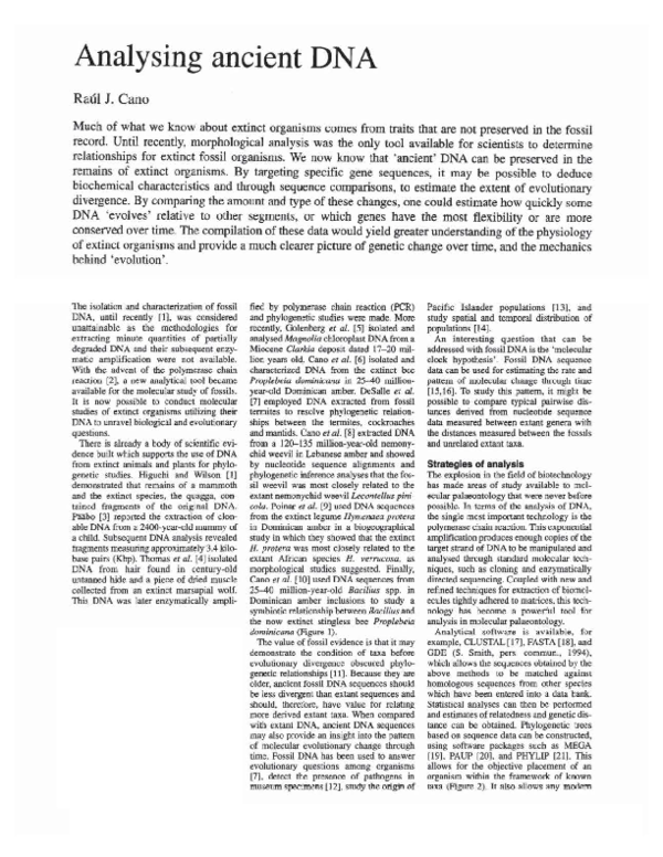
Cano et al. [I 0] used DNA sequences from
25-40 million-year-old Bacillus spp. in
Dominican amber inclusions to study a
symbiotic relationship between Bacillus and
the now extinct stingless bee Proplebeia
dominicana (Figure 1).
The value of fossil evidence is that it may
demonstrate the condition of taxa before
evolutionary divergence obscured phylogenetic relationsh.ips [11]. Because they are
older, ancient fossil DNA sequences should
be less divergent than extant sequences and
should, therefore, have value for relating
more derived extant taxa. When compared
with extant DNA, ancient DNA sequences
may also provide an insight into the pattern
of molecular evolutionary change through
time. Fossil DNA has been used to answer
evolutionary questions among organisms
[7], detect the presence of pathogens in
museum specimens [12], study the origin of
Pacific Islander populations [13], and
study spatial and temporal distribution of
populations [14].
An interesting question that can be
addressed with fossil DNA is the ' molecular
clock hypothesis'. Fossil DNA sequence
data can be used for estimating the rate and
pattern of molecular change through time
[15,16]. To study this pattern, it might be
possible to compare typical pairwise distances derived from nucleotide sequence
data measured between extant genera with
the distances measured between the fossils
and unrelated extant taxa.
Strategies of analysis
The explosion in the field of biotechnology
bas made areas of study available to molecular palaeontology that were never before
possible. In terms of the analysis of DNA,
the single most important technology is the
polymerase chain reaction. Th.is exponential
amplification produces enough copies ofthe
target strand of DNA to be manipulated and
analysed through standard molecular techniques, such as cloning and enzymatically
directed sequencing. Coupled with new and
refined techniques for extraction of biomolecules tightly adhered to matrices, this technology has become a powerful tool for
analysis in molecular palaeontology.
Analytical software is available, for
example, CLUSTAL[I7], FASTA [18], and
ODE (S. Smith, pers. comrnun., 1994),
which allows tbe sequences obtained by the
above methods to be matched against
homologous sequences from other species
wh.ich have been entered into a data bank.
Statistical analyses can then be performed
and estimates ofrelatedness and genetic distance can be obtained. Phylogenetic trees
based on sequence data can be constructed,
using software packages such as MEGA
[19]. PAUP [20]. and PHYLIP [21]. This
allows for the objective placement of an
organism within the framework of known
raxa (Figure 2). It also allows any modem
�Figure 1 Stingless bee Proplebeia dominicana entombed in Dominican amber
(25--40 myo).
B.SPHAER
BACRRNAGA
BACRRNAGC
.....--- BACRRNAOO
BACRRNAGF
BACRRNAGE
BAC·RRNAGB
BACRRNAGD
. . - - - - - - - B.PASTEURII
L-l{=
";:(~ B.GLOBISPORUS
S.UREAE
..---- --
- - B.CEREUS
r-----r--:-:=:::-: B.SUBTILIS
'-------l
B.PUMILUS
B.MEGATERIUM
BRONCHOTHRlX
0.01
Figure 2 Phylogenetic tree of ancient and extant Bacillus spp., constructed using the
maximum likelihood algorithm (18). The ancient sequence for Bacillus sphaericus, identified
as BCA16CG, appears to be more ancestral (that is, closest to the root) to modern isolates
of B. sphaer;cus (B.SPHAER, BACRRNAGA-BACRRNAGG).
DNA that may be contaminating ancient
tissues to be characterized and possibly
recognized.
Selections of gene sequences for
analysis of fossil DNA
When working with DNA putatively
obtained from fossils, the selection of gene
sequences for amplification and/or analysis
is a crucial step. In the case ofextinct organisms, for which there is no direct living representative, the genomes of the closest living relatives (based on morphological
analysis) ace examined for conserved
sequences. When selected regions of genes
for these taxa are compared, homologous
sequences can be identified. Regions of
homology, where at least 15 bases are
identical between the two groups, are
good places to start when designing primer
molecules.
The size of the amplified target sequence
(amplicon) is also of importance. Generally,
when designing primers to amplify DNA
segments from fossils, it is best to think
small. The chances for successful amplifications of fossil sequences increase as the
size of the amplicon decreases. As a general
rule it is recommended that the selected
primer pair arnpiify a region of the desired
gene to measure S: 200 bp. As the fossil
DNA becomes damaged and degraded, the
resulting fragme.nt length becomes smaller.
Thus, amplification of small DNA segments
will be more successful than that of larger
segments [22}. Table I illustrates the results
of a study conducted in our laboratory
.a imed at demonstrating the reproducibility
of DNA extraction from amber inclusions
(dated 25-40 million years old) of the
extinct bee Proplebeia dominicana, the
·e xtant bee Plebe ia frontalis, and their corresponding Bacillus symbionts.
The chances for successful amplification
are increased if the target gene sequence is
present in multiple copies within each cell.
Nuclear DNA sequences of ribosomal constituents, such as 18s and 28s rONA, are
often used in such studies. Mitochondrial
DNA sequences are also good candidates
because, not only are there several to thousands of mitochondria per cell, but the complete mitochondrial genome for many taxa
have been sequenced and entered into data
banks, and are available for comparative
studies. For phylogenetic significance, it is
desirable that the selected homologous
primer sequences for known taxa flank
regions of relatively high variability. Tllis
allows for better definition of phylogenetic
placement than if there are relatively few
changes across a broad range of taxa. Also,
it is easier to tell if there is contamination
with modem D NA. If DNA fmm ancient
samples can be obtained and amplified, then
analysis ~.:an
reveal if any bast: pair ~.:hangts
in the sequences from ancient materials are
intermediate between the modem taxa being
used for comparison.
Preservation potential of
biomolecules
It is a commonly held belief, based on
experimental evidence as well as extrapo·
lated predictions based on studies of DNA
TABLE 1 AMPLIFICATION EFFICIENCY OF FOSSIL AND EXTANT DNA SAMPLES
Sample to
BCA•
lnt3b
16S0
NS2119d
NS1f4e
P. dominicana
6/161
7/16
0/16
6/16
0/16
Plebeia frontalis
6/6
6/6
416
6/6
4/6
Bacillus sphaericus
Bacillus subtilis
414
414
4/4
414
4/4
4/4
0/4
0/4
0/4
0/4
•The primer pair BCA341F/BCA871R amplifies a 530bp segment of Bacillus spp. 16s rRNA
(see [32)). (BCA341F: 5'-TACGGGAGGCAGCAGTAGGGAAT-3'), (BCA871R: 5'-TACTCC·
CCAGGCGGAGTGCTTAAT-3'). bBCAint3/BCAa71 R amplify a 336 bp segment of Bacillus
spp. 16s rRNA. The sequence of BCAint31s 5'-TGCCAGCAGCCCGCGGTAT-3'. cThis primer
pair amplifies -1400bp segment of eubacterial 16s rRNA. (16sH: 5'-TNANACATGCAAGTCGAICG-3') corresponds to posi2ions 4~8
of E. coli 16s rANA a:nd the reverse primer (16sl:
5'-GGYTACCTIGTTACGACTT-3'). "The primer pair NS21NS19 amplifies a 177-200bp fragment of 18s rANA. (NS2: 5'-GGCTGCTGGCACCAGACTTGC-3'), (NS19: 5'-CCGGAGAAG·
GAGCCTGAGAAAC-3'). •The primer pair NS1/NS4 amplifies -1200 bp fragment of 18s rRNA
(NS1: 5'·GTAGTCATATGCTTGTCTC-3') (N$4: 5' -CTTCCGTCAATTCCTTTAAG·Jl tThe
numerator represents the number of successful amplifications using the primer pair as deter· •
mined by an amplicon of the expected size and all controls yielding the appropriate results.
The denominator represents the total number of samples tested.
�in aqueous solution. that nucleic acids do
not survive in fossil remains oo a geological
time-scale (4J. These assumptions, bowever, are being challenged by researchers
who are continually pushing back the age
for identification and recovery of DNA and
proteins obrained from fossils preserved
under rare and specific conditions [1,3,6-8,
10,13, 23-25).
The double-stranded, helical srructure of
DNA is more resisrant to damage than
single-stranded RNA [4], but its srructure
and chemistry make it susceptible to cenain
types of damage over time. Conversion
of bases through hydrolytic deamination
(guanine changes to xanthine, cytosine to
uracil or its derivatives) and depurination
{removal of the bases guanine and adenine
from the sugar-phosphate backbone) affect
the informational content of the molecule.
Exposure to oxygen free radicals or UV
radiation also damages DNA strands [26].
Mechanisms have evolved in living organisms for repairing such DNA damage as it
occurs. mainraining genetic information,
and preventing accumulation or errors [27].
With the death of the organism, this selfrepair process stops, while enzymatic attack
and exposure to water, oxygen and ultraviolet radiation continue with advancing
decay. There are rare cases. however, where
DNA is protected from such damage.
Exposure to water is probably the single
most destructive force acting on the DNA
molecule. Water has been shown to initiate
strand breaks by attacking the base-sugar
bonds. Where the base is lost, the chain is
weakened, and eventually cleaved [26,28].
Given these facts, a crucial step in the
presc:rvation of DNA is n:lativc:ly rapid
dehydration of tissues. One way that this
occurs is through entrapment of organisms
in amber-forming resins (Figure 3).
Amber is an amorphous polymeric glass,
with mechanical, dielectric and thermal features common to synthetic polymers (29]. It
originates from the resin of woody plants,
and is commonly recognized as sticky,
odoriferous 'pitch'. Natural resins are complex mixtures of terpenoid compounds,
acids, alcohols. and saccharides secreted
from parenchymal cells, some of which
have preservative and antimicrobial properties [30). Resins are not restricted to the
conifers but occur in a wide range of
flowering plants [30]. Through the ageing
processes of oxidation and polymeriation,
the resin becomes harder and ultimately
forms the gemstone known as amber. The
preservative properties of amber make it a
suitable source of tissue with eJttractable
DNA, from which genetic studies can be
conducted [6-10].
What makes amber such a good preservative of DNA? Studies conducted on the
trunk resin of the tree Agathis australis may
provide part of the answer. First, tbe sugars
arabinose, galactose and sucrose are present
in such resins. High concentrations of these
sugars in the resin would make the resin
hyperosmotic to the cell, drawing water out
F.gure 3 (a) Extinct blood-sucking (phlebotomous) fly (order Diptera) in 25-40 millionyear-old Dominican amber. (b) Colony of ants (family Forrnicldae) in 25-40 million-year-old
Dominican amber. (c) Extinct orb-weaving spider (family Oonopidae) in 35-55 million-yearold Baltic amber. (Courtesy of Ambergene Corporation, San Carlos, CA.)
�and achieving tissue dehydration. Under
water-free conditions, biochemical reactions, including those involved in the degradation of nucleic acids and proteins. are
inhibited. Microbial activity which results
in the degradation of cellular components is
also halted, as there is not sufficient water to
carry out microbial metabolism. Secondly,
alcohols such as fenehyl and communol and
terpenes such as alpha-pi nine. limonene and
dipentene may act as fixatives to preserve
tissue. Evidence of such preservative properties can be seen in the electron photomicrographs in Figure 4, which show evidence of chromatin, endoplasmic reticulum,
and mitochondria of a 40 Ma midge fly in
Baltic amber and endospores from the
abdominal cavity of a stingless bee in
Mexican amber. Additionally, one of the
o.rygenated derivatives of terpene hydrocarbons is aldehyde, which may also serve
as a fixative of embedded tissue.
Eflective dehydrauon can also occur w1lb
the removal of DNA from solution. This
process occurs through adsorption of DNA
onto mineral surfaces. Hydroxyapatite is
known to have a very strong binding affm·
icy for DNA [31) and th.is component is. of
course, the mineral which predominateS in
bone. Removal from ~olutin
through
adsorption protects the molecule from
attack by hydronium ions.
Another consideration in the long-term
preservation of DNA is the pH of the environmenL Acidic environments may increase
the rate of degradation of this molecule as
H• ions can attack the OH groups of the
sugars and the nitrogenous bases. contributing to strand breakage. Bone also sets up an
alkaHne environment (hydroxyapatite is a
basic compound). which can favour the
preservation of DNA [31-35]. However,
Lindahl (28) claims that in the vicinity of
7.4, variations in pH do not seem to be a
major factor in the degradation of DNA.
Oxidation is another source of DNA damage, and removing DNA from water as in
amber or bone does not protect the molecule
, I
Figure 4 (a) Endospore of Bacillus sp.
from abdominal tissue of stingless bee in
Mexican amber (18-25 myo). {b) Electron
photomicrograph of tissue from 40 myo fly
in Baltic amber showing smooth
endoplasmic reticulum. (c) Electron
photomicrograph of tissue from 40 myo fly
in Baltic amber showing tracheole,
mitochondrion, and muscle tissue.
(Courtesy of A. Hess·Polnar and G.O.
Poinar Jr.)
from oxidative attac((. Oxygen, in its molecular state, does not attack DNA. but rather
it is the formation of oxygen free radicals
that attack the nitrogenous bases. Oxidative
attack would be rapid at flrst, but then
would level off (3). It is proposed that
chelation of copper or other metal ions [26J
enhances the preservation of this molecule
by contributing to a reducing environment,
and compensating for the production of
oxygen free radicals
Exposure to ultraviolet light also causes
elttensive damage and degradation of DNA.
and rapid burial of an organism is important
to minimize the consequences of UV darnage to DNA. Rapid burial is implied in the
preservation of fossils such as fossil bones.
It is assumed that predation, bloat, bacterial
decay, scavenging, and other taphonomic
processes seen today were equally active
during prehistoric times in the breakdown
of organic remains. To avoid total disintegration or remains by these forces, burial
must have occurred relatively soon after
death. This is panicularly true when skeletons are found fully aniculated. The
assumption is made that burial occurred
before the soft tissues like ligaments.
muscles and skin, which hold the bones
together. had undergone complete decay.
Problema of working with 'fossil'
blomolecules
The extreme sensitivity of PCR. which
opens the door to the direct analysis of DNA
obtained from ancient materials, also poses
the moSt complications. The fact that PCR
technology can amplify as linle as one molecule of DNA means that minute amounts
of contaminating DNA from modern
sources, such as bacteria, soil fungi, or
human skin cells. can also be amplified.
Indeed, any such modem contaminant
would probably be amplified preferentially
over ancient target molecules, owing to the
probable state of degradation of the latter. It
is for this reason that the selection of primer
molecuJes used m amplification is such a
crucial step. as careful design can decrease
or eliminate spunous amplificatton of
contaminating DNA. Through studies of
published sequences of extant species, it is
desirable to build primer molecules from
regions that would prevent the amplification
of DNA from the most common sources of
contamination. Also. it becomes very
important to run several environmental
controls at each step of the isolation and
amplification process. Lf the gene sequences
chosen for amplification flank regions of
variability, or re~10ns
containing insertion
or deletions, then analysis of sequence data
obtained from PCR amplification of ancient
targets makes contamination by modem
DNA much easier to detect.
Limiting access of technicians to ancientDNA laboratories and equipment reduces
potential sources for contamination.
Frequent washing of laboratory surfaces
with a 10 per cent bleach solution. and continual expo~ur
of ~u rfaces
and rea ge nL ~ to
�UV light when not in use also reduces the
potential for contamination, as UV light is
known to cross-link DNA strands, thus
making them unavailable for amplification
by PCR. Keeping laboratories used in
ancient-DNA work separated from any used
in modem analyses is another important
requirement. Likewise, separating areas for
extraction of DNA from areas designated
for setting up PCR reactions also minimizes
the possibility of contamination.
Ultimately. however, the proof of the
authenticity of any DNA presumably
obtained from ancient materials comes from
careful analysis of sequence data. If phylogenetic analysis of the sequences does not
agree with predicted relationships based
on morphological data, particularly with
species such as dinosaurs which leave no
modem representatives, then the DNA data
must be carefully re-evaluated. Also, the
analysis of at least two different genes or
gene regions should be done, and the results
of both should show similar or identical
phylogenies, before any claims can be made
regarding the sources of the DNA.
The polymerase chain reaction
assay
Once the DNA from fossils has been successfully extracted [36,37], it is now ready
for enzymatic amplification. Needless to
say, gene selection and primer design are of
primary importance and will depend upon
the goals ofthe amplification assay. As each
target DNA and its corresponding primer
pair(s) are unique, the reaction and conditions and thermal cycling protocol will vary
with each sequence and therefore the assay
must be optimized each time a new primer
set i~ 11sed. The Stoffel fragtTtent of Taq
polymerase is sometimes used for initial
studies of fossil DNA as this enzyme is
more tolerant to fluctuation in Mg2+ concentrations and, therefore. would increase the
chances for initial success.
Many fossil samples have tannins, porphyrins, hematin, and other inhibitors of the
PCR reaction. For this reason, bovine serum
albumi.n (BSA fraction V, Sigma) in the
reagent mixture at concentrations of 2~J.g/ml
is added to the reaction mixture to palliate
the inhibitory activity of fossil DNA
contaminants.
Also, to reduce spurious hybridization of
primers to non-homologous target DNA
sequences some modification ofa 'hot start'
PCR should be used. We describe a method
that has been largely successful in our laboratory and does not require the separate
addition of polymerase to each tube or the
use of wax beads. In essence, the reaction
mixture and all the reagents are maintained
on ice throughout the preparation and dispensing of the mixture into the tubes and the
addition of the template to the mixture.
While this is done, a soak cycle of 80°C for
five minutes is programmed into the cycler.
When the heat block of the thennocycler
reaches 80°C, the tubes are removed from
the ice and placed immediately on the heat
block. From then on, the thermal cycling
protocol proceeds normally.
Sequencing of amplification
products
There are many suitable protocols available
for determining nucleotide sequencing of
PCR products, both from clones or directly
from PCR reactions. These include single
and double-stranded template sequencing
with Sequenase (USB, Cleveland, OH).
cycle sequencing, and other techrtiques utilizing thermoresistant DNA polymerases.
Each has its advantages and disadvantages,
which must be evaluated by the investigator
as best suited for the intended goal of the
project.
It should be noted, however, that direct
sequencing of PCR products normally
yields a 'consensus' sequence as the PCR
product represents a 'pool' of individual
amplicons reflecting both template variation, template integrity, and polymerase
fidelity. Sequences proceeding from cloned
amplicons represent the sequence of that
single amplicon ligated to the vector. It is
not a c.onsensus sequence and might reflect
both template variations and/or polymerase
errors. When sequencing cloned amplicons
it is recommended that a minimum of six
different clones be used to generate a 'consensus' sequence. Alternatively, purified
plasmid DNA from 10-20 clones may be
pooled and a single sequencing reaction
conducted as this represents a 'consensus'
sequence of the 10-20 clones pooled.
Automation of DNA sequencing including the incorporation of fluorescent dye
chemistry has greatly improved both the
quality and output capabilities of sequencing DNA from all sources. rn dye terminator
chemistry, fluorescent tags are attached to
the chain terminating nucleotides with each
of the four dideoxynucleotides carrying a
spectrally different fluorophore. During
cycle sequencing both dye-labelled dideoxynucleotides and deoxynucleotides are present, resulting in random chain termination
during nucleotide incorporation and labelled molecules of almost every possible
base length. Unincorporated dye-tenninators are then removed from the reaction by
using a spin column or ethanol precipitation
step. Each reaction is subsequently electrophoresed on a polyacrilarnide sequencing
gel, utilizing only one lan.e on the gel for
each primed reaction. The labeUed DNA
fragments are detected by their fluorescence
as they migrate past the detector which
scans horizontally across the gel.
Automated fluorescent DNA sequencing
systems in general offer many advantages
over manual sequencing in accuracy, reproducibility, and ease of use. Both the software and basic chemistry used in automated
fluorescent sequencing have drastically
improved sequence quality and output. The
most significllllt advantage of this system is
the ability of computer software to perform
base-calling and sequence analysis, eliminating the possibility of errors arising when
DNA sequences are read and processed
manually. Automation also permits one to
easily and qualitatively compare multiple
runs of the same sequence for determining
consensus sequences or heterozygous positioning. Analysis software allows the review ofthe run conditions (voltage, wattage,
amperage and temperature) and error and
command logs providing validation and
trouble-shooting of each run. Furthermore,
sequence assembly software can 'clean up'
sequences, identifying and removing
ambiguous stretches and primer or plasmid
sequences.
Several modifications of fluorescent cycle
sequencing chemistry have assisted in
improving automated DNA sequencing and
base-calling. Improvements specifically in
dye terminator chemistry include the incorporation of diTP in place of dGTP in
the reaction mix (Perlcin Elmer/Applied Biosystems Division, Foster City, CA) which
aids in minimizing band compressions for
more accurate base-calling. Five per cent
DMSO included into reaction mixes decreases peak height variability, contributing
to base-calling precision. Development of
Taq polymerase FS (Perkin Elmer/ Applied
Biosystems Division, Foster City, CA), a
variant of Taq DNA polymerase, has further
refined fluorescent dye cycle sequencing
processes. Mutations in the active site and
the amino terminal domain result in less discrimination against the labelled terminating
dideoxynucleotides and decreased 5'-+3'
nuclease activity, respectively. The end result
ofthese mutations is a much more even peak
intensity pattern leading to more accurate
base-calling by the software.
Aside from the general advantages
described above,
autom~ed
fluorescent
sequencing has several benefits when
specifically working with ancient DNA.
Accurate sequence identification is especially crucial when sequencing ancient
DNA, for both classification purposes and
phylogenetic analysis. Manual base-calling
cannot always be completely eliminated
with automated fluorescent systems, but the
degree to which base-calling relies on perception is greatly diminished. Assignment
of International Union of Biochemistry
auB) codes and viewing electropherograms of aligned forward and reverse
strands can also effectively identify potential polymorphisms important in phylogenetic studies. Heterozygous base positions
are more easily observed by viewing an
electropherogram created by automated
tluoresc.e nt system software. Computer
software programs such as Factura
(PEJABI) assign IUB codes to mixed base
positions by using a ratio set by the user to
compare the highest peak with each of the
other three peaks in the same location. If the
ratio between any of the three lower peaks
and the higher one is above the set threshold
percentage, an IUB code is assigned. This
aids in the detection of heterozygous positions (Figure 5), which may be present at
certain positions in multi-copy genes (for
�London Ser. B. Bioi. Sci. 244, 4550, 199l.
[141 Thomas, W.K., Paabo, S., Villablanca, F.X.
and Wilson, A.C. J. Mol. Evol. 31, 10112.
1990.
[lSI Moran. N., Munson, M.A .. Baumann. P.
and Ishikawa, H. Proc. R. Soc. Lond011 Ser.
B. 253. 16771. 1993.
[16] Ochman, H . .and Wilson. A.W. J. Molec.
Evol. 26,7486. 1987.
( 17) Higgins, D.G., Bleasby. A.J. and Fuchs. R.
Comput. Appt. Blose!. 8, 189 91. 1992.
[ 18) Miller, P.L., Nadkarni, P.M. and Carriero,
N.M. Comput. Appl. Blosci. 1. 71 78, !991.
[19] Kumar, S., Tamura. K. and Nei, M. MEGA:
Molecular Evolutionary Geneti'c Analysis,
Version 1.0. Pennsylvania Stare University,
University Park. PA. 1993.
[20) Swofford. D.L. Phylogenetic Analysis
Using Parsimony. Version 3.0.. compurer
program. Illinois Natural History Survey.
Figure 5 Factura™ output from a dye dideoxy terminator cycle sequencing reaction
indicating the presence of a heterozygous position (see arrow) in the 168 rRNA of ancient
Bacillusspp.
example, I6S rRNA gene) and undetectable
by autoradiographic methods.
Regardless of the method used and the
approach to sequencing, sequence reproducibility and comparison with those of
known, related taxa should be performed to
increase the degree of reliability on the sequence data generated from ancient DNA.
References
(I) Higuchi. P. and Wilson, A.C. Fed. Proc. 43.
155759, 1984.
(2] Mullis, K.. Faloona, F.. Scharf, S.,
Saiki, R .. Hom. G. and Erlich, H. Cold
Spri1tg Harbor Symp. Quant. Bioi. 1,
26373, 1986.
[3] Paabo, S. Proc. Narl. Actul. Sci. U. S. A. 86,
193943, 1989.
(4) Thomas, R.H., Schaffner, W., Wilson, A.C.
and Piiiibo, S. Nature 346, 465{)7, 1989.
[5] Golenberg, E.M .. Giannasi, D.E.. Glegg,
M.T. et al. Nature 344, 65&5%, 1990.
(6) Cano, R.J., Poina.r, H.N., Roubik. D.W. ami
Poinar, G.O., Jr. Med. Sci. Res. 20, 619 23,
1992.
(7] DeSalle, R., Gatesy. J., Wheeler, W. and
Grimaldi, D. Science 257, 507881, 1992.
[8) Cano, R.J., Poinar, H., Pie.niazek. N.,
Acra, A. and Poinar, G.O., Jr. Nature 363,
53&38, 1993.
[9] Puioar, H., Poioar, G.O., Jr and Cano, R.J.
Nature 363, 677, 1993.
[10] Cano, R.J. , Borucki, M.. HigbySchweiu.er, M., Poinar, H., Poinar, G.O.. Jr
and Pollard, K. Appl. Environ. Microblol.
60, 216467. 1994.
[II] Donoghue, M.J .. Doyle, J.A., Gauthier, J.,
Kluge, A.G. and Rowe, T. Am>u. Rev. Ecol.
Sysr. 20.43160, 1989.
[12] Persing, D.H., Telford. S.R., Rys. P.N. eta/.
Science 24?, 142023, 1990.
(13] Hagelberg, E. and Clegg, J.B. Proc. R. Soc.
Champain, lL. 1990.
(21) Felsensrein, J. Cladistics 5. 16466. 1989.
[22) Handt. 0 .. Hoss, M., Krings , M. and
Paiibo, S. Experlemia 50. 52429, 1993.
[23] Gurley. L.R .. Valdez, J.G.. Spall. W.D ..
Smith, B.F. and Gillette. D.D. J. Prot.
Chem. 10,75 90, 199l.
(24) Muyzer, G., Sandberg, P., Knapen, M.H.J ..
Vermeer, C.. Collins, M. and Westbroek. P.
Ccology 20. 871 74, 1992.
[25) Paabo, S., Higuchi, R.G. and Wilson, A.C.
J. Bioi. Chem . 264.97091 2. 1989.
[26] Eglinton, G. and Logan. G.A. Phil. Trans.
R. Soc. London Ser. 8 333, 31528, 1991.
(27) Grossman, L. In Encyclopedia of H11man
Biology. Vol. 6 (ed. R. Dulbecco), pp.
54753. Academic Press, New York, 1991 .
[28] Lim.lah), T. Narure 362, 709IS, 1993.
(29) Wert, C.A. and Miller. M. Bull. Am.
Physiol. Soc. 33, 50&13, 1988.
[30] Langenheim. J.H. Am. Sci. 78, !628.
1990
[31) Adegoke, J. A,. lghavini. B.O. and
Onuigbo, R.O. Genetlca 83, 171 80, !991.
[32] Collins. M.J .• Muyzer. G .. Westbroek, P.
et a/. Geochim. Cosmochim. Acta 55.
225357, 1991.
[33) Reid, R.E.H. Symp. Zoo/. Soc. London 52.
629{iJ, 1984.
[34) Reid, R.E.H. J. Paleonto/. 59. 14048.
1985.
[35) Varrichio. D. J. Vert. Paleonrol. 113.
99104. !993.
[36] Hoss. M. and Plilibo, S. Nucleic Acids Res.
21, 3913 14. 1993.
[37] Walsh, P.S.. Metzger, D.A. and Higuchi. R.
BloTechnique:s 10. 50&513. 1991.
�
 Raul Cano