Academia.edu no longer supports Internet Explorer.
To browse Academia.edu and the wider internet faster and more securely, please take a few seconds to upgrade your browser.
Diagnosis and Localisation of Insulinoma: The value of Modern MRI in Conjunction with Calcium Stimulation Catheterisation
Diagnosis and Localisation of Insulinoma: The value of Modern MRI in Conjunction with Calcium Stimulation Catheterisation
 Anju Sahdev
2010
Related Papers
European Journal of Endocrinology
Diagnosis and localisation of insulinoma: the value of modern magnetic resonance imaging in conjunction with calcium stimulation catheterisation2010 •
British Journal of Medicine and Medical Research
Determination of the Localization by Intra-arterial Calcium Stimulation in Insulinoma: A Case Report2015 •
Internal Medicine
Low-dose Selective Arterial Calcium Stimulation Test for Localizing Insulinoma: A Single-center Experience of Five Consecutive Cases1991 •
Annals of Internal Medicine
Localization of Insulinomas to Regions of the Pancreas by Intra-arterial Stimulation with Calcium1995 •
Acta Endocrinologica (Bucharest)
Localization of Pancreatic Insulinomas with Arterial Stimulation by Calcium and Hepatic Venous Sampling - Presentation of a Single Centre Experience2016 •
Langenbeck's Archives of Surgery
Intra-operative quick insulin assay to confirm complete resection of insulinomas guided by selective arterial calcium injection (SACI)2007 •
European Journal of Endocrinology (2010) 162 971–978
ISSN 0804-4643
CLINICAL STUDY
Diagnosis and localisation of insulinoma: the value of modern
magnetic resonance imaging in conjunction with calcium
stimulation catheterisation
Maralyn R Druce1, Vasantha M Muthuppalaniappan1, Benjamin O’Leary1, Shern L Chew1, William M Drake1,
John P Monson1, Scott A Akker1, Michael Besser1, Anju Sahdev2, Andrea Rockall2, Soumil Vyas3,
Satya Bhattacharya3, Matthew Matson2, Daniel Berney4 and Ashley B Grossman1
Departments of 1Endocrinology, 2Radiology, 3Surgery and 4Histopathology, Barts and the London Medical School, St Bartholomew’s Hospital,
London EC1A 7BE, UK
(Correspondence should be addressed to M R Druce; Email: maralyn.druce@bartsandthelondon.nhs.uk)
Abstract
Context: Preoperative localisation of insulinoma improves cure rate and reduces complications, but
may be challenging.
Objective: To review diagnostic features and localisation accuracy for insulinomas.
Design: Cross-sectional, retrospective analysis.
Setting: A single tertiary referral centre.
Patients: Patients with insulinoma in the years 1990–2009, including sporadic tumours and those in
patients with multiple endocrine neoplasia syndromes.
Interventions: Patients were identified from a database, and case notes and investigation results were
reviewed. Tumour localisation by computed tomography (CT), magnetic resonance imaging (MRI),
octreotide scanning, endoscopic ultrasound (EUS) and calcium stimulation was evaluated.
Main outcome measure(s): Insulinoma localisation was compared to histologically confirmed location
following surgical excision.
Results: Thirty-seven instances of biochemically and/or histologically proven insulinoma
were identified in 36 patients, of which seven were managed medically. Of the 30 treated surgically,
25 had CT (83.3%) and 28 had MRI (90.3%), with successful localisation in 16 (64%) by CT and
21 (75%) by MRI respectively. Considered together, such imaging correctly localised 80% of lesions.
Radiolabelled octreotide scanning was positive in 10 out of 20 cases (50%); EUS correctly identified
17 lesions in 26 patients (65.4%). Twenty-seven patients had calcium stimulation testing, of
which 6 (22%) did not localise, 17 (63%) were correctly localised, and 4 (15%) gave discordant or
confusing results.
Conclusions: Preoperative localisation of insulinomas remains challenging. A pragmatic combination of
CT and especially MRI predicts tumour localisation with high accuracy. Radionuclide imaging and EUS
were less helpful but may be valuable in selected cases. Calcium stimulation currently remains useful
in providing an additional functional perspective.
European Journal of Endocrinology 162 971–978
Introduction
Insulinoma is reported to be the most common cause of
hypoglycaemia in patients who are well without systemic
illness, once factitious hypoglycaemia has been excluded
(1). However, it is a rare tumour, with an estimated
incidence of 4 per million population per year (2).
The clinical diagnosis of hypoglycaemia is generally
based on Whipple’s triad: the occurrence of symptoms
consistent with hypoglycaemia, a low plasma glucose
concentration at the time of symptoms and the relief
of symptoms associated with the correction of the
hypoglycaemia. The diagnosis of hyperinsulinaemic
q 2010 European Society of Endocrinology
hypoglycaemia is established by demonstrating
inappropriately high serum insulin concentrations
during fasting hypoglycaemia, specifically a prolonged
supervised fast (3). The original suggested diagnostic
criterion for an insulinoma was an insulin level
O6 mU/l in the presence of hypoglycaemia, measured
by a double-antibody RIA with a lower limit of detection
of 5 mU/l (4, 5). With the introduction of new highly
specific insulin assays, lower levels of insulin have been
detected in patients with insulinomas, and thus, new
lower diagnostic criteria have been proposed by some
groups (1, 6–8). A recent consensus set of guidelines
for the diagnosis of inappropriate hyperinsulinemia in
DOI: 10.1530/EJE-10-0056
Online version via www.eje-online.org
Downloaded from Bioscientifica.com at 12/04/2021 09:36:04PM
via free access
972
M R Druce and others
the presence of documented hypoglycaemia have
proposed a diagnostic threshold of plasma insulin of
3 mU/l (9). The diagnosis of insulinoma rests on the
combination of hyperinsulinaemic hypoglycaemia and
the exclusion of alternative diagnoses (for example,
by measurement of C-peptide to exclude exogenous
insulin administration, and measurement of a plasma
or urine sulphonylurea screen to exclude the covert use
of such medications, although not all oral hypoglycaemic agents, for example repaglinide, can be detected
in this way) (9).
At the time of diagnosis, the vast majority of
insulinomas are small, intra-pancreatic and curable by
surgery. It might be supposed that preoperative
localisation improves the chance of cure and reduces
the likelihood of complications, although there is a lack
of study data to support this contention. Such
localisation can prove to be a clinical challenge. Crosssectional imaging techniques such as computed
tomography (CT) scanning and magnetic resonance
imaging (MRI) have been extensively used, but
published data have not in general shown these to be
particularly accurate, with reported sensitivities in the
range 17–50% (10–12), although occasional series
suggest sensitivities much higher. For example, a CT
sensitivity of 94% using multidetector scanning and fine
reformats has been demonstrated (13), while MRI
sensitivities have been described of 79% for delayed
enhanced T1-weighted images (14) or with the use of
combined sequences up to 85% (15). Endoscopic
ultrasound (EUS) is evolving and improving, and may
provide an opportunity for histological diagnosis, but
this semi-invasive technique is highly dependent on
operator experience (16, 17). Selective intra-arterial
injection of the pancreatic arteries with calcium and
hepatic venous sampling for insulin, first introduced 17
years ago (18), correlates anatomy with function, and
in many series, it appears to be the most sensitive
method for regionalisation of insulinoma. In a previous
series of 25 surgically proven sporadic insulinomas,
calcium stimulation correctly regionalised 88%
of tumours, compared to MRI in 43% and CT in
only 17% (19), and this high diagnostic accuracy
was maintained in a recent update on a further
45 patients (20).
The relative rarity of insulinomas means that the
majority are diagnosed and managed in tertiary referral
centres. The organisation of randomised controlled
trials in such a small patient pool is notoriously difficult.
It therefore remains instructive to review the information collected by centres with the experience of
managing these rare neuroendocrine tumours. Recent
case series have been published examining both secular
trends in the presentation and diagnosis of insulinoma
(21), and have also reported on the relative accuracy of
several methods of localisation (20). We have previously
reported that MRI appears to be increasingly accurate
in demonstrating small pancreatic tumours (22).
EUROPEAN JOURNAL OF ENDOCRINOLOGY (2010) 162
These single-centre series provide useful information,
and we thought it useful to review data for diagnosis
and localisation of insulinoma from our centre, and
to learn lessons from any important similarities
and differences.
Subjects and methods
We carried out a retrospective review as a single-centre
study (audit number 08/85 Barts and the London
NHS Trust).
Subjects
For evaluation of diagnostic data, inclusion criteria were
all patients with a biochemical diagnosis felt to be
consistent with insulinoma (or with sufficiently persuasive data accompanied by a very suspicious clinical
history). For analysis of localisation of insulinoma, the
patient group was restricted to those who had been
treated surgically and in whom subsequent histological
analysis confirmed an insulinoma. We reviewed data
from all eligible patients presenting to our centre over a
19-year period (1990–2009). We included patients with
tumour-prone syndromes such as multiple endocrine
neoplasia (MEN-1 and MEN-2) syndromes, in whom the
relevant data were available, but as the natural history
and more intensive surveillance protocols in these
patients affect the diagnostic and treatment pathways,
many did not fulfil the eligibility criteria. Patients
included were those with a biochemical diagnosis of
insulinoma, in whom localisation studies were carried
out in order to direct the surgical approach.
Biochemical diagnosis
Case notes were reviewed for demographic characteristics, clinical history and relevant biochemical investigations. Symptomatic hypoglycaemia (%2.2 mmol/l)
together with elevated plasma insulin and elevated
C-peptide levels was confirmed by means of a prolonged
supervised fast and/or a glucose tolerance test (this
has more recently been replaced by a mixed-meal test).
Other causes of hypoglycaemia were excluded by the
usual methods.
Anatomical localisation
Non-invasive localisation investigations included CT
and MRI. CT imaging was performed using an eightslice multidetector CT scanner (Lightspeed ultra, GE
Healthcare, Chalfont St Giles, Buckinghamshire, UK) but
several earlier studies were performed on a single-slice
CT scanner (HiSpeed ZXi GE Healthcare). The imaging
protocols were not standardised. Institutional protocols
were revised over the period of data collection, and
www.eje-online.org
Downloaded from Bioscientifica.com at 12/04/2021 09:36:04PM
via free access
Diagnosis and localisation of insulinoma
EUROPEAN JOURNAL OF ENDOCRINOLOGY (2010) 162
973
external studies sent into the hospital were imaged with
variable protocols. Where possible, i.v. contrast had
been administered and a triple-phase CT scan of the
pancreas was obtained. At our institution, MRI was
performed using a 1.5 Tesla scanner (early studies on
GE and later studies on Phillips scanners). The routine
MR sequences including axial T1-weighted, axial
T2-weighted and axial T1-weighted images with fat
saturation before and after i.v. administration of
gadolinium were obtained. The choice of imaging
modalities was pragmatic and dependent on the quality
of available studies, but the majority of patients had
both investigations. The images were routinely reviewed
in a joint radiology/endocrinology clinical meeting.
Octreotide imaging was performed in the Nuclear
Medicine Department using 111Indium-octreotide
(Octreoscan), and the results were reviewed in a
multidisciplinary team meeting including a Nuclear
Medicine specialist. The use of the technique was
physician dependent but not limited to patients with
non-diagnostic cross-sectional imaging. Non-functional
semi-invasive studies were provided by EUS, carried out
in the majority of patients regardless of results from
cross-sectional imaging, due to the surgeon’s preference
for additional anatomical information.
response following splenic artery injection predicts a
lesion in the body or tail of the pancreas. When a
positive response was seen in more than one artery, by
convention, the territory with the greatest insulin rise
was used to predict regionalisation (20).
Functional localisation
Data analysis
Selective arteriography and calcium arterial stimulation
with hepatic venous sampling for insulinoma localisation used a technique adapted from the original
description (19). This technique was used in as many
patients as feasible, regardless of the results of crosssectional imaging, to provide a corroborative functional
perspective. After an overnight fast, non-ionic contrast
was injected into the main branches of arterial supply to
the pancreas, namely the common hepatic artery
(CHA), gastroduodenal artery (GDA), superior mesenteric artery (SMA) and splenic artery. Intravenous 5%
dextrose was used if required to maintain blood glucose
above the hypoglycaemic range, but use was kept to a
minimum to ensure the lowest possible background
stimulation of insulin secretion. After each selective
arteriogram, 10% calcium gluconate diluted to a
volume of 10 ml with normal saline was injected into
the artery at a dose of 0.00125 mmol CaCC per kg body
weight (0.025 mEq/kg). Based on CT/MRI, the artery
most likely to supply the lesion seen on imaging was
injected last. Samples were obtained before and at
intervals after calcium injection from the hepatic vein
and kept on ice until they could be centrifuged, and the
resulting plasma was stored at K20 8C.
To interpret the calcium stimulation test, an increase
in insulin concentration from baseline to peak of twofold
or greater constituted a positive response (19). In the
absence of anatomic variants, a positive response
following injection into the CHA, GDA or SMA predicts
a lesion in the head of the pancreas, while a positive
The basic data were analysed using descriptive statistics
and are presented as meansGS.D.
Surgical localisation and pathological
examination
Surgery was planned and carried out on the basis of the
results of all the investigations in combination. In cases
where there was doubt, full pancreatic exploration was
carried out at surgery including the use of intraoperative ultrasound. Localisation of the lesion at the
time of surgery was noted. The diagnosis was made on
routine haematoxylin and eosin staining and confirmed
immunohistochemically with neuroendocrine markers
including CD56 and chromogranin. Pancreatic hormonal expression was always tested, but the presence or
absence of insulin was not considered to be a diagnostic
feature. Features such as tumour size and number of
mitoses (as a surrogate for malignant potential) were
evaluated by a departmental pathologist. The anatomical and functional localisation techniques were
compared to the intra-operative findings.
Results
Overall demographics and patient flow
Thirty-seven instances of biochemically or histologically
likely insulinoma were identified in 36 different patients.
Of these, 32 cases were thought to be sporadic (one
insulinoma being a recurrence following biochemical
cure), while a further five were detected in patients with
MEN-1, either diagnosed prior to the insulinoma or
concurrent with the diagnosis (Table 1). Data obtained
in our institution were included in the analysis, as were
any investigation results re-evaluated at our centre (for
example, imaging performed elsewhere but reviewed at
our centre).
Diagnostic data Data were available on a total of 34
prolonged supervised fasts, while a further three were
diagnosed serendipitously. Of the 23 patients with data
available regarding the length of the prolonged fast, one
patient had a negative prolonged supervised fast, and
was diagnosed on the basis of reactive hypoglycaemia
with inappropriately elevated insulin: 12 fasts (52.2%)
were terminated within 24 h, 10 (43.5%) between 24
and 48 h, and none between 48 and 72 h. All patients
achieved a blood glucose !2.2 mmol/l in order to fulfil
www.eje-online.org
Downloaded from Bioscientifica.com at 12/04/2021 09:36:04PM
via free access
974
M R Druce and others
EUROPEAN JOURNAL OF ENDOCRINOLOGY (2010) 162
Table 1 Demographic, prolonged fast and insulinoma data
for cohort.
Demographic data (nZ37)
Males
Females
Sporadic
MEN-1
Medically managed
Surgically managed
Metastatic at diagnosis
Data from prolonged supervised
fast (nZ34)
Glucose %2.2 mmol/l
Time to hypo (nZ23)
Time to hypo !24 h
Time to hypo 24–48 h
Time to hypo O48 h
C-peptide O220 pmol/l (nZ24)
Surgical localisation data (nZ30)
Head/uncinate process
Body/tail
Surgical size (nZ26)
!1 cm
1–2 cm
O2 cm
Number
Percentage
of cases in
category
14
23
32
5
7
30
2
37.8
62.2
86.5
13.5
18.9
81.1
5.4
localise the lesion (the exclusion being based on the
absence of surgical localisation against which to
measure the accuracy of the investigations); the
remaining four were managed medically because of a
combination of uncertainties in localisation and patient
preference for, and tolerance of, conservative treatment.
Of the 37 cases, 36 were first presentations, while one
was a recurrence after surgery: 23 were in females and
14 were in males (including the recurrence), and the
mean age at the time of biochemical diagnosis was
46.0G13.7 years, with a range of 20.9–77.3 years.
Tumour information
33
(one
negative fast)
97.1
12
10
0
(one
negative fast)
23
52.2
43.5
0
15
14
(plus one at
junction of
neck/body)
50.0
46.7
3
18
5
95.8
11.5
69.2
19.2
the diagnostic criterion (Table 1). The mean glucose at
the termination of the fast was 1.7G0.3 mmol/l.
Thirty-two of the patients had a diagnostic level of
insulin O3.0 IU/l, with one further borderline result
and one in whom hypoglycaemia was never achieved
during the prolonged fast (and who was diagnosed on
the biochemical data from a meal test). The mean
C-peptide at the time of hypoglycaemia was
1044.6 pmol/l with a range of 297–3215 pmol/l.
Chromogranin A was available for 12 of the cases and
was elevated above the upper limit of the normal range
on at least one occasion in nine of the subjects (75%).
Demographics
Of the 37 cases of insulinoma, seven were managed
medically. These cases were excluded from the analysis
of the accuracy of localisation techniques as they lack
surgical and histological corroboration. One of these
was a patient with MEN-1 who presented with widespread metastases. Of the sporadic cases, one was
excluded because presentation with widespread metastases prompted a non-surgical approach, while one was
managed with diazoxide when open laparotomy, even
with the aid of intra-operative ultrasound, failed to
Thirty cases were treated with surgical resection at open
operation. Overall, 13 patients underwent enucleation
of the tumour, nine patients underwent distal pancreatectomy, six had pylorus-sparing pancreaticoduodenectomy, and two had other or combined procedures: 20
of the patients had intra-operative ultrasound at the
time of surgery. Data on tumour size were available for
26 of these tumours. Three of the tumours were !1 cm
in diameter (11.5%), 18 were 1–2 cm (69.2%) and
5 were O2 cm (19.2%). Fifteen were found in the head
or uncinate process (50%), while 14 were found in the
body or tail of the pancreas (43.3%) and one was found
at the junction of the neck and body (Table 1).
Immunohistochemistry was available for 28 of the
cases, with the stains applied varying slightly over time.
Ninety-six percent of these were positive or weakly
positive for insulin (Table 2), and there was variable
weak positivity for other pancreatic hormones such
as glucagon and pancreatic polypeptide (PP). The
degree of malignant potential as expressed by the
Ki-67 score was determined in 16 of the cases: it was
expressed as !1% in eight cases, !2% in four cases,
and in two cases with each of Ki-67 !5 and !20%.
Localisation data
Cross-sectional imaging Cross-sectional imaging was
available in all 30 cases, either performed prior to
referral and reviewed at our centre, or carried out
during the process of investigation. Twenty-five patients
had CT imaging (83.3%), while 28 had MRI (93.3%),
and therefore, 24 (80%) patients underwent both types
Table 2 Immunohistochemistry for the histological samples after
operation to surgically remove an insulinoma (nZ28) and the
number of samples with the listed finding.
Marker
MNF116
Synaptophysin
CD56
Chromogranin
Insulin
Positive
Weak
positive
Negative
No record
10
11
12
19
19
0
0
1
0
8
0
0
1
0
1
18
17
14
9
0
www.eje-online.org
Downloaded from Bioscientifica.com at 12/04/2021 09:36:04PM
via free access
EUROPEAN JOURNAL OF ENDOCRINOLOGY (2010) 162
of imaging. A pragmatic approach was adopted
depending on quality and localisation information that
could be obtained from prior imaging. Of the CT scans
carried out, 16 correctly localised the lesion (64%),
while eight CT scans (32%) failed to localise the lesion;
one scan gave a confusing result with two lesions
identified. Of the MRI scans carried out, 21 correctly
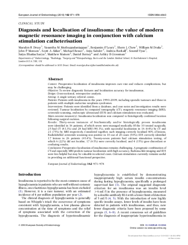
localised the lesion (75%) (example shown in Fig. 1).
Four MRI scans (14.2%) failed to localise the lesion.
However, three scans localised a lesion incorrectly.
The pragmatic use of the combined approach ensured
that 24 of the lesions were localised correctly by
imaging prior to surgery (80%). Five lesions were
correctly localised on MRI but were not visible on CT
scanning, while two lesions were visible on CT scanning
but not detected by MRI (although in one of these
cases, MRI localisation was hampered by marked
gut-movement artefact).
Diagnosis and localisation of insulinoma
975
Re-analysis of the data, to include cases which were
biochemically diagnosed but not histologically proven,
also encompasses cases that were managed medically.
The rationale for this is that the absence of localisation
is likely to have played a part in the withholding of
surgery. Excluding from this the patients with widespread metastases at the time of diagnosis (for whom
localisation was evident), the data demonstrated for CT
a corrected localisation accuracy of 16 cases out of 28
(57%) and for MRI a corrected detection rate of 21 cases
out of 31 patients (66%).
Nuclear medicine Twenty patients had radiolabelled
octreotide scans performed. Of these, ten scans (50%)
were negative (50%) and ten scans (50%) were positive.
Five of the positive scans localised a lesion concordant
with the final surgical location, while five were not
useful predictors of the final surgical location. Octreotide scanning had been carried out in three of the four
cases in which cross-sectional imaging (CT and MRI)
was unable to localise the lesion, and in none of these
cases did the nuclear medicine imaging add further
diagnostic information.
Endoscopic ultrasound Data were available on a total
of 26 EUS scans performed to localise the insulinoma.
Where cross-sectional imaging was also available, the
results would have been available to the endoscopist
prior to the procedure. Seventeen of these procedures
correctly localised a lesion (65.3%) but in five cases no
lesion was seen (19.2%); in addition, there were a
further four cases in which a lesion was correctly
identified but in which a further lesion was also noted,
not subsequently confirmed at operation (false
positives), resulting in some confusion (15.3%). EUS
had been carried out in three of the four cases in which
cross-sectional imaging (CT and MRI) was unable to
localise the lesion. In one of these cases, EUS was also
negative; in one case, the EUS demonstrated a lesion
concordant with the final surgical localisation, and in
one case, the EUS demonstrated a lesion that was in a
pancreatic region discordant with the final surgical
localisation.
Figure 1 Example of the use of MRI in the localisation of insulinoma.
(a) T2-weighted image demonstrating a lesion in the tail of the
pancreas, less well seen in (b) T1-weighted MRI demonstrating
the pancreas in the same patient.
Calcium stimulation catheter Twenty-seven patients
had calcium stimulation testing, although two of these
were incomplete as it was not technically possible
to cannulate one of the arteries. Of the studies, six gave
a non-regionalising response to calcium injection
(22.2%); 17 of the findings were concordant with
the final histological localisation (62.9%). However,
four studies (14.8%) gave discordant results with the
final regionalisation of the lesion being different to
the territory of insulin rise or unhelpful. No obvious
technical difficulty with these particular studies
was noted.
For the six patients in whom imaging failed to
confirm localisation of the insulinoma, in three cases
calcium stimulation provided additional helpful
www.eje-online.org
Downloaded from Bioscientifica.com at 12/04/2021 09:36:04PM
via free access
Percentage cases
976
80
70
60
50
40
30
20
10
0
M R Druce and others
EUROPEAN JOURNAL OF ENDOCRINOLOGY (2010) 162
Localisation of insulinoma – all techniques
Octreotide
EUS
Correct localisation
Catheter
No localisation
CT
MRI
Incorrect localisation
information for regionalisation. In three further cases,
the calcium stimulation test did not provide additional
value, or indeed confused the situation further.
Localisation information is summarised in Fig. 2. No
significant adverse events were recorded as a consequence of calcium stimulation testing.
Discussion
The classical diagnostic test for insulinoma has been
provocation of hypoglycaemia by a supervised fast
which may last up to 72 h. However, there is increasing
economic pressure to demonstrate Whipple’s triad
without a 3-day admission, and reports from some
centres have suggested that up to 83% of patients with
subsequent insulinoma diagnoses are hypoglycaemic
within the first 22 h of the fast, enabling the procedure
to be carried out under out-patient supervision (21).
The majority of our patient cohort required a fast
O24 h; this may reflect in part the less stringent criteria
for termination of the fast applied in other cohorts, such
as 3.3 and 2.75 mmol/l (21). For the cohort of patients,
increasingly recognised, with strong post-prandial
symptoms but a negative fast (around 5% of patients),
a mixed-meal test does appear to be the most
physiological investigation (9).
Once the biochemical diagnosis of a probable
insulinoma has been established, localisation
investigations can greatly aid management decisions.
Non-invasive methods such as abdominal ultrasound,
CT scanning and MRI have the advantage of being
simple and quick to perform with few potential
complications. However, non-functional pancreatic
nodules may occur incidentally, and these methods do
not corroborate structural abnormality with hormone
secretion (23). Several series have reviewed the accuracy
of these methods for localising the lesion; for example, in
a recent series of 237 patients, the accuracy of CT was
55% and of MRI was 42% in tumour localisation (21).
In another series of 39 sporadic cases of insulinoma, the
accuracy of CT scanning was 35% with a false negative
rate of 49% and a false positive localisation rate of 16%.
For MRI, the accuracy was 30% with a false negative
rate of 57% and a false positive rate of 13% (20). While
some small radiological series have suggested greater
Figure 2 Summary of localisation accuracy
for insulinoma in our study population
(nZ30 treated surgically).
sensitivity for both CT and MRI (13, 15), in general, the
low accuracy of cross-sectional imaging has in part
driven the development of more invasive methods.
The differences in the series may relate to the relatively
small size of many insulinomas at the time of diagnosis.
Indeed, in our series, nearly 80% of histologically
proven tumours were !2 cm in diameter, broadly
compatible with other series. Interestingly, however, in
our group of patients, the accuracy of cross-sectional
imaging for localisation was higher, with 65% accuracy
for CT and 74% for MRI. This is comparable to another
recent small UK series of 20 patients (10), in which the
sensitivity of MRI was noted to be 71%. However, in this
study, when the accuracy of localisation by this method
was evaluated, the accuracy was only 56%. In a further
UK series of 28 patients, the tumour detection rate for
MRI was also 71% (12). In our series, when both CT and
MR modalities were used together as part of a pragmatic
approach in which almost all patients with a sporadic
tumour were investigated, 80% of patients with a
confirmed histological diagnosis had their insulinoma
correctly localised.
EUS was introduced as a more invasive procedure
which, while also being an anatomical rather than a
functional technique, with the advent of biopsy via the
endoscope can in some cases provide histological
information. The accuracy in one large series of
insulinoma patients was calculated to be 75% (21).
However, this modality is highly operator dependent. In
our series, 80.7% of the tumours were localised. This
included 13.3% of the total who had confusing results
in that in addition to the lesion correctly identified, a
further lesion was incorrectly noted elsewhere in the
pancreas. In addition, there were 24% false negative
localisations. In the case of radiolabelled octreotide
scanning, the octreotide uptake prevalence of 50% is
less operator dependent, but instead the low localisation
rate is contingent upon the majority of tumours lacking
a high density of somatostatin receptors. Our findings
are concordant with most of the published literature
(24). The relative frequency of confusing results could
suggest that this is a tool best kept in reserve for cases in
which localisation is proving difficult, and requires the
maximum possible weight of evidence, but even in such
cases in our series, octreotide scanning added little in
terms of localisation. More recent literature suggests
www.eje-online.org
Downloaded from Bioscientifica.com at 12/04/2021 09:36:04PM
via free access
Diagnosis and localisation of insulinoma
EUROPEAN JOURNAL OF ENDOCRINOLOGY (2010) 162
additional sensitivity with the use of 18F-DOPA PET
scanning (25), while preliminary evidence suggests a
role for scanning with a radiolabelled GLP-1 analogue
exendin (26, 27).
Selective pancreatic intra-arterial calcium injections
to localise islet cell tumours were introduced in the
1980s (18). The success of the investigation relies on
several assumptions, including that the tumour will
have a dominant arterial supply, that calcium evokes a
characteristic response of coordinated discharge of
vesicles from the entire syncytium of tumour cells,
and that the remainder of the b-cells will be suppressed
from the chronic hormone hypersecretion. As well as
the theoretical advantage of corroboration of structure
and function, accuracy rates are reported to be very
high, with figures of 93% (21) and 89% (20) being
quoted. However, it should be pointed out that to be
accurate this is regionalisation rather than true
localisation. A number of series have reported the
additional value of this test in localisation, particularly
in cases where non-invasive methods have been
unsuccessful (12) Our series provides some cautionary
evidence that accuracy is not always 100%, and given
that the procedure is not without risk, there may be
merit in reserving this investigation for cases where
non-invasive localisation results have been contentious.
Given the relatively high accuracy for MRI in our series,
prospective studies evaluating radiological confidence
in the findings against the final outcome may allow
prediction of which patients require invasive investigations in addition. This may in turn reduce the
radiation exposure to the patients and also the overall
cost of the diagnostic episode (10).
The results from the MEN patients are difficult to
compare due to the small numbers within the cohort. In
general, many such patients present with incidental
findings of pancreatic nodules and undergo one or more
prior pancreatic resections, making further preoperative
localisation strategies challenging. For this group of
patients, the conclusions regarding insulinoma localisation in sporadic cases are unlikely to be suitable for
extrapolation.
In summary, while the diagnosis of an insulinoma
has become increasingly consensual, and diagnostic
algorithms are well established, preoperative localisation remains difficult. Individual institutions have
developed particular expertise in specific techniques,
such as EUS, but these may not readily transfer to other
centres where such techniques are less commonly used.
We and others have increasingly come to rely on the
calcium stimulation catheter technique as the final
arbiter, and indeed, it is one of the most useful means of
functional regionalisation. However, our present series
demonstrates that it too is not always accurate, and we
have been impressed at the increasing usefulness
of cross-sectional imaging, especially recent trends in
MRI. We suggest that increasing confidence with MRI,
novel sequences including diffusion-weighted imaging
977
and novel software programs, will lead to this technique
being used as an anatomical localisation technique
complementary to functional imaging with calcium
stimulation.
Declaration of interest
The authors declare that there is no conflict of interest that could be
perceived as prejudicing the impartiality of the research reported.
Funding
This research did not receive any specific grant from any funding
agency in the public, commercial or not-for-profit sector.
References
1 Service FJ. Diagnostic approach to adults with hypoglycemic
disorders. Endocrinology and Metabolism Clinics of North America
1999 28 519–532, vi.
2 Service FJ, McMahon MM, O’Brien PC & Ballard DJ. Functioning
insulinoma – incidence, recurrence, and long-term survival
of patients: a 60-year study. Mayo Clinic Proceedings 1991 66
711–719.
3 Service FJ & Natt N. The prolonged fast. Journal of Clinical
Endocrinology and Metabolism 2000 85 3973–3974.
4 Service FJ, Dale AJ, Elveback LR & Jiang NS. Insulinoma: clinical
and diagnostic features of 60 consecutive cases. Mayo Clinic
Proceedings 1976 51 417–429.
5 Service FJ. Hypoglycemic disorders. New England Journal of Medicine
1995 332 1144–1152.
6 Vezzosi D, Bennet A, Fauvel J, Boulanger C, Tazi O, Louvet JP &
Caron P. Insulin levels measured with an insulin-specific assay in
patients with fasting hypoglycaemia related to endogenous
hyperinsulinism. European Journal of Endocrinology 2003 149
413–419.
7 Chia CW & Saudek CD. The diagnosis of fasting hypoglycemia
due to an islet-cell tumour obscured by a highly specific insulin
assay. Journal of Clinical Endocrinology and Metabolism 2003 88
1464–1467.
8 Vezzosi D, Bennet A, Fauvel J & Caron P. Insulin, C-peptide and
proinsulin for the biochemical diagnosis of hypoglycaemia related
to endogenous hyperinsulinism. European Journal of Endocrinology
2007 157 75–83.
9 Cryer PE, Axelrod L, Grossman AB, Heller SR, Montori VM,
Seaquist ER, Service FJ & Endocrine Society. Evaluation and
management of adult hypoglycemic disorders: an Endocrine
Society Clinical Practice Guideline. Journal of Clinical Endocrinology
and Metabolism 2008 94 709–728.
10 Ravi K & Britton BJ. Surgical approach to insulinomas: are preoperative localisation tests necessary? Annals of the Royal College of
Surgeons of England 2007 89 212–217.
11 Pasieka JL, McLeod MK, Thompson NW & Burney RE. Surgical
approach to insulinomas. Assessing the need for preoperative
localisation. Archives of Surgery 1992 127 442–447.
12 Morganstein DL, Lewis DH, Jackson J, Isla A, Lynn J, Devendra D,
Meeran K & Todd JF. The role of arterial stimulation and
simultaneous venous sampling in addition to cross-sectional
imaging for localisation of biochemically proven insulinoma.
European Journal of Radiology 2009 19 2467–2473.
13 Gouya H, Vignaux O, Augui J, Dousset B, Palazzo L, Louvel A,
Chaussade S & Legmann P. CT, endoscopic sonography, and a
combined protocol for preoperative evaluation of pancreatic
insulinomas. AJR. American Journal of Roentgenology 2003 181
987–992.
www.eje-online.org
Downloaded from Bioscientifica.com at 12/04/2021 09:36:04PM
via free access
978
M R Druce and others
14 Ichikawa T, Peterson MS, Federle MP, Baron RL, Haradome H,
Kawamori Y, Nawano S & Araki T. Islet cell tumour of the
pancreas: biphasic CT versus MR imaging in tumour detection.
Radiology 2000 216 163–171.
15 Thoeni RF, Mueller-Lisse UG, Chan R, Do NK & Shyn PB. Detection
of small, functional islet cell tumours in the pancreas: selection of
MR imaging sequences for optimal sensitivity. Radiology 2000 214
483–490.
16 McLean AM & Fairclough PD. Endoscopic ultrasound in
the localisation of pancreatic islet cell tumours. Best Practice &
Research. Clinical Endocrinology & Metabolism 2005 19 177–193.
17 Chang F, Chandra A, Culora G, Mahadeva U, Meenan J &
Herbert A. Cytologic diagnosis of pancreatic endocrine tumours by
endoscopic ultrasound-guided fine-needle aspiration: a review.
Diagnostic Cytopathology 2006 34 649–658.
18 Doppman JL, Miller DL, Chang R, Shawker TH, Gorden P &
Norton JA. Insulinomas: localisation with selective intraarterial
injection of calcium. Radiology 1991 178 237–241.
19 Doppman JL, Chang R, Fraker DL, Norton JA, Alexander HR,
Miller DL, Collier E, Skarulis MC & Gorden P. Localisation
of insulinomas to regions of the pancreas by intra-arterial
stimulation with calcium. Annals of Internal Medicine 1995 123
269–273.
20 Guettier JM, Kam A, Chang R, Skarulis MC, Cochran C,
Alexander HR, Libutti SK, Pingpank JF & Gorden P. Localisation
of insulinomas to regions of the pancreas by intraarterial calcium
stimulation: the NIH experience. Journal of Clinical Endocrinology
and Metabolism 2009 94 1074–1080.
21 Placzkowski KA, Vella A, Thompson GB, Grant CS, Reading CC,
Charboneau JW, Andrews JC, Lloyd RV & Service FJ. Secular trends
in the presentation and management of functioning insulinoma
EUROPEAN JOURNAL OF ENDOCRINOLOGY (2010) 162
22
23
24
25
26
27
at the Mayo Clinic, 1987–2007. Journal of Clinical Endocrinology
and Metabolism 2009 94 1069–1073.
Owen NJ, Sohaib SA, Peppercorn PD, Monson JP, Grossman AB,
Besser GM & Reznek RH. MRI of pancreatic neuroendocrine
tumours. British Journal of Radiology 2001 74 968–973.
Service FJ. Classification
of hypoglycemic disorders.
Endocrinology and Metabolism Clinics of North America 1999
28 501–517, vi.
Virgolini I, Traub-Weidinger T & Decristoforo C. Nuclear medicine
in the detection and management of pancreatic islet-cell tumours.
Best Practice & Research. Clinical Endocrinology & Metabolism 2005
19 213–227.
Kauhanen S, Seppanen M, Minn H, Gullichsen R, Salonen A,
Alanen K, Parkkola R, Solin O, Bergman J, Sane T, Salmi J,
Valimaki M & Nuutila P. Fluorine-18-L-dihydroxyphenylalanine
(18F-DOPA) positron emission tomography as a tool to localize
an insulinoma or beta-cell hyperplasia in adult patients.
Journal of Clinical Endocrinology and Metabolism 2007 92
1237–1244.
Wild D, Macke H, Christ E, Gloor B & Reubi JC. Glucagonlike peptide 1-receptor scans to localize occult insulinomas.
New England Journal of Medicine 2008 359 766–768.
Christ E, Wild D, Forrer F, Brändle M, Sahli R, Clerici T, Gloor B,
Martius F, Maecke H & Reubi JC. Glucagon-like peptide-1 receptor
imaging for localisation of insulinomas. Journal of Clinical
Endocrinology and Metabolism 2009 94 4398–4405.
Received 24 February 2010
Accepted 4 March 2010
www.eje-online.org
Downloaded from Bioscientifica.com at 12/04/2021 09:36:04PM
via free access
RELATED PAPERS
Revista Ceres
Symptomatology associated with “Purple top”, an emerging disease of solanaceous fruit species2022 •
Medical Journal Armed Forces India
Hyperperfusion Syndrome after Carotid Artery Stenting2015 •
Annals of Emergency Medicine
Getting Lay Rescuers to Use Public Access Defibrillators2011 •