D A I R Y FOODS TECHNICAL NOTES
Separation of Casein Micelles from Milk for Rapid Determination
of Casein Concentration I
R O B E R T N. C A R P E N T E R 2 and R O D N E Y J. B R O W N
Department of Nutrition and Food Sciences
Utah State University
Logan 84322
ABSTRACT
Size exclusion chromatography was
used to separate casein micelles from
whey protein in milk for rapid measurement of casein. Casein concentration
was measured by size exclusion separation
monitored by ultraviolet spectroscopy
and by acid precipitation followed by
Kjeldahl estimation of protein. The
correlation coefficient between the two
methods was .92. Effects of temperature,
pH, and calcium on separation were
evaluated. Addition of calcium was used
to incorporate all casein into micelles
before separation.
INTRODUCTION
Cheese producers have begun using yield
formulas to calculate the price paid to producers
for milk (6, 8). Both fat and casein percentages
in milk must be known to predict accurately
cheese yield. Casein concentration is estimated
routinely as a percentage of total protein
because protein percentage is obtained more
easily and rapidly than casein percentage.
Casein concentration as a percentage of total
milk protein (casein number) is variable (3, 15,
20). Anything that influences percent casein,
percent whey proteins, or percent noncasein
nitrogen affects casein number, so accuracy of
cheese yield predictions based on estimated
casein is questionable.
Casein micelles are spherical, highly hydrated,
and sponge-like (5) protein particles suspended
in milk. Ninety-five percent of casein micelles
in milk are between 40 and 22 nm in diameter
(4, 17). A small micelle of 250,000 daltons is
Received June 12, 1984.
1Utah State Agricultural Experiment Station
Journal Article No. 2984. Approved by the Director.
2Kraft Inc., Box T, Albany, MN 56307.
1985 J Dairy Sci 68:307--311
307
relatively large compared to whey proteins,
which are from 15,000 to 70,000 daltons.
Immunoglobulins are larger, 150,000 to 300,000
daltons, but exist in low concentrations (about
4% of total protein) in milk (10).
Calcium is important in structural stability
of casein micelles (2). Casein micelles dissociate
as calcium ion activity is reduced. Small additions of calcium ions cause transfer of soluble
casein to micelles without changing micelle
radii, and addition of more calcium ions causes
larger micelles to form (4).
Casein usually is separated from whey
proteins before measurements can be made.
Casein then may be determined by protein
measured in the casein fraction or the difference
in percent protein between milk and whey. Size
exclusion chromatography separates molecules
in solution according to size (22) and has been
used in the study of physiical and chemical
properties and composition of casein miceIles
(7, 11, 16, 18, 21).
This study was to determine if size exclusion
chromatography could be used to separate
casein micelles from whey proteins for rapid
casein determination. Casein micelles separated
in this way remain in suspension and allow
direct casein measurement b y various protein
assays. An ultraviolet spectrophotometer operating at 280 nm wavelength was used to measure
protein. Infrared spectroscopy would be ideally
suited for these measurements and would make
rapid casein testing possible on instruments
now being used by the dairy industry.
MATERIALS
AND METHODS
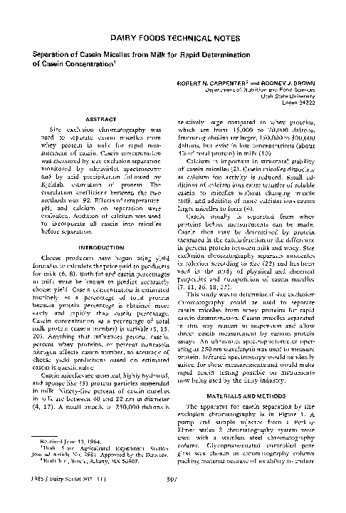
The apparatus for casein separation by size
exclusion chromatography is in Figure 1. A
pump and sample injector from a PerkinElmer series 2 chromatography system were
used with a stainless steel chromatography
column. Glycophase-coated controlled pore
glass was chosen as chromatography column
packing material because of its ability to endure
�308
C A R P E N T E R AND BROWN
Eluent
Pump
~
Sample
Column
Computer
Monitor
I
Figure 1. Flow diagram of equipment for separating
casein micelles from whey protein and monitoring
elution.
faster flow rates and higher pressures than gels.
Pore diameter and particle size were selected to
separate casein micelles from whey proteins.
Both 2,000 and 4,000 nm pore diameter, 37 to
74 /am diameter beads were evaluated. The
2,000 nm beads were chosen because of better
separation of casein and whey protein. Separation was possible in columns as short as 15 cm;
however, a 100-cm column was used in the
experiments described in this paper. Inside
diameter of the column is not a critical separation characteristic but was selected at .4 cm so
small sample sizes (10 to 150/al) could be used.
The column flow rate was held constant at 1 to
6 ml of 40 mM CaC12/min. Before measurement,
the system was equilibrated by passing several
milk samples through the column. A Beckman
DU-8B spectrophotomer was used to monitor
protein at 280 nm as it eluted from the column.
The contribution of light scattering does not
interfere with the usefulness of the readings. At
4-s intervals absorbances were sent from the
spectrophotometer to a Tektronix 4052 computer, which produced an elution plot and
stored data on tape to be evaluated later.
The reference method for determining casein
concentration in milk consisted of separation of
casein by acid precipitation followed by Kjeldahl
nitrogen determination of the precipitate (1,
19). Sample size and solutions of the separation
step were reduced to 1/20th so that microKjeldahl procedures could be followed. A
Journal of Dairy Science Vol. 68, No. 2, 1985
solution of 10% mercuric sulfate was used as
catalyst. Boric acid solution containing methyl
red/bromcresol green indicator and the ammonia
distillate were titrated with .0258 34 HCI to a
pink-grey end point. Percent casein was obtained
by percent nitrogen, minus reagent blank,
multiplied by 6.52 (12).
Whole milk samples from individual cows
had potassium dichromate tablets added at the
farm as preservative. Samples were filtered
through Whatman No. 1 filter paper and forced
twice at 40°C through a small sample homogenizer. Each 50-ml sample was allowed to
equilibrate for 24 h at 4°C. Some samples had
.1 ml of 5 M CaClz added before equilibration.
This could be added as a tablet at the farm
along with the preservative tablet. Before
injection, samples were held at 40°C for at least
2 h. A typical elution plot of milk produces
two peaks (Figure 2). The computer was used
to calculate the volume at which each peak
eluted, maximum absorbance reading of each
peak, and area under the curve according to
Simpson's approximation. Area of the first
peak was calculated from base line to a point
midway between the two peaks. Area of the
second peak was calculated from the same
midpoint to a point after the second peak at
which the curve returned to the baseline. All of
these measures were stored on computer tape
and later used in statistical analyses.
Casein was obtained by precipitation from
milk with 1 N HC1. The precipitate was redissolved in simulated milk ultrafiltrate (13).
A solution of a-lactalbumin and fl-lactoglobulin
also was prepared. Both of these solutions were
injected into the column to determine their
elution rates.
Effluent containing casein and whey protein
peaks collected from the column was analyzed
by polyacrylamide gel electrophoresis following
the procedure used by Heth and Swaisgood
(11). The samples were dried in a vacuum oven
at 50°C. Modified Poulik's buffer (50 pl) was
added to each sample. Mercaptoethanol (100/al
of 1:9 dilution) was added, and samples were
refrigerated for 2 h. Dye marker, acrylamide
solution, preserving, and staining solutions were
prepared according to LKB Application Note
306 (9). Buffer solution and gel solutions were
made according to Kiddy (14). The gel was
prepared and applied to a flat bed electrophoresis unit at least 12 h before use. Samples
�DAIRY FOODS TECHNICAL NOTE
4.0
3.S
3.0
j
2.5
2.0
-
dL
6
,S
I
IX
10
VOLUME CmL~,
4.0
$.5
2.S
~
2.0
i
1.0
.5
added, the eluent was distilled water. Samples
were introduced randomly into the column,
except that calcium-treated samples were run
separately from untreated samples. Thirty six
elution plots were obtained from which characteristics were calculated. Characters were
evaluated by analysis of variance to determine each treatment effect.
Casein percentage was estimated from
triplicate elutions of eight milk samples from
individual cows. Fat and protein percentages
for each sample were obtained with a Multispec
infrared instrument. Casein percentage was
obtained by the standard acid precipitation
and Kjeldahl nitrogen method. Measures from
column elution and fat and protein percentages
were tested for correlation with percent casein.
Analysis of covariance and stepwise regression
were performed, and a regression equation to
estimate casein from elution measures was
derived.
RESULTS A N D DISCUSSION
0
-
309
,S
(!
i
i
6
I0
IE
VOLUME On,L)
Figure 2. Characteristic protein elution plots of
nontreated (top) and calcium treated (bottom) milk
samples.
(10/21) were injected into gel slits and 20 m A of
current was applied for 15 rain; then voltage
was maintained at 200 V for about 4 h.
Changes of the elution pattern caused by
pH, temperature, and addition of calcium were
evaluated. A factorial design experiment
used a single commingled milk sample. Treatments were run through the column in duplicate.
Temperature treatments were: milk injected
cold at 4°C, milk held at 40°C for at least 2 h
before injection, and milk heated to 72°C for 1
rain then cooled to 40 C. The pH treatments
were milk at pH 6.58 (no treatment), pH 6.7
(100/21 1 N NaOH/50 ml), and pH 6.4 (50/21 1
N HC1/50 ml). Each temperature and pH
combination also was run with calcium (100/21
CaCt2/50 ml milk) and without calcium. When
calcium was added to milk, the eluent was a 40
mM CaC12 solution; when calcium was not
A peak appeared at 5.1 ml and a slight rise in
absorbance near 10 ml when casein was passed
through the size exclusion column (Figure 3).
The larger peak contained 97% of the total area
under the elution curve and consisted of casein
micelles. The small peak contained whey
protein not separated by casein precipitation or
nonmicellar casein. A solution of two whey
proteins (~-lactalbumin and /3-1actoglobulin)
also was eluted from the column (Figure 3).
These proteins both eluted at 10.2 ml. The two
peaks of an elution of milk are in the same
places as these casein aggregate and whey
protein peaks (Figures 2 and 3).
Electrophoresis patterns of proteins in the
first peak of elution with or without added
calcium had dark casein bands and no whey
protein bands. Electrophoresis of proteins in
the whey protein peak from samples to which
calcium had been added displayed clearly
visible whey protein bands and faint casein
bands.
Calcium treatment had significant (~<.01)
effects on elution volumes of casein and whey
protein peaks, maximum absorbances of casein
and whey protein peaks, areas of casein and
whey protein peaks, ratio of casein peak
maximum absorbance to whey protein peak
maximum absorbance, and casein peak area as a
Journal of Dairy Science Vol. 68, No. 2, 1985
�310
CARPENTER A N D BROWN
I.S
.S
-
WHEY PRO'rEZNS
I
I~
.S
I
18
IK
I
18
l&
VOLUME ( m L )
I.S
CASEIN
.S
-
I
G
.5
VOLUPIE C m L )
Figure 3. Protein elution curves of a-lactalbumin
and t3-1actoglobulin (top) and casein (bottom).
90-
80-
percentage of total area under the curve.
Calcium treatment did n o t have a significant
effect (a>.01) on the sum of maximum absorbances of both peaks or on the sum of the
areas of both peaks. The casein peak eluted
earlier, and the whey protein peak etuted later
when calcium was added to the sample, Also,
casein peaks increased in area and maximum
absorbance, and whey protein peaks decreased
in area and maximum absorbance. A comparison
between casein peak areas as percentages of
total areas under elution curves with respect to
calcium addition and temperature treatments is
in Figure 4. Calcium addition forced soluble
casein into micetles so that temperature had less
effect when calcium was added. Casein in the
first peak as a percentage of total protein was
between 78 and 80% when calcium was added,
irrespective of temperature treatment. This is
approximately the casein number of normal
milk (3). The pH did not have a significant
effect in any of the analyses of variance. This
may be partly, due to the small pH range
studied.
Eight samples of milk from individual cows
ranging in fat from 2.1 to 3.85% and in protein
from 2.19 to 4,42% were run through the
column. Elution plots were obtained, and
characteristics of each plot were calculated. The
NO CALCIUM
CALCIUM ADDED
2.6-
~,
2.4-
~
~-~\ \ - ~
%\\~,
\ \ "-,\ \ \
~\\-.~
i~ ~k kk
kk k k kh
0
70 -
\\ \\ \\
I ~\\~
I
......
\ \ \ \ \\
- -
--~
z
2-
i ~\\%
~\\~
~\\~
%\\%
4
2.2-
/
\\ \\ ~
C,O ~
!
%\\'%
40
TEMPERATURE
7Z
(C)
Figure 4. Calcium addition and temperature
treatment effects on percent area of first (casein)
peak.
Journal of Dairy Science Vol. 68, No. 2, 1985
//
1.8t
1.6
t6
t
1.8
i
2
i
2.2
I
2.4
' - - I
2.6
ESIIMATEDCASEINcolumn
Figure 5. Correlation of column and standard
methods of casein estimation in milk.
�DAIRY FOODS TECHNICAL NOTE
c o r r e l a t i o n c o e f f i c i e n t o f casein w i t h m a x i m u m
a b s o r b a n c e o f t h e first p e a k was .77 a n d w i t h
t h e area u n d e r t h e first p e a k was .65. C o r r e l a t i o n
coefficients of casein w i t h m a x i m u m a b s o r b a n c e
a n d area of t h e s e c o n d p e a k were .03 a n d .25.
T h e r e l a t i o n s h i p b e t w e e n p e a k area a n d maxim u m a b s o r b a n c e o f t h e first peak w i t h p e r c e n t
casein was d e m o n s t r a t e d b y analysis o f covariance. Stepwise regression i n d i c a t e d t h a t area
a n d m a x i m u m a b s o r b a n c e of t h e first p e a k
e s t i m a t e d casein w i t h a c o r r e l a t i o n c o e f f i c i e n t
o f .92. E s t i m a t i o n o f casein b y this regression
e q u a t i o n is p l o t t e d versus s t a n d a r d casein
m e a s u r e m e n t s in Figure 5.
CONCLUSIONS
G l y c o p h a s e c o a t e d p o r o u s glass b e a d s w i t h
37 to 7 4 / 2 m particle size a n d 2 0 0 0 n m p o r e
d i a m e t e r in a 1 0 0 b y .4 cm c o l u m n can b e u s e d
to separate casein micelles f r o m w h e y p r o t e i n s
b y size e x c l u s i o n c h r o m a t o g r a p h y . Best separat i o n o f t o t a l casein f r o m w h e y p r o t e i n in milk
was a c h i e v e d b y a d d i t i o n o f 100 /21 o f 5 M
CaC12/50-ml sample 24 h b e f o r e testing. This
could b e a d d e d as a t a b l e t at t h e same t i m e as
preservative. Casein c a n b e e s t i m a t e d f r o m
m a x i m u m a b s o r b a n c e a n d u r e a o f t h e first peak
e l u t e d w h e n m o n i t o r i n g at a w a v e l e n g t h o f 2 8 0
nm. S e p a r a t i n g casein micelles f r o m w h e y
p r o t e i n s w i t h o u t c o a g u l a t i o n is a d v a n t a g e o u s
for f u r t h e r t e s t i n g of t h e casein f r a c t i o n . Using
t h e s e p a r a t i o n m e t h o d d e s c r i b e d in t h i s paper,
casein m a y be m e a s u r e d d i r e c t l y b y i n f r a r e d
milk testing i n s t r u m e n t s .
ACKNOWLEDGMENTS
T h e a u t h o r s t h a n k A. A. H e t h f o r assistance
with the electrophoresis.
REFERENCES
1 Association of Official Analytical Chemists. 1980.
Official and tentative methods of analysis. 13th ed.
Assoc. Of tic. Anal. Chem., Washington, DC.
2 Bingham, E. W., H. M. Farrell, Jr., and R. J.
Carroll. 1972. Properties of dephosphorylated
asl-casein. Biochemistry 11:2450.
3 Blake, R. W., I. B. Nmai, and R. L. Richter. 1980.
Relationship between distribution of major milk
proteins and milk yield. J. Dairy Sci. 63:141.
4 Bloomfield, V. A., C. V. Morr. 1973. Structure of
casein micelles: physical methods. Neth. Milk
Dairy J. 27:103.
5 Bloomfield, V. A., and R. J. Mead, Jr. 1975.
Structure and stability of casein micelles. J. Dairy
311
Sci. 50:592.
6 Brown, R. J. 1981. Computerized cheese yield
pricing of milk. 2nd Bienn. Marschall Int. Cheese
Conf, Madison, WI.
7 Eckstrand, B., M. Larsson-Raznikiewicz, E. Brannang, and C. Swensson. 1981. Size distribution of
casein micelles related to coagulation properties: A
comparison between different breeds of cattle.
Swed. J. Agric. Res. 11:57.
8 Ernstrom, C. A. 1980. A workable pricing system
based on cheese yield. Utah State Univ. 4th Bienn.
Cheese Ind. Conf., Logan.
9 Fehrnstrom, H., and V. Moberg. 1977. SDS and
conventional polyacrylamide gel electrophoresis
with LKB 2117 multiphor. Application note 306.
LKB Producter AB, Bromma, Sweden.
10 Gordon, W. G., and E. B. Kalan. 1974. Proteins of
milk. Pages 8 7 - 1 2 4 in Fundamentals of dairy
chemistry. 2nd ed. B. H. Webb, A. H. Johnson, and
J. A. Alford, ed. Avi Publ. Co., Inc., Westport, CT.
11 Heth, A. A., and H. E. Swaisgood. 1982. Examination of casein micelle structure by a method for
reversible covalent immobilization. J. Dairy Sci.
65 : 2047.
12 Jenness, R. 1970. Protein composition of milk.
Milk proteins. Vol. 1. H. A. McKenzie, ed. Academic
Press, New York, NY.
13 Jenness, R., and J. Koops. 1962. Preparation of a
salt solution which simulates milk ultrafiltrate.
Neth. Milk Dairy J. 16:153.
14 Kiddy, C. A. 1974. Gel electrophoresis in vertical
polyacrylamide beds. Pages 1 4 - 1 5 in Methods of
gel electrophoresis of milk proteins. H. E. Swaisgood, ed. Dep. Food Sci., North Carolina State
Univ., Raleigh.
15 Larson, B. L., G. D. Rolleri, and K. A. Kendall.
1956. Protein production in the bovine. Comparison
of daily protein, fat, and milk production during
the entire lactation period. J. Dairy Sci. 39:204.
16 McGann, T.C.A., R. D. Kearney, and W. J. Donnelly.
1979. Developments in column chromatography
for the separation and characterization of casein
micelles. J. Dairy Res. 46: 307.
17 McMahon, D. J., and R. J. Brown. 1984. Composition, structure and integrity of casein micelles:
A review. J. Dairy Sci. 67:499.
18 Ono, T., S. Odagiri, and T. Takagi. 1983. Separation
of submicelles from micellar casein by high performance liquid chromatography on a TSK-Gel
G40OOSW column. J. Dairy Res. 50:37.
19 Rowland, S. J. 1938. The determination of the
nitrogen distribution in milk. J. Dairy Res. 9:42.
20 Waite, R., J.C.D. White, and A. Robertson. 1956.
Variations in the chemical composition of milk
with particular reference to solids-not-fat 1. The
effect of stage of lactation, seasOn on year and age
of cow. J. Dairy Res. 23:65.
21 Yaguchi, M., and D. Rose. 1971. Chromatographic
separation of milk proteins: a review. J. Dairy Sci.
54:1725.
22 Yau, W. W., J. J. Kirkland, and D. D. Bly. 1979.
Modern size exclusion liquid chromatography:
Practice of gel permeation and gel filtration chromatography. John Wiley & Sons, Inc., New York,
NY.
-
-
Journal of Dairy Science Vol. 68, No. 2, 1985
�
 Rodney Brown