Revista Brasileira de Entomologia 66(2):e20210092, 2022
Ants associate with microlepidoptera galleries in leaves of Acrostichum danaeifolium
Langsd. & Fisch.
Marcelo Guerra Santos1* , Isabella Rodrigues Lancellotti1 ,
Gemagno Marinho Ribeiro1, Rennan Leite Martins Coutinho1,
Rodrigo Machado Feitosa2
1
2
Universidade do Estado do Rio de Janeiro, Faculdade de Formação de Professores, Departamento de Ciências, Laboratório de
Biodiversidade, São Gonçalo, RJ, Brasil.
Universidade Federal do Paraná, Departamento de Zoologia, Laboratório de Sistemática e Biologia de Formigas, Curitiba, PR, Brasil.
ARTICLE
INFO
Article history:
Received 25 August 2021
Accepted 20 April 2022
Available online 23 May 2023
Associate Editor: Lucas Kaminski
Keywords:
Biological interactions
Arthropods
Fern-insect interactions
Focal species
ABSTRACT
Acrostichum danaeifolium, a Neotropical fern, occurs preferentially in marshy areas or at the margins of lakes and
mangroves. Microlepidoptera larvae burrow through the petioles of the fern, preferentially on the non-expanded
leaves. The galleries in the petiole create a new microhabitat, harboring a rich fauna of arthropods. The aim of
the present study was to assess the richness of ants associated with its petiole. The study was conducted in a
population of A. danaefolium from the Atlantic Forest in Rio de Janeiro state, Southeastern Brazil. Six collections
were carried out every two months (2009-2010), three in the dry and three in the rainy season. The leaves were
divided into three development stages: non-expanded leaves (NEL), expanded leaves (EL) and senescent leaves
(SL). Seven leaves from each phase were randomly collected from seven individuals. A total of fifteen ant species
were recorded. The species with the highest frequency and density in fern petioles were Camponotus crassus
and Crematogaster curvispinosa. The highest ant richness and abundance was found in senescent leaves. The
high number of ants found in the petioles of Acrostichum danaefolium qualifies it as a potential key species in
the marshes and flooded areas where it occurs.
Introduction
Since ferns have no flowers, most researchers have long ignored
the potential of fern-animal interactions (Watkins Junior et al., 2008).
However, these interactions may occur via herbivory (Mehltreter, 2010),
with the presence of domatia (Gómez-Pignataro, 1974), leaf nectaries
(Koptur et al., 1982), crypticity (Santos and Wolff, 2015) and galls
(Santos et al., 2019a). Mutualistic (Jermy and Walker, 1975; GómezPignataro, 1977; Walker, 1986; Gay, 1993), antagonistic (Farias et al.,
2018) and commensal interactions (Mehltreter et al., 2003; Santos et al.,
2019b) have been recorded between ferns and ants. The ants have also
established poorly understood relationships with fern leaf nectaries
(Page, 1982; Koptur et al., 1982, 1998; Tempel, 1983; Heads and Lawton,
1984, 1985).
In ferns, there are few records of ants using the cavities produced
by microlepidoptera larvae on leaf petioles as shelter (Mehltreter et al.,
2003; Santos and Mayhé-Nunes, 2007), as well as on senescent galls
after the inducing insect hatch (Santos et al., 2019b). Despite their
*Corresponding author.
E-mail: marceloguerrasantos@gmail.com (M.G. Santos).
scarcity, studies demonstrate the importance of cavities and galleries
in the stems and petioles of plants, and fallen twigs on the soil as a
source of shelter and expansion for ant colonies (Fernandes et al., 2019).
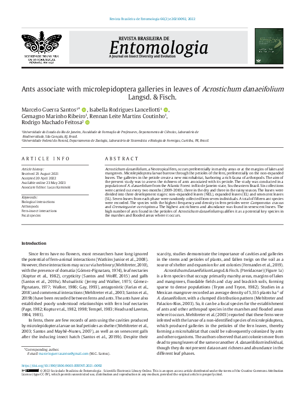
Acrostichum danaeifolium Langsd. & Fisch. (Pteridaceae) (Figure 1a)
is a fern species that occupy primarily marshy areas, margins of lakes
and mangroves, floodable fields and clay and brackish soils, forming
sparse to dense populations (Tryon and Tryon, 1982). Studies in a
Mexican mangrove recorded an average density of 5,555 plants ha-1 of
A. danaeifolium, with a clumped distribution pattern (Mehltreter and
Palacios-Rios, 2003). So, it can be a focal species for the establishment
of ants and other arthropod species in the marshes and flooded areas
where it occurs. Mehltreter et al. (2003) reported that these ferns were
infested with the larvae of a non-identified species of microlepidoptera,
which produced galleries in the petioles of the fern leaves, thereby
forming a microhabitat that could be subsequently colonized by ants
and other organisms. The authors observed that ant colonies move from
dead to young leaves of the same or another A. danaeifolium individual,
though they do not present data on ant richness and abundance in the
different leaf phases.
https://doi.org/10.1590/1806-9665-RBENT-2021-0092
© 2022 Sociedade Brasileira de Entomologia - Scientific Electronic Library Online. This is an open-access article distributed under the terms of the Creative Commons Attribution
License (type CC-BY), which permits unrestricted use, distribution and reproduction in any medium, provided the original article is properly cited.
�2-7
M.G. Santos et al. / Revista Brasileira de Entomologia 66(2):e20210092, 2022
Figure 1 A- The fern species Acrostichum danaeifolium, habit. B- Crozier (non-expanded leave). C- Petiole of the fern leaf with holes and galleries excavated by the microlepidoptera larvae. D- Longitudinal section of the petiole showing the microlepidoptera pupa inside. E- Microlepidoptera adult.
The present study aimed to analyze ant richness and abundance
associated with the petiole of a Brazilian population of the fern A.
danaeifolium, at different leaf phases (non-expanded, expanded, and
senescent) and seasons of the year (rainy and dry) in order to test ant
use regarding leaf stage and periodicity.
Material and methods
Study area
The study was based on a population of A. danaeifolium in a marsh
belonging to the Engenho Pequeno Environmental Protection Area
(APAEP), municipality of São Gonçalo, Rio de Janeiro state, Brazil (22º
50’ 55.74”S 43º 2’ 25.73”W). The APAEP encompasses several Atlantic
Forest fragments, at an altitude above 75m and different stages of
ecological succession, with a total area of 10.05 km2 (Santos and
Pinto, 2006). According to the classification of Veloso et al. (1991)
and subsequent analysis by the Brazilian Institute of Geography and
Statistics (IBGE, 2012), this area is classified as a submontane dense
ombrophilous forest. The climate is type AW, with the driest period
between May and October and the rainy season occurring from
November to April. Average annual temperature, relative humidity
and precipitation are around 26ºC, 74% and 1,060mm, respectively
(Bertolino et al., 2016).
Collection and laboratory procedures
Collections were carried out every two months in different
individuals of the same population, between March 2009 and January
2010, totaling six collections, divided into the dry (May, July and
September) and rainy (November, January and March) seasons. Leaves
of A. danaeifolium were divided into three development stages: nonexpanded leaves (NEL), that is, those with the rachis fully expanded, but
the pinnae still curled; expanded (EL) and senescent leaves (SL) that
are characterized as dry, albeit still attached to the plant (Figure 1AB).
Seven leaves from each phase were randomly collected from seven
individuals, totaling 126 leaves. The number of non-expanded leaves
(crozier and expanding leaves) of each fern was accounted and
inspected for traces of microlepidoptera herbivory (galleries and
cavities). All non-expanded leaves with signs of microlepidoptera
herbivory were also counted.
Leaves sclerophylly or toughness is as important trait to evaluate the
preference of the herbivorous in leaf attack (Coley, 1983). The petiole
sclerophylly was quantified by the specific dry leaf weight per unit
area (Choong et al., 1992). Petiole samples with 4cm long of NEL and
EL leaves were taken. The volume (unit area in cm3) was calculated by
the following equation: πr2h, where r= petiole radius and h= petiole
height. After that the petioles were oven dried and their weight (g)
noted. The petiole sclerophylly (S) was expressed by g/cm3.
Leaves (NEL, EL, SL) were packed in plastic bags and the material was
screened in the laboratory. Petioles were carefully cut with razor blades
�M.G. Santos et al. / Revista Brasileira de Entomologia 66(2):e20210092, 2022
in the search for microlepidoptera (larva and pupa) and ants. All ants
were euthanized and fixed in 70◦GL alcohol. They were identified by Dr.
Rodrigo M. Feitosa, in the Laboratory of Ant Systematics and Biology at
Universidade Federal do Paraná. Botanical vouchers were deposited in the
herbarium of the Faculdade de Formação de Professores da Universidade
do Estado do Rio de Janeiro (RFFP) and zoological vouchers in the Padre
Jesus Santiago Moure Entomological Collection, Universidade Federal
do Paraná, Department of Zoology (DZUP).
3-7
were found on twenty senescent leaves, nine expanded leaves and
one non-expanded leaves. The highest ant richness and abundance
also was found on senescent leaves (Table 3). There was a significant
difference in ant abundance between the dry and rainy seasons, with
the dry season exhibiting the highest abundance (χ2=7.629; P=0.022).
This difference was not found for ant richness (χ2=1.790; P=0.408)
(Table 3). The observed and estimated richness were similar in four
indicators (Table 3).
Statistical analyses
Data distribution was examined using the Shapiro-Wilk test.
The Kruskal-Wallis and Dunn’s post hoc tests were applied to investigate
the following relations: production of non-expanded leaves and seasons;
non-expanded leaves with traces of microlepidoptera herbivory and
seasons; and sclerophylly of the petiole of non-expanded and expanded
leaves. For frequency data of abundance and richness of ants in the
petioles, the Pearson’s χ2 test was applied. Principal Coordinates Analysis
(PCoA) ordination was performed based on presence and absence of
the ants in leaves into different development stages (NEL, EL, SL) and
seasons (dry and rainy), using the Sørensen similarity index. The expected
richness of ants was performed using the estimators Chao 2, Jackknife 1,
Jackknife 2 and Bootstrap. The statistical tests were conducted applying
the PAST (PAleontological STatistics) program, version 3.10.
Results
The petioles of A. danaeifolium are excavated by the larvae of a
non-identified species of microlepidoptera (Figure 1). These larvae
were most frequent on non-expanded leaves (Table 1). These leaves
displayed less sclerophylly than expanded leaves in the dry and rainy
seasons (Kruskal-Wallis H test, χ2=17.8, P=0.001, N=14 – Figure 2).
There was no significant difference in microlepidoptera herbivory in
the period analyzed (χ2 =8.05, P=0.076 - Figure 3A). However, there
was a significant difference in the production of non-expanded leaves
(χ2 =29.51, P=0.001), with leaf production greater in September 2009 (end
of the dry season), and November (2009) and January (2010), both in
the rainy season (Figure 3B).
The tunnels excavated by microlepidoptera larvae in the petioles of
fern leaves (Figure 1CD) provide a suitable microhabitat occupied by a
rich ant fauna (Table 2). Fifteen ant species, belonging to nine genera and
three subfamilies were recorded (Table 2). Except for Camponotus sp.
1, Camponotus sp. 2, Cephalotes minutus (Fabricius, 1804), Cephalotes
pinelii (Guérin-Méneville, 1844), Monomorium floricola (Jerdon, 1851)
and Solenopsis sp. 1, all the ant species established nests inside the fern
petioles. Among ant colonies found, four species were recorded in only
one leaf, while Crematogaster curvispinosa Mayr, 1862 was reported in
10 leaves, Camponotus crassus Mayr, 1862 in eight, Brachymyrmex sp. 1 in
six, Pheidole sp. 1 in five and Brachymyrmex sp. 2 in three leaves (Table 2).
The species with the highest frequency and density in fern petioles
were Camponotus crassus and Crematogaster curvispinosa (Table 2).
Most ants (10 species) were recorded exclusively inside senescent
leaves. Only Pheidole sp. 1 was found in all leaf phases (Table 2). Ants
Figure 2 Sclerophylly (S=g/cm3) of the petiole of non-expanded and expanded leaves
of Acrostichum danaeifolium in the dry and rainy seasons. NELR=non-expanded leaves
of rainy season; NELD=non-expanded leaves of dry season; ELD=expanded leaves of
dry season; ELR=expanded leaves of rainy season. Values with the same letter do not
differ (P<0.05) according to the Kruskal-Wallis and Dunn’s post hoc tests.
Table 1 Microlepidoptera larval and pupal abundance in each leaf phase by season. NEL=
Non-expanded leaves; EL=Expanded leaves; SL=Senescent leaves. (N=126 leaves).
Dry Season
Microlepidoptera
Rainy Season
NEL
EL
SL
NEL
EL
SL
Larva
7
0
2
3
0
0
Pupa
5
0
0
2
0
0
Figure 3 A- Non-expanded leaves of Acrostichum danaeifolium with traces of microlepidoptera herbivory. There is no significant difference between sample medians
according to the Kruskal-Wallis H test (χ2) =8.05 and P=0.076. B- Non-expanded leaf
production (crozier and expanding leaves) of A. danaeifolium. Values with the same
letter do not differ (P<0.05) according to the Kruskal-Wallis and Dunn’s post hoc tests.
�4-7
M.G. Santos et al. / Revista Brasileira de Entomologia 66(2):e20210092, 2022
Table 2 Ants associated with the petioles of Acrostichum danaeifolium leaves. NEL=Non-expanded leaves; EL=Expanded leaves; SL=Senescent leaves. Number of leaves
analyzed (N= 126). Number of leaves with ants (30). Number of ants (N=1893).
Subfamilies
Species
Leaf phase
Occurrence
(No. of
leaves)
Absolute
frequency
(%)
Relative
frequency
(%)
Number of
ants per leaf
Absolute
density
Relative
density (%)
Formicinae
Brachymyrmex sp.1
EL/SL
6
4.76
14.29
13.5±37.11
81
Brachymyrmex sp.2
EL/SL
3
2.38
7.15
23.67±29.02
Camponotus crassus
Mayr, 1862
SL
8
6,35
19.07
108.63±162.99
Camponotus
(Myrmaphaenus) sp. 1
SL
1
0.79
2.37
Colonies
Season
4.28
Yes (larvae
and pupae)
Rainy
71
3.75
Yes (pupae)
Rainy
869
45.91
Yes (eggs,
immature
and sexual
individuals)
Rainy
No
Dry
Dry
Dry
Myrmicinae
Pseudomyrmecinae
1
1
0.05
Dry
Camponotus sp. 2
SL
1
0.79
2.37
1
1
0.05
No
Dry
Camponotus sexguttatus
(Fabricius, 1793)
SL
1
0.79
2.37
118
118
6.23
Yes (Eggs
and sexual
individuals)
Rainy
Nylanderia sp.
SL
1
0.79
2.37
67
67
3.54
Yes (sexual
individuals)
Dry
Cephalotes minutus
(Fabricius, 1804)
SL
1
0.79
2.37
1
1
0.05
No
Rainy
Cephalotes pinelii
(Guérin-Méneville, 1844)
SL
1
0.79
2.37
2
2
0.11
No
Dry
Crematogaster
curvispinosa Mayr, 1862
SL/EL
10
7.94
23.84
47.20±95.18
472
24.93
Yes (Eggs,
pupae and
sexual
individuals)
Rainy
Monomorium floricola
(Jerdon, 1851)
SL
1
0.79
2.37
36
36
1.9
No
Rainy
Pheidole sp. 1
NEL/EL/SL
5
3.97
11.92
6.80±6.19
34
1.8
Yes (Larvae
and pupae)
Rainy
Pheidole sp. 2
SL
1
0.79
2.37
112
112
5.92
Yes (Larvae)
Rainy
Solenopsis sp. 1
EL
1
0.79
2.37
1
1
0.05
No
Dry
1.43
Yes (Eggs
and sexual
individuals)
Rainy
Pseudomyrmex
phyllophilus
(Smith, F., 1858)
SL
1
0.79
2.37
27
27
Dry
Dry
Table 3 Abundance, richness and estimated richness of ants in the petioles of Acrostichum danaeifolium leaves. NEL=Non-expanded leaves; EL=Expanded leaves; SL=Senescent
leaves. n=21 leaves of each phase by season. (N=126 leaves).
Dry
Rainy
χ2 (DF=2)
NEL
EL
SL
NEL
EL
SL
Abundance
5
158
914
0
95
721
7.629 (P=0.022)
Richness
1
4
9
0
4
8
1.790 (P=0.408)
Chao 2
0.6±0.4
3.3±1.6
8.4±2.8
0
3.5±1.3
7.5±3.4
Jackknife 1
1.0±0.8
4.0±1.6
9.3±2.2
0
4.2±1.4
8.1±2.3
Jackknife 2
1.2±1.3
4.5±2.6
10.4±3.6
0
4.4±2.4
9.2±3.9
Bootstrap
1.3±0.6
3.5±1.1
8.2±1.6
0
5.1±1.0
5.6±1.6
In accord with the ant community in leaves of different development
stages (NEL, EL, SL) and seasons (dry and rainy), three groups were
generated. One group composed by expanded leaves in the dry season
(ELD), expanded leaves in the rainy season (ELR), senescent leaves in
the dry season (SLD). A second group formed by non-expanded leaves
in the dry season (NELD) and non-expanded leaves in the rainy season
NELR. Finally, a third group formed by senescent leaves in the rainy
season (SLR). In the PCoA, the axis 1 explains 55.3% and the axis 2 27.9%
of the variance (total=83.2%) (Figure 4).
Discussion
In our analyses, we recorded larvae and pupae of a non-identified
microlepidoptera species in petioles of A. danaeifolium. Many fern
species may have their tissues foraged by moth borer larvae (Balick et al.,
1978; Mehltreter et al., 2003). This moth larvae attack seems to be
correlated with the nutritional composition of the tissues, the presence
of secondary defense metabolites and the diameter and age of the
rhizomes and the petiole. According to Portugal (2011), the petiole tissues
of A. danaeifolium are rich in mucilage, a rich source of carbohydrates.
The microlepidoptera larvae were found mostly in petioles of nonexpanded leaves (Table 1), which exhibit less sclerophylly (Figure 2).
Young leaves with low toughness have high rates of herbivory (Kursar
and Coley, 2003). Despite the fact that larvae were found on senescent
leaves, the largest number was observed on their non-expanded leaves.
Similar results were found by other authors, which observed a preference
of herbivores for recently expanded fern leaves or those in the expanding
stage (Mehltreter et al., 2003; Schmitt and Windisch, 2005).
The leaf production of A. danaeifolium was greater in the end of
dry season and the rainy season (Figure 3B), and crozier and senescent
�M.G. Santos et al. / Revista Brasileira de Entomologia 66(2):e20210092, 2022
5-7
Figure 4 PCoA (Principal Coordinates Analysis) ordination diagram for the presence and absence of the ants in the different leaf stages of Acrostichum danaeifolium collected in
the dry and rainy seasons. NELD=non-expanded leaves of dry season; NELR=non-expanded leaves of rainy season; ELD=expanded leaves of dry season; ELR=expanded leaves of
rainy season; SLD=senescent leaves of dry season; SLR=senescent leaves of rainy season.
leaves were present in all the seasons of the year. Similar results were
found on phenology studies of A. danaeifolium growing in Mexican
mangrove (Mehltreter and Palacios-Rios, 2003), and Brazilian Atlantic
Rain Forest (Farias and Xavier, 2011). Thus, microlepidoptera activity
and the subsequent colonization of empty galleries by ants and other
arthropods are recurrent in all seasons. After microlepidoptera herbivory,
the non-expanded leaves survive and develop. Thus, the holes and
galleries excavated by the microlepidoptera larvae could be visualized,
and their presence confirmed leaf herbivory. In our study, petioles with
traces of microlepidoptera herbivory, ants and other arthropods were
observed during the entire observation period (Figure 3A), in line with
Mehltreter et al. (2003). These authors also reported that the maximum
size of A. danaeifolium leaves attacked or not by moth larvae was not
significantly different, indicating that the damage to the petiole may
not have been harmful to the fern.
Fifteen ant species, belonging to nine genera were recorded in
the tunnels excavated by microlepidoptera larvae in the petioles of
Acrostichum danaeifolium. Form these, nine species established colonies
inside the fern petioles, five of them with high frequency of colonies in A.
danaeifolium leaves, as Crematogaster curvispinosa, Camponotus crassus,
Brachymyrmex sp. 1, Pheidole sp. 1 and Brachymyrmex sp. 2 (Table 2).
Even though species of Crematogaster used nesting in cavities of
standing plants, most species of the referred genera are well-known
for being extremely generalist regarding their nesting strategies, with
colonies found from the soil to the canopy of tropical environments
(Baccaro et al., 2015). Future studies could answer if these nests are
polydomic or monodomic, because the two patterns can be identified in
tropical ants of these genera (Pfeiffer and Linsenmair, 1998; Nakano et al.,
2013). In comparison to the 15 ant species found here, Mehltreter et al.
(2003) recorded 10 species belonging 10 genera of ants in the petioles
of A. danaeifolium in Mexico, in most cases forming colonies.
Ants occurred on all leaf types; however, the highest ant richness
and abundance was found on senescent leaves (Table 2, 3), differing
from the results obtained by Mehltreter et al. (2003), where ants
transferred from one old dry leaf to another younger leaf on the same
or another plant. This result refutes the hypothesis that ants prefer the
young leaf stages of A. danaeifolium.
The ant community of the SLR was different of the others leaf
stages by presenting the exclusive ant species Camponotus sexguttatus
(Fabricius, 1793), Cephalotes minutus, Monomorium floricola, Pheidole
sp. 2, and Pseudomyrmex phyllophilus (Smith, F., 1858) (Figure 4, Table 2).
The senescent leaves of rainy season were characterized by the highest
abundance of the ant species Crematogaster curvispinosa, Camponotus
sexguttatus, and Pheidole sp. 2. On the other hand, in senescent leaves of
dry season, the more abundant species was Camponotus crassus (Table 2).
The fertile leaves of this fern species last for approximately four months and
sterile leaves 10 months (Mehltreter and Palacios-Rios, 2003). However,
there are no data about how long the senescent leaves of A. danaeifolium
persist on the environment as nesting resources for the ants, and neither
why they prefer the senescent leaves. Fernandes et al. (2019) pointed that
the twig morphology (length and diameter) and the presence and size of
its holes can structure the occupation of twigs by ants. A similar process
could be involved in ant occupation of A. danaeifolium leaves.
Although there was a higher abundance in the dry season, ant richness
did not differ between the dry and rainy seasons (Table 3). In the dry
season there are fewer plant structures that act as shelter, foraging or
nidification areas, which increases visitation and ant establishment
in the few plants that provide these resources (Belchior et al., 2016).
This work approached the system formed by fern (Acrostichum
danaeifolium), microlepidoptera (non-identified species) and ants
(fifteen species). We can conclude that A. danaeifolium, a fern species
that occurs in floodable fields, has an elevated richness of ants associate
with microlepidoptera galleries in the petiole of its leaves, especially in
senescent ones. Battirola et al. (2004) report the importance of some
key plant species in floodable systems, as refuge and breeding ground
for different groups of arthropods. The high densities of A. danaeifolium
populations (Mehltreter and Palacios-Rios, 2003), and the elevated
number of ants found in its petioles could qualify it as a key species in
the marshes and flooded areas where A. danaeifolium occurs.
Acknowledgements
MGS thanks CNPq (Conselho Nacional de Desenvolvimento Científico
e Tecnológico grant 308045/2017-3), FAPERJ (Fundação de Amparo à
�6-7
M.G. Santos et al. / Revista Brasileira de Entomologia 66(2):e20210092, 2022
Pesquisa do Estado do Rio de Janeiro grant E-26/203.236/2017), and
PROCIÊNCIA (Programa de Incentivo à Produção Científica, Técnica e
Artística) of UERJ (Universidade do Estado do Rio de Janeiro) for financial
support. RMF was supported by the CNPq (grant 301495/2019-0). We
are indebted to Rafael Pontes, Bianca da Silva, André Siqueira, and
José Luiz Soares Pinto for their help in collecting and screening the
material. We thank Dr. Gilson Rudinei Pires Moreira for identifying the
microlepidoptera. We also acknowledge two anonymous reviewers for
their suggestions that greatly improved this article.
References
Baccaro, F. B., Feitosa, R. M., Fernandez, F., Fernandes, I. O., Izzo, T. J.,
Souza, J. L. P., Solar, R., 2015. Guia para os gêneros de formigas do
Brasil. INPA, Manaus.
Balick, M. J., Furth, D. G., Cooper-driver, G., 1978. Biochemical and
evolutionary aspects of arthropod predation on ferns. Oecologia
35, 55-89. https://doi.org/10.1007/BF00345541.
Battirola, L. D., Marques, M. I., Adis, J., Brecovit, A. D., 2004. Aspectos
ecológicos da comunidade de Araneae (Arthropoda, Arachnida) em
copas da palmeira Attalea phalerata Mart. (Arecaceae) no Pantanal
de Poconé, Mato Grosso, Brasil. Rev. Bras. Entomol. 48, 421-430.
https://doi.org/10.1590/S0085-56262004000300020.
Belchior, C., Sendoya, S. F., Del-Claro, K., 2016. Temporal variation in
the abundance and richness of foliage-dwelling ants mediated by
extrafloral nectar. PLoS One 11, e0158283. https://doi.org/10.1371/
journal.pone.0158283.
Bertolino, A. V. F. A., Bertolino, L. C., Merat, G. S., Lemes, M. W., 2016.
Movimentos de massa no município de São Gonçalo. In: Santos,
M.G. (Ed.), Biodiversidade e sociedade no leste metropolitano do
Rio de Janeiro. EdUERJ, Rio de Janeiro, pp. 243-263.
Choong, M. F., Lucas, P. W., Ong, J. S. Y., Pereira, B., Tan, H. T. W., Turner, I.
M., 1992. Leaf fracture toughness and sclerophylly: their correlations
and ecological implications. New Phytol. 121, 597-610. https://doi.
org/10.1111/j.1469-8137.1992.tb01131.x.
Coley, P. D., 1983. Herbivory and defensive characteristics of tree species
in a lowland tropical forest. Ecol. Monogr. 53, 209-233. https://doi.
org/10.2307/1942495.
Farias, R. P., Costa, L. E. N., Oliveira, A. F. M., Mehltreter, K., 2018. Selective
fern herbivory by leaf-cutter ants of Atta cephalotes (L.) in Brazil.
Braz. J. Bot. 41, 923-929. https://doi.org/10.1007/s40415-018-0499-z.
Farias, R. P., Xavier, S. R. S., 2011. Fenologia e sobrevivência de três
populações de samambaias em remanescente de Floresta Atlântica
Nordestina, Paraíba, Brasil. Biotemas 24, 13-20. https://doi.
org/10.5007/2175-7925.2011v24n2p13.
Fernandes, T. T., Da’ttilo, W., Silva, R. R., Luna, P., Oliveira, C. M., Morini,
M. S. C., 2019. Ant occupation of twigs in the leaf litter of the atlantic
forest: influence of the environment and external twig structure. Trop.
Conserv. Sci. 12, 1-9. https://doi.org/10.1177/1940082919852943.
Gay, H., 1993. Rhizome structure and evolution in the ant-associated
epiphytic fern Lecanopteris Reinw. (Polypodiaceae). Bot. J. Linn.
Soc. 113, 135-160.
Gómez-Pignataro, L. D., 1974. Biology of the potato-fern Solanopteris
brunei. Brenesia 4, 37-61.
Gómez-Pignataro, L. D., 1977. The Azteca ants of Solanopteris brunei.
Am. Fern J. 67, 31.
Heads, P. A., Lawton, J. H., 1984. Bracken, ants and extrafloral nectarines.
II. The effect of ants on the insect herbivores of bracken. J. Anim.
Ecol. 53, 1015-1031.
Heads, P. A., Lawton, J. H., 1985. Bracken, ants and extrafloralnectaries.
III. How insect herbivores avoid ant predation. Ecol. Entomol. 10,
29-42. https://doi.org/10.1111/j.1365-2311.1985.tb00532.x.
Instituto Brasileiro de Geografia e Estatística – IBGE, 2012. Manual
Técnico da Vegetação Brasileira. Instituto Brasileiro de Geografia
e Estatística, Rio de Janeiro.
Jermy, A. C., Walker, T. G., 1975. Lecanopteris spinosa. A new ant-fern
from Indonesia. Fern Gaz. 11, 165-176.
Koptur, S. A., Smith, S., Baker, I., 1982. Nectaries in some neotropical
species of Polypodium (Polypodiaceae): preliminary observations and
analyses. Biotropica 14, 108-113. https://doi.org/10.2307/2387739.
Koptur, S., Rico-gray, V., Palacios-rios, M., 1998. Ant protection of the
nectaried fern Polypodium plebeium in Central México. Am. J.
Bot. 85, 736-739.
Kursar, T. A., Coley, P. D., 2003. Convergence in defense syndromes of
young leaves in tropical rainforests. Biochem. Syst. Ecol. 31, 929949. https://doi.org/10.1016/S0305-1978(03)00087-5.
Mehltreter, K. 2010. Interactions of ferns with fungi and animals.
In: Mehltreter, K., Walker, L.R., Sharpe, J.M. (Eds.), Fern Ecology.
Cambridge University Press, Cambridge, pp. 220-254.
Mehltreter, K., Palacios-Rios, M., 2003. Phenological studies of Acrostichum
danaeifolium (Pteridaceae, Pteridophyta) at mangrove site on the
Gulf of México. J. Trop. Ecol. 19, 155-162. https://doi.org/10.1017/
S0266467403003171.
Mehltreter, K., Rojas, P., Palacios-Rios, M., 2003. Moth Larvae-damaged
giant leather-fern Acrostichum danaeifolium as host for secondary
colonization by ants. Am. Fem J. 93, 49-55. https://doi.org/10.1640/00028444(2003)093[0049:MLGLAD]2.0.CO;2.
Nakano, M. A., Miranda, V. F. O., Souza, D. R., Feitosa, R. M., Morini, M.
S. C., 2013. Occurrence and natural history of Myrmelachista Roger
(Formicidae: Formicinae) in the Atlantic Forest of southeastern
Brazil. Rev. Chil. Hist. Nat. 86, 169-179. https://doi.org/10.4067/
S0716-078X2013000200006.
Page, C. N., 1982. Field observations on the nectaries of bracken, Pteridium
aquilinum, in Britain. Fern Gaz. 12, 233-240.
Pfeiffer, M., Linsenmair, K. E., 1998. Polydomy and the organization of
foraging in a colony of the Malaysian giant ant Camponotus gigas
(Hym. / Form.). Oecologia 117, 579-590. https://doi.org/10.1007/
s004420050695.
Portugal, A. S. 2011. Caracterização das estratégias ecofisiológicas de
samambaias em resposta à inundação na restinga de Maricá, Rio
de Janeiro, Brasil. Master’s thesis. Universidade do Estado do Rio
de Janeiro.
Santos, M. G., Hanson, P., Maia, V. C., Mehltreter, K., 2019a. A review of
galls on ferns and lycophytes. Environ. Entomol. 48, 53-60. https://
doi.org/10.1093/ee/nvy172.
Santos, M. G., Mayhé-Nunes, A. J., 2007. Contribuição ao estudo das
interações entre pteridófitas e formigas. Rev. Bras. Biocienc. 5, 381-383.
Santos, M. G., Pinto, L. J. S., 2006. Riqueza biológica da Área de Proteção
Ambiental do Engenho Pequeno, São Gonçalo, estado do Rio de
Janeiro. Interagir. 9, 39-44.
Santos, M. G., Porto, G. F., Lancellotti, I. R., Feitosa, R. M., 2019b. Ant
fauna associated with Microgramma squamulosa (Kaulf.) de la
Sota (Polypodiaceae) fern galls. Rev. Bras. Entomol. 63, 101-103.
Santos, M. G., Wolff, V. R. S., 2015. Two species of armored scale insects
(Hemiptera: Diaspididae) associated with sori of ferns. EntomoBrasilis
8, 232-234. https://doi.org/10.12741/ebrasilis.v8i3.492.
Schmitt, J. L., Windisch, P. G., 2005. Aspectos ecológicos de Alsophila
setosa Kaulf. (Cyatheaceae, Pteridophyta) no Rio Grande do Sul,
Brasil. Acta Bot. Bras. 19, 859-865. https://doi.org/10.1590/S010233062005000400021.
Tempel, A. S., 1983. Bracken fern (Pteridium aquilinum) and nectarfeeding ants: a nonmutualistic interaction. Ecol. 64, 1411-1422.
https://doi.org/10.2307/1937495.
�M.G. Santos et al. / Revista Brasileira de Entomologia 66(2):e20210092, 2022
Tryon, R. M., Tryon, A. F., 1982. Ferns and Allied Plants with Special
Reference to Tropical America. Springer-Verlag, New York.
Veloso, H. P., Rangel, A. L. R. F., Lima, J. C. A., 1991. Classificação da
vegetação brasileira adaptada a um sistema universal. IBGE, Rio
de Janeiro.
7-7
Walker, T. G., 1986. The ant-fern Lecanopteris mirabilis. Kew Bull. 41,
533-545. https://doi.org/10.2307/4103115.
Watkins Junior, J. E., Cardelús, C. L., Mack, M. C., 2008. Ants mediate
nitrogen relations of an epiphytic fern. New Phytol. 180, 5-8. https://
doi.org/10.1111/j.1469-8137.2008.02606.x.
�
 Rodrigo Feitosa