Proteomics 2006, 6, 6263–6273
6263
DOI 10.1002/pmic.200600499
RESEARCH ARTICLE
Proteomic analysis of glutathione transferases from the
liver fluke parasite, Fasciola hepatica
Gustavo Chemale1, Russell Morphew2, Joseph V. Moxon2, Alessandra
L. Morassuti3, E. James LaCourse1, John Barrett2, David A. Johnston4 and Peter M. Brophy1
1
School of Biological Sciences, University of Liverpool, Liverpool, UK
Institute of Biological Sciences, University of Wales, Aberystwyth, Ceredigion, UK
3
Centro de Biotecnologia, Universidade Federal do Rio Grande do Sul, Porto Alegre, Brasil
4
Biomedical Parasitology Division, Department of Zoology, Natural History Museum, Cromwell Road, London, UK
2
The parasite Fasciola hepatica causes major global disease of livestock, with increasing reports of
human infection. Vaccine candidates with varying protection rates have been identified by pregenomic approaches. As many candidates are part of protein superfamilies, sub-proteomics
offers new possibilities to systematically reveal the relative importance of individual family proteins to vaccine formulations within populations. The superfamily glutathione transferase (GST)
from liver fluke has phase II detoxification and housekeeping roles, and has been shown to
contain protective vaccine candidates. GST were purified from cytosolic fractions of adult flukes
using glutathione- and S-hexylglutathione-agarose, separated by 2-DE, and identified by MS/MS,
with the support of a liver fluke EST database. All previously described F. hepatica GST isoforms
were identified in 2-DE. Amongst the isoforms mapped by 2-DE, a new GST, closely related to the
Sigma class enzymes is described for the first time in the liver fluke. We also describe cDNA
encoding putative Omega class GST in F. hepatica.
Received: July 11, 2006
Revised: July 27, 2006
Accepted: August 12, 2006
Keywords:
2-DE / F. hepatica / Glutathione transferases / Omega / Sigma
1
Introduction
The liver fluke Fasciola hepatica infects a wide variety of
mammalian hosts, particularly sheep, goats and cattle, causing economic losses of over US$ 3 billion worldwide per
annum through mortality, reduction in host fecundity, decrease in meat, milk and wool production and condemnation
of livers. Recently, a 12-fold disease increase was recorded in
European Union member states, possibly associated with
Correspondence: Dr. Gustavo Chemale, School of Biological
Sciences, Biosciences Building, Crown Street, University of Liverpool, Liverpool, L69 7ZB, UK
E-mail: g.chemale@liverpool.ac.uk
Fax: +44-151-7954408
Abbreviation: GST, glutathione transperase
2006 WILEY-VCH Verlag GmbH & Co. KGaA, Weinheim
climate change. Moreover, fasciolosis is now recognised as
an emerging human disease, with 2.4 million people infected and a further 180 million at risk of infection [1]. There are
no commercial vaccines for the disease, and control relies on
a few classes of chemicals with toxicity and resistance issues.
Possibly, a major reason for variable vaccine protection rates
in field trials of defined antigens relates to a lack of understanding of the sub-proteome of candidate liver fluke protein
families. The GST superfamily provides vaccine candidate
for parasitic flatworms, including liver fluke [2]. Apart from
their vaccine potential, sub-proteomic analysis of the major
detoxification system in the liver fluke might contribute to a
better understanding of drug resistance mechanisms and the
host-parasite relationship.
The GST superfamily (EC 2.5.1.18) includes phase II
detoxification enzymes that catalyse the conjugation of the
tripeptide glutathione (GSH, g-Glu-Cys-Gly) to a wide variety
www.proteomics-journal.com
�6264
G. Chemale et al.
of non-polar electrophilic, endogenous and xenobiotic toxic
compounds [3], such as products of oxidative stress, chemical
carcinogens, therapeutic drugs, pesticides and herbicides [3].
GST are present in vertebrates and invertebrates, fungi,
plants and bacteria as dimeric enzymes, and occur mainly in
the cytosol with the exception of three membrane-bound
glutathione transferases, i.e. microsomal GST I and II and
leukotriene C4 synthase, all of which are found in microsomal and mitochondrial membrane of human and rat liver
[4]. The family of cytosolic GST is currently subdivided into
eight different and often species-independent classes,
including Alpha, Mu, Pi, Omega, Sigma, Theta, Phi and Zeta
classes [5].
GST are major detoxification enzymes in adult helminths, as these organisms appear to lack the important
cytochrome P-450-dependent detoxification reactions [6]. In
the liver fluke F. hepatica, GST account for as much as 4% of
the total soluble protein, with a widespread distribution in
the parasite’s tissues, especially the parenchyma [7, 8], suggesting important physiological roles for these enzymes. Five
acidic/neutral cytosolic GST isoforms were previously identified from F. hepatica by a combination of GSH-affinity
chromatography and chromatofocusing [9] of which none
showed clear biochemical relationships to any of the mammalian GST families previously characterised. Further to
this, four cDNA encoding different Mu class GST (FhGST-1,
-7, -47 and -51) from F. hepatica have been previously isolated
[10]. Immunocytochemical studies and protein sequencing
of affinity purified GST showed that these isoforms are
expressed in the adult worm [8], and confirmed heterogeneity between fluke GST isoforms. Biochemical analysis of
the four recombinant GST clones revealed overlapping, but
unique substrate specificities with different sensitivities to
inhibitors [11]. A crystal structure of GST Fh-47 was determined that confirmed overall resemblance to the Mu class in
general and to the Sj26 GST of Schistosoma japonicum in
particular [12]. Molecular modelling of the other three
enzymes based on this structure showed critical differences
in the xenobiotic substrate-binding site, which may explain
substrate/inhibitor differences between these isoenzymes as
well as differences in the non-substrate-binding site. To date,
only GST belonging to the Mu class have been isolated and
identified from F. hepatica, with all GenBank deposited
sequences grouping into several isoforms of four different
GST. Besides their importance in detoxification processes, F.
hepatica GST are also immunogenic in infected hosts. Several attempts to use F. hepatica GST as protective antigens in
sheep and cattle have been described with varying levels of
protection related to worm burden [13–15]. However, a systematic global sequence-based approach has not been
undertaken to characterise the GST-ome superfamily of F.
hepatica in order to identify the most appropriate GST for
future vaccine formulations.
For the first time we describe the characterisation of F.
hepatica GST using a systematic proteomics approach coupled to an EST database. GST were purified from cytosolic
2006 WILEY-VCH Verlag GmbH & Co. KGaA, Weinheim
Proteomics 2006, 6, 6263–6273
fractions of adult flukes using GSH- and S-hexylglutathione-agarose, separated by 2-DE and identified by
MS/MS. Amongst the 11 isoforms mapped by 2-DE, a new
GST, closely related to the Sigma class enzymes is described
for the first time in the liver fluke. Moreover, we also
describe cDNA encoding putative Omega class GST in F.
hepatica.
2
Materials and methods
2.1 Cytosolic protein extracts
Adult F. hepatica were obtained from freshly slaughtered
sheep at a local abattoir. Flukes were extensively washed in
PBS and protein extracts obtained by homogenisation in a
glass grinder in lysis buffer, containing 50 mM Tris-Cl, pH
7.4, 0.1% Triton-X100, 5 mM DTT and a cocktail of protease
inhibitors (Roche, Complete Mini, EDTA-free, 11836170001).
After homogenisation, samples were sonicated with four
pulses of 30 s on ice and centrifuged at 100 0006g for 1 h at
47C. Supernatants containing cytosolic proteins from adults
were quantified by Bradford method (Sigma Aldrich) and
stored at 2207C until needed.
2.2 GST purification
Proteins were applied to a glutathione-agarose (SigmaAldrich) or an S-hexylglutathione-agarose (Sigma-Aldrich)
affinity column and purified at 47C according to the manufacturer. Eluted proteins were concentrated using 10-kDa filters (Amicon Ultra, Millipore) and quantified by the Bradford method (Sigma-Aldrich).
2.3 2-DE
Cytosolic protein extracts were precipitated with an equal
volume of ice-cold 20% TCA in acetone w/v and washed
twice in ice-cold acetone before solubilisation into sample
buffer consisting of 7 M urea, 2 M thiourea, 4% w/v
CHAPS, 66 mM DTT and 0.5% carrier ampholytes v/v (Biolyte 3/10, Bio-Rad). Samples were in-gel rehydrated and
focused on 17-cm pH 3-10NL IPG strips (Bio-Rad) for a total
of 60 000 Vh, using the Protean IEF Cell (Bio-Rad). After
focusing, strips were equilibrated for 15 min in reducing
equilibration buffer (30% v/v glycerol, 6 M urea, 1% DTT)
followed by 15 min alkylating equilibration buffer (30% v/v
glycerol, 6 M urea, 4% iodoacetamide). IPG strips were run
in the second dimension on 20620 cm SDS-PAGE gels
(12.5% acrylamide) using the Protean II xi 2-D Cell (BioRad). Gels were CBB stained (Phastgel Blue R, Amersham
Biosciences) and scanned using the GS-800 calibrated densitometer (Bio-Rad).
Quantitative differences between protein spots were analysed using Progenesis PG220 software, version 2006
(Nonlinear Dynamics). Spots were manually detected on gels
www.proteomics-journal.com
�Animal Proteomics
Proteomics 2006, 6, 6263–6273
and normalisation performed using total spot volume multiplied by total area. Quantitative analysis was based on average gels created from three gel replicates. Twofold differences between spots with a p ,0.05 were considered significant when average gels were compared.
2.4 Protein identification
Protein spots were manually excised from the gels and in-gel
digested with trypsin according to the following protocol. Gel
plugs were treated in three washing steps with 180 mL of 50%
ACN and 50 mM ammonium bicarbonate for 15 min, followed by 180 mL of ACN. Gel plugs were then dried by
vacuum centrifugation and digested for 18–24 h at 377C
using 10 mL of porcine trypsin (Proteomics grade trypsin,
Sigma-Aldrich) diluted to 10 mg/mL in 25 mM NH4HCO3.
After tryptic digestion, peptides were extracted in two steps
with 50 mL of 50% ACN and 5% TFA for 1 h. Extracted peptides were dried and re-suspended in 10 mL of 1% formic
acid.
Peptides from digested protein spots were desalted for
analysis using C18 ZipTips (Millipore) according to manufacturer’s protocol. Samples were manually loaded into a
gold-coated nanovial (Waters, UK) and sprayed under atmospheric pressure at 800–900 V into a Q-Tof-1.5 hybrid
TOF mass spectrometer (Waters). Initially, data were collected from the whole sample to provide an overview of the
full range of observable peptides present in the digest, and
peptides were selected individually for further MS/MS
analysis. MassLynx software was used to control the mass
spectrometer voltages, collect and process data, MaxEnt3
software to deconvolute multiple charged ions to singly
charged species and BioLynx software to process MS/MS
data (MassLynx, MaxEnt3 and Biolynx all from Waters).
Peptides selected for sequencing were fragmented using
CID with argon. The spectra acquired were combined and
smoothed twice using the Savitzky-Golay method.
Smoothed data were then subjected to MaxEnt3 (Micromass) using a minimum mass of 50 Da and maximum of
2000 Da.
MS/MS data from each protein spot were analysed
using Peptide Sequencing (PepSeq) in the Masslynx version
3.5 software package (Micromass) to provide fragmentation
spectra and peptide sequences. For automatic peptide sequencing an intensity threshold of one was set and a fragment ion tolerance of 0.1 Da. Standard peptide modifications such as methionine oxidation and carbamidomethylation of cysteines were considered. Peptide sequences were
searched separately using BLAST [16] short nearly exact
matches against the GenBank database (NCBI – www.ncbi.
nlm.nih.gov) and an in-house translated F. hepatica
EST database (ftp://ftp.sanger.ac.uk/pub/pathogens/fasciola/
hepatica/ESTs/). Only protein entries with complete identity
to that of the peptides sequenced were considered for the
identification of the GST.
2006 WILEY-VCH Verlag GmbH & Co. KGaA, Weinheim
6265
2.5 Protein sequences alignment and phylogenetic
tree
Translated cDNA sequences from the ‘in-house’ translated
EST database showing complete identity to peptides
sequenced from spots 9 and 10 were aligned using ClustalW
[17] and assembled as a contig, named FhepGSTs.
To classify the cDNA sequences found in the library,
protein sequences from different classes of GST from several
species were aligned with F. hepatica GST obtained from
GenBank and from the ‘in-house’ translated EST library
database using ClustalW [17], and neighbour joining trees
produced using BioEdit Sequence Alignment Editor version
7.0.5.2. [18]. Tree was viewed within TREEVIEW [19] to
visualise the relationship between sequences and provide
direction upon assigning the F. hepatica GST to an existing
GST family class. Sequences obtained from the three-element signature of Omega class GST (PRINTS PR01625),
were aligned with several Omega GST, including the F.
hepatica sequences found in the ‘in-house’ translated EST
database. Consensus sequences were constructed using the
PRATT consensus sequence search tool (http://us.expasy.org/tools/pratt/) [20, 21]. Omega class GST signature
sequences PR01625 were obtained from PRINTS data bank
of protein family fingerprints (http://umber.sbs.man.ac.uk/
dbbrowser/sprint/).
2.6 Immunoblot
S-hexylGSH-affinity purified GST samples were subjected to
2-DE, electro-transferred to NC membrane and immunoblotted with GST class antibodies to further characterise isolated GST. Mu class GST antibody was represented by the
anti-Schistosoma japonicum Mu class antibody (PharmaciaBiotech 27-4577). A GST antibody to the Sigma-like GST
class of the nematode helminth Haemonchus contortus
recombinant GST polyclonal antibody (rHcon GST Ab) was
also used [22].
Proteins were transferred according to standard procedures [23, 24] using Bio-Rad criterion blotter overnight at 47C
to a NC membrane (Hybond-P, Amersham, UK). Membranes were incubated in blocking buffer, 1% skimmed-milk
powder w/v in TTBS (20 mM Tris-HCl, 154 mM NaCl, 0.1 %
v/v Tween 20, pH 7.2) for 2 h at 207C. After blocking, the
membrane was washed three times for 5 min with TTBS
before 1-h incubation at 207C with antibody diluted according to manufacturer’s instructions (1:400 for rHcon GST Ab;
1:1000 for anti-Mu antibody) in the blocking buffer. The
membrane was washed as before and incubated for 1 h at
207C with the appropriate secondary antibody according to
manufacturer’s instructions diluted in blocking buffer (antirabbit IgG conjugated with alkaline phosphatase, produced
in goat, A3687 Sigma, UK for rHcon GST Ab, anti-goat IgG
conjugated with alkaline phosphatase, produced in rabbit,
A4187 Sigma for anti-Mu GST). Blots were washed as previously described and immunogenic proteins visualised with
www.proteomics-journal.com
�6266
G. Chemale et al.
BCIP/NBT (5-bromo-4-chloro-3-indoyl phosphate/nitro blue
tetrazolium) liquid substrate system (Sigma). The development reaction was terminated by briefly washing the membranes with distilled water, and incubating for 10 min in
20 mM EDTA.
3
Results
3.1 2-DE mapping of F. hepatica GST
Adult F. hepatica flukes were removed from sheep livers, and
following homogenisation, 50 mg of soluble proteins were
passed separately through a GSH- and an S-hexylglutathione
agarose column to isolate 1.7 mg of GST protein. Purification
using the GSH-agarose column yielded eight prominent
proteins spots on 2-DE (Fig. 1A), and purification with the Shexylglutathione-agarose yielded ten protein spots on 2-DE
(Fig. 1B). Comparison of the 2-D gels of the two purification
matrices using the Progenesis PG220 gel analysis software
showed highly significant quantitative differences (Fig. 1C).
The greatest differences were found at the basic end of the
gel, with proteins present in spots 9 and 10 being six- and
tenfold, respectively, more efficiently purified using the Shexylglutathione-based matrix.
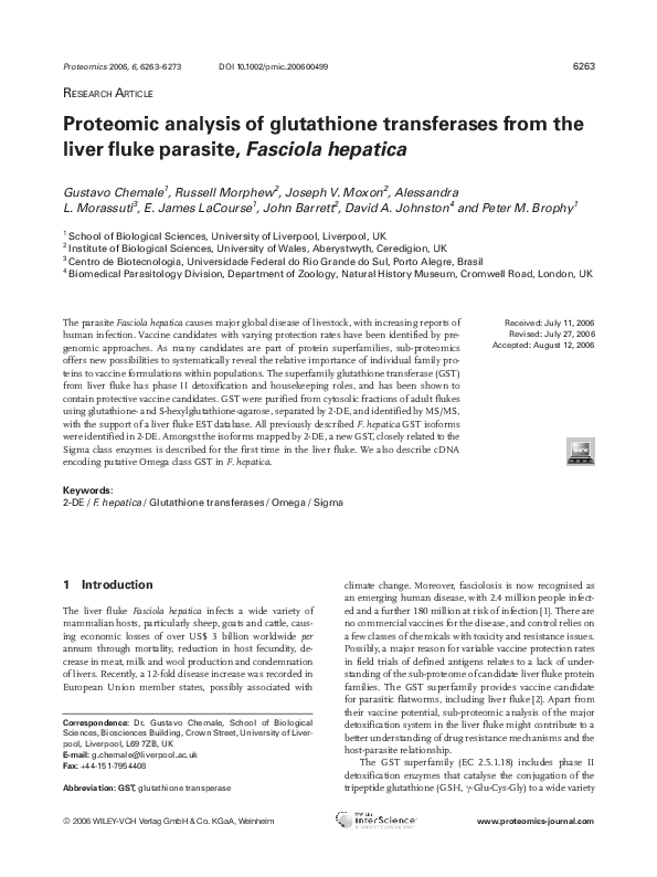
Figure 1. 2-DE mapping of F. hepatica affinity purified GST. Glutathione transferases were affinity purified using a GSH (A) or
S-hexylglutathione-agarose (B). Numbered protein spots were
identified by MS/MS (Table 1). Strips were rehydrated with 50 mg
of eluted proteins. IEF was carried out until 60 000 Vh at 207C in
17-cm IPG strips pH 3-10NL (Bio-Rad). Second dimension was
carried out in 12.5 % SDS-PAGE. Gels were subsequently stained
with CBB. Quantitative gel analysis was performed using Progenesis PG220, version 2006 software. The 3-D view of the GSHagarose purified GST 2-D gel (C) showing the quantitative differences per spot in number of fold compared to the S-hexylglutathione-agarose purified GST.
2006 WILEY-VCH Verlag GmbH & Co. KGaA, Weinheim
Proteomics 2006, 6, 6263–6273
The identification of the GST separated by 2-DE was performed by MS/MS with the sequencing of three to eight
peptides per protein spot. Peptide sequences obtained from
each spot were searched against GenBank (www.ncbi.nlm.
nih.gov) or an ‘in-house’ translated F. hepatica EST database
(ftp://ftp.sanger.ac.uk/pub/pathogens/fasciola/hepatica/ESTs/)
using BLAST algorithm [16]. Hits presenting total identity to
the peptide sequences were considered for analysis. All
identified protein spots were shown to be GST (Table 1). The
three protein spots at the acidic end of the gel, spots 1 to 3,
were shown to be the previously isolated FhGST51. Peptides
corresponding to FhGST7 were identified in spot 5 and
FhGST51, FhGST1 and FhGST7 were identified in spot 4,
probably a result of a spot overlapping in the gels. FhGST47
was identified at spot 8, at the more basic end of the gel. We
were not able to find complete identity hits in GenBank with
peptides from spots 9 and 10 (Fig. 1, Table 1). However, ten
translated cDNA sequences were found in the ‘in-house’
translated F. hepatica EST database with amino acid sequences showing complete identity to the peptides sequenced.
One peptide found in these spots was also found in spot 7
(Fig. 2), together with peptides corresponding to FhGST1
and FhGST47.
3.2 New F. hepatica Sigma and Omega GST
As many of the translated cDNA sequences from the ‘inhouse’ EST database showing complete identity to peptides
sequenced from spots 9 and 10 were partial sequences, they
were aligned using ClustalW [17] and assembled as a contig,
named FhepGSTs (Fig. 2A). Besides these cDNA clones,
which were identified in spots 9 and 10 (Fig. 1) and had not
been previously deposited in the GenBank database, we also
found two cDNA clones with partial similarity to some of
the peptides sequenced, Fhep54b04 and Fhep49c06. To
classify the GST encoded by these cDNA, they were aligned
with existing classified GST from seven species-independent classes within mammals, helminths and insects,
including F. hepatica sequences previously described. To
visualise the relationship between sequences and provide
direction upon assigning the F. hepatica GST to an existing
GST family class, a phylogram tree was built from the
alignment (Fig. 3). EST clones from the ‘in-house’ F. hepatica library cluster within GST classes previously unrepresented in F. hepatica. FhepGSTs clustered with GST
belonging to the Sigma class, suggesting that a Sigma/
prostaglandin-D-synthase GST is present and expressed in
F. hepatica. This contig shows similarities ranging from 26
to 45% to other Sigma class GST (Table 2). Highest identities were observed with S. mansoni, S. japonicum and Clonorchis sinenis Sigma GST, ranging from 42 to 45%. This
Sigma GST is also supported by proteomic sequence data
(Fig. 1 and Table 1).
Fhep54b04 and Fhep49c06 partial cDNA clones clustered with GST belonging to the Omega class. Overall identity with other sequences was low, ranging from 9 to 32%
www.proteomics-journal.com
�Proteomics 2006, 6, 6263–6273
Animal Proteomics
6267
Figure 2. F. hepatica Sigma GST amino acid deduced sequence from cDNA contig assembly. (A) Ten cDNA clones encoding protein
sequences matching peptides sequenced from spots 9 and 10 (Fig. 1, Table 1) were assembled as a contig, named FhepGSTs, containing
the complete peptide sequence of this protein. (B) MS/MS spectra resulting from the analysis of a peptide sequenced from spots 7, 9 and
10, belonging to the F. hepatica Sigma GST.
(Table 2), with S. mansoni Omega GST showing the highest
identity. Sequence identity for these F. hepatica sequences
is lower than that typically expected to place a GST into
a given class (.40%, [25]). Between classes, GST may typically be expected to share ~25% identity. This class structure is historically derived from mammalian classifications,
and thus might be less applicable to invertebrate GST.
It may be better to consider ‘class-specific’ domains in
alignments, biochemical specificity and modelled 3-D
structure, rather than overall identity, which may incorrectly exclude GST from assignment to given classes. In
order to better classify F. hepatica putative Omega GST, we
made a comparison of the consensus three-element signature of the Omega GST. This showed a much higher
relationship of the F. hepatica Omega GST (Fig. 4A) to
other members of this class, with similarity ranging from
47 to 65% similarity (Fig. 4B). Additionally, the F. hepatica
sequences were found to contain the characteristic N-terminal extension of Omega GST and the highly conserved
cysteine at the catalytic site, exclusive to Omega class GST
that replaces the typical serine or tyrosine of all other GST
classes [25].
2006 WILEY-VCH Verlag GmbH & Co. KGaA, Weinheim
3.3 Immunological classification of F. hepatica GST
To further characterise F. hepatica GST isolated in this work,
2-D gels containing the S-hexylglutathione-agarose purified
GST were transferred to PVDF membranes and assayed with
anti-S. mansoni Mu-class GST and anti-H. contortus Sigmaclass antibodies (Fig. 5). Spots containing FhGST51 and
FhGST7 GST and spot 6 were recognised by the anti-Mu class
antibody (Fig. 5A). As peptides belonging to FhGST1 and
FhGST7 were also found in spot 6, and that this antibody did
not recognise Mu-class GSTpresent in spots 7 and 8 (FhGST1
and FhGST47), the protein recognised in spot 6 is probably
FhGST7 or a very similar isoform not identified yet. Surprisingly, proteins present in spots 9 and 10, identified in this work
as F. hepatica Sigma GST, were not recognised by the antiSigma class antibody (Fig. 5B). Moreover, proteins in spots 6–8
were recognised by this antibody and were all identified as Muclass GST, apart from one peptide belonging to the newly
described F. hepatica Sigma class GST (Table 1, Fig. 2). We also
tried to identify Pi class GST in the F. hepatica affinity purified
GST by immunoblot using an anti-human GST1 antibody, but
no reactions were observed (data not shown).
www.proteomics-journal.com
�6268
G. Chemale et al.
Proteomics 2006, 6, 6263–6273
Table 1. Identification of GST from F. hepatica by MS/MS. Peptide sequences from spots were used to search against GenBank or the
translated EST library for the identification of the specific members of the GST superfamily. Numbers in brackets following GenBank accession numbers correspond to the peptides that share complete identity with the amino acid sequences
Spot
MS/MS peptidesa), b)
GenBank entries
E value
Coverage
(%)
Theoretical
pI/MW (Da)
Class
Observed
pI/MW (Da)
1, 2, 3
1-ISMIEGAAMDLR
2-YLAPQCLEDFPK
3-MWSNFLGDR
4-FEEVQGDYLK
5-FNMGLDLPNLPYYIDDK
6-LTQSVAIMR, 7-LGYWK
8-LLLEYLGEEYEEHLYGR
AAB28746 (1-8)
AAA29141 (1-8)
P30112 (1-8)
1905266A (1-8)
7e-25
41%
FhGST51
5.88/25373.36
m
5.4-5.82/
5196-25665
4
1-LTQSLAILR
2-VSMIEGAAVDLR
3-PAKLGYWK
4-ISMIEGAAMDLR
5-YLAPQCLEDFPK
6-IGFGLTCYNPK
7-YLAPQCLDDFPK
P56598 (1-3)
1905266D (1-3)
8e-03
13%
FhGST1
6.61/25729.55
m
5.95/26000
AAB28746 (3-5)
AAA29141 (3-5)
P30112 (3-5)
1905266A (3-5)
8e-04
14%
FhGST51
5.88/25373.36
m
P31671 (3,4,6,7)
1905266B (4,6,7)
7e-08
20%
FhGST7
6.13/25327.27
m
5
1-ISMIEGAAMDLR
2-MWSDFLGDR
3-IGFGLTCYNPK
4-YLAPQCLDDFPK
P31671 (1-4)
1905266B (1-4)
3e-15
20%
FhGST7
6.13/25327.27
m
6.03/25582
6
1-LTQSLAILR
2-VSMIEGAAVDLR
3-PAKLGYWK
4-ISMIEGAAMDLR
P56598 (1-3)
1905266D (1-3)
8e-03
13%
FhGST1
6.61/25729.55
m
6.51/25554
P31671 (3-4)
1905266B (3-4)
2e-03
9%
FhGST7
6.13/25327.27
m
AAA29140 (1-2)
P31670 (1-2)
1905266C (1-2)
2e-03
10%
FhGST47
6.54/25412.38
m
P56598 (3)
1905266D (3)
8e-03
5.5%
FhGST1
6.61/25729.55
m
FhepGSTs (4)
1e-02
5.6%
local database
s
7
1-MWSDFLGDR
2-ISMIEGAAMDLR
3-VSMIEGAAVDLR
4-VPLLDVTGPDGK
6.84/25360
8
1-YLAPHCLDEFPK
2-FNMGLDLPNLPYYIDDK
3-ISMIEGAAMDLR
AAA29140 (1-3)
P31670 (1-3)
1905266C (1-3)
7e-06
18%
FhGST47
6.54/25412.38
m
7.1/25200
9, 10
1-EVYTLFR, 2-LLLTCAGVK
3-LVSESLESSGGK
4-VPLLDVTGPDGK,
5-IIGECEDLYR
6-MMGETDEEYYLIER,
7-LWYFQFR
FhepGSTs (1-7)
9e-03
34%
Local database
8.86/24534.41
s
8.54/25223
a) Alignment of F. hepatica GST showing peptides that differentiate between isoforms, see Supplementary Figure I.
b) MS/MS spectra from peptides specific to each isoform, see Supplementary Figures 2–15.
4
Discussion
The 2-DE mapping of GST can be a very useful tool, as these
proteins play a major role in phase II detoxification, especially
in drug and oxidative stress response. To provide a 2-DE
mapping of F. hepatica GST, we purified GST from adult F.
hepatica using GSH- and S-hexylGSH agarose columns. For
2006 WILEY-VCH Verlag GmbH & Co. KGaA, Weinheim
the first time, we combined purification using two affinity
matrix isolation approaches, EST database searching and
2-DE array (Fig. 1B), resulting in the detection of ten predominant GST spots. This approach produced a more complete
GST-ome profile compared to the classical approach of GSHagarose purification alone, with significant quantitative differences between the two methods, especially when spots 9
www.proteomics-journal.com
�Proteomics 2006, 6, 6263–6273
Animal Proteomics
6269
Figure 3. Classification of F. hepatica
GST. Phylogenetic bootstrap tree of
F. hepatica GST aligned with existing
classified GST from seven species-independent classes within mammals, helminths and insects. Protein sequences
from different classes of GST from seven
species were aligned with F. hepatica
GST obtained from GenBank and from
‘in-house’ EST library database using
ClustalW [17], and neighbour joining
trees produced using BioEdit Sequence
Alignment Editor version 7.0.5.2. [18].
Tree was viewed within TREEVIEW [19]
to visualise the relationship between
sequences and provide direction upon
assigning the F. hepatica GST to an
existing GST family class. Accession
numbers: GenBank, gb; Swiss-Prot, sp.
F. hepatica (gb), Fhep, P31671, P56598,
1905266A,
1905266B,
1905266C,
1905266D,
AAA29139,
AAA29140,
AAA29141, AAB28746, P30112,P31670.
Ascaris summ (gb), asum, GST1,
P46436; Caenorhabditis elegans (sp),
Cele, GST1 pi, P10299; GST7, P91253;
GST43 zeta, Q9N4H6; omega, P34345.
Clonorchis sinensis (gb), Csin, GST28,
O97096. Drosophila melanogaster (sp),
Dmel, GSTT1, P20432; Haemonchus
contortus (sp), Hcon, Q9NAW7. Homo
sapiens (sp), Hsap, GSTM1.1, P09488;
GSTA1, P08263; GSTP1, Q5TZY3; PGD2,
Q6FHT9; GSTZ1.1, Q6IB17; GSTO1,
P78417; GSTT1, Q5TZY2. Mus musculus
(sp), Mmus, GSTM1, Q58ET5(sp);
GSTA1, Q6P8Q0; GSTP1, P19157; PGD2,
Q8CA80; GSTZ1, Q9WVL0; GSTO1,
O09131; GSTT1, Q91X50. Schistosoma
haematobium (gb), Shae, GST28,
P30114. S. japonicum (gb), Sjap, mu,
P08515. S. mansoni (gb), Sman, mu,
P35661;
GST28,
P09792;
omega,
Q86LC0.
and 10 are compared (Fig. 1C). All previously described F.
hepatica GST were identified in the 2-DE gels, many of them
present in more than one spot (Table 1). FhGST51 was
identified in spots 1 to 3, indicating that this protein may be
modified after translation or during detoxification or that
very similar isoforms with slightly different pI exist. We were
not able to sequence peptides belonging to other isoforms,
even after determining the sequence of eight peptides from
these spots, covering approximately 35% of the protein. If
they are different isoforms, regions responsible for the difference in the pI observed were not covered by these peptides. Peptides belonging to different GST were found in the
same protein spot, as can be seen in spots 4, 6 and 7 (Fig. 1
2006 WILEY-VCH Verlag GmbH & Co. KGaA, Weinheim
and Table 1). This might be the result of spot overlapping in
the 2-DE gels, as these proteins present very similar pI, or
possible modification and co-migration. Peptides corresponding to FhGST7 and FhGST47 were identified in single
spots, 5 and 8, respectively. We were not able to find complete
identity hits in GenBank with peptides from spots 9 and 10.
However, cDNA sequences were found in the ‘in-house’
translated F. hepatica EST database matching the peptides
sequenced and were used to assemble a contig (Fig. 2A). One
peptide found in these spots was also found in spot 7
(Fig. 2B), together with peptides corresponding to FhGST1
and FhGST47. When aligned with GST from other organisms, FhepGSTs sequence clustered with Sigma class GST
www.proteomics-journal.com
�6270
G. Chemale et al.
Proteomics 2006, 6, 6263–6273
Figure 4. Comparison of F. hepatica Omega GST to conserved signature motifs. Sequences obtained from the three-element signature of
Omega class GST (PRINTS PR01625), were aligned with several Omega GST including the F. hepatica sequences found in the ‘In-house’
EST database (A). Consensus sequences to these three Omega domains were found to match well with the F. hepatica sequences, ranging
from 47 to 65% similarity (B). Consensus sequences were constructed using the PRATT consensus sequence search tool (http://us.
expasy.org/tools/pratt/) [20, 21]. Omega class GST signature sequences PR01625 were obtained from PRINTS data bank of protein family
fingerprints http://umber.sbs.man.ac.uk/dbbrowser/sprint/. Accession numbers are same as in Figure 3.
(Fig. 3). As well as being identified in adults, FhepGSTs was
also shown to be highly expressed in F. hepatica eggs (data
not shown), showing that this protein is expressed in both
parasite stages.
Overall identities of the F. hepatica Sigma and Omega
GST were higher when compared to other trematode GST,
varying from 27 to 45%. There are no clearly established and
definitive criteria for placing a GST in a particular class. The
extent of sequence identity from which to assign a GST to a
given class varies in the literature from greater than 40%
[3, 25], to greater than 60% [26]. GST with less than 30% [26]
or 25% [3, 25] identity are taken to belong to separate classes.
These criteria are typically met when mammalian GST are
compared, but not with invertebrates enzymes. When
2006 WILEY-VCH Verlag GmbH & Co. KGaA, Weinheim
Sigma GST from humans and rodents are compared, they
present an overall identity of about 80% (Table 2). However,
when compared to trematode GST, identity levels are much
lower, presenting less than 30% of identity with S. mansoni,
S. japonicum and C. sinensis Sigma GST. Identity levels are
much higher when trematode Sigma GST are compared,
varying from approximately 41% to 77% (Table 2), meaning
that overall identity values for mammalian GST cannot be
used to classify trematode GST. This was observed when we
only compared signature motifs for Omega GST with F.
hepatica Omega class enzymes. Overall, identity levels were
low when compared to mammalian GST, (Table 2) but
when compared to conserved motifs, identity levels were
rose to 45%-65%, showing that trematode GST cannot
www.proteomics-journal.com
�Animal Proteomics
Proteomics 2006, 6, 6263–6273
6271
Table 2. Sequence identity matrix of F. hepatica GST from proposed Sigma and Omega classes obtained from the ‘in-house’ translated EST
database compared to mammalian, helminth and insect GST from these two classes using ClustalW
Sigma-class glutathione transferases
Sequence
Fhep
GSTs
Sman
GST28
Sjap
GST
Csin
GST28
Hsap
PGD2
Mmus
PGD2
Cele
GST7
Asum
GST1
Hcon
GST
FhepGSTs
SmanGST28
SjapGST
CsinGST28
HsapPGD2
MmusPGD2
CeleGST7
AsumGST1
HconGST
ID
0.450
0.425
0.450
0.262
0.275
0.280
0.310
0.270
ID
0.770
0.430
0.300
0.290
0.300
0.300
0.320
ID
0.415
0.293
0.298
0.251
0.288
0.288
ID
0.273
0.264
0.240
0.282
0.259
ID
0.804
0.300
0.378
0.344
ID
0.300
0.364
0.330
ID
0.519
0.601
ID
0.475
ID
Omega-class glutathione transferases
Sequence
Fhep
54b04
Fhep
49c06
Sman
omega
Hsap
GST1
Mmus
GST1
Cele
P34345
Fhep54b04
Fhep49c06
Sman omega
HsapGST1
MmusGST1
CeleP34345
ID
0.319
0.322
0.174
0.171
0.118
ID
0.278
0.096
0.093
0.093
ID
0.189
0.182
0.154
ID
0.721
0.318
ID
0.303
ID
Figure 5. Immunoblot of 2-DE affinity purified F. hepatica GST.
Affinity purified glutathione transferases were separated by 2-DE
in 17-cm IPG strips pH 3-10NL (Bio-Rad). Second dimension was
carried out in 12.5 % SDS-PAGE. Gels were subsequently transferred to PVDF membranes and incubated with anti-S. mansoni
Mu class GST (A) and anti-H. contortus Sigma class GST (B)
antibodies as described in Section 2.
be excluded from a class when similarity is lower than 40%
to mammalian GST. Additional evidence for Omega classification was found in the presence of the ‘Omega-typical’ Nterminal extension and catalytic cysteine. Other invertebrate
and trematode sequences must be used for classification
and conserved motifs and structures considered during
analysis, so as not to exclude GST from classes based only
on overall identity to mammalian GST. This method of
2006 WILEY-VCH Verlag GmbH & Co. KGaA, Weinheim
matching GST to conserved motifs within classes may be
more robust than global alignment and overall identity percentages.
Further characterization of F. hepatica purified GST by
immunoblot analysis using antibodies raised against classspecific GST was unable to confirm the presence of the new
liver fluke Sigma class GST. However, the relatively low identity shared between H. contortus Sigma GST and FhepGSTs
(27%, Table 2) could be responsible for the weak recognition.
However, three spots (6-8, Fig. 4B) identified as Mu GST were
recognised by this antibody, suggesting that the use of classspecific polyclonal antibodies for class differentiation might
be misleading. Spot 3, identified as FhGST51 was also recognised by the Sigma antibody, reflecting in part the relatively
large amount of protein present in that spot, i.e. spots 1 and 2,
identified as the same isoform, were not recognised. Moreover, anti-Mu class antibody raised against S. mansoni GST
was not able to recognise all F. hepatica Mu class GST (Fig. 4A),
even though they share more than 75% identity. Our data
confirms that antibodies to generic GST classes are problematic in cross-species identifications. Highly specific monoclonal antibodies raised against GST class-specific epitopes,
are probably required for confidently using immunology as a
validation tool for GST classification.
Sigma class GST have been strongly implicated in
prostaglandin synthesis. A relationship between Sigma class
GST of helminths and a GSH-dependent prostaglandin-H
www.proteomics-journal.com
�6272
G. Chemale et al.
E-isomerase was revealed by comparison of partial sequence
data [27]. This enzyme was later purified from the parasitic
nematode Ascaridia galli and confirmed to be a Sigma class
GST [28]. It is thought that this enzyme promotes the endogenous synthesis of prostaglandin E in multicellular
parasites. The production of prostaglandins by these parasites could be an important component of their mode of
subversion or suppression of host immunity [29]. The identification of this highly expressed GST in F. hepatica adults
tentatively suggest that flukes could be using prostaglandin
synthesis in such a way as to suppress the host’s protective
immune response. Although only recently described as a
new class of GST [30], Omega GST appears widespread in
the animal kingdom. Human, pig, mouse, rat, C. elegans, S.
mansoni, and D. melanogaster were all found, via bioinformatic analysis, to possess Omega GST [30, 31, 32, 33, 34, 35].
As sequence annotation progresses, further Omega GST
continue to emerge, as for example here, in the parasitic liver
fluke, F. hepatica. In humans, Omega GST have recently
received much attention after their linkage to age onset of
Alzheimer’s and Parkinson’s diseases [36], as well as roles in
arsenic metabolism [37]. Other roles are also proposed; Human Omega GSTO1-1 has been shown to modulate ryanodine receptors, calcium channels in the endoplasmic reticulum [38] and was found to be up-regulated in radiationresistant cancer cell lines [33]. Sei et al. (1999) [39] and Xu et
al. (1998) [40] propose [Ca2+] dependant immune modulation may also arise as a result of ryanodine receptor interaction in T and B lymphocytes.
Newly uncovered F. hepatica cDNA belonging to Sigma
and Omega class GST are currently being produced as
recombinants in order to further characterise their biochemical properties and class-specific activities prior to challenge vaccinations.
BBSRC and DEFRA UK for grant BB/C503638/1 to support Gustavo Chemale, and the EU (STREP DELIVER) for a
grant to support James LaCourse are acknowledged. The authors
are also grateful to Jim Heald at UWA for technical assistance
related to tandem mass spectrometry and the staff and UK meat
inspectorate at Oriel Jones abattoir, West Wales for providing
fresh F. hepatica infected livers.
Proteomics 2006, 6, 6263–6273
[5] Pearson, W. R., Methods Enzymol. 2005, 401, 186–204.
[6] Precious, W. Y., Barrett, J., Biochim. Biophys. Acta 1989,
992, 215–222.
[7] Howell, M. J., Board, P. G., Boray, J. C., J. Parasitol. 1988,
74, 715–718.
[8] Wijffels, G. L., Sexton, J. L., Salvatore, L., Pettitt, J. M. et al.,
Exp. Parasitol. 1992, 74, 87–99.
[9] Brophy, P. M., Crowley, P., Barrett, J., Mol. Biochem. Parasitol. 1990, 39, 155–161.
[10] Panaccio, M., Wilson, L. R., Crameri, S. L., Wijffels, G. L.,
Spithill, T. W., Exp. Parasitol. 1992, 74, 232–237.
[11] Salvatore, L., Wijffels, G., Sexton, J. L., Panaccio, M. et al.,
Mol. Biochem. Parasitol. 1995, 69, 281–288.
[12] Rossjohn, J., Feil, S. C., Wilce, M. C., Sexton, J. L. et al., J.
Mol. Biol. 1997, 273, 857–872.
[13] Morrison, C. A., Colin, T., Sexton, J. L., Bowen, F. et al.,
Vaccine 1996, 14, 1603–1612.
[14] Sexton, J. L., Milner, A. R., Panaccio, M., Waddington, J. et
al., J. Immunol. 1990, 145, 3905–3910.
[15] Sexton, J. L., Wilce, M. C., Colin, T., Wijffels, G. L. et al., J.
Immunol. 1994, 152, 1861–1872.
[16] Altschul, S. F., Gish, W., Miller, W., Myers, E. W., Lipman, D.
J., J. Mol. Biol. 1990, 215, 403–410.
[17] Thompson, J. D., Higgins, D. G., Gibson, T. J., Nucleic Acids
Res. 1994, 22, 4673–4680.
[18] Hall, T. A., Nucleic Acids. Symp. Ser. 1999, 41, 95–98.
[19] Page, R. D., Comput. Appl. Biosci. 1996, 12, 357–358.
[20] Jonassen, I., Collins, J. F., Higgins, D., Protein Sci. 1995, 4,
1587–1595.
[21] Jonassen, I., Comput. Appl. Biosci. 1997, 13, 509–522.
[22] van Rossum, A. J., Jefferies, J. R., Rijsewijk, F. A.,
LaCourse, E. J. et al., Infect. Immun. 2004, 72, 2780–2790.
[23] Towbin, H., Staehelin, T., Gordon, J., Proc. Natl. Acad. Sci.
USA 1979, 76, 4350–4354.
[24] Harlow, D., Lane, D., Antibodies: A Laboratory Manual, Cold
Spring Harbor, New York 1988.
[25] Board, P. G., Anders, M. W., Methods Enzymol. 2005, 401,
61–77.
[26] Sheehan, D., Meade, G., Foley, V. M., Dowd, C. A., Biochem. J. 2001, 360, 1–16.
[27] Meyer, D. J., Thomas, M., Biochem. J. 1995, 311, 739–742.
[28] Meyer, D. J., Muimo, R., Thomas, M., Coates, D., Isaac, R.
E., Biochem. J. 1995, 313, 223–227.
5
References
[1]
Anon. Control of foodborne trematode infections, WHO,
Geneva 1995.
[2]
Brophy, P. M., Pritchard, D. I., Exp. Parasitol. 1994, 79, 89–96.
[3]
Hayes, J. D., Flanagan, J. U., Jowsey, I. R., Annu. Rev.
Pharmacol. Toxicol. 2005, 45, 51–88.
[4]
Armstrong, R. N., Chem. Res. Toxicol. 1997, 10, 2–18.
2006 WILEY-VCH Verlag GmbH & Co. KGaA, Weinheim
[29] Maizels, R. M., Bundy, D. A., Selkirk, M. E., Smith, D. F.,
Anderson, R. M., Nature 1993, 365, 797–805.
[30] Board, P. G., Coggan, M., Chelvanayagam, G., Easteal, S. et
al., J. Biol. Chem. 2000, 275, 24798–24806.
[31] Wilson, R., Ainscough, R., Anderson, K., Baynes, C., et al.,
Nature 1994, 368, 32–38.
[32] Ishikawa, T., Casini, A. F., Nishikimi, M., J. Biol. Chem. 1998,
273, 28708 –28712.
www.proteomics-journal.com
�Proteomics 2006, 6, 6263–6273
Animal Proteomics
6273
[33] Kodym, R., Calkins, P., Story, M., J. Biol. Chem. 1999, 274,
5131–5137.
[37] Zakharyan, R. A., Sampayo–Reyes, A., Healy, S. M., Tsaprailis, G. et al., Chem. Res. Toxicol. 2001, 14, 1051–1057.
[34] Rouimi, P., Anglade, P., Benzekri, A., Costet, P., et al., Biochem. J. 2001, 358, 257–262.
[38] Dulhunty, A., Gage, P., Curtis, S., Chelvanayagam, G.,
Board, P., J. Biol. Chem. 2001, 276, 3319–3323.
[35] Girardini, J., Amirante, A., Zemzoumi, K., Serra, E., Eur. J.
Biochem. 2002, 269, 5512–5521.
[39] Sei, Y., Gallagher, K. L., Basile, A. S., J. Biol. Chem. 1999,
274, 5995–6002.
[36] Kolsch, H., Linnebank, M., Lutjohann, D., Jessen, F. et al.,
Neurology 2004, 63, 2255–2260.
[40] Xu, D. P., Washburn, M. P., Sun, G. P., Wells, W. W., Biochem. Biophys. Res. Commun. 1996, 221, 117–121.
2006 WILEY-VCH Verlag GmbH & Co. KGaA, Weinheim
www.proteomics-journal.com
�
 Alessandra Morassutti