Hindawi
BioMed Research International
Volume 2018, Article ID 6432742, 11 pages
https://doi.org/10.1155/2018/6432742
Research Article
In Vivo Evaluation of Different Collagen Scaffolds in
an Achilles Tendon Defect Model
Carolin Gabler ,1 Jan-Oliver Saß,1 Susann Gierschner,1 Tobias Lindner ,2
Rainer Bader ,1 and Thomas Tischer1
1
Rostock University Medical Center, Department of Orthopedics, Biomechanics and Implant Technology Laboratory, Rostock, Germany
Rostock University Medical Center, Core Facility Multimodal Small Animal Imaging, 18057 Rostock, Germany
2
Correspondence should be addressed to Carolin Gabler; carolin.gabler@med.uni-rostock.de
Received 16 March 2018; Revised 4 July 2018; Accepted 18 July 2018; Published 8 August 2018
Academic Editor: Berardo Di Matteo
Copyright © 2018 Carolin Gabler et al. This is an open access article distributed under the Creative Commons Attribution License,
which permits unrestricted use, distribution, and reproduction in any medium, provided the original work is properly cited.
In the present study, a newly introduced bovine cross-linked collagen scaffold (test material) was investigated in vivo in an Achilles
tendon defect model and compared to a commercially available porcine collagen scaffold (control material). In total, 28 male
Sprague Dawley rats (about 400 g) were examined. The defined Achilles tendon defect of 5 mm of the right hind limb was replaced
by one of the scaffold materials. After euthanasia, the hind limbs were transected for testing. Biomechanical evaluation was carried
out via tensile testing (n = 8 each group, observation time: 28 days). Nonoperated tendons from the bilateral side were used as a
control (native tendon, n = 4). For the histological evaluation, 12 animals were sacrificed at 14 and 28 days postoperatively (n = 3
each group and time point). Stained slices (Hematoxylin & Eosin) were evaluated qualitatively in terms of presence of cells and cell
migration into scaffolds as well as structure and degradation of the scaffold. All transected hind limbs were additionally analyzed
using MRI before testing to verify if the tendon repair using a collagen scaffold was still intact after the observation period. The
maximum failure loads of both scaffold materials (test material: 54.5 ± 16.4 N, control: 63.1 ± 19.5 N) were in the range of native
tendon (76.6 ± 11.6 N, p ≥ 0.07). The stiffness of native tendons was twofold higher (p ≤ 0.01) and the tear strength was approximately
fivefold higher (p ≤ 0.01) compared to the repaired tendons with both scaffolds. Histological findings indicated that neither the test
nor the control material induced inflammation, but the test material underwent a slower remodeling process. An overall repair
failure rate of 48% was observed via MRI. The experimental data of the newly developed test material showed similar outcomes
compared to the commercially available control material. The high repair failure rate indicated that MRI is recommended as an
auxiliary measurement tool to validate experimental data.
1. Introduction
Tendon regeneration, e.g., after rotator cuff tears, is known
to be a complex and slow process, and the healing of
tendon repair still remains a clinical challenge. Depending on
individual factors (e.g., patient’s age, tendon quality, and tear
size) high rerupture rates can be observed [1–3]. Therefore,
scaffold devices for tendon augmentation, whether biologic
or synthetic, have been introduced to increase healing rates
[4–6]. It is important to note that the absence of approval
from the health authorities limits the clinical use of some graft
materials in many countries. For example, in Germany, allografts are subjected to regulations based on transplantation
law. In Japan, the use of allografts is also not approved and
therefore the use of autografts is common [7].
The biomechanical behavior of graft materials in vitro
was characterized by means of uniaxial mechanical tests
[8, 9]. The results can be adjusted by varying the sample
characteristics of the material tested such as thickness [10]
or the processing method of the material [11], independently
from graft source. Therefore, clinically relevant scaffold constructs should be performed to produce qualitative evidence.
In recent years, several clinical trials have evaluated the
functional outcome of the augmented rotator cuff repair
(RCR) with the use of different scaffold devices. Most of these
trials were quite limited as they were often retrospective case
series with small patient populations, without control groups,
and produced controversial results [12, 13]. Based on the
recent literature, allografts made of acellular human dermis
are thought to provide the most beneficial clinical outcome
2
with low rerupture rates compared to other scaffolds [14–16].
In the current literature, the clinical outcome of xenografts is
discussed controversially [7, 17, 18]. Thus, the source of the
graft plays an important role [4, 12]. In particular, collagenbased grafts made from porcine small intestine submucosa
(SIS) are known to end up in suboptimal results and may
promote postoperative inflammatory reactions. Therefore,
the use of porcine SIS xenografts (Restore, DePuy) for
augmentation in RCR is not recommended [12, 19]. Ciampi
et al. [20] reported that RCR using bovine pericardiumderived patches (Tutopatch, RTI Surgical, Inc. Alachua, FL,
USA) showed significantly lower healing rates compared to
synthetic grafts (Repol Angimesh, Angiologica BMSrl, Pavia,
Italy). However, the healing rate was not significantly lower
than RCR without a graft. Further clinical data showed that
xenografts, based on dermal collagen, led to more promising
results [5, 21–23]. However, it is not known if there are
differences in clinical outcome with respect to the dermal
graft source (porcine versus bovine).
Currently, there are two graft materials based on bovine
dermis available for clinical use (TissueMend, Stryker Corp.,
Mahwah, NJ, USA, and Bio-Blanket, Kensey Nash Corp., PA,
USA), but only few results have been published on clinical
outcomes. Sears et al. [24] compared the clinical outcome
of an allograft (GraftJacket, Wright Medical Arlington, TN,
USA), a porcine dermal extracellular matrix (ECM) (Conexa,
Tornier Inc., Bloomington, MN, USA), and a bovine dermal
ECM (TissueMend, Stryker Corp.) in a retrospective case
study. Significant differences in clinical outcome were not
found between the different patches. However, the study was
strongly limited by the small sample size.
The inconsistent clinical outcomes using scaffolds for
tendon augmentation underline that there is still a need for
new scaffold materials. Therefore, we have developed a new
scaffold material, based on bovine collagen and a chemical
cross-linking process. The material has been already tested
in vitro and showed favorable biomechanical, biochemical,
and cell biological properties [25]. In the present pilot
animal study, the newly introduced bovine collagen scaffold
was investigated with regard to functional outcomes and
remodeling using an Achilles tendon defect model in rats.
2. Materials and Methods
2.1. Scaffolds. Two collagen scaffold materials were evaluated.
As the test material, a newly developed scaffold based on
bovine collagen was used, as described previously [25].
Briefly, for scaffold preparation, bovine dermal collagen was
treated chemically with NaOH, H2 O2 , and HCl in order
to remove noncollagenous proteins, fatty acids, and cells
and to inactivate viruses. The purified dermal collagen was
processed into a matrix with longitudinally orientated fibrils
consisting mainly of collagens type I, type III, and type
V. This predefined matrix structure was first stabilized by
freeze-drying within a temperature range of 55 to 65∘ C and
further by chemical cross-linking. Then, the freeze-dried
matrix was subjected to an aqueous epoxide solution with
a concentration of 0.19% (w/w). Before further use, the
BioMed Research International
cross-linked collagen matrix was successively washed with
reverse osmosis (RO) water to remove any free epoxide.
For the control group, DX Reinforcement scaffolds (Arthrex,
Inc., Naples, FL, USA) based on porcine dermal extracellular
matrix with no cross-linking were used.
Before testing, the test material was hydrated in saline
solution (NaCl, 0.9%) for at least 30 min (thickness after
rehydration: 0.89 ± 0.04 mm). The control material was
delivered hydrated (thickness: 1.43 ± 0.16 mm). Test samples
for in vitro and in vivo evaluation were obtained from two
patches of one charge for the test material and from two
patches of two charges for the control material.
2.2. Biomechanical Testing of Scaffold Materials In Vitro.
As we used samples from new charges, we repeated the
initial biomechanical testing [25] according to Barber and
Aziz-Jacobo [8]. Before animal testing, additionally a suture
retention test was performed according to Barber and AzizJacobo [8]. Samples were bisected and one vertical stitch with
No. 2 FiberWire (Arthrex, Inc., Naples, FL, USA) was passed
to the distal end of the scaffold with a distance of 5 mm from
the tissue edge. The scaffold was mounted on the upper grip
with roughened chuck jaws in the testing machine; the suture
was fixed with a sample grip with corrugated chuck jaws. The
start length was 3 cm and the predetermined tearing location
was centered. The destructive test was conducted again with
a distraction rate of 12.5 mm/s.
For all biomechanical evaluations of the test material,
care was taken that the load was applied longitudinally to
the orientation of collagen fibers (according to native in vivo
situation). Six samples of the test material were tested. Only
four samples of the control material were tested due to the
limited quantity of the material. The orientation of the control
material during testing was not required, since this graft has
no directional fiber alignment.
2.3. Animal Testing. For in vivo testing of both scaffold
materials, a total of 28 male Sprague-Dawley rats (Janvier
Labs, Le Genest-Saint-Isle, France) weighing 404 ± 20 g were
used. The study was approved by the local animal research
committee (LALLF MV, reference number 7221.3-1-036/15).
Rats were kept in an animal facility, where temperature and
light/dark cycle (12:12 hours) were controlled, and access to
standard food and water was provided ad libitum. Animals
were randomly divided into two groups: the test material
group, where the Achilles tendon defect was replaced by the
bovine collagen scaffold, and the control group, where the
DX Reinforcement graft material was used as the scaffold. For
functional and biomechanical evaluations, the postoperative
observation time was 28 days (n = 8 each group). To determine the optimal number of animals, an a priori analysis was
performed for the mean of two independent samples using
G∗Power (version 3.1.9.2). For the histological evaluation, in
total 12 animals were sacrificed 14 and 28 days postoperatively
(n = 3 each group and each time point). The allocation of the
rats is shown in Figure 1.
Surgery was performed by an experienced orthopedic surgeon (TT), who underwent additionally a training
BioMed Research International
3
total
(n = 28)
Test material
(n = 14)
Biomechanical testing
(n = 8)
4 weeks
(n = 8)
Control material
(n = 14)
Histology
(n = 6)
2 weeks
(n = 3)
4 weeks
(n = 3)
Biomechanical testing
(n = 8)
4 weeks
(n = 8)
Histology
(n = 6)
2 weeks
(n = 3)
4 weeks
(n = 3)
Figure 1: Overview of the allocation of rats and experiments used in present study.
on cadaveric rats before. For surgery, the animals were
anesthetized with medetomidine (150 𝜇g/kg), midazolam
(2 mg/kg), and fentanyl (5 𝜇g/kg) due to an intraperitoneal
injection. The right Achilles tendon was dissected and freed
from soft tissue and the M. plantaris tendon was removed.
Each rat underwent a transection of 5 mm of the Achilles
tendon. Due to differences of sizes of the animals, the
transection was set individually in the middle part of the
tendon. A 5 mm scaffold of either the new bovine material
(n = 14) or the porcine control material (n = 14) was replaced
in the defect and the remaining ends were refixed with two
single stitches (Vicryl 4-0, Ethicon, Somerville, NJ, USA)
(Figure 2). Collagen fibers of the test material scaffold device
were aligned longitudinally to the orientation of the Achilles
tendon.
Finally, anesthesia was antagonized by a subcutaneous
(s.c.) dose of atipamezole (750 𝜇g/kg), flumazenil (200 𝜇g/
kg), and naloxone (120 𝜇g/kg). Postoperative analgesia was
provided through an intramuscular injection of 0.5 ml
metamizol (0.5 g/ml) immediately after surgery, as well as
orally via the drinking water (30 drops 500 mg/ml metamizol per 0.5 l) for three days. Animals were allowed to
move freely in their cages after surgery. In the first postoperative week, they were kept individually per cage to
prevent adverse events like fighting or mutually gnawing
on surgical sites. At the end of the observation period,
the animals were euthanized by an intracardiac injection
with an overdose of pentobarbital sodium (80 mg/kg) under
anesthesia.
2.4. Imaging Analysis. After scarification of the animals, hind
limbs were transected proximal to the knee joint, and magnetic resonance imaging (MRI) scans were acquired using 7.0
Tesla scanner (BioSpec 70/30, Bruker, Ettlingen, Germany).
T2 -weighted TurboRARE sequences in axial, coronal, and
sagittal plane were recorded (TE/TR: 28/4400 ms, spatial inplane resolution: 0.12 mm, slice thickness: 0.7 mm, matrix
size (sagittal/frontal/transversal): 338 × 304 / 320 × 166 / 280
× 196, FoV (sagittal/frontal/transversal): 40.5 mm x 36.5 mm
/ 38.3 mm × 20 mm / 33.5 mm x 23.5 mm, RARE factor: 8,
averages: 5). Afterwards, specimens for biomechanical testing
were wrapped in gauze soaked with NaCl solution (0.9%) and
stored at −20∘ C. Specimens for histomorphometric analyses
were fixed in buffered formalin (4%).
A
B
C
D
Figure 2: Schematic illustration of implantation procedure. A:
preparation of the Achilles tendon, B: marking defined defect length
of the Achilles tendon, C: setting the defined defect, D: implantation
of the scaffold material, suturing with two single stitches at each end.
For analysis, the software Amira 5.4.1 (Thermo Fisher
Scientific, USA) was used. The distance between the distal
edge of the scaffold and the tendon insertion on the calcaneus was measured. Three measurements per view were
executed, and mean value was calculated for each sample,
as shown in Figure 3. Additionally, the junction between
the scaffold and the native tendon was evaluated. It was
determined whether the reconstruction was still intact or
if it had failed during the observation time. Failure was
defined if either the position of the scaffold was obviously
slipped into the proximal direction (distance to calcaneus
> 10 mm) and/or the musculotendinous junction was elongated.
2.5. Functional Evaluation. A gait analysis was performed for
the animals planned for biomechanical testing. A modified
method for the Achilles Functional Index (AFI) described
by Murrel et al. [26], according to Kurtz et al. [27], was
used. Therefore, the hind paws of the rats were colored by
dipping a sponge soaked with nontoxic food dye. Animals
were then allowed to walk a confined walkway prepared
with white paper on the floor of the corridor, leaving paw
prints on the paper. Gait was recorded preoperatively, at
day 4 and day 7, and then at weekly intervals up to day
28. The papers were scanned and measurements of from
paw prints were performed with GIMP 2.8.20 (GIMP, the
GIMP Team). Measurements included print length (PL),
total spreading (TS, distance between first and fifth toes),
and intermediary spreading (IT, distance between second
and fourth toes). Three left and three right paw prints were
4
BioMed Research International
evaluated and averaged each time point to calculate the
corresponding factors (PLF, TSF, and ITF) according to [21].
Murrel’s formula was used for determination of AFI:
AFI = 74 (PLF) + 161 (TSF) + 48 (ITF) − 5
(1)
[26].
2.6. Biomechanical Testing In Vivo. Prior to testing, the
fresh-frozen animal specimens were thawed in a bath of
NaCl overnight at 4∘ C and stored at room temperature for
at least 4 hours before preparation. The Achilles tendoncalcaneus-foot complex was dissected from the hind limb.
The gastrocnemius-soleus muscle was removed with the
blunt end of a scalpel, as described in literature [28]. The
proximal end of the tendon was spread out of some paper.
The paper was then folded two times and fixed with tape. The
foot was mounted with a cyanoacrylate adhesive (LOCTITE
4902, Henkel, Düsseldorf, Germany) at 45∘ to the surface of
a custom-made aluminum block and additionally fixed due
to a clamping unit via screws. The specimens were mounted
on a custom-made test setup in a materials testing machine
(Z1.0, Zwick, Ulm, Germany) for tensile testing (Figure 4). All
tendons were preloaded with 1 N, and width and thickness
were measured with a caliper at three measuring points.
Cross-sectional area was calculated from the averaged values
under the assumption that the area is oval. Subsequently, the
tendons were stretched at a rate of 1 mm/s until complete
rupture was observed. Care was taken that the specimens
were kept moist with NaCl throughout the procedure. Nonoperated tendons from the bilateral side (left leg) were used
as a control (n = 4). Load-displacement curves were recorded
and evaluated.
2.7. Histological Analysis. The specimens were dehydrated
in a graded series of alcohol and embedded in polymethylmethacrylate. Slices in the longitudinal direction of the
implant were cut with a laser microtome (TissueSurgeon,
LLS ROWIAK GmbH, Hannover, Germany) and stained
with Hematoxylin & Eosin (HE). Slice thickness was 10 𝜇m.
Scanning and digitalizing for evaluation were performed
using a digital microscope (VHX-6000, Keyence, Osaka,
Japan) at 500x (objective VH-Z250T) magnification. Samples
were evaluated qualitatively in terms of structure and degradation of the scaffold (preserved fiber structure), reaction of
surrounding tissue (cell infiltration), and cell migration into
scaffolds.
2.8. Data Analysis. Statistical analysis was performed using
IBM SPSS Statistics 22 software (IBM, Ehningen, Germany).
The statistical significance of differences was calculated by
Mann–Whitney test within two independent groups. The
level of significance was set to p < 0.05.
3. Results
3.1. Biomechanical Testing of Scaffolds In Vitro. Biomechanical data of the new charges of scaffolds used in this study
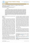
Figure 3: T2 -weighted MR image of an experimental hind limb
repaired with a collagen scaffold (dorsal view). Arrows indicate
measurement of the distance between the distal edge of the scaffold
and tendon insertion into the calcaneus.
are displayed in Table 1. The test material shows initial higher
mean maximum failure load (Fmax) compared to the control
material, but the difference is not significant (p < 0.05). There
is also no significant difference of stiffness. Mean tear strength
(tensile load normalized to cross section) and elastic modulus
of the test material were significantly higher compared to the
control material (p < 0.05).
The retention strength of single vertical stitches in both
scaffold materials (maximum failure load) is demonstrated in
Figure 5. The test material showed a lower maximum failure
load (41.5 ± 2.2 N) compared to the control material with 77.0
± 21.0 N. Differences were significant (p < 0.05).
3.2. Analysis of MRI Data. All animals tolerated the surgical
procedure. No dropouts or adverse events occurred during
the observation period. MR images showed that the location
of the scaffolds relative to the tendon insertion on the
calcaneus differed considerably in some cases. Some of the
scaffolds were located in the proximal part of the lower leg,
which was an indication that the reconstruction might have
failed distally. Total distances ranged from 3.2 mm to 18 mm
(Figure 6).
An overall failure rate of refixation of 48% (13/27) was
observed. In one case in the test material group, failure could
not be determined due to MR image artifacts. MR analysis
indicated that all failures were caused due to suture tearout. No material defects of scaffolds themselves were visible,
independent of the scaffold material. An overview of the
location of scaffolds and failures are displayed in Table 2.
3.3. AFI. Achilles Functional Index values are shown in
Figure 7. Differences between the Achilles tendon repairs
BioMed Research International
5
Table 1: Biomechanical data (mean ± standard deviation) of the two collagen scaffold materials. Fmax: maximal failure load, Rm: tear strength,
S: stiffness, EM: elastic modulus. ∗ p < 0.05.
Scaffold
Test Material
Control Material
Rm (N/mm2 )
22.7 ± 4.9∗
10.4 ± 5.5
Fmax (N)
395.6 ± 70.5
309.8 ± 193.9
S (N/mm)
75.0 ± 10.1
79.4 ± 30.6
EM (MPa)
90.4 ± 12.1∗
61.7 ± 13.5
Table 2: MRI analysis: location of the scaffolds and failure rates of the samples (n.d.: nondescript).
Material
position in vivo
proximal
distal
proximal
distal
Test Material
Control Material
n
6
8
3
11
n (failure)
5
2
3
3
n (no failure)
6
8
M
n (n.d.)
1
-
M
C
S
C
S
C
Figure 4: Custom-made setup for tensile testing with mounted
Achilles tendon-calcaneus-foot complex.
120
∗
100
Load (N)
80
60
40
20
0
Test material
Control material
Figure 5: Suture retention strength (maximum failure load, mean ±
standard deviation) using a single vertical stitch. ∗ p < 0.05.
C
Figure 6: T2 -weighted MR images and photographs (dorsal view)
of experimental hind limbs repaired with collagen scaffolds after
28 days of implantation. Left MRI: scaffold is located distally as
implanted and expected. Right MRI: scaffold is located proximally
in the gastrocnemius muscle. Both prepared samples (for biomechanical testing) with analogues scaffold position gave no evidence
about this position macroscopically. Arrows mark the position of
calcaneus (C), scaffold (S), and gastrocnemius muscle (M).
with the two different scaffold materials were not significant
for all time points (p > 0.05).
The results of both groups showed typical curves, as the
AFI is neutral preoperatively, decreased significantly (p <
0.01) in the first days after surgery, and recovers over time. The
lowest AFI over time was seen on day 4 for the test material
group and on day 7 for the control group, respectively.
Postoperatively, the increase in AFI of the total sample at two
consecutive time points was significant from day 14 to 21 and
from day 21 to 28 for the test material (p ≤ 0.05) and from day
14 to 21 for the control material (p < 0.05). The AFI on day 28
was significantly higher compared to all other postoperative
days (p ≤ 0.05) for the test material group, and for the control
material group AFI on day 28 was higher compared to day 4
up to day 14 (p ≤ 0.01). The AFI on day 28 of both groups was
still decreased compared to the preoperative AFI (p < 0.01).
When the failed samples (detected within MRI) were
excluded from the analysis retrospectively, both groups still
showed a significant difference between preoperative evaluation and day 28 postoperatively, but the difference between
the materials was also still not significant (p ≥ 0.126). Only
6
BioMed Research International
Table 3: Cross section area (mean ± standard deviation) of explanted Achilles tendons treated with both collagen scaffold materials compared
to contralateral native Achilles tendon. ∗∗ p < 0.01.
Cross section (mm2 )
Test Material
22.93 ± 4.71
Control Material
22.75 ± 4.69
Native Tendon
6.76 ± 3.01∗∗
Table 4: Biomechanical data (mean ± standard deviation) of the Achilles tendon defects treated with collagen scaffolds after 28 days
postoperatively compared to native tendons. Sample sizes: native) n = 4; Test Material) total: n = 7, not failed: n = 4, failed: n = 3; Control
Material) total: n = 8, not failed: n = 6, failed: n = 2. ∗∗ p < 0.01.
Test Material
Control Material
Native Tendon
Failure load (N)
total
not failed
failed
54.5 ± 16.4
55,7 ± 18.7
52.9 ± 16.7
63.1 ± 19.5
67.7 ± 20.2
49.2 ± 10.3
76.6 ± 11.6
Stiffness (N/mm)
total
not failed
failed
9.0 ± 2.8
8.6 ± 1.8
9.6 ± 4.3
10.7 ± 2.7
11.1 ± 2.5
9.4 ± 4.0
20.2 ± 6.6∗∗
Tear strength (N/mm2 )
total
not failed
failed
2.5 ± 0.8
2.2 ± 0.9
2.8 ± 06
2.8 ± 1.0
2.9 ± 1.2
2.6 ± 0.5
13.3 ± 5.9∗∗
a slight tendency of improved AFI for the test material was
found.
3.4. Biomechanical Testing. Preparation of samples revealed
higher cross section areas of repaired tendons compared
to contralateral native tendons (p < 0.01; see Table 3). The
surrounding connective tissue could not be distinguished
from the scaffold material (Figure 6).
During tensile testing, one sample of the test material
group was not correctly mounted in the test setup, so slipping
occurred. The sample was excluded from evaluation. In
total, healed tendon defects replaced with the test material
(n = 7) and the control material (n = 8) showed almost
similar maximum tensile loads. The respective native tendons
showed only slightly higher tensile loads. Data did not differ
significantly between the three groups (p ≥ 0.07). The stiffness
of the samples showed no significant differences between the
two scaffold materials (p > 0.23). The stiffness of native group
was significantly higher (p < 0.01). Tear strength was also
significantly reduced in the tendons treated with collagen
scaffolds compared to the respective native tendons (p ≤
0.01). There was no significant difference between the test
and control material (p > 0.69). In total, there were only
slight differences in the biomechanical data between failed
and successful repairs (according to MRI). No significant
difference was detected (p > 0.40; see Table 4).
3.5. Histology. At two weeks postoperatively, the fiber structure of both scaffold materials was clearly visible. Low cell
reaction could be observed overall, although the cell reaction
on the ventral side was higher compared to the dorsal side.
There was only slight to no visible cell migration into both
scaffold materials. Furthermore, bridging between the native
tendon and scaffold materials with tendon tissues could not
be found (Figure 8, first row).
One sample of the test material implanted for four weeks
could not be analyzed as the histological preparation failed.
One of the remaining two scaffolds was located in the distal
part of the lower limb and looked similar compared to the
two-week samples. Scaffold structure was well preserved and
little cell reaction was observed. Only at the outside margins
of the scaffold, some cell migration could be detected. The
other sample moved in the proximal part of the limb after
failure and showed an obvious rebuilding process. Scaffold
fibers were visible at only a few locations. Overall, cell
migration into the scaffold was seen (Figure 8, second row).
At two weeks postoperatively, samples of the control
material showed a higher surrounding cell reaction, compared to the test material. The scaffolds were in rebuilding
process, as there were only few structures visible. Cell migration into the scaffolds was mainly visible at the outer margin
(Figure 8, third row).
Within the four-week samples of the control material,
there was one failed scaffold located next to the calcaneus.
The remodeling process was visible and some ossification
occurred on the distal side of the scaffold. The two successful
samples showed less remodeling. The fiber structure of the
scaffolds was visible and cell migration was seen in deeper
scaffold regions (Figure 8, fourth row).
4. Discussion
In the present study, a newly introduced scaffold material for
tendon augmentation based on bovine cross-linked collagen
BioMed Research International
7
AFI (total sample)
∗
AFI (no failures)
∗
∗
∗
∗
∗
∗
∗
∗
∗
∗
20
AFI
0
AFI
−20
−40
−60
−80
−100
−120
pre
4
7
14
21
Days after operation
28
20
0
−20
−40
−60
−80
−100
−120
Test material (n=8)
Control material (n=8)
∗
∗
pre
4
7
14
21
Days after operation
28
Test material (n=5)
Control material (n=6)
Figure 7: Achilles Functional Index (AFI) at different postoperative observation times after Achilles tendon repair with collagen scaffolds.
Symbols represent mean ± standard deviation. ∗ p ≤ 0.05.
Su
Su
AT
Sc
Sc
Sc
AT
Su
Su
Su
Su
Su
Su
Sc
Sc
distal
Control material
Test material
4 weeks
2 weeks
4 weeks
2 weeks
proximal
dorsal
Su
Su
Sc
Sc
Sc
Su
Su
Su
Su
Su
Su
Sc
Sc
Sc
O
C
ventral
Figure 8: Histological specimens of test material (first two rows) and control material (bottom two rows) obtained two and four weeks after
surgery, respectively (HE-staining, original magnification 500x); bar in survey views: 500 𝜇m; bar in detailed views: 100 𝜇m. Sc: scaffold; Su:
suture; AT: Achilles tendon; C: calcaneus, O: ossification.
was tested and compared to a control material based on
porcine collagen (DX Reinforcement).
Before animal testing, the scaffold material was tested
in vitro, showing promising biomechanical and cell biological properties [25]. The biomechanical data of the test
material used in the present study were superior to the DX
Reinforcement Matrix material and were in the range of
the GraftJacket allograft [8]. In the present study, we used
two patches of the control material which differed in their
mechanical properties, resulting in high standard deviations
in vitro. Also the means of stiffness and tensile modulus
were about three times higher compared to the control
material [25]. Due to high costs and long delivery times of
the commercial control material, the patches were used for
both in vitro and in vivo trials to save time and material.
The retention strength of single stiches was significantly lower
for the used test material compared to the control material.
It should be noted that results of the suture retention test
[10] are influenced by sample characteristics of the material
tested such as thickness; the control material was 1.5 times
thicker than the test material. However, in clinical use, single
stitch sutures are not commonly used to augment RCR [29].
Therefore, future studies should be carried out with clinically
more relevant suture techniques.
MRI allowed a qualitative evaluation of the Achilles
tendon repair before the samples were prepared for further
testing. In the present study, an overall failure rate of 48%
was observed. Most of these failures were not visible during
8
sample preparation (Figure 6), as all animals showed rebuilt
tendon and new connective tissue (e.g., scar tissue). By means
of MR imaging conducted postmortem at the transected
hind limb, elongation of the native tendon was detected,
causing dislocation of the scaffolds. The implanted scaffolds
themselves remained intact. It was assumed that failures were
caused due to suture tear-out. While the evaluation of the
distal failure was quite obvious (big distance to calcaneus),
the classification of proximal failure was more difficult,
because the elongation of the musculotendinous junction was
subjected a higher variability. Although the problem of suture
failure rates in tendon repair in humans is known [10, 30, 31],
this issue is rarely discussed with respect to the outcome of
tendon repair in animal models. Therefore, MRI may be a
suitable auxiliary tool to validate functional, biomechanical,
and histological outcomes. Another enhancement, such as
contrast-agent enhanced MRI as described by Cutlip et al.
[32], could be suitable for in vivo experiments.
The AFI was shown to be valuable for quantifying the
functional performance of the repair over time in the rat
model [26]. We used a simple setup using white paper on
the floor of the walkway and food dye to color the hind
paws. Even after conditioning trials, the rats often stopped
and walked backwards to explore the corridor. Therefore, they
were sent over the walkway up to three times each time point
to obtain at least three left and right printed hind paws for
the evaluation. Compared to other studies [28, 33–35], we
observed similar functional outcomes with a sharp decrease
in AFI in the first postoperative days with improvement over
time. In the literature the time to improvement varied from
15 days [28, 33] to 40 days [34], depending on factors like
defect size or scaffold material. Return to complete function
was nowhere to be found. In our study, AFI also did not
achieve initial preoperative values after 28 postoperative days
of healing. However, differences in Achilles tendon repair
with both different scaffold materials could not be observed.
Murrell et al. [26] showed that AFI is sensitive for different
groups such as sham-op, repair, and defect. AFI seems to
be not sensitive enough to differentiate treatment groups,
which differ only in the scaffold material used. Liang et al.
[35] therefore introduced a video-based gait analysis with
higher sensitivity. Nevertheless, the results coincide with our
biomechanical data.
The biomechanical data of the newly developed test
material showed similar outcomes compared to the control
material. The maximum failure loads of both scaffold materials were in the range of native tendon. This is in agreement
with the results of Best el al. [28] who investigated a simple
repair of a division of the Achilles tendon in rats. However,
in a study by Webb et al. [34], the maximum failure loads of
the repaired tendons at 40 days postoperatively using several
synthetic scaffolds in an Achilles tendon defect model were
significantly decreased compared to the native control.
In our study, the tear strength and stiffness of native
tendons were significantly higher compared to the repaired
tendons. In this context, our animal study was limited by
missing a negative control group. Aspenberg and Virchenko
[36] showed in their investigation that a 3 mm defect without
repair achieved 70% of the force at failure of unoperated
BioMed Research International
tendons after 28 days postoperatively. For further investigations, negative control groups (i.e., tendon repair without a
scaffold) should be attempted to determine the biomechanical properties of the native scar and connective tissue.
Our histological findings are limited due to a small sample
size and high failure rates. We assume that cell infiltration,
remodeling, and tissue organization of newly formed ECM
are highly dependent on whether the Achilles tendon repair
using scaffolds was intact over time, particularly at the
junction with native tendon tissue, and whether it transferred
tensile load. The histological evaluation of the in vivo host
response to several collagen scaffold materials was performed
in a defect in the musculotendinous tissue of the abdominal
wall by Valentin et al. [37], but this model lacked tensile
and strain loads applied to the grafts, like in tendons [38].
Although our results with respect to the remodeling and
degradation process were inconsistent in some cases, the test
material seemed to undergo a slower remodeling process,
which was expected due to the processing of the test material
by means of cross-linking. ECM material that is further
processed to minimize its degradation rate, e.g., through
cross-linking, is associated with fibrous encapsulation and
chronic inflammation [39]. Therefore, removal of potential
free epoxide was carried out by successive washing. However,
neither the test material nor the control material showed any
signs of inflammation. For further histological investigations
not only a higher sample size, but also a preparation with
additional staining (e.g., Picrosirius red) is recommended,
which allows a more detailed analysis, like quantitative
analysis of cell migration and evaluation of new versus old
collagen.
The relatively high implant failure rate observed in our
study was caused by the limitations of the animal model.
The scaffolds were applied as an interposition in a large
tendon defect. For tendon repair of the Achilles tendon in
rats, defect sizes range from 3 mm [36] to 5 mm [34]. The
defect size of 5 mm was considered suitable as we used
comparatively big rats (mean weight operation about 400 g).
Thus, healthy tissue, which is important for secure anchoring
of the sutures, was resected. Furthermore, the M. plantaris
tendon was removed to prevent any negative impact as
splints [26]. As the space in small animals is limited, we
used only a simple stitch suture technique as described in
[34, 40]. Suture techniques using more stiches preventing
suture tear-out are recommended [41]. Our animals were
allowed to move free in their cages postoperatively. Some
animal models examine if postoperative immobilization may
improve the outcome of tendon regeneration [38, 42], but
in rodent studies, immobilization due to several casting
methods resulted in skin irritation, weight loss, slipping out
of the cast, or muscle trophy due to the resting position of
the limbs [43]. In addition, we used anesthesia that could be
directly antagonized after the surgical procedure. Thus, the
animals spent less time under anesthesia after the operation,
which incurred fewer perioperative risks like hypothermia.
Since the animals were supplied with analgesics, this allowed
them to stress their operated leg immediately postoperatively and may have promoted rupture of the sutures. In
a subsequent investigation we tested the primary fixation
BioMed Research International
stability of different suture techniques for the described
animal model [44]. The results support the findings that
almost all samples failed due to suture tear-out of the tendon
and simple sutures performed poorly against techniques with
more suture strands. Therefore, it is important to use a secure
suture technique to decrease the risk and prevent suture
tear-out or other defects in vivo. In our defect model we
did not consider the normal anatomy of the native Achilles
tendon of the rat, which consist of three subtendons. It
was reported that they cause nonuniform behavior (relative
displacement and differential strains) within the tendon [45].
Further investigation may include influence of the subtendon
organization and properties on tendon defect models. For
further investigations, large animal models should also be
considered to assess the application of scaffolds for tendon
repair in a situation closer to that of humans.
5. Conclusion
We analyzed a newly introduced bovine collagen scaffold
material for tendon repair in an Achilles tendon defect model
in rats. The experimental data revealed that the bovine scaffold material had comparable biomechanical and biological
properties in vitro and in vivo compared to a commercially
available porcine scaffold material. In order to detect possible
failure of the replaced tendon and therefore to validate the
functional, biomechanical, and histological outcomes, MRI
as an auxiliary measurement tool is recommended. Further
in vivo investigations should be carried out to assess the
degradation and remodeling process of the scaffolds in detail.
Data Availability
The data used to support the findings of this study are
included within the article.
Disclosure
The results were presented in part at the annual meeting of
the Orthopaedic Research Society in 2017 (ORS 2017, March
19-22, 2017, San Diego, California).
Conflicts of Interest
The authors declare that they have no conflicts of interest.
Acknowledgments
This work was supported by the Federal Ministry for
Economic Affairs and Energy within the ZIM Program
(KF2100703AJ2). The authors thank MedSkin Solutions Dr.
Suwelack AG (Billerbeck, Germany) for providing the test
material made of bovine collagen. The digital microscope
(VHX-6000, Keyence, Osaka, Japan) for histological evaluation was funded by the EFRE Program of the Ministry of
Education, Science and Culture, Mecklenburg-Vorpommern
(GHS-16-0002). Furthermore, the authors would like to
thank Mario Jackszis, Biomechanics and Implant Technology
9
Research Laboratory, University Medical Center Rostock, for
support during the animal investigations. The authors also
would like to thank Reinhard Schwärmer and Anne Möller,
Central Laboratory Animal Facility, University Medical Center Rostock, for supporting the animal investigations.
References
[1] R. R. Clark, B. D. Dierckman, M. S. Bahk, N. S. Ghodadra,
S. J. Snyder, and J. P. Burns, “Patch augmentation for rotator
cuff repair: indications, techniques, and outcomes,” Operative
Techniques in Sports Medicine, vol. 20, no. 3, pp. 224–232, 2012.
[2] S. Choi, M. K. Kim, G. M. Kim, Y.-H. Roh, I. K. Hwang,
and H. Kang, “Factors associated with clinical and structural
outcomes after arthroscopic rotator cuff repair with a suture
bridge technique in medium, large, and massive tears,” Journal
of Shoulder and Elbow Surgery, vol. 23, no. 11, pp. 1675–1681,
2014.
[3] A. M. Müller, M. Flury, H. N. Alsayed, and L. Audigé, “Influence
of patient and diagnostic parameters on reported retear rates
after arthroscopic rotator cuff repair,” Knee Surgery, Sports
Traumatology, Arthroscopy, vol. 25, no. 7, pp. 2089–2099, 2017.
[4] Y. Ono, D. A. Dávalos Herrera, J. M. Woodmass, R. S. Boorman,
G. M. Thornton, and I. K. Lo, “Can grafts provide superior
tendon healing and clinical outcomes after rotator cuff repairs?”
Orthopaedic Journal of Sports Medicine, vol. 4, no. 12, p.
232596711667419, 2016.
[5] J. H. James Choi, E. Alentorn-Geli, J. J. Stuart, G. E. Garrigues,
and A. P. Toth, “Rotator cuff reconstruction and augmentation
using polymer, allograft, and xenograft constructs,” Techniques
in Orthopaedics, vol. 31, no. 2, pp. 102–107, 2016.
[6] C. Jung, G. Spreiter, L. Audigé, S. J. Ferguson, and M. Flury,
“Patch-augmented rotator cuff repair: influence of the patch
fixation technique on primary biomechanical stability,” Archives
of Orthopaedic and Trauma Surgery, vol. 136, no. 5, pp. 609–616,
2016.
[7] Y. Ono, D. A. Dávalos Herrera, J. M. Woodmass, R. S. Boorman,
G. M. Thornton, and I. K. Lo, “Healing rate and clinical
outcomes of xenografts appear to be inferior when compared to
other graft material in rotator cuff repair: a meta-analysis,” Journal of ISAKOS: Joint Disorders & Orthopaedic Sports Medicine,
vol. 1, no. 6, pp. 321–328, 2016.
[8] F. A. Barber and J. Aziz-Jacobo, “Biomechanical testing of
commercially available soft-tissue augmentation materials,”
Arthroscopy: The Journal of Arthroscopic and Related Surgery,
vol. 25, no. 11, pp. 1233–1239, 2009.
[9] K. A. Derwin, A. R. Baker, R. K. Spragg, D. R. Leigh, and J. P.
Iannotti, “Commercial extracellular matrix scaffolds for rotator
cuff tendon repair: biomechanical, biochemical, and cellular
properties,” The Journal of Bone & Joint Surgery, vol. 88, no. 12,
pp. 2665–2672, 2006.
[10] E. T. Ricchetti, A. Aurora, J. P. Iannotti, and K. A. Derwin,
“Scaffold devices for rotator cuff repair,” Journal of Shoulder and
Elbow Surgery, vol. 21, no. 2, pp. 251–265, 2012.
[11] J. E. Reing, L. Zhang, J. Myers-Irvin et al., “Degradation
products of extracellular matrix affect cell migration and proliferation,” Tissue Engineering Part A, vol. 15, no. 3, pp. 605–614,
2009.
[12] M. H. Amini, E. T. Ricchetti, J. P. Iannotti, and K. A. Derwin, “An
update on scaffold devices for rotator cuff repair,” Techniques in
Shoulder & Elbow Surgery, vol. 18, no. 3, pp. 101–112, 2017.
10
[13] T. Thangarajah, C. J. Pendegrass, S. Shahbazi, S. Lambert, S.
Alexander, and G. W. Blunn, “Augmentation of rotator cuff
repair with soft tissue scaffolds,” Orthopaedic Journal of Sports
Medicine, vol. 3, no. 6, 2015.
[14] F. A. Barber, J. P. Burns, A. Deutsch, M. R. Labbé, and R. B.
Litchfield, “A prospective, randomized evaluation of acellular
human dermal matrix augmentation for arthroscopic rotator
cuff repair,” Arthroscopy: The Journal of Arthroscopic and Related
Surgery, vol. 28, no. 1, pp. 8–15, 2012.
[15] G. J. Gilot, A. M. Alvarez-Pinzon, L. Barcksdale, D. Westerdahl,
M. Krill, and E. Peck, “Outcome of large to massive rotator cuff
tears repaired with and without extracellular matrix augmentation: a prospective comparative study,” Arthroscopy—Journal of
Arthroscopic and Related Surgery, vol. 31, no. 8, pp. 1459–1465,
2015.
[16] J. P. Yoon, S. W. Chung, J. Y. Kim et al., “Outcomes of combined
bone marrow stimulation and patch augmentation for massive
rotator cuff tears,” The American Journal of Sports Medicine, vol.
44, no. 4, pp. 963–971, 2015.
[17] M. E. Steinhaus, E. C. Makhni, B. J. Cole, A. A. Romeo, and
N. N. Verma, “Outcomes after patch use in rotator cuff repair,”
Arthroscopy—Journal of Arthroscopic and Related Surgery, vol.
32, no. 8, pp. 1676–1690, 2016.
[18] M. Flury, D. Rickenbacher, C. Jung, M. M. Schneider, D.
Endell, and L. Audigé, “Porcine dermis patch augmentation of
supraspinatus tendon repairs: a pilot study assessing tendon
integrity and shoulder function 2 years after arthroscopic repair
in patients aged 60 years or older,” Arthroscopy—Journal of
Arthroscopic and Related Surgery, vol. 34, no. 1, pp. 24–37, 2018.
[19] C. G. Ziegler, C. Edgar, M. Cote, and A. D. Mazzocca, “Biological augmentation in repair and reconstruction of the rotator
cuff,” Operative Techniques in Sports Medicine, vol. 23, no. 1, pp.
2–10, 2015.
[20] P. Ciampi, C. Scotti, A. Nonis et al., “The benefit of synthetic
versus biological patch augmentation in the repair of posterosuperior massive rotator cuff tears: a 3-year follow-up study,” The
American Journal of Sports Medicine, vol. 42, no. 5, pp. 1169–1175,
2014.
[21] J. A. Neumann, M. H. Zgonis, K. D. Rickert et al., “Interposition
dermal matrix xenografts: a successful alternative to traditional
treatment of massive rotator cuff tears,” The American Journal
of Sports Medicine, vol. 45, no. 6, pp. 1261–1268, 2017.
[22] S. P. Badhe, T. M. Lawrence, F. D. Smith, and P. G. Lunn, “An
assessment of porcine dermal xenograft as an augmentation
graft in the treatment of extensive rotator cuff tears,” Journal of
Shoulder and Elbow Surgery, vol. 17, no. 1, pp. S35–S39, 2008.
[23] E. S. Lederman, A. P. Toth, G. P. Nicholson et al., “A prospective,
multicenter study to evaluate clinical and radiographic outcomes in primary rotator cuff repair reinforced with a xenograft
dermal matrix,” Journal of Shoulder and Elbow Surgery, vol. 25,
no. 12, pp. 1961–1970, 2016.
[24] B. W. Sears, A. Choo, A. Yu, A. Greis, and M. Lazarus, “Clinical
outcomes in patients undergoing revision rotator cuff repair
with Extracellular matrix augmentation,” Orthopedics, vol. 38,
no. 4, pp. e292–e296, 2015.
[25] C. Gabler, J. Spohn, T. Tischer, and R. Bader, “Biomechanical,
biochemical, and cell biological evaluation of different collagen
scaffolds for tendon augmentation,” BioMed Research International, vol. 2018, Article ID 7246716, 11 pages, 2018.
[26] G. A. C. Murrell, E. G. Lilly, H. Davies, T. M. Best, R. D. Goldner,
and A. V. Seaber, “The achilles functional index,” Journal of
Orthopaedic Research, vol. 10, no. 3, pp. 398–404, 1992.
BioMed Research International
[27] C. A. Kurtz, T. G. Loebig, D. D. Anderson, P. J. DeMeo, and P. G.
Campbell, “Insulin-like growth factor I accelerates functional
recovery from Achilles tendon injury in a rat model,” The
American Journal of Sports Medicine, vol. 27, no. 3, pp. 363–369,
1999.
[28] T. M. Best, A. Collins, E. G. Lilly, A. V. Seaber, R. Goldner, and G.
A. C. Murrell, “Achilles tendon healing: a correlation between
functional and mechanical performance in the rat,” Journal of
Orthopaedic Research, vol. 11, no. 6, pp. 897–906, 1993.
[29] A. Aurora, J. A. McCarron, A. J. van den Bogert, J. E. Gatica, J. P.
Iannotti, and K. A. Derwin, “The biomechanical role of scaffolds
in augmented rotator cuff tendon repairs,” Journal of Shoulder
and Elbow Surgery, vol. 21, no. 8, pp. 1064–1071, 2012.
[30] M. T. Rodrigues, R. L. Reis, and M. E. Gomes, “Engineering
tendon and ligament tissues: Present developments towards
successful clinical products,” Journal of Tissue Engineering and
Regenerative Medicine, vol. 7, no. 9, pp. 673–686, 2013.
[31] G. Walden, X. Liao, S. Donell, M. J. Raxworthy, G. P.
Riley, and A. Saeed, “A clinical, biological, and biomaterials perspective into tendon injuries and regeneration,” Tissue
Engineering—Part B: Reviews, vol. 23, no. 1, pp. 44–58, 2017.
[32] R. G. Cutlip, M. S. Hollander, G. A. Johnson, B. W. Johnson,
S. A. Friend, and B. A. Baker, “Magnetic resonance imaging of
graded skeletal muscle injury in live rats,” Environmental Health
Insights, vol. 8, supplement 1, pp. 31–39, 2014.
[33] G. A. C. Murrell, E. G. Lilly, R. D. Goldner, A. V. Seaber, and T.
M. Best, “Effects of immobilization on achilles tendon healing
in a rat model,” Journal of Orthopaedic Research, vol. 12, no. 4,
pp. 582–591, 1994.
[34] W. R. Webb, T. P. Dale, A. J. Lomas et al., “The application of
poly(3-hydroxybutyrate-co-3-hydroxyhexanoate) scaffolds for
tendon repair in the rat model,” Biomaterials, vol. 34, no. 28,
pp. 6683–6694, 2013.
[35] J. Liang, M. Chen, T. Hsieh et al., “Video-based gait analysis for
functional evaluation of healing achilles tendon in rats,” Annals
of Biomedical Engineering, vol. 40, no. 12, pp. 2532–2540, 2012.
[36] P. Aspenberg and O. Virchenko, “Platelet concentrate injection
improves Achilles tendon repair in rats,” Acta Orthopaedica, vol.
75, no. 1, pp. 93–99, 2004.
[37] J. E. Valentin, J. S. Badylak, G. P. McCabe, and S. F. Badylak,
“Extracellular matrix bioscaffolds for orthopaedic applications:
a comparative histologic study,” Journal of Bone and Joint
Surgery—Series A, vol. 88, no. 12, pp. 2673–2686, 2006.
[38] T. W. Lin, L. Cardenas, and L. J. Soslowsky, “Biomechanics of
tendon injury and repair,” Journal of Biomechanics, vol. 37, no.
6, pp. 865–877, 2004.
[39] L. M. Delgado, Y. Bayon, A. Pandit, and D. I. Zeugolis, “To crosslink or not to cross-link? cross-linking associated foreign body
response of collagen-based devices,” Tissue Engineering—Part
B: Reviews, vol. 21, no. 3, pp. 298–313, 2015.
[40] T. Zantop, T. W. Gilbert, M. C. Yoder, and S. F. Badylak, “Extracellular matrix scaffolds are repopulated by bone marrowderived cells in a mouse model of Achilles tendon reconstruction,” Journal of Orthopaedic Research, vol. 24, no. 6, pp. 1299–
1309, 2006.
[41] R. Y. Ikemoto, J. Murachovsky, L. G. P. Nascimento, R. S. Bueno,
L. H. Almeida, and E. Strose, “Study on the resistance of the
supraspinous tendon using simple, matress and mason allen
stitches,” Acta Ortopédica Brasileira, vol. 18, no. 2, pp. 100–103,
2010.
[42] C. Güngörmüş, M. A. Çetinkaya, and A. Demirutku, “A new
model for partial immobilization of rat hind limb after Achilles
BioMed Research International
tendon excision/reinterposition,” Turkish Journal of Veterinary
& Animal Sciences, vol. 37, pp. 546–552, 2013.
[43] E. L. Coutinho, A. R. S. Gomes, C. N. Fança, and T. F. Salvini,
“A new model for the immobilization of the rat hind limb,”
Brazilian Journal of Medical and Biological Research, vol. 35, no.
11, pp. 1329–1332, 2002.
[44] C. Gabler, S. Gierschner, T. Tischer, and R. Bader, “Comparison
of different suture techniques for Achilles tendon repair in
rat model using collagen scaffolds,” Acta of Bioengineering and
Biomechanics, vol. 20, no. 2, pp. 73–77, 2018.
[45] T. Finni, M. Bernabei, G. C. Baan, W. Noort, C. Tijs, and
H. Maas, “Non-uniform displacement and strain between the
soleus and gastrocnemius subtendons of rat Achilles tendon,”
Scandinavian Journal of Medicine & Science in Sports, vol. 28,
no. 3, pp. 1009–1017, 2018.
11
MEDIATORS
of
INFLAMMATION
The Scientific
World Journal
Hindawi Publishing Corporation
http://www.hindawi.com
www.hindawi.com
2013
Volume 2018
Gastroenterology
Research and Practice
Hindawi
www.hindawi.com
Journal of
Diabetes Research
Hindawi
www.hindawi.com
Volume 2018
Volume 2018
Hindawi
www.hindawi.com
Volume 2018
Hindawi
www.hindawi.com
Volume 2018
International Journal of
Journal of
Endocrinology
Immunology Research
Hindawi
www.hindawi.com
Disease Markers
Hindawi
www.hindawi.com
Volume 2018
Volume 2018
Submit your manuscripts at
www.hindawi.com
BioMed
Research International
PPAR Research
Hindawi
www.hindawi.com
Hindawi
www.hindawi.com
Volume 2018
Volume 2018
Journal of
Obesity
Journal of
Ophthalmology
Hindawi
www.hindawi.com
Volume 2018
Evidence-Based
Complementary and
Alternative Medicine
Stem Cells
International
Hindawi
www.hindawi.com
Volume 2018
Hindawi
www.hindawi.com
Volume 2018
Journal of
Oncology
Hindawi
www.hindawi.com
Volume 2018
Hindawi
www.hindawi.com
Volume 2013
Parkinson’s
Disease
Computational and
Mathematical Methods
in Medicine
Hindawi
www.hindawi.com
Volume 2018
AIDS
Behavioural
Neurology
Hindawi
www.hindawi.com
Research and Treatment
Volume 2018
Hindawi
www.hindawi.com
Volume 2018
Hindawi
www.hindawi.com
Volume 2018
Oxidative Medicine and
Cellular Longevity
Hindawi
www.hindawi.com
Volume 2018
In Vivo Evaluation of Different Collagen Scaffolds in an Achilles Tendon Defect Model
BioMed Research International, 2018
In the present study, a newly introduced bovine cross-linked collagen scaffold (test material) was investigated in vivo in an Achilles tendon defect model and compared to a commercially available porcine collagen scaffold (control material). In total, 28 male Sprague Dawley rats (about 400 g) were examined. The defined Achilles tendon defect of 5 mm of the right hind limb was replaced by one of the scaffold materials. After euthanasia, the hind limbs were transected for testing. Biomechanical evaluation was carried out via tensile testing (n = 8 each group, observation time: 28 days). Nonoperated tendons from the bilateral side were used as a control (native tendon, n = 4). For the histological evaluation, 12 animals were sacrificed at 14 and 28 days postoperatively (n = 3 each group and time point). Stained slices (Hematoxylin & Eosin) were evaluated qualitatively in terms of presence of cells and cell migration into scaffolds as well as structure and degradation of the scaffold. A......Read more