NATURAL HISTORY NOTES
455
NATURAL HISTORY NOTES
GYMNOPHIONA — CAECILIANS
POTAMOTYPHLUS KAUPII (Kaup’s Caecilian). PREDATION
ATTEMPT. The natural history of caecilians is poorly known. Although the number of studies has grown over the last few years,
there are few publications about their natural history and interspecific interactions. Potamotyphlus kaupii (Typhlonectidae) is
an aquatic caecilian, occurring throughout the Amazon River basin and the Orinoco River in northern South America (Maciel and
Hoogmoed 2011. Zootaxa 2984:1–53). Knowledge of the ecology
of P. kaupii is scarce, and there are no records of its diet or predators. Leucophaeus atricilla (Laughing Gull) is a typical coastal bird
which breeds in North America and overwinters along the coast
and parts of the interior of South America (Lima et al. 2010. Rev.
Bras. Ornitol. 18:199–206). It forages along beaches and bays, and
has a generalist and opportunistic diet, feeding mainly on aquatic prey such as fishes and crabs (Burger 1988. Colon. Waterbird.
11:9–23).
Here, we report the first observation of an interaction between
P. kaupii and L. atricilla. At 1458 h on 12 March 2014, while photographing birds on the margin of the Guajará Bay in the Municipality of Belém, Pará, Brazil (1.4545°S, 48.5059°W; WGS 84) we observed three L. atricilla flying close to each other, one of them with
a P. kaupii in its beak, and the other two trying to take the caecilian from the first (Fig. 1A). The chase continued for a few seconds,
until the caecilian was released (Fig. 1B) and fell into the water,
where it swam to the bottom.
We believe this was a predation attempt by L. atricilla since it
forages opportunistically on riverbanks and bays (Washburn et al.
2013. Condor 115:67–76) and may have spotted the P. kaupii while
it was at the surface to breathe. The P. kaupii spent a few seconds
floating on the surface while breathing, as we have observed while
studying their natural history in the region over the last five years.
Potamotyphlus kaupii is probably not part of the regular diet of
L. atricilla and this was most likely an atypical and opportunistic
attempt of predation.
Why the L. atricilla released the P. kaupii is unknown. It is possible that the L. atricilla dropped the P. kaupii when pressured by
the other two birds. The L. atricilla may have dropped the P. kaupii because of the large amount of mucus which P. kaupii sheds
when it is manipulated (ECMA, pers. obs.). Potamotyphlus kaupii
may also be unpalatable and toxic for L. atricilla. Although there
are no toxicological studies on the mucus of P. kaupii, we have
observed in an ongoing study that fishes collected with this species that have bitten its skin are likely to die of intoxication. The
same has been observed in a closely related species, Typhlonectes
compressicauda (Cayenne Caecilian), which has toxins in its mucus that can lead to the death of its predators (Moodie 1978. Can.
J. Zool. 56:1005–1008). However, toxicological studies in P. kaupii
are required to confirm this hypothesis.
ELISIA CLARA MENEZES ARAUJO, Laboratório de Ecologia e Zoologia de Vertebrados, Instituto de Ciências Biológicas, Universidade Federal do
Pará, Belém, Pará, Brazil (e-mail: elisiacma@gmail.com); ADRIANO OLIVEIRA MACIEL, Coordenação de Zoologia, Programa de Capacitação Institucional, Museu Paraense Emílio Goeldi, Belém, Pará, Brazil (e-mail: aombiologo@
yahoo.com.br).
CAUDATA — SALAMANDERS
Fig. 1. A captured Potamotyphlus kaupii in the beak of a Leucophaeus
atricilla being chased by two others (A) and released after a few seconds (B) in Pará, Brazil.
AMBYSTOMA MABEEI (Mabee’s Salamander). MAXIMUM
SIZE. The maximum size for A. mabeei has been reported as 114
mm total length (Beane et al. 2010. Amphibians and Reptiles of
the Carolinas and Virginia. Second Edition. University of North
Herpetological Review 53(3), 2022
�456 NATURAL HISTORY NOTES
Table 1. Measurements of Ambystoma mabeei specimens >114 mm
total length in the North Carolina State Museum of Natural Sciences
(NCSM) collection.
Sex
SVL (mm) Total length (mm) State, county
NCSM #
Male
Female
Female
Male
Male
Male
70
77
70
65
63
65
Female 64
126
>123
122
119
116
115
NC, New Hanover
NC, Dare
NC, New Hanover
NC, Scotland
NC, New Hanover
NC, Bertie
16054
1903
16060
7314
16047
106539
115
NC, Hokee
28973
Fig. 1. Hindlimbs and feathers of the duck Anas platyrhynchos (or A.
poecilorhyncha) and parts of the crab Geothelphusa dehaani regurgitated from an Andrias japonicus from Kyoto Prefecture, Japan.
Fig. 1. Male and female Ambystoma mabeei measuring 126 mm and
>123 mm, respectively, in total length.
Carolina Press, Chapel Hill, North Carolina. 274 pp.; Powell et al.
2016. A Field Guide to Reptiles and Amphibians of Eastern and
Central North America. Fourth Edition. Houghton Mifflin Harcourt, Boston, Massachusetts. 494 pp.). At least seven specimens
in the North Carolina State Museum of Natural Sciences (NCSM)
collection exceed that size (Table 1). The two largest are a male
(NCSM 16054) collected ca. 5.6 km north of Sea Breeze, New Hanover County, North Carolina, USA (ca. 34.1199°N, 77.8866°W;
WGS 84), on 5 January 1976, by W. M. Palmer, A. L. Braswell, and
M. George and measuring 126 mm total length (70 mm SVL), and
a female (NCSM 1903) collected on Roanoke Island, Dare County, North Carolina, USA (ca. 34.9275°N, 75.6993°W; WGS 84) on
16 May 1960 by J. F. Parnell and C. Gifford and measuring >123
mm total length (tail tip missing; 77 mm SVL); see Fig. 1. Thus, I
report a new maximum recorded total length of 126 mm for A.
mabeei.
JEFFREY C. BEANE, North Carolina State Museum of Natural Sciences,
Research Laboratory, MSC #1626, Raleigh, North Carolina 27699-1626 USA;
e-mail: jeff.beane@naturalsciences.org.
ANDRIAS JAPONICUS (Japanese Giant Salamander). DIET. Andrias japonicus feeds on a variety of both aquatic and terrestrial
prey items. On the morning of 21 May 2021, a local found an A.
japonicus on the left bank of the Katsura River in Kyoto City, Kyoto Prefecture, Japan, after a heavy rain (34.9992°N, 135.7060°E;
WGS 84; 26 m elev.) and called the police. The policeman captured the salamander, and we temporarily kept it in a tank at
our laboratory at Kyoto University for genetic analysis. Because
hybrid individuals between A. japonicus and introduced A. davidianus have been found in Kyoto City, we need to examine
the genetic identity of individuals whenever they are available
to help conserve pure A. japonicus, a Japanese endemic species.
The A. japonicus was 635 mm in total length and weighed 1840
g (adult, but sex was unknown), and was identified genetically
as A. japonicus by the use of microsatellite markers (Yoshikawa
et al. 2011. Cur. Herpetol. 30:177–180). Three days later (24 May
2021) the A. japonicus regurgitated bird feathers, duck webbings,
and a Geothelphusa dehaani (Japanese Freshwater Crab; Fig. 1).
The webbed limbs were difficult to identify to species morphologically, but we could extract total DNA from the muscle tissue. We sequenced short (687 bp) mitochondrial DNA of Cytochrome oxidase subunit 1 and identified the sample as an Anas
platyrhynchos (Mallard) by use of the DNA barcoding system
(Ratnasingham and Hebert. 2007. Mol. Ecol. Notes 7:355–364).
The nucleotide sequence data reported are available in the DDBJ
Sequenced Read Archive under the accession number LC651455.
However, given the location and timing of the capture of the A.
japonicus, it is possible that it was an A. poecilorhyncha (Indian
Spot-billed Duck), since the sequence used for this DNA barcoding system cannot distinguish between the two species.
YUUKI KONOMI, Graduate School of Global Environmental Studies,
Kyoto University, Yoshida-hon-machi, Sakyo-ku, Kyoto 606-8501, Japan (email: konomi.yuuki.66e@st.kyoto-u.ac.jp); KOUHEI MATSUBARA, Graduate School of Human and Environmental Studies, Kyoto University, Yoshida-nihonmatsu-cho, Sakyo-ku, Kyoto 606-8501, Japan (e-mail: matsubara.
kouhei.37r@st.kyoto-u.ac.jp); KANTO NISHIKAWA, Graduate School of
Global Environmental Studies, Kyoto University, Yoshida-hon-machi, Sakyo-ku, Kyoto 606-8501, Japan (e-mail: nishikawa.kanto.8v@ kyoto-u.ac.jp).
DESMOGNATHUS AENEUS (Seepage Salamander). DIURNAL
ACTIVITY. At ca. 1530 h on 5 December 2021, I encountered
an adult Desmognathus aeneus perched atop a downed log in
the Chattahoochee National Forest in Union County, Georgia, USA (34.7537°N, 83.9978°W; WGS 84). Upon my approach,
the salamander jumped off the log and disappeared into the
adjacent leaf litter. This observation took place on a mild and
Herpetological Review 53(3), 2022
�NATURAL HISTORY NOTES
overcast day, directly following a light afternoon rain. Brandon
and Huheey (1975. Herpetologica 31:252–255) report observations of diurnal activity in most Desmognathus from the southern Appalachians, noting the exception of D. wrigthi and D. aeneus. To the best of my knowledge, no other reports of diurnal
behavior in D. aeneus have been published, and the diminutive
size of this species likely decreases the probability of detecting
this relatively uncommon behavior.
TODD W. PIERSON, Department of Ecology, Evolution, and Organismal Biology, Kennesaw State University, Kennesaw, Georgia 30144, USA;
e-mail: tpierso3@kennesaw.edu.
counted larvae in seven of those clutches. Average clutch size was
43.8 (SD = 10.7). All clutches were attached to the undersurface of
hard packed partially or fully submerged clay chunks in flowing
water at 11.0°C. These clay chunks are eroded clay fragments
from the Miocene Pascagoula formation. We found females in
association with three of the clutches. We believe the rest of the
clutches had attending females but due to the clutches being
in areas of high stream flow the females were likely displaced.
Most of the clutches were at an early stage of development
(Fig. 1A) and one clutch was slightly more developed (Fig. 1B).
We revisited the site on 1 April 2021 and checked the status of
a previously discovered clutch, finding that development had
progressed (Fig. 1C). Oviposition sites ranged from ca. 4 cm to
over 15 cm below the water surface.
Streams in this region are clear, have steep banks, lack
vegetation, exhibit low productivity, and have sandy bottoms
with small pebbles and gravel. This site has no large rocks
and has very limited woody debris. To our knowledge, this is
the southernmost E. cirrigera clutch documentation and the
first report of E. cirrigera clutches located on the underside of
clay chunks. These clay chunks have weak structural integrity;
when placing back the first observed clay chunk with a clutch
it broke in half and disturbed the clutch. Nest site selection of
clay chunks may indicate limited availability of higher quality
oviposition sites.
We thank Van Smith for information about the geology of the
region.
BRITTANY R. MALDONADO (e-mail: brittany.maldonado@selu.edu),
TYLER L. BROCK, CLAIRE M. CROOKSTON, COREY S. SAMPLES, and
CHRISTOPHER K. BEACHY, Department of Biological Sciences Southeastern Louisiana University, Hammond, Louisiana 70402, USA.
EURYCEA CIRRIGERA (Southern Two-Lined Salamander). REPRODUCTIVE MORPHOLOGY. Males from the Eurycea bislineata species complex (Two-Lined Salamanders) exhibit marked
geographic variation in secondary sexual characters and reproductive behaviors (Sever 1979. J. Herpetol. 13:245–253; Pierson et
PHOTOS BY BRITTANY MALDONADO AND CHRISTOPHER BEACHY
EURYCEA CIRRIGERA (Southern Two-lined Salamander).
NEST SITE. Female Eurycea cirrigera typically attach eggs to
the undersides of submerged rocks in a tight monolayer (Wilder
1899. Am. Nat. 33:231–246; Richmond 1945. Copeia 1945:170;
Wood 1950. Virginia J. Sci. 1:348–349; Duellman 1951. Ohio J. Sci.
51:335–341; Wood and Duellman 1951. Copeia 1951:181; Wood
and McCutcheon 1954. Amer. Midl. Nat. 52:433–436; Baumann
and Huels 1982. J. Herpetol. 16:81–83). Nesting usually occurs
in running water, but nests have been observed in stagnant water on occasion (Wood 1953. Chicago Acad. Sci., Nat. Hist. Misc.
122:1–7). Females may nest in submerged cavities on the undersides of embedded rocks (Petranka 1998. Salamanders of the
United States and Canada. Smithsonian Institution Press. Washington, D.C. 483 pp.) and even within caves (Niemiller and Miller
2007. Herpetol. Conserv. Biol. 2:106–112). In streams that lack
rocks, eggs have been found attached to root fibers, leaves, vegetation, logs, and woody debris (Richmond 1945, op. cit.; Wood
1953, op. cit.). At a spring in northern Mississippi, eggs were
found attached to the undersides of logs or buried in sediments
with little water flow and low oxygen levels (Marshall 1996. Herpetol. Rev. 27:75).
On 23 February 2021, between 1130 and 1400 h, while
collecting larval E. cirrigera, we observed multiple E. cirrigera
clutches along an unnamed creek in Amite County, Mississippi,
USA (31.2993°N, 90.9566°W; WGS 84). We photographed and
457
Fig. 1. Eurycea cirrigera clutches of varying stages of development found in Amite County, Mississippi, USA: A) ca. Harrison stage 11; B) ca.
Harrison stage 32; C) ca. Harrison stage 41.
Herpetological Review 53(3), 2022
�458 NATURAL HISTORY NOTES
Table 1. Locations of field observations of guarding male Eurycea cirrigera (Lineage L) from the Georgia Piedmont, USA. All reported coordinates are based on the World Geodetic System (WGS 84).
Location
Clarke
Clarke
Clarke
Clarke
Cobb
Cobb
Coweta
DeKalb
DeKalb
Rockdale
33.9042°N, 83.3797°W
33.9267°N, 83.3876°W
33.9478°N, 83.3802°W
33.9908°N, 83.3719°W
34.0345°N, 84.5861°W
34.0348°N, 84.6183°W
33.2548°N, 84.5630°W
33.7727°N, 84.3211°W
33.8020°N, 84.3167°W
33.6234°N, 84.1677°W
PHOTO BY TODD W. PIERSON
County
Fig. 1. An example of a guarding male Eurycea cirrigera (Lineage L)
found in Clarke County, Georgia, USA.
al. 2019. Am. Nat. 193:608–618). In some populations, alternative
reproductive tactics—“searching” and “guarding” (a.k.a., “Morph
A”) males—coexist and appear to be specialized for courtship in
terrestrial and aquatic environments, respectively. Previously,
this polymorphism was thought to be limited to Lineages E, J,
and M (Sever 1979, op. cit.; Pierson et al. 2019, op. cit.). In contrast, Lineage L of E. cirrigera (Southern Two-Lined Salamander), which is widespread throughout the Piedmont and Coastal
Plains (Kozak et al. 2006. Mol. Ecol. 15:191–207), was thought
to consist of populations of only searching males (Pierson and
Bayona-Vásquez 2019. Herpetol. Rev. 50:544). Here, we report
guarding males in Lineage L from five counties in the Georgia
Piedmont (Table 1). We did not genotype these individuals, but
all come from a region only known to be inhabited by Lineage
L, and we have previously genotyped animals from some of the
same localities (e.g., Pierson 2019. Ph.D. Dissertation, University
of Tennessee, Knoxville, Tennessee. 146 pp.). We located guarding males through chance encounters and opportunistic surveys
during the breeding seasons from 2019–2022; in most of these
locations, we have also observed searching males. We identified
salamanders as guarding males using the following gross morphological features: lack of ova visible through the abdominal
skin, lack of an easily discernible mental gland, lack of elongate
cirri, and presence of enlarged jaw musculature (Fig. 1). To support our field identifications, we collected, fixed, preserved, and
dissected males from one location in Clarke County and one
location in Coweta County and confirmed the presence of male
reproductive organs. Together, our records represent the first
clear documentation of guarding males in Lineage L. Although
this demonstrates that Lineage L is not monomorphic for male
secondary sexual characters, further field surveys are necessary
to determine whether there is variation in the relative frequency
of guarding males across the distribution of this species.
Field observations and collections described here were
made while conducting research approved by the Lock Haven
University IACUC (#01501) and Kennesaw State University
IACUC (ACUP #21-001), permitted with a Scientific Research
and Collection Permit from Georgia State Parks (#232020), and
with field site access granted by the Sandy Creek Nature Center,
Deepdene Park, and Emory University.
TODD W. PIERSON (e-mail: tpierso3@kennesaw.edu), LEAH T. RITTENBURG, and YATIN KALKI, Department of Ecology, Evolution, and
Organismal Biology, Kennesaw State University, Kennesaw, Georgia
30144, USA; NOAH K. FIELDS, Newnan, Georgia 30263, USA; KEVIN G.
HUTCHESON, Warnell School of Forestry and Natural Resources, University of Georgia, Athens, Georgia 30602, USA.
NOTOPHTHALMUS VIRIDESCENS (Eastern Newt). MANDIBULAR HYPOPLASIA. On 20 March 2020 at 1400 h, Powdermill Nature Reserve, Westmoreland County, Rector, Pennsylvania, USA
(40.16373°N, 79.26713°W; WGS 84; 530 m elev.), we found an adult
male N. viridescens that exhibited a skeletal anomaly known as
mandibular hypoplasia, where this individual lacked most of its
upper and lower jaws (Fig. 1). The N. viridescens (47.7 mm SVL,
46.5 mm tail length, 2.45 g) had a swollen cloaca and prominent
nuptial pads. Observations of the N. viridescens from a distance
showed it to be fully ambulatory and, when approached, it did not
appear to behave abnormally. After capture for closer examination, the upper jaw to nearly the eye and lower jaw to the middle
of the eye were missing, almost exhibiting agnathia. The missing
portions of the mandible did not appear to represent an old injury,
but rather, seemed to be developmental micrognathism based on
a lack of visual scar tissue and complete pigmentation in the remaining portion of the upper mandible (Fig. 1).
Reports of mandibular hypoplasia in amphibians are
rare (Lanoo 2008. Malformed Frogs: the Collapse of Aquatic
Fig. 1. Views of an adult male Notophthalmus viridescens, exhibiting
mandibular hypoplasia at Powdermill Nature Reserve, Westmoreland
County, Pennsylvania, USA.
Herpetological Review 53(3), 2022
�NATURAL HISTORY NOTES
Ecosystems. University of California Press, Berkeley, California.
288 pp.), but at least 26 different species across 70 natural
populations have been affected (Henle et al. 2017a. Mertensiella
25:57–164). From a quarry in Germany, more than 10 Bufotes
viridis (European Green Toad) were found exhibiting various
degrees of mandibular hypoplasia from individuals with a minor
reduction of jaw elements to some that entirely lacked a lower jaw
(Henle et al. 2017b. Mertensiella 25:185 –242). From the United
States, several Lithobates pipiens (Northern Leopard Frog) were
found exhibiting jaw abnormalities from Minnesota populations
(Hoppe 2000. J. Iowa Acad. Sci. 107:86–89). At least two newt
species have been reported exhibiting mandibular hypoplasia:
adult Triturus carnifex (Italian Crested Newt) in laboratory
experiments (Zavanella et al. 1989. Herpetopathologica 1:51–
56) and haploid larvae of Pleurodeles waltl (Iberian Ribbed
Newt; Fankhauser 1945. Q. Rev. Biol. 20:20–78). To the best of
our knowledge, this observation represents the first record of
mandibular hypoplasia in N. viridescens.
DANIEL F. HUGHES, Department of Biology, Coe College, Cedar Rapids, Iowa 52402, USA (e-mail: dhughes@coe.edu); CULLEN HANES, Avian
Research Center, Powdermill Nature Reserve, 1847 Route 381, Rector,
Pennsylvania 15677, USA (e-mail: hanesc@carnegiemnh.org).
OEDIPINA ALLENI (Allen’s Worm Salamander). HABITAT USE.
Oedipina alleni occurs in lowland tropical forests throughout its
narrow distribution in southern Central America. Like many Oedipina species, O. alleni has been cited as a leaf litter salamander
with fossorial habits (Savage 2002. The Amphibians and Reptiles
of Costa Rica: a Herpetofauna between Two Continents, Between
Two Seas. University of Chicago Press, Chicago, Illinois. 934 pp.).
Only one record of O. alleni exists above the forest floor, actively
moving along a fallen log at a height of 1.25 m (Leenders 2016.
Amphibians of Costa Rica. Cornell University Press, Ithaca, New
York. 544 pp.).
Here, I provide observations of extended habitat and
microhabitat use of O. alleni at the Osa Conservation Campus in
the Osa Peninsula, Costa Rica. All of the following observations
took place in lowland tropical forest above the forest floor. At
2100 h on 23 November 2021, I observed an adult (4.5 cm SVL,
10.8 cm total length, 0.96 g) on a mossy rock (0.24 m diameter,
Fig. 1. Riparian habitat on the Osa Peninsula, Costa Rica, where the
first Oedipina alleni was found. Photographed during the dry season, two months after the O. alleni observation.
459
1.03 m from the bank) in a small river (Fig. 1) near the Cerro Osa
Trail (8.41381°N, 83.32257°W; WGS 84; 176 m elev.). At this time
during the transition to the dry season, the maximum river depth
was 0.59 m and the maximum river width was 6.44 m within 25
m of the salamander sighting. At 2030 h on 14 December 2021, I
observed another adult (3.3 cm SVL, 8.4 cm total length, 0.63 g)
along the Ajo Trail perched on the leaf stem of a palm (Geonoma
sp.; 2.97 m height, 2 cm diameter; 8.40991°N, 83.33034°W;
WGS 84; 48 m elev.), 87 cm above the ground. At 2030 h on 19
December 2021, I observed four individuals (1: 3.8 cm SVL, 8.9
cm total length, 0.91 g; 2: 3.6 cm SVL, 9.3 cm total length, 0.86 g;
3: 4.1 cm SVL, 9.4 cm total length, 1.05 g; 4: 4.1 cm SVL, 10.4 cm
total length, 1.15 g) on a giant Ajo Tree (Caryocar costaricense;
ca. 30 cm trunk diameter, recorded at a height of 2.5 m; 1.97 m
maximum buttress root height) along the Ajo Trail (8.40965°N,
83.33472°W; WGS 84; 96 m elev.). At the same Ajo Tree, I observed
two more unique O. alleni individuals (1: 3.2 cm SVL, 8.7 cm total
length, 0.67 g; 2: 3.5 cm SVL, 9.8 cm total length, 0.72 g) at 1945
h on 6 January 2022. All individuals spotted on this tree were
observed at heights between 0.77 and 1.44 m. Individuals were
observed as close as 0.74 m from one another.
Contrasting previous observations of O. alleni residing
between tree buttress roots, I observed these six individuals fully
exposed on mossy tree buttress roots, partially exposed under
leaf litter mats on tree buttress roots and perched on woody
lianas adjacent to the bare trunk. Three individuals (one from 19
December 2021 and both from 6 January 2022) were observed
on a woody liana containing a termite trail with active termites.
Termites may serve as an important food source for O. alleni;
however, termite predation was not observed.
It is possible that large trees with buttresses provide ideal
microhabitat conditions for O. alleni because they collect deep
pockets of leaf litter for egg deposition and prey foraging. It is
at least possible, perhaps only in association with large tree
buttresses, for this species to occur at relatively high densities.
Further, it is evident that O. alleni is not as strictly associated
with the leaf litter as previously thought, at least in part of its
geographic distribution. However, it remains unclear what drives
activity above the forest floor.
BENJAMIN T. CAMPER, Department of Biology, Clemson University,
Clemson, South Carolina 29631, USA; e-mail: bcamper@g.clemson.edu.
PLETHODON CINEREUS (Eastern Red-backed Salamander).
MORTALITY. Vertebrate mortality due to entrapment in discarded
bottles has been documented since the early 1960s (Morris and
Harper 1965. Proc. Zool. Soc. London 145:148–153). Bottle mortality occurs when an animal becomes trapped inside a bottle and
is unable to escape. The animal will sometimes drown if there is
liquid in the bottle, but in most reports for small mammals, starvation is thought to be the cause of death (Benedict and Billeter
2004. Southeast. Nat. 3:371–377). Most observations of this type
of mortality involve small mammals, but there have been a few
reports of plethodontid salamander mortality in discarded containers. Brannon and Bargelt (2013. J. North Carolina Acad. Sci.
129:126–129) found a single Eurycea wilderae (Blue Ridge Twolined Salamander) carcass in a bottle. Bottle mortality surveys
conducted in North Carolina, South Carolina, and Georgia, USA,
identified Plethodon serratus (Southern Red-backed Salamander)
and Plethodon metcalfi (Southern Gray-cheeked Salamander) remains within bottles (Bost et al. 2010. Southeast. Nat. 9:781–794).
On 17 March 2021, we observed two deceased Plethodon
cinereus (38.2 and 41.4 mm SVL) in a discarded 354.8 mL (12
Herpetological Review 53(3), 2022
�460 NATURAL HISTORY NOTES
oz), brown glass bottle on Forest Service Road 721 in Giles
County, Virginia, USA (37.46583°N, 80.53556°W; NAD 83).
The bottle contained ca. 5% of leftover beer. Upon further
examination, one salamander was a female (41.4 mm SVL)
with 10 eggs. To our knowledge, this is the first report of a P.
cinereus experiencing mortality due to discarded bottles. This
observation demonstrates the potential impact of discarded
bottles on small amphibian species, such as P. cinereus. United
States Forest Service Roads often bisect forested salamander
habitats. In addition to the known impacts of forest service roads
on salamander populations, associated container litter could
further increase negative impacts (Semlitsch et al. 2008. Conserv.
Biol. 21:129–167).
KALIN J. DAVIS (e-mail: kjdavis424@vt.edu), NATHAN W. FERGUSON, AUSTIN W. HOLLOWAY, and M. KEVIN HAMED, Department of
Fisheries and Wildlife Conservation, Virginia Polytechnic Institute and State
University, 100 Cheatham Hall, Blacksburg, Virginia 24061, USA.
PLETHODON CINEREUS (Eastern Red-backed Salamander).
ERYTHRISM. Moore and Ouellet (2014. Can. Field Nat. 128:250–
259) reviewed color phenotypes in Plethodon cinereus in North
America, reporting that the striped, lead-backed, and erythristic
morphs, were the most widespread among eight documented
color morphs. Apparently, the erythristic color morph is most
common in the United States, where prevalence can reach 50%
in some populations (Pauley et al. 2001. Northeast. Nat. 8:355–
358). In Canada, the erythristic form has been reported from
four provinces, including Ontario (Brown 1928. Can. Field Nat.
42:125–127), Quebec (Rosen 1971. Can. Field Nat. 85:326–327),
New Brunswick (Cook and Bleakney 1961. Can. Field Nat. 75:53;
Ekstrom 1973. NB Nat. 4:50), and Nova Scotia (Bleakney and
Cook 1957. Copeia 1957:143). Jongsma (2012. Herpetol. Rev.
43:318) reported a prevalence of 12.9% (11 of 85) erythristic individuals among a sample from a single site in New Brunswick.
Here, we report the first occurrences of erythrism in P. cinereus
from Prince Edward Island, Canada.
On 25 May 2021, during local watershed stream assessments,
RJP collected an erythristic P. cinereus under a rock at streamside
in a hardwood-dominated mixed forest ca. 2.5 km SSW of Hunter River, Queens County, Prince Edward Island (46.33373°N,
63.36124°W; WGS 84). Identification was confirmed by NJM, the
specimen photographed by REC, and then released at the capture site (Fig. 1; New Brunswick Museum [NBM] photo voucher
file NBM-Pc-2021-001). On 21 June 2021, RJP collected a second
erythristic P. cinereus under a decaying log at a streamside in
similar hardwood dominated mixed forest 3.4 km SW of Hunter
River (46.34492°N, 63.3912°W; WGS 84), ca. 2.6 km NNW of the
first site. Both wooded areas are <5 km2 and situated among a
mosaic of small woodland areas and agricultural lands. The
second specimen (39.4 mm SVL, 73.4 mm total length) was deposited in the collection of the New Brunswick Museum (NBMAR-12829).
Pauley et al. (2001, op. cit.) suggested that the erythristic P.
cinereus morph was limited to cooler climates in glaciated northeastern North America. However, Moore and Ouellet (2014, op.
cit.) note that the apparent general rarity of the erythristic form
in eastern Canada seems to contradict this. Thurow (1955. Ph.D.
Dissertation, Indiana University, Bloomington, Indiana. 250
pp.) felt that genetic rather than environmental factors were involved, while several studies have suggested that erythristic P.
cinereus may be Batesian mimics of the toxic eft stage of Notopthalmus viridescens. Although Bleakney (1954. Can. Field Nat.
Fig. 1. Erythristic Plethodon cinereus collected 25 May 2021 near
Hunter River, Queens County, Prince Edward Island.
68:165–171) reported N. viridescens as widespread on Prince Edward Island, Cook (1967. Nat. Mus. Canada Bull. 212, Biol. Ser.
75:1–60) found the species abundant at only 1/53 sites examined
on the Island. Cook (1967, op. cit.) examined a sample of 86 specimens of P. cinereus from three sites on Prince Edward Island,
reporting both striped (97.7%) and lead-backed (2.3%) color
phases. He also suggested that conversion of most of the original forest to agricultural land may have reduced the abundance
and distribution of P. cinereus on Prince Edward Island. Likewise,
Silva et al. (2003. Can. J. Zool. 81:563–573) only encountered P.
cinereus at a single site across 11 forest remnants on the Island.
While P. cinereus appears to now have limited distribution on
Prince Edward Island, further sampling will be required to determine the true prevalence of the erythristic form among the
Prince Edward Island population.
Stream surveys were done with the support of the HunterClyde Watershed Group.
DONALD F. MCALPINE, New Brunswick Museum, 277 Douglas Avenue, Saint John, New Brunswick, Canada, E2K 1E5 (e-mail: donald.mcalpine@nbm-mnb.ca); RILEY J. PINO (e-mail: rileypino@hotmail.com), NICOLE J. MURTAGH (e-mail: nicole_murtagh@hotmail.com), and ROBYN
E. CASELEY, Hunter-Clyde Watershed Group, 19137 PE-2, Greenvale,
Prince Edward Island, C0A 1Y0 (e-mail: recaseley@gmail.com); DWAINE
C. OAKLEY, Wildlife Conservation Technology Program, Holland College,
Charlottetown, Prince Edward Island, Canada, C1A 4Z1 (e-mail: doakley@
hollandcollege.com).
ANURA — FROGS
ANAXYRUS AMERICANUS (American Toad). ARBOREAL BEHAVIOR. Anaxyrus americanus is a primarily terrestrial (e.g.,
Jermakowicz et al. 2004. J. Morphol. 261:225–248) species, found
throughout the eastern United States and Canada (Conant and
Collins, 1998. A Field Guide to Reptiles and Amphibians. Eastern and Central North America. Third Edition. Houghton Mifflin Company, Boston, Massachusetts. 616 pp.). Few accounts
of climbing or arboreal behavior have been reported for North
American bufonids (but see Kornilev 2007. Herpetol. Rev. 38:319
for A. terrestris).
We observed consistent arboreal behavior by an A. americanus (ca. 4.5 cm SVL) over the course of 14 d in a mixed deciduous
Herpetological Review 53(3), 2022
�NATURAL HISTORY NOTES
461
PHOTO BY JEFFERY T. WILCOX
GREBOM LUAP YB OTOHP
Fig. 1. Anaxyrus americanus resting in the v-shaped crotch of two
Liriodendron tulipifera trunks, 1.67 m above the forest floor in Pennsylvania, USA.
forest in Delaware County, Pennsylvania, USA (39.9300°N,
75.3732°W; WGS 84; Fig. 1). We first observed the A. americanus
at 1200 h on 16 July 2020, 0.68 m from the forest floor in the ushaped crotch of a conjoined four-trunk Tulip Tree (Liriodendron
tulipifera). Upon closer examination, the A. americanus retreated further into the crotch of the tree and burrowed into the leaf
detritus. A few hours later, we observed the same A. americanus
perched in a higher v-shaped crotch between two of the tree
trunks 1.67 m from the forest floor. While we did not observe the
A. americanus climb the interior of the tree, its markings matched
the individual we photographed that same day. In regular visits
to the tree over the course of the next 13 d, we repeatedly observed the A. americanus perched in the same v-shaped crotch
feeding on insects that climbed up and down the trunk. Hourly
visits over the course of 2 d revealed the A. americanus spent ca.
5–6 h each day in this v-shaped notch. The A. americanus was
not seen in the tree after this two-week period.
EMILY A. MOBERG, World Wildlife Fund, 1250 24th Street NW, Washington, D.C. 20037, USA (e-mail: emily.moberg@wwfus.org); PAUL J. MOBERG, Department of Psychiatry, University of Pennsylvania Perelman
School of Medicine, 10th Floor Gates Building, 3400 Spruce Street, Philadelphia, Pennsylvania 19104, USA (e-mail: moberg@pennmedicine.upenn.
edu).
ANAXYRUS BOREAS (Western Toad). DEATH FEIGNING. Amphibians use a variety of behaviors and mechanisms in order to
avoid potential predation, including encounter behavior, escape
behavior, and toxicity and noxiousness (Duellman and Trueb
1994. Biology of Amphibians. McGraw-Hill, New York, New York.
670 pp.). Although most anurans typically utilize escape behavior, encounter behaviors include several specific mechanisms
used by amphibians. Some frogs and toads will excessively inflate their lungs to appear larger, assume postures elevating noxious or toxic glands, use escape calls, bite the potential predator,
or may feign death (Marchisin and Anderson 1978. J. Herpetol.
12:151–155; Stebbins and Cohen 1995. A Natural History of Amphibians. Princeton University Press, Princeton, New Jersey. 336
pp.; Ferreira et al. 2019. Behav. Ecol. Sociobiol. 73:1–21). Toledo
et al. (2010. J. Nat. Hist. 44:1979–1988) considered death feigning as a form of tonic immobility, which they separated into two
Fig. 1. Anaxyrus boreas in thanatosis when in close proximity to an
adult Rana boylii in Sonoma County, California, USA.
categories: death feigning (thanatosis) and shrinking or contracting the body. Death feigning, as described by Toledo et al.
(2010, op. cit.) included complete immobility, extended limbs,
and the eyes typically remaining open. Herein, we report an
observation of thanatosis, or death feigning in Anaxyrus boreas
when confronted by a Rana boylii (Foothill Yellow-legged Frog).
Concurrent with efforts to control Lithobates catesbeianus
(American Bullfrog) at Stewart Pond, in Sonoma County,
California, USA (38.64682°N, 122.66130°W; WGS 84), we regularly
encountered R. boylii (Alvarez and Wilcox 2021. West. N. Am. Nat.
81:293–299), Pseudacris regilla (Pacific Treefrog), and A. boreas.
During a night survey on 22 October 2020, we had completed
control efforts of L. catesbeianus and were finalizing counts of
R. boylii. Present at the time were numerous post-metamorphic
A. boreas, and adult and post-metamorphic R. boylii, along the
margin of the pond. While using eye shine to detect individuals,
we noted a distant adult frog that required closer scrutiny to
positively identify it to species. Upon our approach we noted a
post-metamorphic A. boreas which lay ca. 1 cm immediately in
front of a large female R. boylii. Under close inspection, we noted
that the A. boreas was exhibiting a position that is similar to that
described by Toledo et al. (2010, op. cit.) as thanatosis. The A.
boreas was completely motionless, had all four limbs extended,
and had its eyes open (Fig. 1); the body was rigid, straightened,
and pressed toward the ground. We observed the event over
several minutes. Neither the R. boylii nor the A. boreas moved
for a period of 12 min, when the R. boylii suddenly rotated in
place, quartering to the A. boreas, and leapt ca. 60 cm into the
pond. The A. boreas remained prostrate for a few minutes, and
then raised itself to its feet and, in short bursts of hops and walks,
made its way to the pond.
Honma et al. (2006. Proc. R. Soc. B 273:1631–1636) conducted
experiments to determine the effectiveness of thanatosis as a
method to avoid predation. They determined that thanatosis
is likely effective for prey that encounter predators that use a
sit-and-wait method of foraging, which is the case for R. boylii.
Endler (1991. In Krebs and Davies [eds.], Behavioural Ecology:
An Evolutionary Approach, pp. 169–196. Blackwell Scientific
Publications, Oxford, UK) added that the effectiveness of
thanatosis is based on use of the behavior early in the encounter.
Herpetological Review 53(3), 2022
�462 NATURAL HISTORY NOTES
We speculate that the A. boreas detected the presence of R. boylii
and engaged in thanatosis in order to remove the movement
stimulus that is typically required for frogs to feed (Duellman
and Trueb 1994, op. cit.; Stebbins and Cohen 1995, op. cit.).
We believe this is the first record of A. boreas engaged in
thanatosis, and we were unaware of R. boylii apparently targeting
A. boreas as a food item prior to this observation. Thanatosis
is an intriguing behavioral display for the juvenile A. boreas to
employ since adult A. boreas possess toxins in their dorsal skin
rendering them either unprofitable prey, or possibly lethal, to
some predators (Stebbins and Cohen 1995, op. cit.). If the toxicity
is developed in the juvenile life stage of A. boreas, we would have
anticipated that it would instead assume postures elevating
noxious or toxic glands. This observation provides insight into
previously unobserved interactions between two sympatric
anurans within the habitat of a pond margin.
We are grateful to the Peter Michael Winery and Tom Eakin
for access to the Stewart Pond site, and for support of the bullfrog
control project.
JEFF A. ALVAREZ, The Wildlife Project, P.O. Box 188888, Sacramento,
California 95818, USA (e-mail: jeff@thewildlifeproject.com); JEFFERY T.
WILCOX, Sonoma Mountain Ranch Preservation Foundation, 3150 Sonoma Mountain Rd, Petaluma, California 94954, USA (e-mail: jtwilcox@
comcast.net).
ANAXYRUS BOREAS (Boreal Toad). EGG PREDATION. Chemical defenses are a mechanism used by amphibians to prevent
predation (Daly 1995. In Eisner and Meinwald [eds.], Chemical
Ecology: The Chemistry of Biotic Interaction, pp. 17–28. National
Academy Press, Washington, D.C.). Adult bufonids are known
to contain bufotoxins that make them unpalatable to predators
(Üveges et al. 2019. Ecol. Evol. 9:6287–6299). In extreme cases,
fish starved for 13 days would still not consume a single egg,
tadpole, or adult of one bufonid species (Anaxyrus canorus) due
to unpalatability (Grasso et al. 2010. Copeia 2010:457–462). Although all life stages contain the bufotoxin, eggs have the highest concentrations and most diverse library of bufadienolide
toxins compared to all other life stage (Hayes et al. 2010. J. Chem.
Ecol. 35:391–399), a logical evolutionary tactic given the immotile vulnerability of the egg strings. Eggs and hatchlings are more
distasteful than motile life stages, particularly in wetlands with
permanent water capable of sustaining fish and alternative prey
(Gunzberger and Travis 2005. J. Herpetol. 39:547–571).
During night surveys on 27 May 2021 at a constructed
ephemeral wetland near Grand Teton National Park, USA
(43.83289°N, 110.35463°W; WGS 84; 2089 m elev.), we observed
multiple Anaxyrus boreas in amplexus and depositing egg strings
among the vegetation. The following night (2230 h on 28 May
2021) a convergence of over a dozen Ambystoma mavortium
(Western Tiger Salamander) individuals was observed consuming A. boreas eggs that had been deposited 24–48 h earlier (Fig.
1). We observed the salamanders barrel rolling among the egg
strands and tugging side-to-side to rip apart the egg strands for
consumption. Upon capture we noticed that the midsection of
multiple salamanders was considerably turgid, suggesting significant egg consumption.
Consumption of A. boreas eggs by A. mavortium may be feasible because salamanders engulf rather than masticate their food.
This may reduce the distastefulness of the eggs. Furthermore,
the gel-like matrix that holds the eggs (in the strings) together
may add an additional protective and more palatable barrier
around the A. boreas eggs. An alternative hypothesis is that the
Fig. 1. Anaxyrus boreas egg strings being consumed by two different
Ambystoma mavortium at Grand Teton National Park, USA.
bufadienolide concentration and subsequent toxicity decreases
as a function of increasing elevation, as has been shown for tetrodotoxin of Taricha granulosa (Stokes et al. 2015. Northwest. Nat.
96:13–21). Given the relatively high elevation of the observation
(2089 m elev.), the distasteful toxins may be at lower concentrations than expected. Although previous accounts have reported
larval and adult tiger salamanders (Ambystoma spp.) preying on
A. boreas tadpoles (Dodd 2013. Frogs of the United States and
Canada. The Johns Hopkins University Press, Baltimore, Maryland. 460 pp.; Swartz et al. 2014. Herpetol. Rev. 45:303), we provide an observation from the field demonstrating A. mavortium
predation on A. boreas eggs.
We thank A. Ray for edits and comments that improved the
manuscript. This manuscript is contribution #826 of the USGS
Amphibian Research and Monitoring Initiative. Any use of trade,
firm, and product names is for descriptive purposes only and
does not imply endorsement by the U.S. Government.
BENJAMIN LAFRANCE (e-mail: bjlafrance@gmail.com) and NINA
MOORE, Northern Rockies Conservation Cooperative, 185 North Center
Street, Suite D, Jackson, Wyoming 83001, USA (e-mail: ninamoore134@
gmail.com); DAVID S. PILLIOD, U.S. Geological Survey, Forest and Rangeland Ecosystem Science Center, 970 Lusk Street, Boise, Idaho 83706, USA
(e-mail: dpilliod@usgs.gov); ERIN MUTHS, U.S. Geological Survey, Fort
Collins Science Center, 2150 Centre Avenue Bldg C, Fort Collins, Colorado
80526, USA (e-mail: muthse@usgs.gov).
ANSONIA LEPTOPUS (Matang Stream Toad). PARASITES.
There is little published information regarding the endoparasites of the genus Ansonia which contains 37 species, mainly
in southeast Asia with two restricted to the Philippines (Frost
2022. Amphibian Species of the World: an Online Reference. Version 6.1; https://amphibiansoftheworld.amnh.org; 3 Jan 2021).
In this paper we establish the initial helminth list for A. leptopus. Ansonia leptopus is widely distributed in Borneo where it
inhabits hilly primary forest or old secondary growth below 600
m; adults move to clear, medium sized streams for breeding,
males usually call in groups (Inger et al. 2017. A Field Guide to
the Frogs of Borneo. Third Edition. Natural History Publications,
Borneo, Kota Kinabalu, Malaysia. 228 pp.). Seventeen A. leptopus
(mean SVL: 33.4 mm ± 2.9 SD; range: 28–39 mm) were loaned
from the Field Museum of Natural History (FMNH). The sample
consisted of: FMNH 138822, 138826, 138827, 138834, 138838
(collected 1962), FMNH 138841 (collected 1963) from Malaysia,
Herpetological Review 53(3), 2022
�NATURAL HISTORY NOTES
Sarawak, 3rd Division, Kapit District, Mengiong River, Nanga
Tekalit camp (1.63333°N, 113.5667°E; WGS 84); FMNH 194711,
194719, 194729, 194742, 194750, 194755 (collected 1970) from
Malaysia, Sarawak, 3rd Division, Kapit District, Sungai Mengiong
(1.63333°N, 113.5667°E; WGS 84); and FMNH 244636, 244637,
244645, 244647, 244651 (collected 1990) from Malaysia, Sabah,
Lahad Datu District, Danum Valley Field Center, Sungai Palum
Tambun (5.2°N, 117.8333°E; WGS 84). Specimens had been fixed
in 10% formalin and stored in 70% ethanol. The body cavity of
each specimen was opened by a longitudinal incision, and the
gastrointestinal tract was removed, and the contents were examined using a dissecting microscope. Only nematodes were found.
Each nematode was removed with jeweler’s forceps, cleared in
lactophenol, examined under a compound microscope, and
identified as seven Cosmocerca ornata from the intestines. Prevalence (infected specimens/examined specimens × 100) was
29% and mean intensity of infection (mean number of nematodes per infected individual) was 1.2 (range: 1–2). We identified
C. ornata utilizing Anderson et al. (2009. Keys to the Nematode
Parasites of Vertebrates. CABI Publishing, Oxfordshire, United
Kingdom. 463 pp.), Bala (2016. J. Environ, Appl. Biores. 4:49–51)
and Kirillova and Kirillov (2021. Inland Water. Biol. 14:316–330).
The C. ornata were deposited in the Harold W. Manter Laboratory (HWML), University of Nebraska, Lincoln as HWML 112273.
Cosmocerca ornata is widespread and has been reported
from Europe (Baker 1987. Mem. Univ. Newfoundland, Occas.
Pap. Biol. 11:1–325), Africa (Aisien et al. 2003. Acta. Parasitol.
48:47–54) and Asia (Goldberg et al. 2017. Pac. Sci. 72:367–375). It
has previously been found in frogs from Borneo (Goldberg and
Bursey 2019. Comp. Parasitol. 86:149–152). Cosmocercidae are
parasites of the gut of amphibians and reptiles and have direct
development (no intermediate host) (Anderson 2000. Nematode
Parasites of Vertebrates: Their Development and Transmission.
CABI Publishing, Wallingford, Oxon, UK. 650 pp.). Kirillova and
Kirillov (2021, op. cit.) reported infection by C. ornata occurs in
the water when larvae penetrate through the conjunctiva into
the eyes of their hosts. They moult and then enter the digestive
tract, move to the posterior intestine; longevity is 45 days for females and 14–23 days for males (Kirillova and Kirillov 2021, op.
cit.). Cosmocerca ornata in A. leptopus is a new host record.
We thank Joshua Mata (FMNH) for permission to examine A.
leptopus and for facilitating the loan.
STEPHEN R. GOLDBERG, Whittier College, Department of Biology, Whittier, California 90608, USA (e-mail: sgoldberg@whittier.edu);
CHARLES R. BURSEY, Pennsylvania State University, Shenango Campus,
Department of Biology, Sharon, Pennsylvania 16146, USA (e-mail: cxb13@
psu.edu).
ANSONIA LONGIDIGITA (Long-fingered Stream Toad). PARASITES. The genus Ansonia contains 37 species, mainly in southeast Asia with two occurring in the Philippines: (Frost 2022. Amphibian Species of the World: an Online Reference. Version 6.1;
https://amphibiansoftheworld.amnh.org; 3 Jan 2022.). There is
little published information on their endoparasites (see Shipley
1903. Proc. Zool. Soc. Lond. 2:145–156). In this note we establish the initial helminth list for A. longidigita which is found in
western and central parts of Sabah, Brunei and Sarawak, Borneo
where it lives in primary and old secondary or selectively logged
forests. Adults move to clear swift, rocky streams to breed and
males form calling aggregations (Inger et al. 2017. A Field Guide
to the Frogs of Borneo. Third Edition. Natural History Publications, Borneo, Kota Kinabalu, Malaysia. 228 pp.). Twenty-five
463
A. longidigita (mean SVL: 40.7 mm ± 2.2 SD; range: 36–44 mm)
were loaned from the Field Museum of Natural History (FMNH).
The sample consisted of: FMNH 234512, 234513, 234516, 234519,
234523, 234527, 234529–234531, 234538, 234540, 234546, 234549,
234554 (collected 1987), 242528–242530, 242533, 242540, 242545,
242547, 242548, 242553, 242554, 242565 (collected 1990), all
from Malaysia, Sabah, Sipitang District, Mendolong camp, Sungai Mendolong (4.9°N, 115.75°E; WGS 84).
The specimens had been preserved in 10% formalin and
stored in 70% ethanol. The body cavities were opened by a longitudinal incision, and the gastrointestinal tracts were removed
and opened. Incisions were made using a stainless-steel razor
blade. The body cavity was pinned with insect pins to remain
open, and the contents were examined using a dissecting microscope. Two nematodes were removed from the small intestine
of FMNH 234523 with jeweler’s forceps, cleared in lactophenol,
examined under a compound microscope, and identified as Falcaustra dubia utilizing Anderson et al. (2009. Keys to the Nematode Parasites of Vertebrates. CABI Publishing, Oxfordshire, UK.
463 pp.) and by comparisons with the original description. The
F. dubia were deposited in the Harold W. Manter Laboratory
(HWML), University of Nebraska, Lincoln, as HWML 112274. Falcaustra dubia was described from Limnonectes (as Rana) macrodon from Selangor State, Malaysia by Yuen (1963. J. Helminthol.
37:241–250). The life cycle of Falcaustra is not known, however,
invertebrates are believed to serve as paratenic (= transport)
hosts (Anderson 2000. Nematode Parasites of Vertebrates: Their
Development and Transmission. CABI Publishing, Wallingford,
Oxon, UK. 650 pp). The adult presumably develops in frogs that
eat an infected invertebrate paratenic host. Falcaustra dubia in
A. longidigita is a new host record.
We thank Joshua Mata (FMNH) for permission to examine A.
longidigita and for facilitating the loan.
STEPHEN R. GOLDBERG, Whittier College, Department of Biology, Whittier, California 90608, USA (e-mail: sgoldberg@whittier.edu);
CHARLES R. BURSEY, Pennsylvania State University, Shenango Campus,
Department of Biology, Sharon, Pennsylvania 16146, USA (e-mail: cxb13@
psu.edu).
ANSONIA MUELLERI (Muller’s Stream Toad). PARASITES. Ansonia muelleri is widely distributed on Mindanao Island, Philippines, where, because of specific larval requirements, it is limited
to montane habitats and lowlands adjacent to mountains with
high gradient stream flow. In suitable habitat, this species has
been observed in large numbers (Sanguila et al. 2016. ZooKeys
624:1–132). We know of no published records of helminths found
in A. muelleri and herein establish the initial helminth list.
Twelve A. muelleri (mean SVL: 29.0 mm ± 2.6 SD; range:
27–36 mm) were loaned from the Field Museum of Natural History (FMNH). The sample consisted of: FMNH 50802, 50812,
50815, 50817 (collected 1946) from the Philippines, Mindanao,
Davao Province, Mt. McKinley (14.66667°N, 121.0667°E; WGS
84); FMNH 50834, 50851 (collected in 1946) from the Philippines, Mindanao, Davao Province, Mt. Apo, Todaya (6.98333°N,
125.2667°E; WGS 84); and FMNH 96084, 96113, 96126, 96154,
96156, 96157 (collected in 1956) from the Philippines, Mindanao, Zamboanga Province, Mt. Malindang, Gandawan (8.21667°N,
123.6333°E; WGS 84). Specimens had been fixed in 10% formalin
and stored in 70% ethanol. The body cavity of each specimen
was opened by a longitudinal incision, and the gastrointestinal
tract contents were examined using a dissecting microscope.
Only nematodes were found. Each nematode was removed with
Herpetological Review 53(3), 2022
�jeweler’s forceps, cleared in lactophenol, examined under a compound microscope, and identified as seven Cosmocerca leytensis
(large intestine), prevalence (infected specimens/examined specimens × 100) = 33%, mean intensity (mean number of nematodes
per infected individual) = 1.8 (range: 1–4); five Cosmocerca ornata
(small and large intestines) prevalence = 17%, mean intensity =
2.5 (range: 2–3). The third nematode species found in A. muelleri
was four Falcaustra dubia (small intestine), prevalence = 8%. The
nematodes were deposited in the Harold W. Manter Laboratory
(HWML), University of Nebraska, Lincoln, as C. leytensis (HWML
112277), C. ornata (HWML 112275), and F. dubia (HWML 112276).
Cosmocerca leytensis was described from the gecko Cyrtodactylus
gubaot from Leyte Island, Philippines by Bursey et al. (2015. Acta
Parasitol. 60:675–681). Ansonia muelleri is the second host reported to harbor it. As additional frogs and lizards from the Philippines
are examined for helminths, we expect the host list for C. leytensis
to grow. Cosmocerca leytensis in A. muelleri is a new host record.
Cosmocerca ornata is widespread and has been reported from
Europe (Baker 1987. Mem. Univ. Newfoundland, Occas. Pap. Biol.
11:1–325), Africa (Aisien et al. 2003. Acta. Parasitol. 48:47–54), and
Asia (Goldberg et al. 2017. Pac. Sci. 72:367–375). Cosmocercidae
are parasites of the gut of amphibians and reptiles and have direct
development (no intermediate host) (Anderson 2000. Nematode
Parasites of Vertebrates: Their Development and Transmission.
CABI Publishing, Wallingford, Oxon, UK. 650 pp.). Kirillova and
Kirillov (2021. Inland Water Biol. 14:316–330) reported infection by
C. ornata occurs in the water when larvae penetrate through the
conjunctiva into the eyes of their hosts. They moult and then enter
the digestive tract, move to the posterior intestine; females may
live for 45 d and males 14–23 d (Kirillova and Kirillov 2021, op. cit.).
Cosmocerca ornata in A. muelleri is a new host record. Cosmocerca
leytensis can be distinguished from C. ornata by number of plectanes: eight in C. leytensis and ten in C. ornata. Falcaustra dubia
was described from Limnonectes (as Rana) macrodon from Selangor State, Malaysia by Yuen (1963. J. Helminthol. 37:241–250). The
life cycle of Falcaustra is not known, however, invertebrates are
believed to serve as paratenic (= transport) hosts (Anderson 2000,
op. cit.). The adult parasite presumably develops in frogs that eat
an infected invertebrate paratenic host. Falcaustra dubia in A.
muelleri is a new host record.
We thank Joshua Mata (FMNH) for permission to examine A.
leptopus and for facilitating the loan.
STEPHEN R. GOLDBERG, Whittier College, Department of Biology,
Whittier, California 90608, USA (e-mail: sgoldberg@whittier.edu); CHARLES
R. BURSEY, Pennsylvania State University, Shenango Campus, Department
of Biology, Sharon, Pennsylvania 16146, USA (e-mail: cxb13@psu.edu).
BOANA XEROPHYLLA (Emerald-eyed Treefrog). CLOACAL PROLAPSE. Boana xerophylla is a nocturnal and arboreal frog found
across northern South America and Trinidad and Tobago. They are
found naturally in savanna and forest edge habitats, but also in
disturbed urban areas (Murphy et al. 2018. A Field Guide to the
Amphibians and Reptiles of Trinidad and Tobago. Trinidad and
Tobago Field Naturalists’ Club, Port of Spain, Trinidad and Tobago. 336 pp.). Here, I report an observation of cloacal prolapse
in B. xerophylla from an urban setting. Cloacal prolapse is a type
of abnormality that appears to be relatively common in amphibians, for example in the South American frog Dendropsophus rhodopeplus (Tipantiza-Tuguminago and Medrano-Vizcaino 2021.
Herpetol. Rev. 52:369). On 26 July 2015, at 1830 h, an adult B. xerophylla was observed in a concrete drain near a high traffic road
in the town of St. Augustine, Trinidad and Tobago (10.64528°N,
PHOTO BY RENOIR J. AUGUSTE
464 NATURAL HISTORY NOTES
Fig. 1. Cloacal prolapse observed in Boana xerophylla in Trinidad
and Tobago.
61.39667°W; WGS 84; 30 m elev.). The individual (not measured)
had what appeared to be a red protruding abnormality from its
cloaca; cloacal prolapse (Fig. 1). The underlying cause of the cloacal prolapse is unknown in this individual, but pathogens are potentially the likely cause as noted in other frog species (Bertelsen
and Crawshaw 2003. Exotic DVM 5:23–26). Documenting signs
of abnormalities in frogs is important as it offers insights into the
state of the environment and how environmental conditions, like
those in urban areas, may affect the health of frogs.
RENOIR J. AUGUSTE, Trinidad and Tobago Field Naturalists’ Club, Port
of Spain, Trinidad and Tobago; e-mail: renguste@gmail.com.
BRACHYCEPHALUS HERMOGENESI (Flea Toad). DIET. X-ray
micro-computed tomography generates high resolution images
that are used to generate three-dimensional (3D) models of biological specimens (Baird and Taylor 2017. Curr. Biol. 27: R283–
R293). Here, we provide information on the diet of Brachycephalus
hermogenesi using high resolution microcomputed tomography
images of a preserved museum specimen. The specimen was preserved in 10% formalin and stored in 70% ethanol and is deposited at the Museu de Zoologia da Universidade Estadual de Campinas, “Adão José Cardoso”, São Paulo, Brazil. The tomography was
made using the phoenix v|tome|x m 300 GE tomography system
at the Laboratório de Instrumentação Nuclear at COPPE, Universidade Federal do Rio de Janeiro, Rio de Janeiro, Brazil. Parameters
for image acquisition were 55 kV voltage and 250 µA current for
each frame. We made an average of five frames (skipping 2), with
250 ms exposure time and a total of 1200 projections with 8 µm
pixel size. The 3D reconstruction of the individual specimen was
obtained with the software Phoenix Datos/X v. 2.2 (GE). VGStudio
MAX 3.3 was used for 3D visualization and image generation.
Brachycephalus hermogenesi is a miniaturized species of the
family Brachycephalidae (Trueb and Alberch 1985. Functional
Morphology in Vertebrates. Gustav Fisher Verlag, Stuttgart, Germany. 752 pp.). Brachycephalus hermogenesi is known to occur in
Herpetological Review 53(3), 2022
�NATURAL HISTORY NOTES
465
We are indebted to Luís F. T. R. Pereira, Michela Borges, and
Karina R. Gomes for loaning us specimens under their care. We
thank Wesley A. C. Godoy for reading the manuscript and greatly
contributing to its clarity. Research was supported by grants to
RTL, CFBH, and SFDR (FAPESP: 2017/17357-0).
Fig. 1. High resolution computed tomography image of a mite of the
suborder Oribatida consumed by a Brachycephalus hermogenesi in
São Paulo, Brazil. Enlarged images of lateral and dorsal views of the
mite are also shown. Scale bar = 0.5 mm.
Rio de Janeiro and São Paulo in southeastern Brazil at elevations
ranging from 0–1090 m (Bornschein et al. 2019. Diversity 11:150).
Brachycephalus hermogenesi is mostly found on the leaf litter
from sea level in the sandy soil of secondary forests to primary
forests at higher elevations (Giaretta and Sawaya 1998. Copeia
1998:985–987). The tomography of B. hermogenesi (ZUEC 23204),
collected in the Municipality of Ubatuba, São Paulo, in southeastern Brazil, revealed a mite of the suborder Oribatida (Fig. 1; scale
bar = 0.5 mm). Figure 1 shows the location of the mite inside the B.
hermogenesi, in addition to enlarged images of lateral and ventral
views of the mite. Apparently, there is no information on the diet
of B. hermogenesi. The finding of a mite in the digestive tract of B.
hermogenesi is not unexpected though, as mites of the suborder
Oribatida are one of the most predominant prey types in the diet
of leaf litter dwelling amphibians (Lopes et al. 2017. Biota Neotrop.
17:e20170323).
CAIO M. S. F. F. DOS SANTOS (e-mail: caio_santos@id.uff.br) and RICARDO T. LOPES, Laboratório de Instrumentação Nuclear, Programa de
Engenharia Nuclear, Universidade Federal do Rio de Janeiro/COPPE, Rio
de Janeiro, 21941-972, Rio de Janeiro, Brazil (e-mail: rlopes@coppe.ufrj.
br); RUTE B. G. CLEMENTE-CARVALHO, Hakai Institute/Tula Foundation,
1713 Hyacinthe Bay Rd, BC V0P 1H0, Canada (e-mail: rute.carvalho@hakai.
org); CÉLIO F. B. HADDAD, Departamento de Biodiversidade e Centro de
Aquicultura, Instituto de Biociências, Universidade Estadual Paulista Júlio de
Mesquita Filho, Avenida 24-A, 1515, Rio Claro, 13506-900, São Paulo, Brazil
(e-mail: haddad1000@gmail.com); RODRIGO M. FEITOSA, Departamento
de Zoologia, Universidade Federal do Paraná, Curitiba, 81531-980, Paraná,
Brazil (e-mail: rsmfeitosa@gmail.com); GILBERTO JOSÉ DE MORAES, Departamento de Entomologia e Acarologia, Universidade de São Paulo, Escola
Superior de Agricultura Luiz de Queiroz, Piracicaba, 13418-900, São Paulo,
Brazil (e-mail: moraesg@usp.br); REINALDO JOSÉ DA SILVA, Departamento de Parasitologia, UNESP, Botucatu, 18618-689, São Paulo, Brazil (e-mail:
reinaldo.silva@unesp.br); S. F. DOS REIS, Departamento de Biologia Animal,
Universidade Estadual de Campinas, Campinas, 13083-970, São Paulo, Brazil
(e-mail: sfreis@unicamp.br).
BUFO BUFO (Common Toad). UNUSUAL SPAWN. On 2 March
2021, a single strand of Bufo bufo spawn was found in a garden
pond in north London, England, which differed significantly from
what is normal. Instead of being a cylindrical strand with the ova
spaced evenly, this strand was a flattened gelatinous sheet with
the ova positioned extremely close to one another (Fig. 1A). Less
than 50% of the ova appeared to be normal, however, both the
beginning and end of the strand were abnormal, in that the ova
were not discrete, but instead present as a semi-continuous filament (Fig. 1B). On 24 March 2021, a small number of the developing embryos were released from the spawn and some slight
twitching was observed. The remainder of the embryos were still
contained within the spawn, continuing to develop. On 31 March
Fig. 1. Abnormal Bufo bufo spawn from London, England, with the ova abnormally close together (A) and not discrete, but present as a thin
filament (B).
Herpetological Review 53(3), 2022
�466 NATURAL HISTORY NOTES
2021, the first tadpole was observed swimming in the pond. By the
beginning of April, an estimated 10–20 had hatched and were free
swimming. By mid-July, at least 10–15 had developed hind legs.
The ends of the spawn where there were no distinct ova failed to
develop. The pond (created in July 2019) containing the spawn is
a small wildlife pond about 1.5 m2 in size, with shallow margins
and an abundance of native plant cover. There is a healthy Rana
temporaria (Common Frog) population in both this and the surrounding gardens. The B. bufo spawn was found among several
clumps of R. temporaria spawn, which had also been laid in the
pond.
After it was discovered, the B. bufo spawn was moved into a
shallow protected area in a new pond to reduce the risk of predation from the R. temporaria tadpoles or other predators such as
birds. This is the first evidence of B. bufo using the pond as no B.
bufo have previously been observed or heard in the garden. Normally in B. bufo, the ova receive their gelatinous envelope while
in the oviducts (Rostand 1934. Toads and Toad Life. Methuen and
Company Ltd., London, England. 192 pp.). The oviducts are very
long, with thick walls roughened by longitudinal ridges. Within
the grooves dividing these ridges there are tubular glands which
secrete the envelope which encapsulates the ova. This thin envelope expands several times over when it comes in contact with
water. Given that the envelope of the spawn described within was
thicker than usual, and the ova were deformed, it is likely that the
spawn spent too long within the oviducts before being laid. There
is also the possibility that the B. bufo were predated upon during
the act of amplexus, leading to the abnormal segmentation of the
spawn (King 1909. Biol. Bull. 16:27–43). This is unlikely as no B.
bufo remains were observed in the pond. This is the first time that
this abnormality has been described from wild B. bufo spawn, although the cause of which is still yet to be determined. It was assumed that due to its deformation, none of the ova would develop.
However, a small number of tadpoles did emerge, although their
growth was stunted.
BARBARA SQUIRE (e-mail: bsquire@btinternet.com) and STEVEN J. R.
ALLAIN, 11 Trafalgar Way, Braintree, Essex, United Kingdom (e-mail: steveallain@live.co.uk).
TAO LIANG, College of Forestry, Nanjing Forestry University, Nanjing,
Jiangsu 210037, China (e-mail: liangtrep@126.com); AMAËL BORZÉE, Laboratory of Animal Behaviour and Conservation, College of Biology and the
Environment, Nanjing Forestry University, Nanjing, Jiangsu 210037, China
(e-mail: amaelborzee@gmail.com); XUANLONG LIN, Institute of Forest
Ecology, Xinjiang Academy of Forestry, Urumqi 830063, China; DIANXUE
CHANG, Xinjiang Bird Watching Society, Urumqi 830011, Xinjiang, China.
DENDROBATES LEUCOMELAS (Yellow-banded Poison Frog).
COMMUNAL AESTIVATION. On 15 March 2007, at 2137 h within
a small ravine a short distance from Puerto Ayacucho, Amazonas,
Venezuela (5.58151°N, 67.49423°W; WGS 84; 80 m elev.), 12 adult
Dendrobates leucomelas were found within a granite crevice measuring ca. 10 mm in height and 30 mm in width. The individuals
were removed, counted, and released back into the crevice. The
depth of the crevice was not measured. Communal aestivation of
a “dozen or more individuals’’ has been documented for this species during the dry months of January to February, and aestivation
sites have been found under boulders and fallen logs (Lötters et
al. 2007. Poison Frogs: Biology, Species and Captive Husbandry.
PHOTO BY DIANXUE CHANG
BUFOTES PEWZOWI (Xinjiang Toad). HIBERNATION. Bufotes
pewzowi occurs in central Asia, including China, Kazakhstan,
Kyrgyzstan, Mongolia, and Uzbekistan (Stöck et al. 2015. The
IUCN Red List of Threatened Species 2015:e.T161757A74503748).
In China, B. pewzowi is widespread in Xinjiang, where it is found
in dry steppes and semi-deserts (Amphibia China 2022. http://
www.amphibiachina.org/; 20 Jan 2022).
In general, B. pewzowi in Xinjiang hibernate when the temperature drops below 10°C (end of October), and the peak activity is during the breeding season, from April to June (Wang 2017.
For. Humankind 4:98–101). However, there is no data regarding
the hibernation habitat of this species. On 1 January 2022, we observed seven B. pewzowi individuals in a small pond without a full
ice cover (Fig. 1A) in Tuoli, Xinjiang, China (45.9907°N, 83.6698°E;
WGS 84; 988 m elev.). The air temperature was ca. -14°C. On 12
January 2022, we found the small pond to have totally frozen over,
with two individuals dead within the ice (Fig. 1B) and the other
individuals still alive under the ice cover.
Some amphibian species may hibernate under water to breed
earlier in the season when compared to species breeding in terrestrial habitats. This record shows that this may be the strategy
followed by B. pewzowi. To our knowledge, this is the first documentation of B. pewzowi hibernation behavior in the wild.
Fig. 1. A) Bufotes pewzowi individuals found in a patch of unfrozen water in Tuoli, Xinjiang, China; B) one dead individual after the water froze.
Herpetological Review 53(3), 2022
�NATURAL HISTORY NOTES
Serpent’s Tale/Edition Chimaira, Frankfurt am Main, Germany.
668 pp.). Our observation occurred during the dry season one
month beyond the previously reported timeframe for aestivation.
There was evidence of recent forest fires in the area. The use of
such communal refugia can provide protection from these fires as
well.
CARL J. FRANKLIN, Texas Turtles, 1001 Denmark, Grand Prairie, Texas
75050, USA (e-mail: turtlesoftexas@gmail.com); COLEMAN M. SHEEHY
III, Florida Museum of Natural History, Division of Herpetology. Gainesville,
Florida 32611, USA (e-mail: coleman3@ufl.edu).
mamushi; Kim 2010. Ph. D. Thesis, Jeju National University, Jeju,
Republic of Korea. 98 pp.). However, detailed accounts of avian
predation on D. japonicus are rarely reported.
Falco amurensis (Amur Falcon) is rarely observed as a seasonal migrant in Korea, where they forage along grasslands
and agricultural landscapes (Lee et al. 2020. A Field Guide to
the Birds of Korea. LG Evergreen Foundation, Seoul, Korea. 404
pp.). Previous studies have identified the diet of F. amurensis to
be composed primarily of invertebrates, including odonates,
orthopterans, coleopterans, myriapods, isopterans, dermapterans, blattodeans, solifuges, and hymenopterans (Pietersen and
Symes 2010. Ostrich 81:39–44).
Here, we report a case of predation on D. japonicus by a F.
amurensis, observed on 5 October 2021, at 1450 h, in Galhyeonri, Paju-si, Republic of Korea (37.76326°N, 126.72412°E; WGS 84;
6 m elev.). The weather was slightly overcast, with an air temperature of 24.2°C and relative humidity of 90%. The observation
took place in an agricultural landscape primarily composed of
rice paddies. Immediately prior to the observation, we observed
two male and one female F. amurensis foraging near rice paddies.
We observed these individuals feeding on various prey items, including grasshoppers (tentatively identified as Oxya chinensis)
PHOTOS BY DAMI JEONG
DRYOPHYTES JAPONICUS (Japanese Treefrog). PREDATION. Dryophytes japonicus is a hylid widespread in northeast
Asia (Borzée et al. 2018. Amphibia-Reptilia 39:163–175). Dryophytes japonicus is abundant in agricultural and forest landscapes where they forage and reproduce. Due to its small size
and abundance across various landscapes, the species is preyed
upon by a variety of predators including birds, snakes, other
amphibians, and mammals (Kang et al. 2016. Sci. Rep. 6:1–12).
More specifically, previous studies reported predation on D. japonicus by Fejervarya kawamurai (Marsh Frog; Doi 2014. Curr.
Herpetol. 33:129–134) and the snake Gloydius ussuriensis (Ussuri
467
Fig. 1. Predation on Dryophytes japonicus by a female Falco amurensis observed in Galhyeon-ri, Paju-si, Republic of Korea: A) F. amurensis
inspecting D. japonicus (circled; enlarged in square); B) F. amurensis biting the frog’s leg while holding it with its right foot; C) F. amurensis
preparing for takeoff. Note the frog still held in its right foot; D) F. amurensis taking off with its prey item.
Herpetological Review 53(3), 2022
�468 NATURAL HISTORY NOTES
and fish (tentatively identified as Misgurnus mizolepis). The female then landed on one of the rice paddies and started foraging
for what we first perceived as a grasshopper, a known prey item
for F. amurensis (Pietersen and Symes 2010, op. cit.). Later, the prey
item was photographically reidentified as D. japonicus. The whole
sequence from initial prey detection (Fig. 1A) to prey capture (Fig.
1B, C) and the F. amurensis flying off with the D. japonicus (Fig.
1D) took >1 min.
While F. amurensis is also known to occasionally consume
small vertebrate prey items, such as birds and rodents (Alexander
and Symes 2016. J. Raptor Res. 50:276–288), anurans have not been
reported in the diet of this species. Therefore, our observation is
the first account of D. japonicus as a prey item of F. amurensis.
DAMI JEONG, Interdisciplinary Program of EcoCreative, Ewha Womans
University, Seoul 03760, Republic of Korea; BONGHEE LIM, The Ggulook
Institute of Ornithology, Gyeonggi-do 10864, Republic of Korea; YUCHEOL SHIN, Department of Biological Sciences, College of Natural Sciences,
Kangwon National University, Chuncheon 24341, Republic of Korea (e-mail:
brongersmai2@gmail.com).
PHOTOS BY ALBERT M. VAN DER HEIDEN
INCILIUS MARMOREUS (Marbled Toad). SEXUAL DICHROMATISM and REPRODUCTIVE BEHAVIOR. Incilius marmoreus
is endemic to Mexico. It mainly occurs along the Pacific coast,
from northern Sinaloa (near Los Mochis) southward to eastern
Chiapas, in tropical deciduous to semi-deciduous forest at elevations between 0–800 m. The species is listed as Least Concern
given its relatively wide distribution, tolerance of some habitat
modification, and presumed large population (IUCN SSC Amphibian Specialist Group. 2020. The IUCN Red List of Threatened
Species 2020:e.T54702A53950253; 21 Nov 2021; www.naturalista.
mx/taxa/65846-Incilius-marmoreus; 21 Nov 2021). It is not included in the Mexican Official Environmental Standard List of
species at risk, NOM-059 (SEMARNAT 2019. D.O.F.; 14 Nov 2019).
On 19 June 2016 at 1020 h, we observed a mating frenzy of
small toads gathering in the shallow parts of a sand and rock
pool in the arroyo San Pablo at the Community of La Guásima,
situated in the Priority Area for Conservation “Monte Mojino”,
Municipality of Concordia, southern Sinaloa (Paso de Lisa:
23.32333°N, 105.94833°W; WGS 84; 194 m elev.; maximum depth
ca. 1.3 m; Fig. 1A). Bright, yellow-colored males came hopping
out of the nearby vegetation (sparse low tropical deciduous forest) and over the rocky edge of the pool to jump upon the cryptically colored brown females (Fig. 1A–D). From photographs, I
identified the species as I. marmoreus (Wiegmann 1833. Isis von
Oken 26:651–662) and became interested in the evident sexual
dichromatism of these toads.
Fig. 1. A) Permanent pool at the start of the rainy season (maximum depth ca. 1.3 m) with tropical dry forest in the background in Arroyo San
Pablo, Comunidad La Guásima, Municipality of Concordia, Sinaloa, Mexico; B) “golden” male Incilius marmoreus; C) scramble situation: two
amplectant pairs and one “golden” male I. marmoreus; D) amplectant pair.
Herpetological Review 53(3), 2022
�469
PHOTOS BY ALBERT M. VAN DER HEIDEN
NATURAL HISTORY NOTES
PHOTOS BY ALBERT M. VAN DER HEIDEN
Fig. 2. Incilius marmoreus males collected from Arroyo Colorado, Comunidad La Guásima, Municipality of Concordia, Sinaloa, Mexico, on
12 July 2016 displaying a less yellow nuptial coloration: yellow green on most of the upper sufaces (especially in panel A) with underlying
blotches and oblique bar on upper eyelids.
Fig. 3. Incilius marmoreus collected from Arroyo Colorado, Comunidad La Guásima, Municipality of Concordia, Sinaloa, Mexico, on 20 July
2019: A) amplectant pair; B) four males and four females shortly after separation from amplexus.
About three weeks later, on 12 July 2016, I was able to examine and photograph two I. marmoreus males collected by César
and Jessica Terán-Olivas at the confluence of the Arroyo San Pablo and its tributary Arroyo Colorado (23.32574°N, 105.94558°W;
WGS 84; 210 m elev.) near Rancho San Isidro (RSI). Compared to
the males from the first observation, they were yellow-greenish
on most of the upper surfaces and presented the faint oblique
bar on the upper eyelids that is typical of females as mentioned
in further detail below (Fig. 2).
Some details about sexual dichromatism in this species
have been reported by several authors. Wiegmann (1833, op.
cit.) described coloration but did not make reference to sexual
dichromatism. Smith and Taylor (1948. Bull. U.S. Natl. Mus. 194:1–
118) mentioned the existence of sexual dimorphism in markings
in I. marmoreus but gave no details, and according to Duellman
and Trueb (1986. Biology of Amphibians. McGraw-Hill, New York,
New York. 670 pp.), I. marmoreus exhibits constant color differences between adult males and females, the latter being more
boldly marked by a broad green mid-dorsal mark. Hardy and McDiarmid (1969. Univ. Kans. Publ., Mus. Nat. Hist. 18:39–252) noted
the presence of a pale-colored, diagonal lateral stripe in females
and a narrow mid-dorsal line (or none) in males. According to
Ramírez-Bautista (1994. Cuadernos del Inst. de Biól. 23, UNAM,
Mexico City, Mexico. 127 pp.), coloration of the species may vary
Herpetological Review 53(3), 2022
�PHOTO BY VIRIDIANA OLIVAS-MENDOZA
470 NATURAL HISTORY NOTES
Fig. 4. Temporary rock pool created by the rain (maximum depth ca.
15 cm) in Arroyo Colorado, Comunidad La Guásima, Municipality of
Concordia, Sinaloa, Mexico on 12 July 2020 visited by I. marmoreus.
slightly between the sexes, but the author erroneously mistook
the coloration of the male for that of the female. Bell and Zamudio (2012. Proc. R. Soc. B 279:4687–4693) classified the sexual dichromatism of I. marmoreus as ontogenetic (either males or females undergo a permanent color and/or color pattern change,
generally at onset of sexual maturity). Many images of the species are available on the internet and in field guides but without specifically mentioning sexual dimorphism. Based on our
observations of live individuals from La Guásima, I expand the
descriptions of sexual dichromatism in I. marmoreus and its reproductive behavior.
After an interval of several years of not visiting Arroyo San
Pablo due to the presence of heavily armed drug traffickers, on
10 July 2019, César Terán-García, keeper of RSI and assistant for
many years in our conservation projects as well as all members
of his family, observed a huge aggregation of ca. 400 individuals of I. marmoreus at the first locality described above. Then, on
20 July 2019, he caught four and, one day later, two amplectant
pairs in Arroyo Colorado and transported them to the nearby
village of Chupaderos where I was able to photograph two amplectant pairs as well as four males and four females which had
separated from amplexus shortly before (Fig. 3A, B).
The coloration differences between the sexes are striking: the
head and body as well as the upper parts of the fore- and hind
limbs of the males are bright yellow, whereas the females show
a pale mid-dorsal line and broad diagonal bands of the same
color on the flanks separated by a series of dark brown blotches
(known as “dead leaf” pattern). This mid-dorsal line, although
narrow, was present in two males, and supraorbital and postorbital cranial crests of most males are black in stark contrast to the
surrounding yellow color of the head. Also, typically, an interorbital light bar or space is present in females, followed by a pair of
symmetrically placed oblique dark brown blotches that continue
on the upper eyelids as faint brown oblique bars.
On 3 July 2020, Viridiana Olivas-Mendoza observed some 40
individuals of I. marmoreus in a shallow, temporary rock pool
created by recent rains in Arroyo Colorado (1200 h; 23.32263°N,
105.94194° W; WGS 84; 214 m elev., some 660 m from the first
observation site; maximum depth ca. 15 cm), all “fighting among
themselves.” After sunset, the males began roaming and vocalizing all over the cleared patio around RSI; during the day, the
toads use the loosely constructed boulder retaining walls at RSI
for shelter in company of a few I. mazatlanensis (Sinaloa Toad).
On 12 July 2020, the I. marmoreus were back at the same pool as
well as a small adjacent one, some 100 individuals in total. Most
of them were males, all vigorously trying to mate with a female
and thus forming clusters known as “toad balls” (Fig. 4).
From these observations, it is evident that I. marmoreus
breeds in large aggregations at the onset of the wet season. The
breeding season probably lasts about five to six weeks and is
highly dependent on the weather. The I. marmoreus spawn in
shallow sand-bottom to rocky pools that remain after the arroyos have dried at the end of the dry season (ca. 7–8 mo long),
or they congregate in very shallow, temporary streambed pools
created by rains. The toads disappear from the streambeds when
rain becomes heavy and constant, and creeks and streams start
to flow. Incilius marmoreus is probably a semi-fossorial species that remains hidden underground or among organic debris
throughout most of the year, emerging only for a short breeding season, as is the case with other species of the genus, such
as I. luetkenii (www.amphibiaweb.org/species/224; 21 Oct 2021)
and I. periglenes (https://amphibiaweb.org/species/253; 21 Oct
2021). This interesting species warrants further reproductive and
behavioral research.
As mentioned previously, Bell and Zamudio (2012, op. cit.)
distinguished between two classes of sexual dichromatism: dynamic and ontogenetic. According to them, I. marmoreus displays ontogenic dichromatism, following Duellman and Trueb
(1986. op. cit.). From my observations, I tend to conclude that
the distinction between both classes of sexual dichromatism
may not be as clear-cut as suggested by those authors. Incilius
marmoreus apparently presents both types of sexual dichromatism: dynamic changes in male coloration during the breeding
season and the permanent ontogenetic differences in coloration
and its patterns between sexes.
Because the vernacular name, Marbled Toad, provides little
information about the species’ appearance, and since it is endemic to Mexico, I suggest changing its standard names in English and Spanish to Mexican Golden Toad and Sapo Dorado
Mexicano, respectively. These proposed names are in line with
the criteria used by Crother ([ed.]. 2017. SSAR Herpetol. Circ.
43:1–102) and Liner and Casas-Andreu (2008. SSAR Herpetol.
Circ. 38:1–162).
I am grateful to members of the Terán-Olivas family, especially César Sr., Idalia Viridiana Olivas-Mendoza, César Jr. and
Jessica for providing specimens, data, photographs and videos
of I. marmoreus: their enthusiastic support and interest in nature
always makes our visits to Rancho San Isidro very pleasant and
productive. My gratitude to my son Alwin van der Heiden and
colleague Héctor Plascencia González for assistance in the field.
Lloyd T. Findley made valuable comments and corrections to the
manuscript.
ALBERT M. VAN DER HEIDEN, Centro de Investigación en Alimentación y Desarrollo, A.C. – Unidad Mazatlán en Acuicultura y Manejo Ambiental, Av. Sábalo-Cerritos s/n, Mazatlán CP 82112, Sinaloa, México; e-mail:
albert@ciad.mx.
LEPTODACTYLUS MACROSTERNUM (Miranda’s White-lipped
Frog). REPRODUCTION. Anurans are sensitive to factors such as
temperature and salinity, however, some species are recognized
for their tolerance to salinity and may occupy dune or mangrove
environments (Ferreira et al. 2019. Herpetol. Notes 12:865–868).
In most frogs, fertilization is external and requires water or moist
Herpetological Review 53(3), 2022
�NATURAL HISTORY NOTES
471
PAULO MATEUS CRUZ SANTOS (e-mail: paulomateuscss@gmail.
com), FRANCISCO JOSÉ MARIANO VASCONCELOS (e-mail: fjmv1994@
gmail.com), FRANCISCA BEATRIZ ARAÚJO (e-mail: fbeatrizaraujo@
gmail.com), ROBÉRIO MIRES DE FREITAS (e-mail: roberiodw20@gmail.
com), and AMAURÍCIO LOPES ROCHA BRANDÃO, Instituto Federal de
Educação, Ciência e Tecnologia do Ceará, CEP: 62.680-000, Acaraú, Ceará,
Brazil (e-mail: amauricio.brandao@gmail.com).
Fig. 1. Shoal of Leptodactylus macrosternum tadpoles in a pool of
water in the environment between dunes at the Praia de Arpoeiras
Mangrove in Acaraú, Ceará, northeastern Brazil.
Fig. 2. Foam nest of Leptodactylus macrosternum in a pool of water in
the environment between dunes at the Praia de Arpoeiras Mangrove
in Acaraú, Ceará, northeastern Brazil.
environments, which must remain in place until the metamorphosis of the tadpoles. Leptodactylus macrosternum is a leptodactilyd that is widely distributed in South America and can occur in dune, caatinga, and cerrado environments (Borges-Leite
et al. 2014. Herpetol. Notes 7:405–413). As the fertilization of this
species is external, water bodies are needed for mating and for
tadpoles to complete their development. In a survey of the herpetofauna of the Praia de Arpoeiras Mangrove in Acaraú, Ceará,
northeastern Brazil (2.83614°S, 40.08411°W; WGS 84), one shoal
of tadpoles (Fig. 1) and one foam nest (Fig. 2) were recorded on
5 April and 6 April 2019 in pools of water that accumulated in
the inter-dunes region. This is the first record of L. macrosternum
tadpoles in pools between dunes, which were located ca. 20 m
from the mangrove swamp and 30 m from the beach.
We would like to thank the malacologist Rafaela Camargo
Maia for making this work easier and for always giving us her
house to carry out the collections in the mangroves.
LEPTODACTYLUS VASTUS (Northeastern Pepper Frog). DIET.
Leptodactylus vastus is an anuran (Leptodactilidae) with a wide
distribution in northeastern Brazil, inhabiting open tropical areas including the Caatinga and Cerrado domains (Heyer et al.
2005. Arq. Zool. St. Paul 37:269–348). It is a generalist which feeds
on arthropods and small vertebrates such as anurans and bats
(Santana et al. 2012. Herpetol. Notes 5:449–450; Leite-Filho et al.
2014. Rev. Biotem. 27:205–208; Teles et al. 2017. Herpetol. Rev.
48:410). Here, we describe the first predation record of a Noctilio
leporinus (Greater Bulldog Bat) by L. vastus.
At 0800 h on 15 June 2019 in the Reserva Ecológica Verdes
Pastos located in the Municipality of São Mamede, Paraíba,
northeastern Brazil (6.92694°S, 37.09583°W; WSG 84), an adult L.
vastus preyed upon a N. leporinus that had been captured in a
mist net mounted near a temporary pond. Soon after the event,
the L. vastus regurgitated the bat, due to it being attached to the
mist net (Fig. 1A). The N. leporinus was removed from the net
for proper identification and later marked and released (Fig. 1B).
Bats are often preyed upon by snakes and owls (Strigiformes)
and, opportunistically, by anurans (Castro et al. 2010. Acta Amaz.
41:171–174; Silva et al. 2010. Rev. Biotem. 23:215–218; Leite-Filho et al. 2014, op. cit.; Bigai and Faria 2018. Rev. Biol. Neotrop.
15:96–108). Bat predation by L. vastus usually occurs when bats
are in vulnerable situations, either when they are trapped in mist
nets or when they are unable to fly due to injuries (Gouveia et
al. 2009. Herpetol. Rev. 40:210; Leite-Filho et al. 2014, op. cit.).
Previous reports show that, although bats are not usually part of
the diet of L. vastus, they can be preyed upon when vulnerable.
There are reports of L. vastus preying upon four families of bats:
Mormoopidae, Natalidae, Phyllostomidae and Vespertilionidae
(Gouveia et al. 2009, op. cit.; Leite-Filho et al. 2014, op. cit.), but
this is the first record of predation on a bat in the family Noctilionidae by L. vastus.
ROGÉRIO MARQUES DA COSTA FILHO, Laboratório de Genética Animal, Biodiversidade e Ecologia de Morcegos, Unidade Acadêmica de Ciências Biológicas, Centro de Saúde e Tecnologia Rural, Universidade Federal
de Campina Grande, Centro de Saúde e Tecnologia Rural, Av. Universitária,
s/n - Santa Cecilia, 58.708-110, Patos, PB, Brazil; GABRIEL NÓBREGA DE
ALMEIDA MARINHO and JULIANA DELFINO DE SOUSA, Laboratório de
Herpetologia, Unidade Acadêmica de Ciências Biológicas, Centro de Saúde
e Tecnologia Rural, Universidade Federal de Campina Grande, Centro de
Saúde e Tecnologia Rural, Av. Universitária, s/n - Santa Cecilia, 58.708-110,
Patos, PB, Brazil; MERILANE DA SILVA CALIXTO, Laboratório de Genética Animal, Biodiversidade e Ecologia de Morcegos, Unidade Acadêmica
de Ciências Biológicas, Centro de Saúde e Tecnologia Rural, Universidade
Federal de Campina Grande, Centro de Saúde e Tecnologia Rural, Av. Universitária, s/n - Santa Cecilia, 58.708-110, Patos, PB, Brazil and MARCELO
NOGUEIRA DE CARVALHO KOKUBUM, Laboratório de Herpetologia
and Programa de Pós-Graduação em Ciências Florestais, Universidade
Federal de Campina Grande, Centro de Saúde e Tecnologia Rural, Av. Universitária, s/n - Santa Cecilia, 58.708-110, Patos, PB, Brazil, and Programa
de Pós-Graduação em Ecologia e Conservação, UEPB, Campina Grande,
58429-500, Paraíba, Brazil. (e-mail: mnckokubum@gmail.com).
Herpetological Review 53(3), 2022
�472 NATURAL HISTORY NOTES
LITORIA DAHLII (Dahl’s Aquatic Frog). DRY SEASON AGGREGATION. In the wet-dry tropics the harsh dry season can cause
animals to take extreme measures to survive. The northern Australian dry season typically includes up to six months of little or
no rainfall (Australian Bureau of Meteorology), during which
time frogs inhabiting temporary aquatic habitats must seek refuge. Herein, I report finding a very large number of individual
Litoria dahlii in refuge in a drying billabong during the dry season.
On 20 August 2006, I visited a billabong (i.e., river swamp,
oxbow) along the Daly River, in the Northern Territory, Australia
(14.00479°S, 131.22562°E; WGS 84). The billabong (ca. 900 m long
× 500 m wide) was nearly dry; water was restricted to a few discrete puddles. Typically, the billabong is full (up to ca. 2 m deep)
during the wet season, but by October is either completely dry
or nearly so, depending on the magnitude and duration of the
previous wet season (JSD, pers. obs.). The drying billabong was
dominated by ground vegetation but was dotted with freshwater mangrove trees (Barringtonia actuangula). Walking close to
one such tree that contained hollows I heard unusual sounds
coming from within the tree at ground level. Shining a flashlight
into a hollow above revealed a remarkable number of L. dahlii
moving together en masse in the hollow base of the tree. The
frogs not only covered the entire inside base of the tree (ca. 60
cm in diameter) but were stacked at least 20 cm deep. The frogs
shifted in response to noise and my flashlight, making a sound
similar to running one’s hands through massage oil vigorously.
I was unable to secure any of the frogs because they were out of
reach from the hollow from which I viewed them. It is possible
that there were other species, but I could see only scores of L.
dahlii. Using the equation for a cylinder, and considering that
the radius of the inner tree trunk was ca. 30 cm and that the frogs
were ca. 20 cm deep, we can calculate a volume of 56,549 cm3.
If we divide this by the approximate volume of a single L. dahlii
(7 cm SVL × 2 cm wide × 2 cm high = 28 cm3), we arrive at 2020
individual frogs, assuming negligible air spaces between individuals (if we assume half of the volume was air space, we arrive
at 1010 individual frogs). A quick search found no other hollow
trees with frogs, and no other L. dahlii were seen in the puddles
or anywhere else at the billabong.
The only dry season information published for this species is
its tendency to hide in cracks and crevices in the soil during the
dry season (Tyler and Crook 1989. Frogs of the Magela Creek System. Technical Memorandum No. 19, Supervising Scientist for
the Alligator Rivers Region, Australian Government Publishing
Service, Canberra. 43 pp.). It seems intuitive that the frogs in the
present report had found refuge from the threat of desiccation
during the harsh dry season. Less obvious is the possibility that
masses of frogs could reduce the threat of desiccation as conditions become drier. In experiments, L. dahlii aggregated with
conspecifics in the laboratory and in the field during the late dry
season (Bleach et al. 2013. Austral Ecol. 39:50–59). The soil outside the base of the tree was still moist at the time of observation.
Typically, billabong water levels begin to fall in April and rise in
November, but rainfall starts in October. The large aggregation of
frogs may have also reflected limited suitable refuge sites; further
research is required to determine overall patterns of dry season
refuging in L. dahlii and to determine if aggregating is a typical
survival strategy.
J. SEAN DOODY, Department of Integrative Biology, University of
South Florida, St. Petersburg Campus, 104 7th Ave South, St. Petersburg,
Florida 33701, USA; e-mail: jseandoody@gmail.com.
MYERSIELLA MICROPS (Rio Elongated Frog). ENDOPARASITES. X-ray micro-computed tomography produces high resolution images from which three-dimensional (3D) models of
biological specimens are reconstructed (Baird and Taylor 2017.
Curr. Biol. 27:R283–R293). Here, we provide information on an
endoparasite of Myersiella microps using high resolution microcomputed tomography images of a preserved museum specimen. The specimen was preserved in 10% formalin and stored
in 70% ethanol and is deposited in the amphibian collection
“Célio F. B. Haddad” (CFBH), housed at the Departamento de
Biodiversidade, Instituto de Biociências, Universidade Estadual
Paulista. The tomography was made using the phoenix v|tome|x
m 300 GE tomography system at the Laboratório de Instrumentação Nuclear at COPPE, Universidade Federal do Rio de Janeiro,
Rio de Janeiro, Brazil. Parameters for image acquisition were 55
kV voltage and 250 µA current for each frame. We made an average of five frames (skipping 2), with 250 ms exposure time and
a total of 1200 projections with pixel size within the range of 13
µm to 18 µm per scan. The 3D reconstruction of the individual
specimen was obtained with the software Phoenix Datos/X v. 2.2
(GE). VGStudio MAX 3.3 was used for 3D visualization and image
generation.
Myersiella microps is a fossorial species of the family
Microhylidae, which is considered naturally rare (Dixo and
Verdade 2006. Biota Neotrop. 6:1–20). Myersiella microps is
known to occur at low to moderate elevations (1100 m elev.) in the
Atlantic Forest in the states of Espírito Santo, Minas Gerais, Rio de
Janeiro, and São Paulo in southeastern Brazil (Peixoto et al. 2013.
Check List 9:847–848). The tomography of M. microps (CFBH
10805), collected in the Municipality of Ubatuba, São Paulo,
Brazil, revealed acanthocephalan cysthacanths of the family
Oligacanthorhynchidae (Yamaguti 1963. Systema Helminthum.
Vol. V. Acanthocephala. Interscience Publishers, Geneva,
Switzerland. 423 pp.). The genus and species could not however
be identified (Fig. 1; scale bar = 200 mm). Acanthocephalans have
already been recorded infecting M. microps from Anchieta Island
on the north coast of São Paulo State, southeastern Brazil (Aguiar
et al. 2014. Herpetol. Rev. 45:227–236), but this is the first record
of an oligacanthorhynchid as a parasite of M. microps.
We are indebted to Nadya C. Pupin, curator at the CFBH collection, for kindly helping us in the selection of specimens. We
thank Wesley A. C. Godoy for reading the manuscript and greatly
contributing to its clarity. Research was supported by grants to
RTL, CFBH, and SFDR (FAPESP: 2017/17357-0).
Fig. 1. High resolution computed tomography image of an Acanthocephalan cysthacanth (Oligacanthorhynchidae), an endoparasite of
Myersiella microps. Scale bar = 200 mm.
Herpetological Review 53(3), 2022
�NATURAL HISTORY NOTES
CAIO M. S. F. F. DOS SANTOS (e-mail: caio_santos@id.uff.br) and RICARDO T. LOPES, Laboratório de Instrumentação Nuclear, Programa de
Engenharia Nuclear, Universidade Federal do Rio de Janeiro/COPPE, Rio
de Janeiro, 21941-972, Rio de Janeiro, Brazil (e-mail: rlopes@coppe.ufrj.
br); RUTE B. G. CLEMENTE-CARVALHO, Hakai Institute/Tula Foundation,
1713 Hyacinthe Bay Rd, BC V0P 1H0, Canada (e-mail: rute.carvalho@hakai.
org); CÉLIO B. F. HADDAD, Departamento de Biodiversidade e Centro de
Aquicultura, Instituto de Biociências, Universidade Estadual Paulista Júlio
de Mesquita Filho, Avenida 24-A, 1515, Rio Claro, 13506-900, São Paulo,
Brazil (e-mail: haddad1000@gmail.com); RODRIGO M. FEITOSA, Departamento de Zoologia, Universidade Federal do Paraná, Curitiba, 81531980, Paraná, Brazil (e-mail: rsmfeitosa@gmail.com); GILBERTO JOSÉ DE
MORAES, Departamento de Entomologia e Acarologia, Universidade
de São Paulo, Escola Superior de Agricultura Luiz de Queiroz, Piracicaba,
13418-900, São Paulo, Brazil (e-mail: moraesg@usp.br); REINALDO JOSÉ
DA SILVA, Departamento de Parasitologia, UNESP, Botucatu, 18618-689,
São Paulo, Brazil (e-mail: reinaldo.silva@unesp.br); S. F. DOS REIS, Departamento de Biologia Animal, Universidade Estadual de Campinas, Campinas, 13083-970, São Paulo, Brazil (e-mail: sfreis@unicamp.br).
Bacon et al. 2013. J. Exp. Zool. B Mol. Dev. Evol. 320:218–237). Tail
bifurcations in larval anurans have been reported from Germany,
Austria, Brazil, Australia, Greece, USA, and Argentina from the
genera Pelobates, Bufotes, Elachistocleis, Ranoidea, Bokermannohyla, Hyla, Dryophytes, Scinax, Trachycephalus, Lithobates,
Pseudacris, Boana, and Rana (Henle et al. 2012, op. cit.; Henle et
al. 2017. Mertensiella 25:57–164; de Souza et al. 2021. Herpetol.
Notes 14:31–41). The tadpoles of Nanorana minica are reported
to overwinter in Western Himalayan stream habitats (Jithin 2021.
M.S. Thesis, Saurashtra University, Rajkot & Wildlife Institute of
India, Dehradun, India. 127 pp.) and their larval ecology is poorly
known (Das and Dutta 2007. Hamadryad 31:330–358).
On 19 January 2021, we collected N. minica tadpoles from a
backwater pool (30.46724°N, 78.02704°E; WGS 84; 1647 m elev.)
in the Dhobhighat (Ringali Gad) stream flowing along the Mussoorie Wildlife Sanctuary. Upon microscopic examination, we
encountered an individual (WIIAD T-100) with a tail bifurcation
(17.88 mm body length; Gosner Stage 28; Gosner 1960. Herpetologica 16:183–190; Fig. 1A, B). From the labial tooth row formula
[LTRF: 6(5)/3(1)] and tail end shape, we identified the tadpole
as N. minica. Another individual WIIAD T-104 (16.74 mm body
length; Gosner Stage 28) collected on 19 March 2021 from another shallow natural pool from the same stream (30.46794°N,
78.03093°; WGS 84; 1631 m elev.) showed a bent tail tip (Fig. 1C).
The specimens were collected under a research permit (No:
2144/5-6, 21 January 2021) from the Chief Wildlife Warden, Uttarakhand and were deposited in the Herpetofauna collections
of the Wildlife Institute of India, Dehradun.
PHOTOS BY V. JITHIN
NANORANA MINICA (Small Paa Frog). DEFORMITY. Natural
occurrences of tail bifurcations and duplications in amphibians
are considered rare anomalies (Henle et al. 2012. J. Herpetol.
46:451–455). Amphibian malformations are of high interest considering the frequency of occurrence and the chemical, physical,
and biological stressors with which they are associated (Blaustein and Johnson 2003. Front. Ecol. Environ. 1:87–94; Ballengee
and Sessions 2009. J. Exp. Zool. B Mol. Dev. Evol. 312:770–779;
473
Fig. 1. A) Tail bifurcation in an overwintering tadpole of Nanorana minica (WIIAD-T100); B) microscopic view of the bifurcated tail end in
individual WIIAD-T100; C) bent tail end in individual WIIAD-T104.
Herpetological Review 53(3), 2022
�474 NATURAL HISTORY NOTES
Hyper-regeneration following mechanical damage by predators is often used as an explanation of tail duplication, but staged
experiments did not support this explanation (Henle et al. 2012,
op. cit.). Tail squeezing or partial amputation have been found to
result in tail bifurcations (Tornier 1900. Zool. Anz. 23:233–256.),
but not the complete removal of the tail tip (Barfurth 1891. Arch.
Mikrosk. Anat. Enwicklmech. 37:392–405). Causes of tail duplication in amphibian embryos range from extreme temperatures,
chemical agents, over-ripeness of eggs, and ultrasound to radioactivity (Henle et al. 2012, op. cit.). Bent tail or torsion of the tail is
a common anomaly in amphibian embryos, but relatively little is
known about tadpoles. Bent tails have been reported in tadpoles
of several species including Lithobates pipiens, Pelobates fuscus,
Bufotes viridis, Lithobates catesbeianus, Bufo gargarizans, Rana
amurensis, Rana pirica, and hybrid tadpoles of tetraploid Bufotes
viridis × Bufotes pewzowi (Henle et al. 2017, op. cit.). The causes
for this anomaly are reported to be a wide range of chemicals, radioactivity, intensive light, UV-B, cercariae infection, and genetics (Henle et al. 2017, op. cit.). This is the first observation of tail
bifurcation in the family Dicroglossidae and the first report of a
natural occurrence of anuran larval tail bifurcation from India.
We thank the Forest Department of Uttarakhand, and Wildlife Institute of India for facilitating our research.
V. JITHIN (e-mail: jithinvjyothis@gmail.com) and ABHIJIT DAS, Wildlife Institute of India, Dehradun, Uttarakhand 248001, India (e-mail: abhijit@wii.gov.in).
PELOBATES FUSCUS (Common Spadefoot Toad) and PELOPHYLAX RIDIBUNDUS (Marsh Frog). INTERSPECIFIC AMPLEXUS. During the breeding season for anurans abnormal
amplexus behaviors are sometimes observed. There are many
reports of interspecific amplexus between different species of
anurans (Mollov et al. 2010. Biharean Biol. 4:121–125), between
anurans and salamanders (Koynova and Natchev 2021. Herp.
Notes 14:653–655; Mačát et al. 2019. North-West. J. Zool. 15:112–
113), and between an anuran and a reptile (Jablonski et al. 2021.
Herpetol. Rev. 52:607–608). One report is even on amplexus of a
toad with inanimate objects (Dordević and Simović 2014. Ecol.
Montenegrina 1:15–17).
On 21 April 2020 at 1035 h, we observed an adult male Pelobates fuscus engaged in inguinal amplexus with an adult Pelophylax ridibundus inside one of the six funnel traps (Fig. 1A)
setup the previous day at 1800 h. The traps were placed in a water channel near Dragash Voyvoda Village, Pleven District, Bulgaria (43.6817°N, 24.9927°E; WGS 84; 21 m elev.) as part of a herpetological research project on Natura 2000 Protected areas in
the country (Permit No. 861/13.01.2021). While the two anurans
were gently taken out of the trap, they remained in amplexus on
the hand of one of the authors for ca. 1 min (Fig. 1B), after which
the P. fuscus released the P. ridibundus. Both animals were then
released in the channel.
During the breeding season, amphibians dramatically
change their behavior. The factors that stimulate the sexual impulse are complex, but among the main ones are the release of
pheromones and the vocalizations of the same or other species
of anurans (Lukanov et al. 2013. J. Nat. Hist. 49: 257–272), which
also inhabit the same water basins during the same time of year.
MIROSLAV SLAVCHEV, Institute of Biodiversity and Ecosystem Research at the Bulgarian Academy of Sciences, 2 Gagarin Str., 1113 Sofia,
Bulgaria; National Museum of Natural History at the Bulgarian Academy
of Sciences, 1 Tsar Osvoboditel Blvd, 1000 Sofia, Bulgaria (e-mail: slmiro@
abv.bg); VLADISLAV VERGILOV, National Museum of Natural History at
the Bulgarian Academy of Sciences, 1 Tsar Osvoboditel Blvd., 1000 Sofia,
Bulgaria; ANGEL DYUGMEDZHIEV, Institute of Biodiversity and Ecosystem Research at the Bulgarian Academy of Sciences, 2 Gagarin Str., 1113
Sofia, Bulgaria.
PELOPHYLAX NIGROMACULATUS (Black-spotted Pond Frog).
ALBINISM. Pelophylax nigromaculatus is a common species
found around rice fields and is widely distributed in Japan, Korea, China, and the Amur basin of Russia. This species lays 1800–
3000 eggs from April to June once a year (Matsui 2018. Encyclopedia of Japanese Frogs. Bun-ichi Shogo Shuppan Company,
Tokyo, Japan. 271 pp.). Albinism of wild P. nigromaculatus has
been observed several times, and a total of nine albinism stocks
were obtained from the field during 13 years from 1967 to 1979
(Nishioka and Ueda 1985. Sci. Rep. Lab. Amphibian Biol., Hiroshima Univ. 7:1–122). However, no studies have reported albinism in this species in the same rice field in multiple years. Here,
I report that albino tadpoles of P. nigromaculatus occurred in the
same rice field in two successive years.
On 23 May 2020, I found 51 albino P. nigromaculatus tadpoles
in the rice field in Hatsukaichi, Hiroshima, Japan (34.48385°N,
132.14338°E; WGS 84; 594 m elev.). They were in stages 26–27
Fig. 1. Amplexus between Pelobates fuscus and Pelophylax ridibundus observed on 21 April 2021 in Bulgaria: A) the amplexus position while
the anurans were inside the funnel trap; B) the amplexus position after the anurans were taken out of the trap.
Herpetological Review 53(3), 2022
�NATURAL HISTORY NOTES
Fig. 1. Albino Pelophylax nigromaculatus tadpole, photographed in
captivity.
(Iwazawa and Morita 1980. Zool. Mag. 89:65–75). On 3 July 2021,
I found 53 albino P. nigromaculatus tadpoles in the same rice
field. They were in stages 32–33 (Iwazawa and Morita 1980, op.
cit.). The area of the rice field is ca. 600 m2. I searched for tadpoles visually from the surrounding foot path, and thus, I would
have missed other albino tadpoles that may have been present in
the center of the rice field. Their body color was mainly yellowish
white and partially pinkish white with no noticeable markings.
Their pupils were pink and irises were gold (Fig. 1).
Albinism of P. nigromaculatus is a recessive trait and would
not occur unless both parents possessed at least one recessive
allele (Nishioka and Ueda 1985, op. cit.). Because mating by the
same female and male in successive years is unlikely, the presence of albino tadpoles in two consecutive years implies that the
frequency of the albino allele in the present population may be
high. Because the study area is surrounded by high mountains,
the population may be isolated from other populations. Consequently, the albino allele frequency may have increased by repeated inbreeding.
I thank A. Mori for his valuable comments on the manuscript.
YANAGI HIROAKI, Department of Zoology, Graduate School of Science, Kyoto University, Sakyo, Kyoto 606-8502, Japan; e-mail: yanagi.
hiroaki.82k@st.kyoto-u.ac.jp.
PLECTROHYLA HARTWEGI (Hartweg’s Spikethumb Frog).
DIET. Plectrohyla hartwegi is a large Plectrohyla species (BarrioAmorós et al. 2016. Amphib. Rept. Conserv. 10:11–17) occupying
a discontinuous range from southern Mexico, south into Central America. It is typically a high elevation species, occupying
an elevation range from 925–2700 m (Duellman and Campbell
1992. Hylid Frogs of The Genus Plectrohyla: Systematics and
Phylogenetic Relationships. Misc. Publ. Mus. Zool., Univ. Michican 181:vi + 1–32). Eggs are deposited in pools and streams
near small waterfalls during the dry season months of January
to May. Due to the cool temperatures of these streams, tadpole
metamorphosis typically takes more than a year (Mendelson et
al. 2004. Rev. Biol. Trop. 52: 991–1000). The larvae of P. hartwegi
are somewhat unique among hylid tadpoles of Central America
and Mexico with their enlarged suctorial oral disc which distinguishes them from tadpoles of other species (Campbell and Kubin 1990. Southwest. Nat. 91–94; Duellman and Campbell 1992,
op. cit.). The tadpoles of Plectohyla species feed by catching food
particles flowing in the water or by rasping substrates with their
475
Fig. 1. Plectrohyla hartwegi tadpoles feeding opportunistically on
a dead Drymobius chloroticus in Baja Verepaz Department, Guatemala.
labial teeth (Wells 2010. The Ecology and Behavior of Amphibians. University of Chicago Press, Chicago, Illinois. 1400 pp.; Duellman 2019. Univ. Kansas Mus. Nat. Hist. 15:297–349.). To the
best of our knowledge, there is no information available on the
diet of P. hartwegi tadpoles and we herein report an instance of
P. hartwegi tadpoles opportunistically feeding on the carcass of a
colubrid snake, Drymobius chloroticus.
During a herpetological survey of a private preserve in the
Baja Verepaz Department of Guatemala on 15 June 2019 at 2230
h we came across a deceased D. chloroticus submerged in a
stream. Plectrohyla hartwegi tadpoles present in the stream were
found feeding on the carcass of this dead snake, eating their way
from the inside out, taking advantage of rips in the snake’s skin
as well as feeding upon the inner lining of the snake’s mouth (Fig.
1). Such a feeding incident has not been previously reported. The
tadpoles were observed feeding on the snake for 2 d before the
carcass of the snake was either washed downstream or removed
by a larger scavenger.
JUSTIN ELDEN, Highlands & Islands Conservatory, 78 Granvue Drive,
Belleville, Illinois 62223, USA (e-mail: highlandsislandsconservation@
gmail.com); SAUNDERS S. DRUKKER, Texas State University, 601 University Dr, San Marcos, Texas 78666 (e-mail: ssd50@txstate.edu); ANDRES
NOVALES-AGUIRREZABAL, Universidad del Valle de Guatemala, 18 Avenida 11-95 Guatemala 01015 (e-mail: nov16331@uvg.edu.gt); DANE CONLEY, Christopher Newport University, 1 Avenue of the Arts, Newport News,
Virginia 23606, USA (e-mail: dane.conley.18@cnu.edu); MYLES MASTERSON, Tidewater Community College, 1700 College Crescent, Virginia
Beach, Virginia 23453, USA (e-mail: mtm24337@email.vccs.edu).
PRISTIMANTIS VENTRIMARMORATUS (Tungurahua Robber Frog). PREDATION. On 27 January 2016 at 2015 h, at the
Villa Carmen Research Station in the Cusco Region of Peru
(12.89979°S, 71.40551°W; WGS 84), I encountered an adult Leptodeira annulata (283 mm SVL, 96 mm tail length, 5.5 g) on the
ground in the process of consuming an adult female Pristimantis
ventrimarmoratus (36.81 mm SVL). The head of the frog was entirely in the mouth of the snake. Upon being disturbed, the snake
abandoned its prey and attempted to retreat into the undergrowth before I hand captured it. The frog was already deceased.
Both the snake and frog are deposited as voucher specimens in
the University of Michigan, Museum of Zoology (UMMZ 245057
and 244944, respectively).
Herpetological Review 53(3), 2022
�476 NATURAL HISTORY NOTES
This is the first record of L. annulata preying upon this species
and only the second documented instance of a craugastorid in its
diet (Dos Santos et al. 2018. Herpetol. Rev. 49:99–100). Leptodeira
annulata are also known to eat bufonids, hylids, leptodactylids,
and microhylids, as well as reptiles and insects (Cantor and Pizzatto 2008. Herpetol. Rev. 39:462–463; Graham and Kelehear 2017.
Herpetol. Rev. 48:675–676).
The research and specimen collection were conducted under permits issued by Peru (SERFOR N°029-2016-SERFORDGGSPFFS).
JOANNA G. LARSON, Department of Ecology and Evolutionary Biology
and Museum of Zoology, University of Michigan, 2070 Biological Sciences
Building, Ann Arbor, Michigan 48109, USA; e-mail: jglarson@umich.edu.
RANA CATESBEIANA (American Bullfrog). PREDATION. Lanius
ludovicianus (Loggerhead Shrike) use spines, thorns, barbed
wire, and other sharp objects to impale their prey in larders for
future consumption (Sibley 2003. The Sibley Guide to Birds of
Western North America. Alfred A. Knopf. New York, New York.
474 pp.). Lanius ludovicianus proclivity for impaling its prey
has been observed with numerous invertebrate and vertebrate
taxa, including a wide variety of herpetofauna (Clark 2011. Sonoran Herpetol. 24:20–22). Anurans are common prey items for
L. ludovicianus (Clark 2011, op. cit.; Donahue et al. 2021. Southeast. Nat. 20:427–447). Despite widespread distribution of Rana
catesbeiana throughout much of the range of L. ludovicianus, it
appears that R. catesbeiana have not been previously documented as a prey species for L. ludovicianus.
Here, we report an incidence of impalement of R. catesbeiana by a L. ludovicianus. At ca. 0900 h on 1 November 2021, we
Fig. 1. Rana catesbeiana impaled on razor wire by Lanius ludovicianus in Sacramento County, California, USA.
observed a R. catesbeiana deceased and impaled on a razor wire
security fence in Sacramento County, California, USA (38.7287°N,
121.5852°W; WGS 84; Fig. 1). The desiccated R. catesbeiana was
ca. 4.5 cm SVL and appeared to have been impaled several days
earlier, presumably by L. ludovicianus that have been observed
in close proximity to this location, as opposed to Lanius borealis
(Northern Shrike) which are infrequent winter visitors in this region (Sibley 2016, op. cit.). Adjacent to the location of our observation are former rice fields that have been converted to wetland
and upland grassland habitat that is managed for the benefit of
Thamnophis gigas (Giant Gartersnake), and where invasive R.
catesbeiana are found in abundance.
To the best of our knowledge this is the first documented
occurrence of L. ludovicianus predation and impalement of R.
catesbeiana, though in a study of L. ludovicianus diet (Donahue
et al. 2021, op. cit.) a potential R. catesbeiana was photographed
impaled on a stick by Joseph Youtz, though identification to species was not possible (E. Donahue, pers. comm.).
We thank Emily Donahue for sharing her expertise regarding L. ludovicianus diet and Joseph Youtz for sharing his impaled
anuran observation.
ERIC O. OLSON (e-mail: eolson@cnlm.org) and ALLISON B. TITUS,
27258 Via Industria, Suite B, Temecula, California, USA (e-mail: atitus@cnlm.
org).
RANA CATESBEIANA (American Bullfrog). DIET. Rana catesbeiana food items have been extensively documented in the literature (e.g., Bury and Whelan 1984. USFWS Resource Publ. 155. 23
pp.; King et al. 2002. Herpetol. Rev. 33:130–131), including studies
in southern Nevada (e.g., Cross and Gerstenberger 2002. Herpetol.
Rev. 33:129–130). To determine the predatory impact of R. catesbeiana on endemic and rare Nevada species, 100 adult R. catesbeiana were opportunistically collected from the Ash Meadows National Wildlife Refuge (AMNWR) (36.4015°N, 116.2738°W; WGS 84)
and along the Amargosa River (36.9051°N, 116.7533°W; WGS 84) in
the Oasis Valley, both in Nye County, Nevada, USA. This is the first
report of R. catesbeiana consuming an endangered Cyprinodon
nevadensis mionectes (Ash Meadows Amargosa Pupfish) and an
additional instance for the imperiled Anaxyrus nelsoni (Amargosa
Toad).
Rana catesbeiana were collected by gigging from the pond’s
edge at the AMNWR (N = 13) and river’s edge along the Amargosa
River (N = 87). AMNWR collections took place across six visits in
2002 (12 May, 3 August, and 10 August) and 2004 (1 June, 29 June,
and 20 October). Collections along the Amargosa River took place
in 2003 (28 July and 28 August). Captured R. catesbeiana were euthanized and measured for SVL (± 1 mm) and mass (± 1 g). Specimens were placed in plastic bags on ice and transported to the
laboratory, where they were stored in a freezer. Later, specimens
were thawed to extract stomach contents. Contents were identified as either plant material, inorganic material, or food items.
Food items were identified to the lowest taxonomic level possible.
Rana catesbeiana collected along the Amargosa River had
a wider range in both SVL and mass than those at the AMNWR.
This was likely due to the disparity of sample sizes at the two sites.
Males (N = 9) at the AMNWR ranged from 98–121 mm and 82–227
g. Females (N = 4) ranged from 73–163 mm and 48–65 g. Males (N
= 48) at the Amargosa River ranged from 53–188 mm and 16–664
g, whereas females (N = 39) ranged from 41–204 mm and 7–754 g.
Most dietary contents collected were consistent with past
studies (Table 1). The primary food item at the Amargosa River
was the non-native, and highly invasive, Procambarus clarkii (Red
Herpetological Review 53(3), 2022
�NATURAL HISTORY NOTES
Table 1. Number of individual food items within stomach contents from
100 adult Rana catesbeiana collected from the Ash Meadows National
Wildlife Refuge (AMNWR) and along the Amargosa River in Nye County,
Nevada, USA, from May 2002 through October 2004. All items are adults
unless specified. L = larvae.
Ingested item
AMNWR
Amargosa River
Male Female Male Female
(N = 9) (N = 4) (N = 48) (N = 39)
Annelida
Arthropoda
Araneae
Blattidae
Coleoptera
Crustacea
Ammadillidum vulgare
Procambarus clarkii
Diptera
Hymenoptera
Ichneumonidae
Unknown
Lepidoptera
Odonata (L)
Orthoptera
Chordata
Cyprinodontiformes
Cyprinodon nevadensis mionectes
Gambusia affinis
Anura
Anaxyrus nelsoni
Mammalia (fur)
Mollusca
Gastropoda
Other Items
Plant material
Inorganic material
0
0
0
1
1
0
0
0
0
0
1
0
10
1
1
7
0
1
0
0
4
0
2
34
1
36
18
0
0
0
1
0
0
0
0
0
0
0
0
1
1
0
5
1
0
0
1
1
1
0
0
0
0
0
0
2
0
0
0
0
0
1
1
1
5
0
0
0
1
1
1
1
4
1
2
1
Swamp Crayfish). Procambarus clarkii is indigenous to the USA
Gulf Coast states and Mississippi River basin, where it exists sympatrically with R. catesbeiana. Not surprisingly, P. clarkii has been
documented as a common R. catesbeiana food item in California
(e.g., Carpenter et al. 2002. Herpetol. Rev. 33:130), where both
species have been introduced. It is presumed that P. clarkii and
R. catesbeiana were both introduced at the AMNWR in the 1960s,
although the exact date is unknown (Sada 1990. Recovery plan for
the endangered and threatened species of Ash Meadows, Nevada.
U.S. Fish and Wildlife Service, Reno, Nevada. [8] + 86 + [35] pp.).
Rana catesbeiana also consumed two imperiled species endemic to southern Nevada. On 29 June 2004, a R. catesbeiana
(104.7 mm, 101 g) containing a single C. n. mionectes (ca. 31.8
mm total length) was collected at Roger’s Spring at the AMNWR.
Cyprinodon n. mionectes is endemic to this site and was federally
listed as endangered in 1982. Fish have not been documented to
be a substantial component of R. catesbeiana diet, typically less
than 10% (Korschgen and Moyle 1955. Am. Midl. Nat. 54:332–
341; Cohen and Howard 1958. Copeia 1958:223–225).
On 28 July 2003, a R. catesbeiana (204 mm, 754 g) containing
a single A. nelsoni (27.9 mm) was collected along the Amargosa
477
River. Anaxyrus nelsoni is endemic to the Oasis Valley and the
subject of a conservation agreement in 2000 that precluded it
from being listed as endangered by the U.S. Fish and Wildlife
Service. Previously, Jones et al. (2003. Herpetol. Rev. 34:229) documented retrieving two A. nelsoni ingested by R. catesbeiana in
Oasis Valley in 2002. Moreover, Smith and Green (2002. Herpetol. Rev. 33:125) documented a threatened A. fowleri being consumed by R. catesbeiana in Ontario, Canada; thus, confirming
that R. catesbeiana also consume toads within their native range.
The prevalence of P. clarkii in aquatic environments in southern Nevada and R. catesbeiana diet may be reducing the impact
of R. catesbeiana on native species, such as A. nelsoni and C. n.
mionectes. Bissattini et al. (2018. Aquat. Conserv. 28:1465–1475)
and Liu et al. (2018. J. Anim. Ecol. 87:850–862) found the abundance of native amphibian fauna in R. catesbeiana diet was reduced when P. clarkii was present in both native and non-native
habitats. Substantial efforts from the U.S. Fish and Wildlife Service have been undertaken at the AMNWR to remove R. catesbeiana and P. clarkii since this survey was conducted. While both
are still present, it is unknown if reduction in either species have
had an impact on native fish and anurans.
The capture and euthanasia of R. catesbeiana was approved
by the Institutional Animal Care and Use Committee at UNLV
and authorized under permits by NDOW (fishing license) and
the Ash Meadows National Wildlife Refuge (SUP 03007). Thanks
to the UNLV Graduate Student Association for grant funding for
this project. Thank you to S. Gerstenberger for assistance with
field gigging at AMNWR, C. Cross with stomach content identification, S. Goodchild for providing many of the Oasis Valley
specimens, and R. Saumure for assistance with this manuscript.
JASON R ECKBERG, Southern Nevada Water Authority, 100 City Parkway, Las Vegas, Nevada 89106, USA; e-mail: jason.eckberg@snwa.com.
RANA DALMATINA (Agile Frog). LEUCISM. Rana dalmatina is
a common forest-dwelling ranid species throughout much of
western, central, eastern, and southeastern Europe as well as the
Italian mainland, with isolated occurrences in Germany, southern Sweden, the Danish islands east of Jutland, and the Channel Island of Jersey (Speybroeck et al. 2016. Field Guide to the
Amphibians and Reptiles of Britain and Europe. Bloomsbury,
London, UK. 432 pp.). It is a slender, long-legged brown frog,
generally uniform brown, light brown or beige, with minor areas
of dark blotches. The coloration is often prominently tinged with
red, orange or yellow. As in the congeners R. arvalis and R. temporaria, the dark brown facial mask behind the eye is conspicuous. The lower posterior corner of the mask rarely goes below
the mouth line and the tympanum is relatively large and close
to the eye.
At 1035 h on 2 April 2006, I found a leucistic adult male R.
dalmatina in a breeding pond on the forest edge west of Køge,
Zealand, Denmark (55.44903°N, 12.13125°E; WGS 84; 20 m elev.).
It was bright orange. The mask behind the eye and the cross
bands on the hind legs which are normally dark brown were dark
orange to brownish orange, except for the light pigmentation of
the tympanum (Fig. 1). The eye coloration was normal (brown
and black) and not red. Leucism is the term used for this condition of deficient skin pigmentation, but normal eye coloration.
This phenomenon of yellowish to golden skin color is also called
flavism (Henle et al. 2017. Mertensiella 25:9–48).
For comparison, I photographed the leucistic individual together with a normal male (Fig. 2). During the breeding season,
the males may become very dark, nearly black (Hachtel and
Herpetological Review 53(3), 2022
�478 NATURAL HISTORY NOTES
Fig. 1. Leucistic male Rana dalmatina from eastern Denmark.
Fig. 2. Leucistic male Rana dalmatina photographed together with a
normally colored male displaying its typical nuptial coloration, from
the same breeding pond.
Grossenbacher 2014. In Grossenbacher [Hrsg.], Handbuch der
Reptilien und Amphibien Europas. Bd. 5/III A Froschlurche, pp.
116–186. AULA-Verlag, Wiebelsheim, Germany). The fact that
males remain in the breeding ponds for long periods of time
in the early spring may be an important reason for their dark
cryptic nuptial coloration matching a substrate of dark leaf litter in the ponds. The bright orange coloration made the leucistic
male very conspicuous in the pond where I could easily detect it.
Young and adult R. dalmatina, especially in the terrestrial phase,
are often orange-brown to orange on the dorsal and dorsolateral surfaces, but in such variants the facial mask is always dark
brown, the cross bands on the hind legs are also clearly brown
and the overall coloration is not as bright orange as in this leucistic individual.
I have followed the metapopulation of R. dalmatina around
Køge, and to a lesser extent, other Danish populations, for decades, but I have only seen such an individual once. Although
abnormal coloration in R. arvalis and R. temporaria has been reported regularly, to my knowledge this is the first record of any
anomalously colored R. dalmatina.
HENRIK BRINGSØE, Irisvej 8, DK-4600 Køge, Denmark; e-mail: bringsoe@email.dk.
RANA SIERRAE (Sierra Nevada Yellow-legged Frog). BEHAVIOR and DIET. Rana sierrae is endemic to the Sierra Nevada
of eastern California and western Nevada, USA. It is currently
listed as “Endangered” under both the US and California Endangered Species Acts. A primary cause of extirpations and declines
throughout the species’ range is the disease chytridiomycosis,
caused by the amphibian chytrid fungus (Batrachochytrium
dendrobatidis [Bd]). However, some R. sierrae populations show
resilience and have persisted or recovered despite high prevalence of Bd infection (Knapp et al. 2016. Proc. Natl. Acad. Sci.
USA 113:11889–11894). These persistent populations are important sources of frogs that can be translocated to reestablish extirpated populations. Documenting non-disease drivers of survival
and mortality in these persistent populations is essential to understanding other constraints on frog abundance and population growth rates.
In July 2021, we observed unusual mortality in a persistent
R. sierrae population in northern Yosemite National Park, California, USA. The site is a lake at 3025 m elevation, with a surface
area of 0.96 ha, and a maximum depth of 6.5 m. It is typical habitat for R. sierrae in Yosemite, except for the unusually extensive
beds of the aquatic plant Sparganium angustifolium (Narrowleaved Bur-reed). This plant occurs in water bodies throughout
the Sierra Nevada, and is characterized by conspicuous floating
stems and long, narrow, smooth leaves (ucjeps.berkeley.edu; 14
Oct 2021). While S. angustifolium frequently coexists in lakes
with R. sierrae, it is notably abundant at this site. The R. sierrae
population inhabiting this lake is robust despite ongoing Bd infection of all non-egg life stages, with visual encounter survey
(VES) counts typically exceeding 500 post-metamorphic frogs
and 4000 tadpoles.
We were present at this site from 6–7 July 2021, conducting
VES and collecting frogs for a translocation. We observed four
dead post-metamorphic R. sierrae that appeared to have died in
a consistent and unusual manner. Three were observed on 6 July
2021, and the fourth was found on 7 July 2021. A fifth frog, observed on 6 July 2021, exhibited similar symptoms as the other
four, but remained alive. All five frogs were floating on the water surface, amidst dense S. angustifolium beds. Each frog had
leaves of S. angustifolium in its mouth or esophagus (Fig. 1). With
a bolus of leaves lodged in its mouth and the plant rooted to the
lake bottom, each floating frog was anchored in place. We attempted to remove the bolus of S. angustifolium leaves from two
frogs, and in both cases, it came free with a gentle pull. Amid the
ball of leaves, one bolus also contained an adult damselfly (Fig.
1; likely Enallagma sp.). Damselflies were concurrently emerging en masse, foraging, and laying eggs throughout the site. The
other bolus contained unidentifiable insect legs, and the leaves
appeared entangled with the frog’s tongue. The live frog had also
ingested S. angustifolium leaves, was not moving, and did not
attempt to escape when approached. Left alone, it neither struggled nor attempted to expel the leaves from its mouth. As we
lifted this frog to inspect the bolus, it regurgitated the leaves and
swam off, apparently unimpaired. Three of the four dead frogs
were ≥60 mm SVL, adult males, and the fourth was a subadult
≥39 mm SVL (apparently male). The live frog was estimated to be
50 mm SVL (sex unknown).
On 14 July 2021, we returned to the site and observed an additional three dead R. sierrae in the S. angustifolium beds. Based
on the absence of obvious signs of decomposition, we assumed
that they died in the preceding 1–2 d. As before, each of these
dead frogs had a bolus of S. angustifolium leaves in its mouth.
Herpetological Review 53(3), 2022
�NATURAL HISTORY NOTES
479
PHOTOS BY JEFFREY C. BEANE
PHOTOS BY ROLAND KNAPP
SCAPHIOPUS COUCHII (Couch’s Spadefoot). DIET. Scaphiopus couchii is a sit-and-wait generalist predator, feeding on a
wide range of primarily invertebrate prey (Dodd 2013. Frogs of
the United States and Canada. Johns Hopkins University Press,
Baltimore, Maryland. 982 pp.). Here, we describe a potentially
detrimental prey selection by this species. On 25 August 2021,
at 2249 h, we encountered an adult S. couchii (ca. 70 mm SVL)
prostrate on a paved road, ca. 14.5 km SSW of Continental, Pima
County, Arizona, USA (31.72698°N, 111.01474°W; WGS 84) with
its hind limbs partially extended (Fig. 1A). Our initial impression
was that it had been struck by a vehicle, but closer inspection
revealed that it had partially ingested a Scolopendra polymorpha
(Common Desert Centipede), which we estimated at ≥100 mm
total length based on the visible portion. The centipede’s forcipules were deeply imbedded in the S. couchii’s left forelimb (Fig.
1B). The posture and behavior of the S. couchii strongly suggested it was experiencing paralysis or other stressful effects from
the centipede’s venom.
After observing for ca. 1 min, we removed the S. couchii from
the roadway. It was lethargic, and the centipede retained its hold
on its forelimb while being moved. We opted not to interfere
further and hence did not observe the outcome of the encounter, but large scolopendromorph centipedes certainly represent
formidable prey items and may manage with some regularity to
“turn the tables” on many of their would-be predators.
Most dietary studies on S. couchii have noted relatively small
insects comprising most of their diet (Whitaker et al. 1977.
Fig. 1. A dead Rana sierrae adult floating on the lake surface with
Sparganium angustifolium lodged in its mouth (A) and alongside a
bolus of leaves and damselfly removed from its mouth (B).
Adult damselflies were abundant during this visit. On 11 August
2021, we returned but we saw no dead frogs. Damselflies were
scarce during this late summer visit, which corresponds with
typical damselfly phenology in the Sierra Nevada. Our collective
observations suggest that the observed R. sierrae adults had unintentionally ingested the S. angustifolium leaves while attempting to capture adult damselflies perched on the leaves.
Our observations exemplify how frogs can unintentionally
ingest non-target items during attempts to capture prey (Hayes
and Tennant 1985. Southwest. Nat. 30:601–605; Anderson et
al. 1999. Copeia 1999:515–520; Hothem et al. 2009. J. Herpetol.
43:275–283) and illustrate a potential cost of that error. We suggest that these R. sierrae were unable or unwilling to remove or
release the boluses of ingested S. angustifolium leaves (perhaps
because a bolus contained a prey item), and the resulting inability to close their mouths left them susceptible to drowning. Further, the frequency of this behavior and the associated mortality
appeared to change with prey phenology and availability. Although rare and conspicuously different than chytridiomycosiscaused mortality, this event highlights how other processes may
impact frog population survival.
THOMAS C. SMITH (e-mail: tcsmith@ucsb.edu), ROLAND A. KNAPP
(e-mail: roland.knapp@ucsb.edu), JOHN IMPERATO (e-mail: jti9@cornell.
edu), KIRA MILLER (e-mail: kmmiller09@gmail.com), and DYLAN ROSE,
Sierra Nevada Aquatic Research Laboratory, University of California, Mammoth Lakes, California 93546, USA (e-mail: dylan.rose@colorado.edu).
Fig. 1. Attempted predation on Scolopendra polymorpha by Scaphiopus couchii, Pima County, Arizona, USA.
Herpetological Review 53(3), 2022
�480 NATURAL HISTORY NOTES
Herpetologica 33:468–475; Dimmitt and Ruibal 1980. Copeia
1980:854–862; Punzo 1991. Herpetol. Rev. 22:79–80). Although
centipedes have been occasionally reported, most reports have
been of small individuals not identified to species (e.g., Little and
Keller 1937. Copeia 1937:215–222; Whitaker et al. 1977, op cit.;
Tocque et al. 1995. Herpetol. J. 5:257–265). This may represent
the first published report of S. polymorpha specifically as prey
of S. couchii.
JEFFREY C. BEANE, North Carolina State Museum of Natural Sciences,
Research Laboratory, MSC #1626, Raleigh, North Carolina 27699-1626m
USA (e-mail: jeff.beane@naturalsciences.org); JEFFREY G. HALL, North
Carolina Wildlife Resources Commission, 405 Lancelot Drive, Greenville,
North Carolina 27858, USA (e-mail: jeff.hall@ncwildlife.org); STEPHANIE J.
HORTON, North Carolina Natural Heritage Program, Nature Research Center, 121 W Jones Street, Raleigh, North Carolina 27699-1651, USA (e-mail:
stephanie.horton@ncdcr.gov).
STEREOCYCLOPS INCRASSATUS (Brazilian Dumpy Frog).
DIET. X-ray micro-computed tomography (micro-CT) is an important tool for non-destructive analysis of biological samples
(du Plessis et al. 2017. GigaScience 6:1–11). Micro-CT produces
high resolution image stacks at the micron to sub-micron scales,
which are used to generate three-dimensional (3D) models of
biological specimens (Baird and Taylor 2017. Curr. Biol. 27:R283–
R293). The 3D models generated by micro-CT imaging have
become instrumental in several areas of biological research,
including taxonomy (Faulwetter et al. 2013. ZooKeys 263:1–45),
ecology (Gutiérrez et al. 2017. Ecol. Evol. 8:7717–7732), systematics (Chaplin et al. 2020. Syst. Biol. 62:294–307), and evolutionary biology (Davydenko et al. 2020. Evol. Biol. 48:67–80). Here,
we provide information on the diet of Stereocyclops incrassatus
using high resolution microcomputed tomography images of a
preserved museum specimen. The specimen was preserved in
10% formalin and stored in 70% ethanol and is deposited in the
amphibian collection “Célio F. B. Haddad” (CFBH), housed at
the Departamento de Biodiversidade, Instituto de Biociências,
Universidade Estadual Paulista. The tomography was made using the phoenix v|tome|x m 300 GE tomography system at the
Laboratório de Instrumentação Nuclear at COPPE, Universidade
Federal do Rio de Janeiro, Rio de Janeiro, Brazil. Parameters for
image acquisition were 55 kV voltage and 250 µA current for each
frame. We made an average of five frames (skipping 2), with 250
ms exposure time and a total of 1200 projections with 15 µm
pixel size. The 3D reconstruction of the specimen was obtained
with the software Phoenix Datos/X v2.2 (GE). Threshold-based
and manual segmentation were performed with the AVIZO software (AVIZO Fire 9.1).
Stereocyclops incrassatus (Microhylidae), has a conservation
status that is considered in decline (Moura et al. 2010. Check List
6:71–72). Stereocyclops incrassatus is known to occur in humid
coastal forests from Pernambuco and Alagoas through Bahia in northeastern Brazil to Minas Gerais and Espírito Santo
in southeastern Brazil (Frost 2021. Amphibian Species of the
World: an Online Reference. Version 6.1; https://amphibiansoftheworld.amnh.org; 20 Sept 2021). Stereocyclops incrassatus
is mostly found on the leaf litter (Moura et al. 2010, op. cit.) and is
reported to be a generalist predator of arthropods (Teixeira et al.
2006. Bol. Mus. Biol. Mello Leitão 19:53-58). The tomography of
S. incrassatus (CFBH 22911), collected in the Municipality of Linhares, Espírito Santo, in southeastern Brazil, revealed the consumption of a worker ant of the genus Ectatomma (Formicidae:
Ectatomminae; shown in red), a curculionid beetle (shown in
Fig. 1. High resolution computed tomography image of a curculionid
beetle, an anuran, and an ant of the genus Ectatomma consumed by
Stereocyclops incrassatus (left). Enlarged images of the prey items are
also shown (right). Scale bar = 8 mm.
violet), and an anuran (shown in green; Fig. 1; scale bar = 8 mm).
The spatial distribution of these prey items inside the S. incrassatus is shown on the left side of the figure. Ants and beetles, in
addition to isopods, are known to be the dominant items in the
diet of S. incrassatus (Teixeira et al. 2006, op. cit.). Apparently, no
vertebrates have yet been reported in the diet of S. incrassatus
(Teixeira et al. 2006, op. cit.). Therefore, this is the first record of a
vertebrate (an anuran) in the diet of S. incrassatus.
In closing, we add that the image presented here was obtained as part of a larger project on the evolutionary morphology
of neotropical anurans (Reis et al. 2020. Anat. Rec. 2020:1–15).
Micro-CT imaging allows visualization of both internal and external features of biological specimens. Therefore, images obtained for taxonomic, systematic, or evolutionary studies can
also be used to enhance biological discovery and infer interactions in nature from preserved specimens available in collections of research museums (Keklikoglou et al. 2019. Eur. J. Taxon.
522:1–55; Watanabe 2019. BioScience 69:163–169).
We are indebted to Nadya C. Pupin for kindly helping us in
the selection of specimens. We thank Wesley A. C. Godoy for
reading the manuscript and greatly contributing to its clarity. Research was supported by grants to RTL, CFBH, and SFDR
(FAPESP: 2017/17357-0).
CAIO M. S. F. F. DOS SANTOS (e-mail: caio_santos@id.uff.br) and RICARDO T. LOPES, Laboratório de Instrumentação Nuclear, Programa de
Engenharia Nuclear, Universidade Federal do Rio de Janeiro/COPPE, Rio
de Janeiro, 21941-972, Rio de Janeiro, Brazil (e-mail: rlopes@coppe.ufrj.
br); RUTE B. G. CLEMENTE-CARVALHO, Hakai Institute/Tula Foundation,
1713 Hyacinthe Bay Rd, BC V0P 1H0, Canada (e-mail: rute.carvalho@hakai.
org); CÉLIO F. B. HADDAD, Departamento de Biodiversidade e Centro de
Aquicultura, Instituto de Biociências, Universidade Estadual Paulista Júlio
de Mesquita Filho, Avenida 24-A, 1515, Rio Claro, 13506-900, São Paulo,
Brazil (e-mail: haddad1000@gmail.com); RODRIGO M. FEITOSA, Departamento de Zoologia, Universidade Federal do Paraná, Curitiba, 81531980, Paraná, Brazil (e-mail: rsmfeitosa@gmail.com); GILBERTO JOSÉ DE
MORAES, Departamento de Entomologia e Acarologia, Universidade
de São Paulo, Escola Superior de Agricultura Luiz de Queiroz, Piracicaba,
13418-900, São Paulo, Brazil (e-mail: moraesg@usp.br); REINALDO JOSÉ
DA SILVA, Departamento de Parasitologia, UNESP, Botucatu, 18618-689,
Herpetological Review 53(3), 2022
�NATURAL HISTORY NOTES
481
São Paulo, Brazil (e-mail: reinaldo.silva@unesp.br); S. F. DOS REIS, Departamento de Biologia Animal, Universidade Estadual de Campinas, Campinas, 13083-970, São Paulo, Brazil (e-mail: sfreis@unicamp.br).
TRIPRION (ANOTHECA) SPINOSUS (Coronated Treefrog).
PHRAGMOSIS. Amphibians are commonly associated with defensive strategies involving aposematic signal coloration and
chemical defenses, but structural defenses are also employed.
Skull ossifications and novel cranial elements of the Central and
South American clades of casque-headed frogs (Triprion and
Aparasphenodon, Corythomantis, Osteocephalus, Osteopilus,
and Trachycephalus, respectively) are associated with a defensive
behavior known as phragmosis, where animals position their
head to block access to their body (Smith et al. 2007. Evolution
61:2075–2085) and incur indirect water balance benefits (Jared et
al. 2005. J. Zool. 265:1–8.; Cajade et al. 2017. J. Zool. 302:94–107).
On 20 June 2016, at 2010 h in the tropical forest of Adolfo
Ruiz Cortines along the trail to Las Cavernas, Veracruz, Mexico
(18.5438°N, 95.1415°W; 1058 m elev.) we found two male Triprion
spinosus in a tree hollow ca. 1 m above ground level (Fig. 1). One
was severely wounded (MZFC-HE 32736). This individual was in
the posterior position in the tree cavity and seemingly anchored
in a position which would block access to the bottom portion of
the cavity containing water from the anterior individual (not collected). The posterior individual had extensive and visible external injuries surrounding the left orbital (Fig. 2). The left eye was
ruptured, and the right eye had a clouded appearance.
While Smith et al. (2007, op. cit.) reported a significant relationship between novel cranial modifications and phragmosis,
they had no evidence for the use of this defensive strategy in T.
spinosus and indicated it was absent in the species. The use of
phragmosis as a defensive behavior was predicted in T. spinosus
due to their use of tree holes for breeding and the presence of
cranial spines by Toledo et al. (2011. Ethol. Ecol. Evol. 23:1–25).
In line with this prediction, the injuries incurred by MZFC-HE
32736 are isolated to the head and eyes, suggesting crown to
crown combat.
This observation in T. spinosus presents evidence the entire
clade employs the phragmosis defensive strategy despite differentiated cranial modifications from T. petasatus and T. spatulatus
(Faivovich et al. 2018. S. Am. J. Herpetol. 13:1–32). Behavioral and
ecological uses of defensive observations in hylids with highly
modified skull ossifications appear to be understudied (Oakley
Fig. 2. A severely injured male Triprion spinosus anchored at the base
of a tree cavity in Veracruz, Mexico possibly preventing encroachment of a rival male.
and Theodorou 2021. Herpetol. Rev. 52:123–124) and provide an
opportunity to understand adaptive trait evolution in the clade
(Paluh et al. 2020. Proc. Natl. Acad. Sci. USA. 117:8554–8562).
We are grateful to the Adolfo Ruiz Cortines ejido of Veracruz
for their preservation of critical forest habitat and Adrián Nieto
Montes de Oca for logistical support. Fieldwork was conducted
under the authority of collecting permit FAUT 0243 issued to
UOG-V by the Secretaría de Medio Ambiente y Recursos Naturales.
BRITT A. WHITE, The University of Texas at Austin, Department of Integrative Biology, Austin, Texas 78712, USA (e-mail: bawhite@utexas.edu);
LEVI N. GRAY, U.S. Geological Survey, Fort Collins Science Center, 2150
Centre Ave Bldg C, Fort Collins, Colorado 80526, USA; CARLOS J. PAVÓNVÁZQUEZ, Research Foundation, New York City College of Technology,
City University of New York, Brooklyn, New York 11201, USA; URI O. GARCÍA-VÁZQUEZ, Facultad de Estudios Superiores Zaragoza, Universidad
Nacional Autónoma de México, Batalla 5 de mayo s/n, Ejército de Oriente,
México 09230, D.F., México.
TESTUDINES — TURTLES
Fig. 1. Tree hollow in Veracruz, Mexico ca. 1 m from ground level containing two Triprion spinosus.
CARETTA CARETTA (Loggerhead Sea Turtle). SHELL DEFORMITY. Caretta caretta is the most abundant sea turtle species
known to nest on the Atlantic Coast, USA. In all documented
Herpetological Review 53(3), 2022
�482 NATURAL HISTORY NOTES
Fig. 1. Loggerhead Sea Turtle (Caretta caretta) on Jekyll Island, Georgia, USA exhibiting a significant deformity of the carapace.
Fig. 2. Loggerhead Sea Turtle (Caretta caretta) on Jekyll Island, Georgia, USA exhibiting irregular and asymmetrical vertebral scutes that
curve to the left of the midline.
reports of sea turtles with spinal deformities, the turtles were
observed as stranded, dead, hatchling or sub-adult, or adults
during a non-nesting activity (Dodd 1988. Biol. Rep. 88:1–110;
Drennen 1990. Marine Turtle Newsl. 48:19–20). Here, we report a
successfully nesting adult C. caretta exhibiting a significant shell
deformity, with an abnormal outward curvature in the anterior
region of the carapace (Fig. 1). The vertebral scutes were irregular in size and shape, curving to the left of the midline (Fig. 2). In
previously recorded observations of freshwater turtles with similar malformations, authors have suggested this deformity to be
the result of kyphoscoliosis (Stuart and Painter 2008. Herpetol.
Rev. 39:218–219). In the turtle noted here, radiographic diagnosis
of the deformity was not possible, however, the visual appearance resembled other cases of spinal deformities in sea turtles
and other testudines (Rhodin et al. 1984. Br. J. Herpetol. 6:369–
373; Bell et al. 2005. Env. Pol. 142:457–465; Taylor and Mendyk
2017. Herpetol. Rev. 48:418–419).
At 2153 h on 7 July 2020, researchers encountered an
adult female C. caretta during a nesting emergence on Jekyll
Island, Georgia, USA (31.12026°N, 81.41156°W; WGS 84). As
part of an ongoing mark-recapture study, the turtle was given
identification tags, measured, and skin samples were collected
for genetic analysis (Ondich and Andrews 2013. Marine Turtle
Newsl. 138:11–15). Morphometrics recorded included maximum
carapace width and straight carapace length (SCL) notch to tip.
This turtle’s carapace width, 71.50 cm, was close to the average
width (70.263 cm ± 5.048) of all measured females. However, the
SCL measured 72.2 cm, which is notably shorter than the average
(90.541 cm ± 5.707) for all female C. caretta encountered in 2020
on Jekyll Island. Of the 476 female C. caretta measured within a
10-year range (2012–2021), only 1.89% had a SCL less than 80 cm,
where the individual in question was the only one with a SCL less
than 75 cm.
The Northern Recovery Unit Research Project confirmed
that this turtle’s DNA had never been detected prior to 2020
(unpublished data). Thus, this individual was presumably
encountered as a first-year nesting neophyte (Shamblin et al.
2020. Mar. Biol. 168:16). The turtle showed no signs of distress
or injury that warranted capture for rehabilitation, and therefore
was left to return to the ocean after nesting. Despite the
significant anomalous curvature to its carapace, this female laid
three nests between June and July of 2020 on Jekyll Island Georgia,
USA (31.12026°N, 81.41156°W; WGS 84) and Blackbeard Island,
Georgia, USA (31.51761°N, 81.17832°W; WGS 84). These nests
had an 84% average hatch success rate, determined by the ratio
of hatched eggs to eggs laid. While further study is warranted,
this observation illustrates that successful reproduction and
nesting can occur in C. caretta despite the presence of severe
physical deformities.
Field research was carried out under the Georgia Department
of Natural Resources Scientific Collecting Permit (#29-WJH-1624, CN 14265) issued to Terry M. Norton.
KIRA S. WILSON, Georgia Sea Turtle Center, Jekyll Island, Georgia,
USA (e-mail: kiraskywilson@gmail.com); ELIZABETH J. SUTTON, Georgia
Sea Turtle Center, Jekyll Island, Georgia, USA (e-mail: libby_sutton@yahoo.
com); ELIZABETH M. PEREZ, Nova Southern University, Fort Lauderdale,
Florida, USA (e-mail: ellyperez29@gmail.com); KELLY E. HOLLAND, Georgia Sea Turtle Center, Jekyll Island, Georgia, USA (e-mail: kholland0122@
gmail.com).
CHELYDRA SERPENTINA (Snapping Turtle). NEST STRUCTURE. The nesting ecology of Chelydra serpentina has been
studied extensively, but the distinctive structure of the completed nest has not been described in general reviews (e.g., Carr
1952. Handbook of Turtles: The Turtles of the United States, Canada, and Baja California. Cornell University Press, Ithaca, New
York. 542 pp.; Aresco et al. 2006. Chelydra serpentina – Snapping
Turtle. Chelon. Res. Monogr. 3:44–57; Ernst and Lovich 2008.
Turtles of the United States and Canada. The Johns Hopkins University Press, Baltimore, Maryland. 840 pp.), nor in most detailed
nesting studies (e.g., Congdon et al. 1987. Herpetologica 43:39–
54; Wirsing et al. 2012. Oecologia 168:977–988; Power and Gilhen
2018. Can. Field-Nat. 132:8–17). However, Ewert (1976. Herpetologica 32:150–156) stated that “the [completed] nest is clearly
distinguishable by two mounds of disturbed earth divided by the
turtle’s tail drag”; Iverson et al. (1997. Herpetologica 53:96–117)
described “a mound of earth remaining behind and beyond the
reach of each of the female’s hind legs”; Oddie et al. (2015. Can.
J. Zool. 93:295–305) reported the presence of “two small mounds
on either side of the nest site”; and De Solla and Gugelyk (2018.
Can. Field-Nat. 132:103–107) mentioned that all three nests
they found “had the classic mound and trough characteristics”
Herpetological Review 53(3), 2022
�NATURAL HISTORY NOTES
483
Fig. 1. Female Chelydra serpentina following nest excavation, but
during oviposition in Garden County, Nebraska, USA. Note the two
mounds of excavated soil behind her.
of the species. This note describes and confirms this distinctive
structure, suggests that the behavior may occur range-wide, and
speculates that this behavior may characterize all extant chelydrids.
The nest chamber for C. serpentina has been well described
and measured (reviewed by Aresco et al. 2006, op cit.; Congdon et
al. 2008. In Steyermark et al. [eds.], Biology of the Snapping Turtle
(Chelydra serpentina), pp. 123–134. John Hopkins University
Press, Baltimore, Maryland). However, during nest construction,
the Snapping Turtle piles the soil excavated from the nest cavity
in two mounds on either side of the tail, behind her shell (Fig. 1).
Once oviposition is complete, the female closes the nest opening
by scraping soil over the nest with her hind legs (Ernst and Lovich
2008, op. cit.). Nearly all other turtles completely incorporate the
piles of excavated soil into the nest covering, typically so well
that locating the nest is visually very difficult (Ehrenfeld 1979. In
Harless and Morlock [eds.], Turtles: Perspectives and Research,
pp. 417–434. Wiley-Interscience, New York, New York). However,
C. serpentina does not, but rather leaves two piles of soil in place
when it moves away from the completed nest (Fig. 2). The two
mounds of freshly excavated soil are visibly very obvious, even
to the untrained eye, and can often be located after several days
(and even after heavy rain).
This distinctive nest structure is now known for C. serpentina
from at least Nebraska (pers. obs.), Colorado (Cameron Young,
pers. comm.), southern and upper peninsula Michigan (James
Harding, pers. comm.), Minnesota, Wisconsin, and Minnesota
(Ewert 1976, op. cit.), Indiana (pers. obs.), New Jersey (Kurt
Buhlmann, pers. comm.), and Ontario, Canada (Jackie Litzgus,
pers. comm.; Oddie et al. 2015, op. cit.; De Solla and Gugelyk
2018, op. cit.). This suggests that the behavior might be rangewide, although I could find no published report of nest structure
for the southern half of the species’ distribution, and ten
academic colleagues in Louisiana, Alabama, South Carolina,
Florida, and Texas were uncertain about the final structure of
nests in their areas. In addition, it is not known whether the
congeners C. rossignoni (Legler and Vogt 2013. Turtles of Mexico:
Land and Freshwater Forms. University of California Press,
Berkeley, California. 402 pp.) or C. acutirostris (Acuña Mesen
1996. Las Tortugas Continentales de Costa Rica. 2nd Edition,
Editorial de la Universidad de Costa Rica, San Jose, Costa Rica.
92 pp.) exhibit this behavior, but the sister genus Macrochelys
Fig. 2. Completed nest sites for Chelydra serpentina from Garden
County, Nebraska, USA (A) and Boulder County, Colorado, USA (B).
In each case, the nest itself is located at approximately the apex of
an equilateral triangle formed with the base extending from the top
center of each of the two mounds.
also exhibits a similar nest covering behavior (Ewert 1976, op.
cit.). Further study of southern Chelydra populations is clearly
needed to address the hypothesis that all chelydrids exhibit this
distinctive behavior.
Congdon et al. (2008, op cit.) suggested that because of the
large size of the nest cavity in C. serpentina, there might simply
be too much excavated soil to attempt to disperse. Yet other large
turtles with equal or larger nest volumes successfully level all the
soil around the nest (Ehrenfeld 1979, op. cit.). Hence, the adaptive
significance of this behavior is not clear, but it may be that the
nest site is so obvious to predators in other ways (e.g., odor) that
the mounds don’t significantly increase nest depredation (e.g.,
see Oddie et al. 2015, op. cit.). Perhaps prolonged terrestrial
exposure of the female may incur a greater fitness cost than
taking the time to completely cover the nest.
I thank the staff of the Crescent Lake National Wildlife Refuge
(CLNWR) for allowing me to undertake this research. Turtles
were captured, measured, marked, and released under annual
permits from the CLNWR as well as the Nebraska Game and
Parks Commission. Our field methods adhered to the American
Society of Ichthyologists and Herpetologists’ Guidelines for
use of Live Amphibians and Reptiles in Field and Laboratory
Herpetological Review 53(3), 2022
�484 NATURAL HISTORY NOTES
Research, and in recent years to approved protocols from the
Earlham College Institutional Animal Care and Use Committee.
JOHN B. IVERSON, Department of Biology, Earlham College, Richmond, Indiana 47374, USA; e-mail: johni@earlham.edu.
CHRYSEMYS PICTA BELLII (Western Painted Turtle). LONGEVITY. The field longevity records for Chrysemys picta are 61 years
for a female and 46 years for a male from a population of Midland
Painted Turtles, C. p. marginata, in Michigan, USA (Congdon et
al. 2003. Exp. Gerontol. 38:765–772). However, those for C. p. bellii
are only 29 years for a female and 19 years for a male from western
Illinois (Hoekstra et al. 2018. Evol. Ecol. Res. 19:639–657), and the
records for C. p. picta are 25 years for a female and 22 years for a
male from a New York population (Zweifel 1989. Amer. Mus. Novit.
2952:1–55). The corresponding captive longevity records (Slavens
and Slavens 2000. Reptiles and Amphibians in Captivity Breeding—Longevity and Inventory January 1, 1999. Slaveware, Seattle,
Washington. 400 pp.) are only 23 years, 11 months for a male initially acquired as an adult (marginata), 14 years, 8 months for unsexed turtle initially acquired as an adult (bellii), and 18 years, 3
months for a male initially acquired as a juvenile (picta).
During my mark-recapture study of C. p. bellii at Gimlet Lake
on the Crescent Lake National Wildlife Refuge (Garden County,
Nebraska, USA; Iverson and Smith 1993. Copeia 1993:1–21) from
1981–2018, I individually marked 578 Western Painted Turtles
(and recorded over 1303 recaptures). Because the study was
focused mainly on nesting ecology, only 194 males were marked
(plus 46 recaptures). Female #128 was first marked in 1988 (197
mm carapace length [CL]; 184 mm plastron length), captured 27
times over the next 29 years, and last captured in 2017. Based on
age-CL data from 495 known-age captures, the youngest female
to exceed 195 mm CL was 18 years old. If female #128 was at
least that old in 1988, she would have been at least 47 years old
at last capture. Female #9 was first marked in 1981 (179 mm CL),
recaptured 24 times, and last captured in 2017, after 36 years.
Based on my age-CL data, the youngest female to reach that size
was seven years old (although the average female took 15 years
to reach that size). If female #9 was seven years old in 1981, she
would have been at least 43 years old at last capture.
Eight other females confidently aged at maturity (first nest)
based on plastral annuli were 33, 29, 28, 28, 26, 25, 24, and 23
years old at last capture. Within the smaller male sample, #404
was captured first in 1994 at age 4 (based on annuli), and last
captured in 2018 at an age of 28 years. These data clearly suggest
that female Western Painted Turtles in Nebraska occasionally
live beyond 40 years, and may exceed 50 years. In addition, the
longevity data from across the species range suggest that females
regularly outlive males in C. picta.
I thank the staff of the Crescent Lake National Wildlife Refuge
(CLNWR) for allowing us to undertake this research. Turtles were
captured and held under annual permits from the CLNWR as
well as the Nebraska Game and Parks Commission. Our field
methods adhered to the American Society of Ichthyologists
and Herpetologists’ Guidelines for use of Live Amphibians and
Reptiles in Field and Laboratory Research, and in recent years
to approved protocols from the Earlham College Institutional
Animal Care and Use Committee.
JOHN B. IVERSON, Department of Biology, Earlham College, Richmond, Indiana 47374, USA; e-mail: johni@earlham.edu.
CLEMMYS GUTTATA (Spotted Turtle). NEST PREDATION.
Rodents including Tamias striatus (Chipmunk) and Microtus
Fig. 1. Peromyscus individual depredating a Clemmys guttata hatchling in Georgian Bay, Ontario, Canada, on 31 August 2021 at 2258 h.
The QR code can be scanned to view the timestamped trail camera
footage of Peromyscus depredating two hatchlings.
pennsylvanicus (Meadow Voles) are known to depredate freshwater turtle nests (e.g., Snow 1982. Can. J. Zool. 60:3290–3292;
Zappalorti et al. 2017. Chelon. Conserv. Biol. 16:194–202), but
to our knowledge, rodent depredation of Clemmys guttata nests
has not been previously reported in the literature. Here, we describe nest depredation by Peromyscus sp. (white-footed or deer
mouse) in which hatchling C. guttata were extracted from their
nest chamber 79 d after oviposition.
On 12 June 2021 at 0900 h, a female C. guttata was detected
ovipositing 6 eggs in a crevice-type nest cavity on a rock barren
in Eastern Georgian Bay, Ontario, Canada. On 25 August 2021, we
deployed a trail camera (model STC-BT14, Stealth Cam, Irving,
Texas) 1 m away from the nest and ca. 0.3 m aboveground to
monitor hatching. On 30 August 2021 at 2117 h, camera footage
showed a Peromyscus individual at the C. guttata nest site, but
the mouse did not interact with the nest. On 31 August 2021
at 2257 h, 79 d post-oviposition, a Peromyscus individual dug
into the nest cavity and removed a fully developed C. guttata
hatchling, transporting the hatchling to an unknown location
(Fig. 1), presumably to consume it. The Peromyscus individual
returned on the same night at 0218 h and removed a second
hatchling from the nest, again transporting the hatchling to an
unknown location. The Peromyscus individual returned at 0225
h and 0406 h and spent ca. 1 min each time digging in the nest
cavity before leaving the site. No live hatchlings were recorded
leaving the nest cavity during or following the depredation
event. On 1 September 2021 at 1040 h, we arrived at the nest
and observed what appeared to be an emergence hole, but upon
reviewing the trail camera footage, we discovered that the nest
was depredated. We excavated the nest and found four C. guttata
eggshells in the nest cavity that were consistent with successful
hatching, eggshell fragments putatively from one egg, and one
egg that contained a dead underdeveloped C. guttata embryo
infested with fly maggots.
Herpetological Review 53(3), 2022
�NATURAL HISTORY NOTES
485
Depredation of turtle eggs by Peromyscus spp. has been
hypothesized (e.g., Knoerr et al. 2020. J. Wildl. Manag. 85:293–
302) but to our knowledge not confirmed. Our observation
supports that late term nest depredation by mice can occur
and may be more common than previously believed (Riley and
Litzgus 2014. Can. Field-Nat.128:179–188). The main implication
of our observation is the potential for erroneous conclusions
about nest success based on an apparent hatchling emergence
hole and the lack of obvious predation evidence (i.e., nest dug
up, eggshells removed from nest, etc.). Researchers should
be cautious when assuming nest survival and should always
confirm nest success by trail camera or direct observation, as
false assumptions about nest survival may impact conclusions
about population health and population modelling.
STEPHANIE J DELAY (e-mail: sdelay@laurentian.ca) and JACQUELINE D LITZGUS, Laurentian University, 935 Ramsey Lake Road, Greater
Sudbury, Ontario, Canada (e-mail: jlitzgus@laurentian.ca).
CLEMMYS GUTTATA (Spotted Turtle). USE OF ANTHROPOGENIC MATERIAL. Chelonians are known to have many different types of interactions with anthropogenic material, and
many of these interactions are negative. Much of the published
literature on this topic has focused on marine turtles (Schuyler
et al. 2014. Conserv. Biol. 28:129–139; Duncan et al. 2017. Endanger. Species Res. 34:431–448; Yaghmour 2020. Mar. Pollut. Bull.
153:111031). However, there are also examples of anthropogenic
material being detrimental to freshwater turtles (Ferronato et
al. 2014. J. Nat. Conserve. 22:577–585; Hartzell 2019. Herpetol.
Rev. 50:556; Reding et al. 2020. Herpetol. Rev. 51:108; Pignatelli
and Butler 2021. Herpetol. Rev. 52:623–624; Salmon and Franklin
2021. Herpetol. Rev. 52:841–842).
Clemmys guttata (Spotted Turtle) is a small emydine species
ranging across many states in the eastern United States and into
southern Canada (Ernst and Lovich 2009. Turtles of the United
States and Canada. Second Edition. The Johns Hopkins University
Press, Baltimore, Maryland. 827 pp.). It is listed as Endangered by
the International Union for Conservation of Nature (van Dijk 2011.
The IUCN Red List of Threatened Species 2011:e.T4968A97411228;
30 June 2022). It is associated with an array of habitat types
including marshy pastures, swamps, bogs, ponds, and streams
(Ernst 1976. J. Herpetol. 10:25-33; Haxton and Berrill 1999. Can.
J. Zool. 77:593-599; Litzgus and Brooks 2000. J. Herpetol. 34:178–
185; Oxenrider et al. 2018. Herpetol. Rev. 49:525–526). Here,
we report two cases of C. guttata presumably benefitting from
interaction with anthropogenic material. Specific locations have
been withheld due to conservation concerns.
On 1 and 12 November 2016, we observed two adult (one
male and one female) C. guttata under a piece of rusting metal
(Fig. 1) in a dry forested pool in Lebanon County, Pennsylvania,
USA (143 m elev.). Based on the date of these observations, we
suspected that the individuals were preparing for brumation for
the winter at this location. In the spring of 2017, we also observed
these same two individuals in the same location, indicating
that they did likely spend the winter there; however, one of the
individuals was deceased. On 22 April 2018, we observed an
adult C. guttata basking on a discarded tire (Fig. 2) in Schuylkill
County, Pennsylvania, USA (144 m elev.). Although there was
natural basking habitat in this pool, much of it was along the
edge in the form of small logs and branches. We suspect that
the tire was utilized because it was both wider than much of the
natural basking habitat and was not located near the edge, thus
likely decreasing the threat of predation.
Fig. 1. Two adult Clemmys guttata found underneath a piece of rusting metal in Pennsylvania, USA, on 12 November 2016.
Fig. 2. An adult Clemmys guttata basking on a discarded tire in Pennsylvania, USA.
Basking on a variety of human-made structures, including
tires, has also been reported by Selman (2020. Herpetol. Rev.
51:829–830), for Graptemys oculifera (Ringed Sawback). While
tires may serve as basking structure, it should also be noted that
they have been known to cause entrapment and mortality in
some species (Hartzell 2019, op. cit.; Weber 2021. Herpetol. Rev.
52:388). Chelydra serpentina (Snapping Turtle) has also been
noted to bask on anthropogenic material (Elsey and Platt 2021.
Herpetol. Rev. 52:628).
We are not suggesting that discarded anthropogenic
material is overall a positive benefit for C. guttata; however,
these observations show the adaptability of the species. In an
increasingly anthropogenically altered world, the ability to
utilize such material to its benefit may be of high importance to
this species, especially in areas that have been directly impacted.
A better understanding of the detriment and benefit of different
types of anthropogenic material may hold conservation value for
many species of chelonians.
CHRISTOPHER B. BORTZ, 445 Meadowview Drive, Lebanon, Pennsylvania 17042, USA (e-mail: cbbphoto@gmail.com); VIVIANA RICARDEZ,
Texas Turtles, 1001 Denmark Drive, Grand Prairie, Texas 75050, USA (e-mail:
Herpetological Review 53(3), 2022
�486 NATURAL HISTORY NOTES
turtlesoftexas@gmail.com); ANDREW S. WEBER, theTurtleRoom, P.O. Box
521, Lititz, Pennsylvania 17543, USA (e-mail: andy.weber@theturtleroom.
org).
GOPHERUS BERLANDIERI (Texas Tortoise). MORTALITY. Gopherus berlandieri was once distributed throughout southern
Texas, USA, and historically occurred at densities as high as 15–
16 tortoises/ha (Rose and Judd 1975. Herpetologica 31:448–456).
Tortoise densities have declined to an estimated 0.26 tortoises/
ha and their distribution has become sporadic (Kazmaier et al.
2001. J. Herpet. 35:410–417). Thus, Texas Tortoises are listed as
a threatened species in Texas (Judd and Rose 2000. Occas. Pap.
Mus. TTU 196:1–12). Threats to G. berlandieri include habitat
loss, road mortality, predation, exotic pathogens, and illegal
collection (Judd and Rose 2000, op. cit.). Herein, we describe an
anthropogenic (human-induced) mortality event of an illegally
collected G. berlandieri in southern Texas.
An adult male G. berlandieri was found on the grounds of a
public school within a residential neighborhood on 21 October
2021 by a concerned citizen who gave the tortoise to the Animal
Health Department at the Gladys Porter Zoo in Brownsville,
Texas, USA. The carapace of the tortoise was painted with
several coats of white exterior acrylic latex paint. Although the
reasoning for painting this particular tortoise was unknown,
donators of previous tortoises received by the zoo in similar
condition claimed tortoises were painted so the collector could
avoid striking the tortoise while mowing the lawn. Zoo personnel
removed paint from the carapace with wire brushes. However,
removing much of the paint revealed that the old scutes of the
carapace were not shed during growth periods, but instead,
were held in place by the paint, which caused a thickening of
the carapace (Fig. 1). The plastron was not painted, and thus the
plastron growth appeared unobstructed. The authors obtained
the tortoise on 10 November 2021 from the Gladys Porter Zoo
as part of a repatriation program, where the tortoise was placed
in a 5-ha outdoor enclosure containing grassland-shrubland
habitat typical of southern Texas. At that time, the carapace
measured 16.5 cm and 14.3 cm in length and width, respectively,
and the plastron measured 16.0 cm and 13.8 cm in length and
width, respectively. The tortoise weighed 1250 g. The tortoise
selected a motte of Honey Mesquite (Prosopis glandulosa) with
a dense matte understory of Kleberg Bluestem (Dichanthium
annulatum) during mid-December as its brumation site, which
is typical for the species in southern Texas (Kazmaier et al. 2002.
Chelon. Conserv. Biol. 4:448–496).
The tortoise emerged from its brumation site on 5 April 2022
and was observed by author EDR at ca. 1200 h; however, the
tortoise appeared lethargic and had difficulty holding its head
upright. Upon inspection of the tortoise, a small 5 mm hole and
1.2 cm crack was visible in the medial line within the femoral
scutes of the plastron ca. 3.5 cm from the posterior end of the
plastron (Fig. 2). Upon picking up the tortoise, ca. 300–500 fire
ants (Solenopsis invicta) exited the hole. Water was used to flush
remaining fire ants from the hole. In total, we estimated 800–
1000 fire ants were inside the body cavity of the tortoise, based
upon our attempt to count the number of escaping ants/second
and elapsed time of ants exiting the hole. The tortoise was taken
to an indoor care facility where it died at 1541 h on 5 April 2022.
A necropsy was performed on the tortoise within 30 min
of death. It weighed 1216 g, which constituted a 2.7% decline
in body mass from before its brumation period; however,
tortoises are known to fluctuate in body mass during times of
Fig. 1. Thickening of the scutes on a Gopherus berlandieri collected
from Cameron County, Texas, USA, that were not properly shed on
the carapace due to acrylic latex paint. White specks on the carapace
are remnants of paint that did not scrub off with wire brushes.
Fig. 2. Location of the medial 12 mm crack and 5 mm hole (white
box) that developed within the plastron of a Gopherus berlandieri,
presumably caused by pressure of restricted carapace growth due to
applications of acrylic latex paint on carapace.
low foodconsumption (Nagy and Medica 1986. Herpetologica
42:73–92). The carapace measured 16.5 cm and 14.3 cm in length
and width, respectively, with a height of 9 cm, and the arch
curve measurement of the carapace was 24.1 cm. The plastron
measured 16.1 cm and 14.1 cm in length and width, respectively.
The internal organs appeared normal, although slight redness
Herpetological Review 53(3), 2022
�NATURAL HISTORY NOTES
487
demonstrate that the carapace did not grow during the 4-mo
brumation period; however, the plastron grew 0.6% and 2.2%
in length and width, respectively, during this same time. With
the plastron expanding while the carapace was obstructed from
potential growth, it appears that the plastron weakened under
pressure creating the medial crack. Fire ants are attracted to
moisture (Wojcik 1983. Florida Entomol. 66:101–111), and thus,
entered the body cavity of the tortoise, which ultimately led to
the tortoise’s death.
The public needs to be educated concerning the plight of
the Texas Tortoise. We spoke to several regional game wardens
and anecdotally learned that the illegal collection and painting
of Texas Tortoises is a common problem. The public should be
educated to understand the ecology, biology, and physiological
needs of tortoises for the species to have a chance at population
recovery. Educational material could be developed and made
available to elementary and secondary schools to teach children
that capturing or painting tortoises can ultimately result in
tortoise death.
Funding for this project is provided by the Texas Parks and
Wildlife Department. This is manuscript no. 22-121 of the Caesar
Kleberg Wildlife Research Institute.
Fig. 3. Slight reddening of internal organs and bloating of intestines
of Gopherus berlandieri caused by Solenopsis invicta stings while it
was still alive.
was observed on stomach and small intestines, presumably
due to fire ant stings (Fig. 3). A blood sample was taken with a
3 ml syringe and 22-gauge needle from the subcarapacial vein.
Microhematocrite method determined the packed cell volume
(PCV) was 17.2%; reference interval is 15.6–47.6% with a mean
of 27.8% (Species360, Zoological Information Management
Software, https://zims.species360.org). White blood cell
(WBC) estimate was determined to be 13,600 cells/µl from
blood smears stained with a Wrights-Giemsa stain by using the
equation number of cells/µl = (average number of leukocytes in
10 fields) × (power of the objective)2 (Frye 1991. In Reptile Care:
An Atlas of Diseases and Treatments, TFH Publications, Neptune
City, New Jersey). Reference interval is 900–11,770 cells/µl with
a mean of 4,760 cells/µl (Species360, op. cit.). Differential WBC
counts contained 13% and 5% of eosinophils and basophils,
respectfully, which is indicative of potential parasites and
inflammation due to an allergic reaction. Reference mean levels
of eosinophils and basophils in Texas tortoises are 6.4 and 11.0%,
respectively (Species360, op. cit.). We believe the tortoise died
from anaphylactic shock caused by fire ant stings. Its breathing
appeared rapid and shallow at death, the intestines were bloated
with air, and it appeared dehydrated with a low PCV (Fig. 3;
Martinez-Jimenez and Hernandez-Divers 2007. Veter. Clinics
NA: Exotic Anim. Prac. 10:557–585).
It seems that the carapace was obstructed from normal growth
due to scute adhesion caused by the acrylic latex paint. However,
the plastron was able to grow at a normal rate. Measurements
CHRISTIN MOELLER (e-mail: christin.moeller@students.tamuk.edu),
E. DRAKE RANGEL (e-mail: evan.rangel@students.tamuk.edu), SCOTT
E. HENKE (e-mail: scott.henke@tamuk.edu), and SANDRA RIDEOUTHANZAK, Caesar Kleberg Wildlife Research Institute, MSC 218, Texas A&M
University-Kingsville, Kingsville, Texas 78363, USA (e-mail: sandra.rideout@
tamuk.edu); THOMAS DEMAAR, Gladys Porter Zoo, 500 Ringgold Street,
Brownsville, Texas 78520, USA (e-mail: tdemaar@gpz.org); CORD B. EVERSOLE, Department of Biology and Chemistry, Texas A&M International University, Laredo, Texas 78041, USA.
GRAPTEMYS OUACHITENSIS (Ouachita Map Turtle). HATCHLING EMERGENCE. Camera observations over the past several
years at an Ouachita Map Turtle nesting site in southwest Wisconsin, USA (located within 10 km of Spring Green, Sauk County,
Wisconsin, along the Wisconsin River; 43.1777°N, 90.0679°W;
WGS 84), have revealed several relatively unstudied aspects of
hatchling emergence from natural nests. For example, this site
regularly experiences flooding events before and during nesting periods (23 of 31 years during 15 May-15 July, 1991-2021)
and up to the time of hatchling emergence from nests (10 of 31
years during 16 July-30 Sept, 1991-2021) and provides opportunities for investigating flooding impacts on the survivorship of
turtle embryos. This report relates two instances of emergence
of hatchling turtles from nests in apparent response to inundation by floods.
The nesting habitat is comprised of various xerophytic
herbaceous vegetation covering ca. 20% of the surface,
predominantly common ragweed (Ambrosia artemisiifolia), with
the remainder being open sand. Newly constructed turtle nests
were located beginning in late May of each study year by daily
afternoon review of camera data yielded by digital trail cameras
(RECONYX™ Models, Inc., Holmen, Wisconsin) monitoring the
nesting area via continuous time-lapse (TL) images at 1-min
intervals (see Geller et al. 2020. Chelon. Conserv. Biol. 19:217235). Newly constructed nests were protected by wire cages and
lengths of 1.27 cm hardware cloth secured to the substrate to
prevent predators from digging under them.
Observational data during hatchling emergence periods were
collected from mid-August to early October via dedicated trail
cameras (RECONYX™ models with either low-glow or no-glow
Herpetological Review 53(3), 2022
�488 NATURAL HISTORY NOTES
infrared emissions, programmed to take continuous TL images
at 1-min intervals) suspended ca. 1 m over each nest (Geller et
al. 2020, op. cit.). Other trail cameras positioned around the site
perimeter provided data on the timelines of overall site flooding.
The timeline of inundation for each nest was calculated as the
interval (in min) between the first and last image timestamps
indicating surface water coverage.
Cameras documented the emergence of four hatchlings
from a nest apparently in response to being inundated by water
during a flooding event which began encroaching the nesting
site boundaries at ca. 0015 h on 23 September 2016. Slowly rising
floodwaters began covering the surface within the nest cage
itself several minutes prior to the exit of the first hatchling at
0456 h on 25 September 2016, before which the substrate was
fully saturated and likely had water near the surface level for an
unknown interval while the hatchings were still underground.
After hatchling exit, water was clearly present within the entire
soil column, now visible via the newly made exit hole. The
emergence of three subsequent hatchlings similarly took place
from within fully saturated soils and water within the exit tube as
surface water continued to advance toward the nest. At the time
of this event, the nest was 81 days old and hatchling emergence
was several days later than would be expected by general trends
(e.g., emergence from another nest constructed on the same
date was nine days earlier), but there were no other successful
nests this late in the season available for comparative purposes.
Inspection of this nest in early October indicated that this was
a completely successful nest, with all four of the hatchlings
accounted for in the camera record. A video of this event is
available at: http://dx.doi.org/10.26153/tsw/42499.
A similar, second instance of nest emergence in response
to flooding occurred on 13 August 2021. Floodwater began
to inundate the surface within the nest cage at ca. 0235 h and
eventually shallowly covered the entire substrate to a depth of
ca. 1 cm. The first (and only) hatchling emergence occurred at
1034 h, after a long delay within the flooded exit hole. At the
time of this event, this nest was 71 days old; within typical nestconstruction-to-emergence timelines at this nesting site (Geller
et al. 2020, op. cit.). Inspection of this nest in early October
revealed the presence of three initial eggs: one from the emerged
hatchling (well-slit, with a relatively clean, light-colored shell)
and two others not documented in the camera record which had
uncertain fates (slit, but with darker shells and filled only with
sand).
The rapid exit of apparently fully developed hatchlings
during nest flooding is superficially similar to the behavior
of hatchling Carettochelys insculpta, which typically emerge
during periods of rising river levels and subsequent flooding
of the nest chambers. However, C. insculpta spend several days
underground at a hatchable stage during a period of embryonic
aestivation and then hatch and emerge onto the surface when
activated by anoxia produced by heavy rains or nest flooding
associated with the onset of favorable wet-season conditions
(Webb et al. 1986. J. Zool. B. 1:512–550; Doody et al. 2001. Can.
J. Zool. 79:1062–1072). While G. ouachitensis do not share these
particular life history characteristics, the proximate cue for
the flood-induced behavior observed here may also have been
anoxia.
During many years of study at this site, only these two
successful nests (those producing at least one emergent
hatchling) experienced flooding during the late stages of
embryonic development. However, as hatchlings in both cases
exited nests in apparent response to nest site flooding, these
observations suggest that these behaviors may be typical for
this and other freshwater species nesting in habitats prone
to inundation during late-development stages and may be a
widespread mechanism to promote hatchling survival across
turtle taxa. Similar behavior was also noted for Trachemys scripta
hatchlings by Moll and Legler (1971. Bull. Los Angeles Co. Mus.
Nat. Hist. 11:1-102) who reported that inundation of the nest
produced frenzied activity among hatchlings and floated them
to the top of the cavity or out of it.
As is typical for G. ouachitensis in Wisconsin, hatchlings
at this site emerge from nests in the fall (Geller et al. 2020, op.
cit.), in contrast to Northern Map Turtles (G. geographica),
which sometimes delay emergence until the following spring
(reviewed in Gibbons 2013. J. Herpetol. 47:203-214). Whether
G. geographica would also have exited the nest in response to
late-season flooding is apparently unknown. Unfortunately,
nesting by G. geographica is uncommon at this nesting site and
comparative observations are unavailable. The observations
reported here raise interesting questions about hatchling
survival strategies and the proximate/ultimate fitness tradeoffs
of premature nest emergence.
GREGORY A. GELLER, E7503 County Road C, North Freedom, Wisconsin 53951, USA; e-mail: ggeller54@gmail.com.
HEOSEMYS DEPRESSA (Arakan Forest Turtle). NESTING BEHAVIOR and REPRODUCTION. Heosemys depressa is a medium-sized (up to ca. 300 mm carapace length [CL]) terrestrial
turtle endemic to the mountains of western Myanmar and adjacent areas of Bangladesh (Rhodin et al. 2021. Chelon. Res.
Monogr. 8:1–472) and ranked as Critically Endangered on the
IUCN Red List of Threatened Species (Platt et al. 2020. The IUCN
Red List of Threatened Species 2020:e.T6494A43526147; 30 May
2022). The natural history of H. depressa is poorly known (Platt
et al. 2010. Chelon. Conserv. Biol. 9:114–119) and there is a notable lack of information on nesting and reproduction, both in
the wild and captivity (Platt et al. 2014. Chelon. Conserv. Biol.
13:252–256). Hunters in Myanmar reported finding three enlarged but unshelled follicles when butchering a female in early
February (Platt et al. 2003. Chelon. Conserv. Biol. 4:678–682) and
two intact eggs in a dead female collected in November (Platt et
al. 2014, op. cit.). Platt et al. (2010, op. cit.) found two small juveniles with obvious umbilical scars in early June. The first exsitu reproduction of H. depressa occurred at Zoo Atlanta when
a female deposited a clutch of four eggs in December 2003, two
of which hatched in May 2004 (Lawson 2004. Turtle Survival Alliance News 4:10). According to Wyrwich et al. (2015. Herpetol.
Rev. 46:49–54), females in captivity dig nest holes 10–15 cm deep
and deposit clutches of two to nine eggs. Incubation periods
range from 117–173 days and embryos reportedly only initiate
development “several months” after the eggs are laid (Wyrwich
et al. 2015, op. cit.). Rain (natural or artificial) is said to stimulate courtship behavior in males (Wyrwich et al. 2015, op. cit.).
We here add to this existing body of knowledge with new data on
nest construction, reproductive phenology, and clutch attributes
among a group of captive H. depressa in Myanmar.
Our observations were made at an assurance colony (sensu
Platt et al. 2017. Herpetol. Rev. 48:570–575) established for H.
depressa at the headquarters of the Rakhine Yoma Elephant
Sanctuary (RYES) in Gwa, Rakhine (formerly Arakan) State,
Myanmar. The assurance colony is located on the coastal
plain at the base of the southern Rakhine (formerly Arakan)
Herpetological Review 53(3), 2022
�PHOTO BY NI NI NWE AND AUNG LIN NAING
NATURAL HISTORY NOTES
Fig. 1. Nesting Heosemys depressa at an assurance colony in Gwa,
Myanmar on 12 December 2020. The female began excavating the
nest during the early afternoon and continued into the early evening (A), deposited a clutch of seven eggs (B), and then backfilled
and covered the hole (C).
489
Yoma Mountains and within the natural geographic range of
H. depressa. Indeed, living H. depressa have been captured in
second-growth forest <5 km from the sanctuary headquarters.
The regional climate is under the influence of the southwest
monsoon (Roy and Kaur 2000. Internation. J. Climatol. 20:913–
928) and consequently, southern Rakhine State experiences
three distinct seasons: a wet season extending from late May
and early June through mid- to late October, a cool dry season
from November through late February, and a hot dry season
from March through late May. Mean annual precipitation ranges
from 4500–5300 mm making Rakhine State one of the wettest
regions in continental Southeast Asia (Platt et al. 2010, op. cit.,
and references therein). At the time of this writing (May 2022),
the assurance colony consists of 49 (26 males, 23 females) wildcaught adult H. depressa originally confiscated from wildlife
traffickers by law enforcement authorities. Each turtle is held
in an individual outdoor enclosure (4.5 × 2.1 m) with natural
vegetation and abundant leaf-litter for concealment, and a
shallow concrete basin for drinking and soaking (see Wyrwich et
al. 2015, op. cit.). Males are occasionally placed into pens with
females and kept there for several weeks to provide opportunities
for mating.
On 28 November and 12 December 2020, our curatorial
staff observed and monitored the complete sequence of two
nesting events by female H. depressa in the assurance colony.
During the initial event, nest excavation was first noticed at
1530 h and continued until 2100 h (5.5 h). During this period the
female (252 mm CL) used her rear legs to excavate a hole (ca.
10 cm deep and 7 × 10 cm wide) in compact soil at the base of
a small bamboo clump. Excavation ceased shortly before 2100
h, with egg-laying commencing at 2100 h and continuing until
about 2130 h (0.5 h). During this interval, the female deposited
a clutch of five eggs (0.10 h/egg). Egg-laying ceased at ca. 2130
h and shortly thereafter the female began backfilling the hole,
which was completed at 2320 h (2 h). We determined the body
mass of the female before (2510 g) and after (2280 g) egg-laying,
and estimate the clutch mass was 230 g. Relative clutch mass
(RCM = clutch mass/[gravid female body mass – clutch mass];
Iverson et al. 1991. J. Herpetol. 25:64–72) is therefore 0.10, and
relative egg mass (REM = RCM/clutch size; Iverson et al. 1991,
op. cit.) is 0.02. The clutch was allowed to incubate in-situ and
five offspring were found in the enclosure during the first week
of June 2021, after an incubation period of about seven months.
Nest excavation during the second event was first noticed at
1430 h and ceased about 1945 h (5.25 h). During this period, the
female (238 mm CL) excavated a hole (ca. 13 cm deep and 7 × 13
cm wide) in compact soil at the base of a small tree (Fig. 1A). Egglaying commenced at 1945 h and continued until 2015 h (0.5 h),
during which time the female deposited a clutch of seven eggs
(0.07 h/egg; Fig. 1B). The female began covering the clutch at
2020 h and completed this task by 2330 h (3.1 h; Fig. 1C). We were
unable to determine the body mass of this female. This clutch
was also allowed to incubate in-situ, although the eggs failed to
hatch. The total female time investment in the first and second
nesting events was 8 and 8.85 h, respectively.
Similar to our observations of nesting in November and
December, Lawson (2004, op. cit.) reported nesting in December,
and Wyrwich et al. (2015, op. cit.) stated that females at Zoo
Atlanta “typically” deposited eggs in November and December,
but also noted two instances of egg-laying in March. In Myanmar,
November and December correspond to the early cool dry season,
while March marks the beginning of the hot dry season. The
Herpetological Review 53(3), 2022
�490 NATURAL HISTORY NOTES
seven-month incubation period we recorded for a single clutch of
five eggs incubated in-situ under a natural thermal and moisture
regime is considerably longer than the range of incubation
periods (146–174 d) given by Wyrwich et al. (2015, op. cit.) for eggs
artificially incubated at Zoo Atlanta. We attribute the lengthier
incubation period in Myanmar to low nocturnal temperatures
(ca. 7–10°C) typical of the cool dry season. We also speculate
that upon laying, eggs undergo an embryonic diapause which is
terminated by warming temperatures associated with the hot dry
season, although this is contrary to the experience of Wyrwich et
al. (2015, op. cit.) and Lawson (2004, op. cit.; pers. comm.) under
captive conditions. In Myanmar, hatchling emergence occurred
just prior to the onset of heavy monsoonal rains which generally
begin in late May and early June. Previously, five hatchlings from
another clutch also emerged in late May, although this nesting
event (most likely at night) escaped detection and consequently,
the oviposition date for this clutch is unknown (Platt et al. 2013.
Turtle Survival 2013:215–225).
Based on our recent observations (reported herein) and
previous field studies (Platt et al. 2010, op. cit., 2014, op. cit.),
coupled with reports from other captive facilities (Lawson 2004,
op. cit.; Wyrwich et al. 2015, op. cit.), we propose the following
reproductive phenology for H. depressa in Myanmar: egg-laying
occurs in November and December (cool dry season), and
continues into March (hot dry season). Eggs deposited in the cool
dry season (November–February) undergo embryonic diapause
that is broken by warming temperatures of the hot dry season.
Hatchlings emerge in May and perhaps early June after sporadic
rains that signal the onset of the wet season in late May and June.
Mating probably begins in June and continues throughout the
wet season. Telemetry data indicate minimal activity by adult H.
depressa throughout the dry season with a burst of activity during
the wet season (HT et al., unpubl. data). Finally, we pooled our
data from Myanmar with data presented elsewhere (Lawson 2004,
op. cit.; Wyrwich et al. 2015, op. cit.) to calculate a mean (± 1 SD)
clutch size for H. depressa of 5.3 ± 2.0 egg; range = 2–9; N = 13).
Funding for our assurance colony of Heosemys depressa
is provided by Andrew Sabin and the Andrew Sabin Family
Foundation and Panaphil Foundation. We are indebted to Ni Ni
Nwe and Aung Lin Naing for monitoring and photographing the
events described herein and caring for the turtles in the assurance
colony. We thank Colin Poole, Saw Htun, and Alex Diment for
facilitating our work in Myanmar, Cassandra Paul for providing
literature, Cris Hagen and Dwight Lawson for sharing their
knowledge of embryonic diapause in Asian turtles, and Lewis
Medlock for reviewing an early draft of this manuscript. Support
for SGP and HT was provided by Wildlife Conservation Society.
This paper represents technical contribution number 7064 of the
Clemson University Experimental Station.
STEVEN G. PLATT (e-mail: sgplatt@gmail.com) and HTUN THU, Wildlife Conservation Society - Myanmar Program, No. 12, Nanrattaw St., Kamayut Township, Yangon, Myanmar (e-mail: hthu@wcs.org); NATHAN A.
HAISLIP, Turtle Survival Alliance, 1030 Jenkins Road, Suite D, Charleston,
South Carolina 29464, USA (e-mail: nhaislip@turtlesurvival.org); THOMAS
R. RAINWATER, Tom Yawkey Wildlife Center & Belle W. Baruch Institute
of Coastal Ecology and Forest Science, Clemson University, P.O. Box 596,
Georgetown, South Carolina 29442, USA (e-mail: trrainwater@gmail.com).
LEPIDOCHELYS OLIVACEA (Olive Ridley Sea Turtle). PREDATION. Birds commonly predate all species of sea turtle on nesting beaches; however, they typically either rely on mammalian
predators to excavate and open the nest or consume hatchlings
once they have emerged from the nest chamber (Burger and
Gochfeld 2014. Copeia 2014:109–122). In 1996 on Playa Nancite,
Costa Rica, Caracara plancus (Crested Cara Cara) were observed
opening the nest chambers of Lepidochelys olivicea to consume
the eggs by digging with their long talons. During these two observations, C. plancus would dig to the top of the nest chamber,
take one egg, and consume it in a nearby tree (Nelson and Mo
1996. Marine Turtle Newsl. 74:10–11).
Here, we document several additional instances of C. plancus
predation on L. olivicea nests on the solitary nesting beaches
of playas Pejeperro and Rio Oro on the Osa Peninsula of Costa
Rica. Between November 2021 and February 2022, C. plancus
were observed opening nest chambers on multiple occasions. In
January and February 2022, we further examined the contents
of four nests after C. plancus had finished digging and left. No
mammalian tracks were present near the examined nests nor had
high tides washed over the nests. We found that C. plancus had
opened and consumed nearly all the eggs of two nests and had
dug to depths of 32 cm and 27 cm. When spotted, the full body
of C. plancus was below the surface of the sand (video available
at: http://dx.doi.org/10.26153/tsw/42439). The other two nests
were partially predated and were both excavated to depths of 17
cm. All four nests were observed to contain developing embryos
nearly complete in development (75–100% embryonic growth
stage).
The ability to find turtle nest chambers late in incubation
but before hatchlings emerge suggests a strong reliance on
olfaction for C. plancus; additionally, C. plancus may have the
ability to determine the stage of hatchling development via
olfaction. Caracara plancus may be a commonly overlooked
predator of L. olivicea nests on the Osa Peninsula. Further, like
mammalian predators, they facilitate other predators, such as
Coragyps atratus (Black Vulture), in consuming L. olivicea eggs
and hatchlings.
These observations were supported by Comunidad de
Protectora de Tortugas de Osa (COPROT). Our beach monitoring
activities are under permit granted by SINAC (SINAC-ACOSADASP-PI-R-055-2021).
JADYN M. SETHNA (e-mail: jmsethna@unc.edu), BRUNA FONT
COLL, and CHARLES WHEELER, Comunidad Protectora de Tortugas de
Osa, Dulce Olas, Playa Sombrero, Osa Peninsula, Costa Rica, 60702.
PLATEMYS PLATYCEPHALA (Twist-necked Turtle). PREDATION. Platemys platycephala is a small freshwater turtle found
in flooded forest margins, streams, temporarily flooded forests,
and primary non-flooding forests of central South America
(Ernst 1983. J. Herpetol 17:345–355; Böhm 2013. Chelon. Conserv. Biol. 12:112–118). Avians, particularly raptors, are known
predators of testudines (e.g., Eretmochelys imbricata [Hawksbill
Turtle]: Wilmers and Markey 2006. Floirda Field Nat. 34:115–135;
Sternotherus odoratus [Eastern Musk Turtle], Kinosternon baurii
[Striped Mud Turtle], and Chrysemys picta [Painted Turtle]: Beissinger 1990. The Auk. 107:327–333). However, few animals (e.g.,
Panthera onca [Jaguar]: Emmons 1989. J. Herpetol. 23:311–314)
are reported as predators of P. platycephala. Herein, I describe
an observation of a Urubitinga urubitinga (Great Black Hawk;
Accipitridae) attempting to predate upon a P. platycephala near
the confluence of the Los Amigos and Madre de Dios Rivers in
Southeastern Peru.
At 0705 h on 24 November 2021, I was walking from an
oxbow lake (Cocha Lobo) towards the Estacíon Biologica Los
Amigos (EBLA; 12.563°S, 70.104°W; WGS 84; 275 m elev.) using
Herpetological Review 53(3), 2022
�NATURAL HISTORY NOTES
a seasonally flooded trail. Portions of the trail have wooden
walkways covering areas that occasionally retain depths over
1 m. The walkway was situated at an open gap of softwoods in
an open flooded area. As I approached the wooden walkway, I
observed an adult U. urubitinga perched near the edge of the
wooden planks. I watched for ca. 2 min from ca. 10 m away as the
bird pecked what I thought were the planks of wood. The sound
was not as loud as a hammer but very similar in tone and still
very audible. When the bird noticed my presence, it flew away
across the opening, ca. 30 m away and perched for <30 s and
then flew out of view.
As I approached the walkway, I found an upside-down
juvenile P. platycephala (12.569°S, 70.100°W; WGS 84; 240 m
elev.) in the place that the U. urubitinga had been pecking. I
examined the turtle, which had a total carapace length of ca. 6
cm, and I could see scratches and peck marks on the plastron.
However, the turtle appeared largely unharmed. A noticeable
smell associated with turtle musk was present and in the air.
The turtle remained in a defensive position with its tail, head,
and limbs secured against its body. After briefly photographing
and measuring the turtle, I left it where I had found it, however,
right side up. Approximately 30 min later, another group of
biologists came down the trail and found the turtle still in the
same location.
In 2021, da Frota et al. (2021. Ornithol. Res. 29:29–37) provided
an extensive review of the diet of U. urubitinga using reported
literature and photographs. They found that reptiles compose
35% of the reported diet for the species. Although testudines were
not reported as observed prey items, U. urubitinga is associated
with aquatic systems and usually hunts on the edges of forests
and marshes (Sick 1997. Ornitologia Brasileira. Nova Fronteira,
Rio de Janeiro, Brazil. 862 pp.; Schulenberg et al. 2010. Birds of
Peru. Princeton University Press, Princeton, New Jersey. 664 pp.).
This observation is the first report of testudines as a potential
diet item of U. urubitinga, and a new predator of record for P.
platycephala.
I would like to thank the staff of Amazon Journeys and the
Association para la Conservación y la Cuenca en la Amazonica
(ACCA) for facilitating the site operations at EBLA, as well as the
permitting provided by the Peruvian National Forest and Wildlife
Service (SERFOR).
PATRICK S. CHAMPAGNE, Acadia University Department of Biology
33 Westwood Avenue, Wolfville, Nova Scotia, Canada, B4P 2R6; e-mail: patrickchampgne@gmail.com.
PSEUDEMYS RUBRIVENTRIS (Northern Red-bellied Cooter).
NESTING. Pseudemys rubriventris is a freshwater turtle native
to the mid-Atlantic region of the United States, inhabiting slow
moving bodies of water such as ponds and creeks. Pseudemys rubriventris is classified as threatened in Pennsylvania, USA, due to
the loss of wetland habitat and the introduction of pollutants into
the waterways that the turtles inhabit (Yahner 2003. Northeastern Nat. 10:343–360). The nesting period for this species typically
runs from late May until the middle of July, with females digging
nests in close proximity to water and minimal woody vegetation
(Swarth 2003. In Swarth et al. [eds.], Conservation and Ecology of
Turtles of the Mid-Atlantic Region, pp. 73 –92. Bibliomania!, Salt
Lake City, Utah). In this note we report on observations of these
turtles nesting in an area that had been recently cleared as part
of an invasive plant management program. These observations
were recorded at the John Heinz National Wildlife Refuge at Tinicum (JHNWR), located in southeastern Pennsylvania.
491
A variety of invasive, non-native flora can be found at
JHNWR, including Fallopia japonica (Japanese Knotweed).
Introduced to North America in the late 1800s as an ornamental
plant, F. japonica grows in dense thickets that can out-compete
native plants (see Barney 2006. Biol. Invasions 8:703–717).
JHNWR attempts to combat F. japonica through manual removal
of the plants, as well as chemical herbicide spraying. Beginning
23 May 2022, the refuge began clearing a 0.4-ha patch of
vegetation heavily dominated by F. japonica through mowing
and bulldozing, leaving behind disturbed soil with no plant life
remaining.
On 27 and 28 May and 2 June 2022, P. rubriventris individuals
were observed nesting in the disturbed soil of the F. japonica
clearing. Two of the three P. rubriventris nests were laid in the
sides of loose mounds of dirt that were created during the
removal of the invasive plants, ca. 0.6 m above the surrounding
flat ground. All of the nests were located in sunny locations, with
little to no shade covering the area. In addition, three Chelydra
serpentina (Snapping Turtles) were seen nesting in the area, as
well as a single Trachemys scripta elegans (Red-eared Slider).
Predated nests of unidentified turtle species were also observed
within the F. japonica clearing.
Given the threatened status of P. rubriventris in Pennsylvania,
we believe these observations of nesting behavior in
regions recently cleared of invasive plants to be noteworthy.
Documenting such nesting activity could be useful in future
plans to develop enhanced nesting sites in areas dominated by
invasive plants.
LAUREN SCHNELL, Department of Biology, Saint Joseph’s University,
Pennsylvania, USA (e-mail: ls751975@sju.edu); GARRETT WHITE, John
Heinz National Wildlife Refuge, Tinicum, Pennsylvania, USA (e-mail: garrett_white@fws.gov); SCOTT MCROBERT, Department of Biology, Saint
Joseph’s University, Pennsylvania, USA (e-mail: smcrober@sju.edu).
CROCODYLIA — CROCODILIANS
ALLIGATOR MISSISSIPPIENSIS (American Alligator). DIET and
PREY GUARDING. American Alligators are opportunistic, generalist predators, and their diet has been well-studied throughout
most of their range (Wolfe et al. 1987. NE Gulf Sci. 9:1–8; Shoop
and Ruckdeschel 1990. Am. Midl. Nat. 2:407–412; Elsey et al.
1992. Proc. Annu. Conf. SE Assoc. Fish Wildl. Agencies 46:57–66,
and references therein). Several investigations have noted over
20 mammal species in the alligator diet identified from stomach
contents or scat (Shoop and Ruckdeschel 1990, op. cit.; Elsey et
al. 1992, op. cit., and references therein; Barr 1997. Ph.D. Dissertation. University of Miami, Miami, Florida. 244 pp.; Rice et al.
2007. Southeast. Nat. 6:97–110). Here we provide records of two
mammal species that have rarely or never been reported in previous dietary studies of A. mississippiensis.
Our data were collected at two South Carolina Department of
Natural Resources wildlife management areas (WMA) in coastal
South Carolina, USA, that consist of tidal wetlands, maritime
and pine forests, sand beaches, and tidal managed impounded
wetlands (ponds; Wilkinson et al. 2016. Copeia 104:843–852).
Our first observation occurred on 29 July 2021 on the Botany Bay
Plantation Heritage Preserve WMA, Botany Island in Charleston
County (32.55072°N, 80.23180°W; WGS 84; 3 m elev.). At 2341
h a game camera (Moultrie® Game Spy 2 Plus, Birmingham,
Alabama) photographed an adult (ca. 300 cm total length [TL])
A. mississippiensis walking across a mowed field ca. 100 m from
a tidal marsh with a dead prey item protruding from the left
Herpetological Review 53(3), 2022
�492 NATURAL HISTORY NOTES
side of the alligator’s jaws. The resolution of the photograph
was somewhat low, but by enlarging the image we were able
to discern a carapace with multiple bands, characteristic of
a Nine-banded Armadillo (Dasypus novemcinctus; Loughry
and McDonough 2008. The Nine-banded Armadillo: A Natural
History. University of Oklahoma Press, Norman, Oklahoma. 323
pp.); we were unable to determine if the D. novemcinctus had
been killed or scavenged by the alligator. To our knowledge, this
observation constitutes only the fourth report of D. novemcinctus
in the diet of A. mississippiensis. Previously, McNease and Joanen
(1977. Proc. Annu. Conf. SE Assoc. Fish Wildl. Agencies 31:36–40)
found armadillo remains in 0.9% of alligator stomachs (N = 314)
examined in Louisiana, USA, Shoop and Ruckdeschel (1990, op.
cit.) found armadillo hair in 9% of alligator fecal samples (N =
33) examined in Georgia, USA, and Vliet (2020. Alligators: The
Illustrated Guide to Their Biology, Behavior, and Conservation.
Johns Hopkins University Press, Baltimore, Maryland. 291 pp.)
described an alligator that routinely scavenged armadillos
killed by a dog at a private residence in Florida, USA. Although
consumption of armadillos by alligators appears to be relatively
uncommon, its occurrence will likely increase as the former
continues to extend its range toward the alligator’s northeastern
distributional limit in South and North Carolina (Platt and
Snyder 1995. Brimleyana 23:89–93; Loughry and McDonough
2008, op. cit.).
Our second observation occurred on 7 September 2021 on
the Thomas A. Yawkey Wildlife Center (YWC) in Georgetown
County (33.22953°N, 79.215343°W; WGS 84; 1 m elev.). At 0837
h, we observed the partially submerged carcass of a Coyote
(Canis latrans) at the edge of a small water hole (5.4 m × 4.4 m,
ca. 1 m depth; salinity = 3 ppm) adjacent to an earthen causeway
extending through ca. 2.1 km of tidal marsh separating Cat Island
and South Island. The carcass appeared bloated, with several
white skin patches on the head sloughing off, and on closer
inspection the posterior half of the Coyote was absent. Within 2
min of our arrival, the carcass was pulled underwater by an adult
(ca. 270 cm TL) A. mississippiensis, which rolled multiple times
before both disappeared, a common crocodilian behavior when
subduing or dismembering prey (Fish et al. 2007. J. Exp. Biol.
210:2811–2818). After ca. 5 min we left the area and returned
to the water hole at 1001 h where we found the head (left ear,
left eye, open mouth) and anterior torso (left shoulder and
forelimb) of the Coyote visible at the water’s surface. Within ca.
3 min, the carcass was once again pulled underwater (alligator
not visible), and we left the area immediately. The next day, 8
September 2021, we returned to the water hole multiple times
throughout the day, and during most visits the alligator’s head
(but not the Coyote carcass) was visible at the water’s surface
upon our arrival, but the animal submerged within seconds. On
9 September 2021 at 0848 h, we returned to the water hole to
find the alligator, with what appeared to be the Coyote carcass
grasped in its jaws, visible at the water’s surface. The alligator
then quickly submerged. At 1630 h we placed four game cameras
(Reconyx® XR6 Ultrafire, Holmen, Wisconsin) around the water
hole and while doing so noticed a strong odor of decomposition.
Over the course of the following night, 9 September to the
morning of 10 September 2021, the alligator (but not the Coyote
carcass) was photographed several times by the game cameras
at the water’s surface. On the afternoon of 10 September 2021,
a series of photographs (from ca. 1509 h to 1743 h) showed the
alligator positioned directly adjacent to the edge of the water
hole, its head extended above the water’s surface and jaws
Fig. 1. An adult Alligator mississippiensis in a water hole in coastal
South Carolina, USA, with a portion of a Coyote (Canis latrans) carcass (white mass) in its jaws (A). A Turkey Vulture (Cathartes aura)
arrived at the edge of the water hole presumably to investigate the
carcass (submerged in alligator’s jaws; white arrow) (B = front view; C
= rear view). The A. mississippiensis temporarily released the carcass
and climbed out of the water hole toward the vulture, which departed the area (D = front view; E = rear view).
grasping a white fleshy mass with a canine forelimb attached
(Fig. 1A). At 1719 h, a Turkey Vulture (Cathartes aura), likely
attracted by olfactory cues (Platt et al. 2015. J. Raptor Res. 49:518–
520), appeared on the bank of the water hole and approached
the water’s edge to within ca. 1 m of the alligator and Coyote
carcass (Fig. 1B, C). At 1721 h, the alligator was photographed
partially on land where the Turkey Vulture had been less than 1
min before (Fig. 1D, E), presumably in response to the vulture’s
presence and an attempt to guard the carcass; the vulture was not
seen in subsequent images. The alligator returned to the water,
and the Coyote carcass was last seen (in the alligator’s jaws) at
1745 h. On 11 September 2022, the alligator was photographed
basking on land directly adjacent to the water hole during six
separate events between 0909 h and 1656 h (total of 3.58 h), on
12 September was observed basking only once, from 1155–1240
h, and left the water hole at 2017 h on 14 September.
To our knowledge, this is the first report of A. mississippiensis
consuming a Coyote; however, such occurrences are not
unexpected as alligators commonly prey on Domestic Dogs (Canis
familiaris; Kellog 1929. The habits and economic importance
of alligators. U.S. Department of Agriculture. Technical Bulletin
No. 147. 37 pp.; McIlhenny 1935. The Alligator’s Life History.
Christopher Publishing House, Boston, Massachusetts. 117 pp.;
Neill 1971. The Last of the Ruling Reptiles: Alligators, Crocodiles,
Herpetological Review 53(3), 2022
�NATURAL HISTORY NOTES
on the possibly lethal effects of a C. acutus from consumption of
a pufferfish in south Florida.
On 26 February 2014, a private citizen observed a juvenile
C. acutus (93 cm total length) in a canal on Key Largo, Monroe
County, Florida, USA (25.1722°N, 80.3771°W; WGS 84; 1 m elev.)
floating at an odd angle in apparent distress. The next day on 27
February 2014, the crocodile was found dead floating at the edge
of a private boat ramp. At this time, it was reported to Florida
Fish and Wildlife Conservation Commission officials. The death
was unusual since this young crocodile had been observed on
numerous occasions and was in good physical condition, and a
review of the carcass found the animal was still in good condition
when it died. There were no wounds or visible injuries present,
however, the jaws of the animal were inseparable, and the front
legs were rigid and unbendable. The rest of the carcass was soft
and pliable and did not show any indication of the onset of rigor
mortis.
Repeated attempts to open the jaws resulted in enough space
to see that the soft tissue of the upper palate, lower palate, and
tongue were undamaged, and there was no evidence of the
animal being hooked. The crocodile was held in a head downward
position to drain the water from the belly, which contained a
significant volume of water, and as the water streamed out a
12 cm, partially digested body of S. spengleri came out of the
crocodile’s mouth (Fig. 1A). The fish was removed, measured,
and photographed for identification (Fig. 1B). The stiffness in the
PHOTOS BY WILLIAM BILLINGS
and Their Kin. Columbia University Press, New York, New York. 486
pp.; Taylor 1986. Proc. Annu. Conf. SE Assoc. Fish Wildl. Agencies
40:338–341), and four introduced Red Wolves (Canis rufus) were
killed by alligators on barrier islands in South Carolina and Florida,
including three on Bull Island, ca. 48 km south of YWC (Garris
1988. Cape Romain National Wildlife Refuge: Annual Narrative
Report. U.S. Fish and Wildlife Service, Awendaw, South Carolina.
58 pp.; Garris 1989. Cape Romain National Wildlife Refuge: Annual
Narrative Report. U.S. Fish and Wildlife Service, Awendaw, South
Carolina. 116 pp.; Beeland 2013. The Secret World of Red Wolves.
The University of North Carolina Press, Chapel Hill, North
Carolina. 256 pp.). As with armadillos, the incidence of Coyote
predation by alligators will likely become more common as these
canids continue to expand into the northeastern reaches of the
alligator’s range (Lane. 2016. Coyote Settles the South. University
of Georgia Press, Athens, Georgia. 200 pp.; Hody and Kays 2018.
Zookeys. 759:81–97).
Finally, our observed interaction between the alligator and
Turkey Vulture likely represents an incidence of prey guarding
behavior by the former. Although general accounts refer to
alligators guarding their prey (Vliet 2020, op. cit.), to our knowledge
this is the first reported observation of this behavior. Interestingly,
Platt et al. (2021. J. Raptor Res. 55:455–459) reported an instance
where a dead but uneaten Turkey Vulture was found beside a
fresh Raccoon (Procyon lotor) carcass just beyond the entrance
of an alligator burrow and they speculated the vulture may have
been killed by the resident alligator, presumably while guarding
the raccoon carcass. Both Turkey Vultures and Black Vultures
(Coragyps atratus) probably compete with alligators for uneaten
portions of larger prey items, and active guarding may prevent the
loss of prey items to these scavenging birds.
This project was supported by the Tom Yawkey Wildlife Center
and Clemson University. We thank Sarah Dawsey for providing
references related to alligator predation and Daniel Barrineau for
assistance with the armadillo observation. An early draft of this
manuscript benefited from the thoughtful commentary of Lewis
Medlock. This paper represents technical contribution number
7031 of the Clemson University Experimental Station.
493
THOMAS R. RAINWATER, Tom Yawkey Wildlife Center & Belle W. Baruch
Institute of Coastal Ecology and Forest Science, Clemson University, P.O. Box
596, Georgetown, South Carolina 29442, USA (e-mail: trrainwater@gmail.
com); BAMBI J. MILLER, JACKSON P. BOGARDUS, RANDEEP R. SINGH,
PHILIP M. WILKINSON, Tom Yawkey Wildlife Center, South Carolina Department of Natural Resources, 1 Yawkey Way South, Georgetown, South Carolina 29440, USA; STEVEN G. PLATT, Wildlife Conservation Society - Myanmar
Program, No. 12, Nanrattaw Street, Kamayut Township, Yangon, Myanmar
(e-mail: sgplatt@gmail.com).
CROCODYLUS ACUTUS (American Crocodile). DIET. Reported
prey items of Crocodylus acutus include insects, crustaceans,
fish, and large reptiles (Medem 1981. Los Crocodylia de Sur
America. Volumen I. Los Crocodylia de Colombia. Colciencias. Bogota, Colombia. 398 pp.; Platt et al. 2002. Herpetol. Rev.
33:202–203; Platt et al. 2013. J. Herpetol. 47:1–10; BalagueraReina et al. 2018. Ecosphere 9:e02393). Recently, new potential
prey items such as Ligia spp. (isopod), Python molurus bivittatus
(Burmese Python), and Arius felis (Hardhead catfish) have also
been documented in Florida, USA (Farris et al. 2015. Herpetol.
Rev. 46:85–86; Godfrey et al. 2021. Herpetol. Rev. 52:641–642;
Godfrey et al. 2022. Herpetol. Rev. 53:315–316). However, to our
knowledge the toxic Sphoeroides spengleri (Bandtail Pufferfish)
has not been reported in the diet of C. acutus; herein we report
Fig. 1. A) Juvenile Crocodylus acutus from Key Largo, Florida, USA,
and the Sphoeroides spengleri recovered from the individual’s stomach; B) close up of the S. spengleri.
Herpetological Review 53(3), 2022
�anterior portion of the crocodile’s carcass and flaccid rear limbs
could be an indication that it was poisoned by a neurotoxin, in
this case either saxitoxin or tetrodotoxin from the S. spengleri
(Meyer 1953. New Eng. J. Med. 249:843–852; Kao and Nishiyama.
1965. J. Physiol. 180:50–66; Deeds et al. 2008. Trans. Am. Fish. Soc.
137:1317–1326; Abbott et al. 2009. Harmful Algae 8:343–348).
It appears that the C. acutus might have recently caught the
puffer fish and either succumbed to its expansion after ingestion
and/or the fish’s associated toxins, either of which could have
proven fatal. Sphoeroides spengleri has tetrodotoxin present at
levels dangerous for human consumption and this species is one
of the most toxic puffer fish species found in the Atlantic Ocean’s
waters (Burklew and Morton 1971. Toxicon 9:205–210; Oliveira et
al. 2003. J. Venom. Anim. Toxins incl. Trop. Dis. 9:76–88). Abbott
et al. (2009, op. cit.) sampled several tissues from S. spengleri in
the Florida Keys and identified saxitoxin concentrations above
the action limit in skin, mucous, and muscle tissues; the greatest
level of saxitoxin was found in the skin. Saxitoxin, a neurotoxin
that acts on the peripheral neuromuscular system, effectively
causes severe paralysis, hypotension, and respiratory failure
within a few hours of exposure (Kao and Nishiyama 1965, op. cit.).
Both toxins are documented to similarly affect neuromuscular
impulses in vertebrates and invertebrate subjects which have
been exposed to these toxins in the laboratory due to their similar
chemical structure (Kao and Nishiyama 1965, op. cit.). The
neurotoxins could have inhibited the crocodile’s voluntary and
involuntary motor functions (i.e., swimming, buoyancy control,
and respiration) and caused the animal to drown (Simoes et al.
2014. J. Venom. Anim. Toxins incl. Trop. Dis. 20:1–2). This could
explain the large amount of water present inside the body cavity.
If this death was associated with the pufferfish or its associated
toxins, then this could potentially constitute a novel source of
mortality for C. acutus in south Florida (Brien et al. 2008. Florida
Field Nat. 36:55–82). Yet, due to a lack of necropsy or toxicology
screening post-mortem we cannot rule out that the C. acutus
may have simply drowned or died due to other causes, with the
presence of the S. spengleri being purely coincidental.
We thank Cassidy Klovanish for assistance with initial species
identification. We thank Rob Robins, David Snyder, Richard
Gilmore, and Katherine Bemis for additional assistance with
species identification and provision of the Deeds et al. (2008,
op. cit.) paper. Any use of trade, firm, or product names is for
descriptive purposes only and does not imply endorsement by
the U.S. Government.
SIDNEY T. GODFREY, University of Florida IFAS Fort Lauderdale Research & Education Center, Fort Lauderdale, Florida 3331, USA (e-mail:
sgodfrey1@ufl.edu); MICHAEL S. CHERKISS, U.S. Geological Survey, 3321
College Avenue, Fort Lauderdale, Florida 33314, USA (e-mail: mcherkiss@
usgs.gov); JEFFREY BEAUCHAMP (e-mail: jeffrey_beauchamp@fws.gov),
MICHIKO A. SQUIRES (e-mail: m.squires@ufl.edu), and FRANK J. MAZZOTTI, University of Florida, Fort Lauderdale Research and Education
Center, 3205 College Avenue, Fort Lauderdale, Florida 33314, USA (e-mail:
fjma@ufl.edu); LINDSEY HORD (e-mail: lindsey_hord@yahoo.com) and
WILLIAM BILLINGS, Florida Fish and Wildlife Conservation Commission,
8122 US Hwy 441 SE, Okeechobee, Florida 34974, USA (e-mail: islandhopper3@yahoo.com).
CROCODYLUS ACUTUS (American Crocodile). ECTOPARASITISM. Crocodilians share an ancient co-evolutionary relationship
with their parasites, exemplified by the rich diversity of hostspecific parasites relative to other reptiles (Tellez 2013. Univ.
Calif. Publ. Zool. 136:xi + 1–376). Interestingly, many crocodilian
PHOTO BY JOSEPH. C BROWN
494 NATURAL HISTORY NOTES
Fig. 1. Paratrichosoma sp. trails on the abdomen of Crocodylus acutus in Jamaica.
parasites seem to have evolved a unique relationship with their
host that could even be identified as mutualistic or commensal
(Tellez and Paquet-Durand 2011. Comp. Parasitol. 78:378−381;
Tellez and Merchant 2015. PLoS ONE 10:e0142522). The nematode Paratrichosoma sp. is rare amongst its parasitic group as
it is considered the only nematode ectoparasite of vertebrates.
The parasite is identified by the cutaneous zig-zag lines found
on the ventral side of its crocodilian host and these lines represent thousands of circular scars created by the female nematode
releasing eggs into the environment from underneath the epidermis (Solger 1877. Arch. Naturgesch. 43:19−23). Unlike other
parasites, Paratrichosoma spp. is considered benign or possibly
a commensal of crocodilians given the lack of serious pathology
or inflammation around the site of infection (Tellez and PaquetDurand 2011, op. cit.).
To date, Paratrichosoma spp. has been recorded in nine species
of crocodilians, including Crocodylus acutus (www.iucncsg.org;
1 April 2022). Crocodylus acutus is the most widely distributed
crocodilian in the New World, ranging from the southern tip of
Florida, USA, throughout the Caribbean, and coastal wetlands
from Mexico to northern South America. (Thorbjarnarson et al.
2006. Biol. Conserv. 128:25−36). Despite this large geographic
range, there are only records of Paratrichosoma spp. infecting C.
acutus in Belize, Honduras, and Mexico. Herein, we report the first
observation of Paratrichosoma sp. infecting a crocodilian in the
Caribbean, in addition to reporting the first recorded crocodilian
parasite in Jamaica.
On 27 January 2021, we recorded a case of Paratrichosoma spp.
on a female C. acutus (3 m total length) at the Greater Portmore
sewage treatment plant in St. Catherine, Jamaica (17.9322°N,
76.8964°W; WGS 84; 11 m elev.). This individual’s capture was part
of a rehabilitation program aimed at reverting fed crocodiles back
Herpetological Review 53(3), 2022
�TREYA PICKING, National Environment and Planning Agency, 1011 Caledonia Avenue, Kingston 5, Jamaica and Department of Life Sciences, University of the West Indies, Kingston, Jamaica (e-mail: treya.
picking@gmail.com); MARISA TELLEZ, Crocodile Research Coalition,
Placencia, Stann Creek, Belize (e-mail: marisa.tellez@crcbelize.org); JOSEPH C. BROWN, Hope Zoo Preservation Foundation, 107 Old Hope Road,
Kingston 6, Jamaica (e-mail: brownj@hopezookingston.com); MAX DUPERROUZEL, National Environment and Planning Agency, 10-11 Caledonia
Avenue, Kingston 5, Jamaica (e-mail: max.duperrouzel@nepa.gov.jm).
SQUAMATA — LIZARDS
CHAMAELEO AFRICANUS (Sahel Chameleon). PREDATION.
Chamaeleo africanus is native to the dry Sahelian zone and
the Nile Valley (Tilbury 2018. Chameleons of Africa. Edition
Fig. 1. An adult Aegaeobuthus gibbosus carrying a dead hatchling
Chamaeleo africanus at Gialova, southwestern Peloponnese, Greece.
PHOTO BY BJØRN ROLFES
to their normal behavior. While collecting morphometric data,
cutaneous zig zag lines were observed ventrally on the left side
of the neck, chest, and abdomen. On 8 July 2021, during a body
condition study, we observed a second case of Paratrichosoma
spp. in the same parish of St. Catherine (Fig. 1). Cutaneous zig zag
lines were observed on the abdomen of a male C. acutus (1.4 m
total length) located in a mangrove swamp called ‘The Flashes’
(17.9218°N, 76.8945°W; WGS 84; 9 m elev.).
The Paratrichosoma sp. infected individuals were captured
within 1.25 km of each other, suggesting that the parasite likely is
prevalent amongst the St. Catherine Parish population of C. acutus.
Moreover, the discovery of this parasite further supplements
the hypothesis that Paratrichosoma sp. could be utilized as a
biological indicator of crocodile dispersal and movement (MT,
unpubl. data). It is unknown how long the first crocodile caught
inhabited the sewage ponds, however, the chemicals used for
the wastewater treatment of the sewage ponds likely does not
generate a successful reproductive environment for the parasite.
Although the life cycle of Paratrichosoma spp. has not been fully
studied, preliminary data suggests the life cycle can have a direct
or indirect route, and both routes can expose the vulnerable
nematode egg or juvenile to the external environment, and it is
presumed chemicals used for sewage water treatment would
easily kill these life stages of Paratrichosoma sp. (MT, unpubl.
data). Thus, this crocodile likely became infected by the parasite
in another environment, such as in The Flashes where the second
infected individual was observed, although more research is
warranted to corroborate this theory.
To our knowledge, this is the first record of parasitism in
C. acutus in Jamaica, expanding the known geographic range
of Paratrichosoma sp., as previous records only identified this
parasite from mainland populations, or near offshore cayes (Tellez
et al. 2016. Mesoam. Herpetol. 3:450−460; Charrau et al. 2017.
Dis. Aquat. Organ. 122:205−211). Given parasites can be utilized
to reveal phylogeographic patterns of their host due to their fast
mutation rate, further research into Paratrichosoma sp. in Jamaica
could identify cryptic dispersal patterns of crocodiles in addition
to the identification of a new crocodilian parasite species.
The research activities were permitted by the National
Environment and Planning Agency (NEPA). We thank the Natural
Resource Conservation Authority through NEPA, the University
of the West Indies, Mona, the University of Florida Croc Docs,
Croc Fest and the Jamaica Conservation Partners for support
and funding. We also thank the National Water Commission and
the Urban Development Corporation for site access. We thank
Damany Calder, Ricardo Miller, Damion Whyte, and Flavio
Morrissiey for their assistance during part of this research.
495
PHOTO BY BJØRN ROLFES
NATURAL HISTORY NOTES
Fig. 2. A clutch of hatchling Chamaeleo africanus observed at the
night of 19 August 2007 in the same habitat where the scorpion was
observed preying on a hatchling C. africanus. Sixteen individuals are
visible in this image.
Chimaira, Frankfurt am Main, Germany. 643 pp.), although there
is an introduced population in the Gialova Lagoon in the southwestern Peloponnese, Greece. It is unknown when the introduction was made (Dimaki et al. 2008. Amphibia-Reptilia 29:535–
540) though it was initially believed to be the result of passive
human transport from Egypt during the classical antiquity or
Homeric era (Böhme et al. 1998. Herpetozoa 11:87–91). In its native range, C. africanus inhabits a wide variety of dry to semi-dry
habitats consisting of shrubs, small trees and tall grass (Nečas
2004. Chamäleons – Bunte Juwelen der Natur. Edition Chimaira, Frankfurt am Main, Germany. 382 pp., Tilbury 2018, op.
cit.) whereas in Gialova it lives in a narrow and isolated coastal
dune with shrub vegetation in the Gulf of Navarino, the Ioanian
Sea. Not much is known about this lizard’s predators, although
Böhme et al. (1998, op. cit.) mentioned that egg-laying females
are often killed by patrolling domestic dogs. Here we report on
the predation of a C. africanus by a scorpion in Greece.
On 19 August 2007, at 2320 h, we observed a hatchling C.
africanus being carried by an adult scorpion, Aegaeobuthus
gibbosus, on a sandy beach on the south coast of the Gialova
Lagoon (36.95°N, 21.66°E; WGS 84; 1 m elev.). The total length of
Herpetological Review 53(3), 2022
�the chameleon was estimated to 65 mm whereas the length of the
scorpion was ca. 60 mm. The scorpion was carrying the lizard,
already dead, by the head (Fig. 1). We infer that the scorpion
captured and killed the hatchling lizard as it emerged, or soon
after emergence, from near one of many nearby nests. There
were fresh egg shells lying on the sand within a few centimeters
of the scorpion and we observed several live hatchling C.
africanus nearby around the same time (Fig. 2). The timing of
C. africanus hatchling emergence normally occurs from August
to September and immediately upon hatching they climb small
branches of shrubs and blades of grass (Trapp 2007, Amphibien
und Reptilien des griechischen Festlandes. Natur und Tier Verlag, Münster, Germany. 279 pp.).
To our knowledge this is the first report of scorpions preying
on C. africanus. We suspect predation rates on hatchling C.
africanus are high, especially by an invertebrate predator like
A. gibbosus, which is abundant and often observed during
nocturnal searches for hatching chameleons. In fact, Trapp (2007,
op. cit.) foreshadowed such an event with a photo of a hatchling
C. africanus emerging from a nest with a scorpion nearby.
HENRIK BRINGSØE, Irisvej 8, DK-4600 Køge, Denmark (e-mail: bringsoe@email.dk); BJØRN ROLFES, Østerkæret 36, DK-8990 Fårup, Denmark
(e-mail: bjoernrolfes@gmx.de); JAN GRATHWOHL, Tværvej 10, DK-4700
Næstved, Denmark (e-mail: jan@grathwohl.dk).
EMOIA VERACUNDA (Tropical Emo Skink). REPRODUCTION.
Emoia veracunda (Fig. 1) is a small, brown, diurnal, terrestrial
species found in litter on the forest floor, from the north side of
New Guinea Island from the Wau area to the Vogelkop Peninsula
(Mys 1988. Bull. Inst. Roy. Sci. Nat. Belgique 58:127–183; Brown
1991. Mem. California Acad. Sci. 15:1–94). Little is known about
the species reproductive ecology except that females produce
clutches of two eggs (no months given; Brown 1991, op. cit.).
Herein, I present additional information on the reproductive
cycle of E. veracunda from a histological examination of gonads
from museum specimens.
I examined 10 E. veracunda from multiple Papua New Guinea
provinces that are deposited in the vertebrate collection of the
Bishop Museum (BPBM), Honolulu, Hawaii, USA. The series
consisted of four mature males (mean SVL: 38.3 mm ± 4.8 SD;
range = 34–45 mm), five mature females (mean SVL: 41.2 mm
± 4.7 SD; range: 35–47 mm) and one unsexed subadult (29
mm SVL) collected between 1988 and 2011. Five specimens
(BPBM 13465, 13466, 13472, 13474, 13475) were from East Sepik
Province (4.3333°S, 143.2500°E; WGS 84; 8 m elev.), one (BPBM
13625) from Madang Province (5.1667°S, 145.3333°E; WGS 84;
86 m elev.), one (BPBM 11516) from Morobe Province (6.8333°S,
146.6666°E; WGS 84; 830 m elev.), one (BPBM 39669) from
Northern (Oro) Province, (8.8980°S, 148.1892°E; WGS 84; 720 m
elev.), and two (BPBM 13514, 23304) from West Sepik Province
(3.7126°S, 141.6834°E; WGS 84; 550 m elev.).
Lizards were initially preserved with 10% neutral buffered
formalin and later maintained in 70% ethanol. A slit was made
on the abdomen, and a gonad was removed from each lizard.
The gonads were embedded in paraffin, and sections were cut
at 5 µm and stained by Harris hematoxylin followed by eosin
counterstain. All histology slides were deposited at BPBM.
All mature males exhibited full spermiogenesis in which
lumina of the seminiferous tubules were lined by rows of
metamorphosing spermatids or sperm. Monthly distribution of
mature males was: March (N = 1), May (N = 1), and September
(N = 2), and the smallest mature male was 34 mm SVL from
PHOTO BY FRED KRAUS
496 NATURAL HISTORY NOTES
Fig. 1. Emoia veracunda from the Torricelli Mountains, West Sepik
Province, Papua New Guinea, May 2005.
September (BPBM 13475). The presence of males undergoing
spermiogenesis at widely separated months (March and
September) may suggest E. veracunda has a prolonged period
of spermiogenesis although examination of additional males
are needed to ascertain the monthly distribution of stages in the
testis cycle. Males of the congener E. nigra are reproductively
active throughout the year on American Samoa (Schwaner 1980.
Occas. Pap. Mus. Nat. Hist. Univ. of Kansas 86:1–53).
One female (BPBM 13465) with two enlarged ovarian follicles
(5 mm) and another female (BPBM 13514) with two oviductal
eggs were present in specimens collected in September. Both
of these gravid females measured 43 mm SVL. One larger
female (BPBM 39669) from April, measuring 47 mm SVL,
was not reproductively active (no yolk deposition) and may
have been between clutches. Neither smaller females BPBM
13466 (38 mm SVL) nor BPBM 13472 (35 mm SVL) both from
September exhibited yolk deposition and were considered to be
nonreproductive.
This is the first information on body size for mature females
and males of E. veracunda and months of reproductive activity.
As was the case for males, additional E. kordoana females need
to be examined to elucidate monthly distribution of stages in the
ovarian cycle.
I thank Molly E. Hagemann for permission to examine C.
veracunda, Nicholas Walvoord for facilitating the loan, and Fred
Kraus for Fig. 1.
STEPHEN R. GOLDBERG, Whittier College, Department of Biology,
Whittier, California 90608, USA; e-mail: sgoldberg@whittier.edu.
HEMIDACTYLUS FLAVIVIRIDIS (Indian House Gecko). CANNIBALISM. Hemidactylus flaviviridis is a large bodied and widely
distributed gecko occurring in northern, central and eastern
India (Das et al. 2011. N.-W. J. Zool. 7: 98–104). Their diet normally includes various insects, but there are reports of saurophagy (Parves and Alam 2015. Herpetol. Bull. 132:28–29) and
cannibalism in captivity and in the wild (Rao 1924. J. Bombay
Nat. Hist. Soc. 30:228; Marathe et al. 2022. IRCF Rept. Amphib.
29:318–319). Here, I report two observations of cannibalism in
wild H. flaviviridis from India.
The first observation was made at 2142 h on 22 October
2021 in a city park (22.63406°N, 88.43760°E; WGS 84; 9 m elev.)
Herpetological Review 53(3), 2022
�NATURAL HISTORY NOTES
in Kolkata, India. I observed an adult H. flaviviridis with a small
conspecific juvenile in its mouth on a wall adjacent to a park.
The juvenile appeared to thoroughly subdued, and possibly
dead, as it did not show any movement. It took 5–6 min for the
adult to completely ingest the juvenile headfirst, and afterward
it retreated behind a bamboo structure on the wall. The second
observation occurred at 1825 h on 8 May 2022, on the floor inside
a home (22.61638°N, 88.38908°E; WGS 84; 9 m elev.) in Kolkata,
India. In this instance I saw a juvenile H. flaviviridis attacking a
smaller conspecific, holding the smaller lizard by the left hind
leg. The larger gecko made slight movements while holding the
smaller gecko, with the latter biting the attackers head behind the
eye (video: https://doi.org/10.5063/F1BG2MFD). The attacking
gecko repeatedly thrashed the smaller gecko on the floor until
it no longer moved, it then readjusted its grip towards the head
and swallowed headfirst. The entire event took 11–12 min to
completely ingest the prey. To my knowledge, this is the first
report of cannibalism in H. flaviviridis under natural conditions.
ANIRUDDHA MITRA, Department of Zoology, Sarojini Naidu College
for Women, 30 Jessore Road, Kolkata – 700030, India; e-mail: aniruddha@
sncwgs.ac.in.
HOLBROOKIA LACERATA (Plateau Spot-tailed Earless Lizard)
and HOLBROOKIA SUBCAUDALIS (Tamaulipan Spot-tailed
Earless Lizard). BURYING BEHAVIOR. Holbrookia lacerata and
H. subcaudalis are phrynosomatid lizards that are documented
to bury (Neuharth et al. 2018. Herpetol. Rev. 49:536–537), most
likely for thermoregulation, predator avoidance, and to lay eggs
(Axtell 1956. Bull. Chicago Acad. Sci. 10:163–179). Neuharth et al.
(2018, op. cit.) only states that Holbrookia species bury, but did
not describe their behavior in doing so. Herein, we describe the
burying behavior for both species and propose explanations as
to the difference in their two approaches of burying.
On the afternoon of 29 June 2021, at 1145 h, we observed an
adult-sized, male H. lacerata (61 mm SVL) on sparsely vegetated
ground between a dirt road and cotton field, near San Angelo,
Tom Green County, Texas, USA (31.38180°N, 100.16092°W;
WGS 84; 563 m elev.). We had surrounded the lizard and were
ca. 2 m from it when it ran towards the base of a forb ca. 1 m
away, which was growing in loosely packed sandy loam soil.
Immediately upon reaching the forb the lizard began vigorously
shaking its head side-to-side and dove head-first into the soil,
simultaneously shimmying its body and tail side-to-side while
using its legs to propel itself forward as it submerged itself under
the loose soil. This process happened quickly and we estimate
it required <2 s to become completely buried by ca. 2–3 mm of
soil at its tail-end and ca. 10–20 mm underground towards the
head. The burying trajectory was ca. 20° downward angle from
the soil surface and once buried the H. lacerata was completely
camouflaged exhibiting an effective anti-predation strategy, in
this case to our group conducting lizard surveys. If we had not
observed this behavior the lizard would not have been detectable
by our visual survey techniques.
For a companion study to determine Holbrookia sp.
detectability, we collected 17 H. lacerata from the same vicinity
near San Angelo, Tom Green County, Texas, USA (31.39325°N,
100.24550°W; WGS 84; 563 m elev.) and 35 H. subcaudalis from
the same vicinity near Agua Dulce, Nueces County, Texas, USA
(27.71525°N, 97.85425°W; WGS 84; 39 m elev.). Each lizard
was placed in individual 38 L aquaria equipped with 10 cm
of sandy loam soil substrate, heat lamps, UV lights, and video
monitoring cameras. While in captivity, all H. lacerata (N = 17)
497
Table 1. Comparison of time required to initiate burial behavior (s)
and burial time (s) between Holbrookia lacerata (N = 17) and H.
subcaudalis (N = 35) and between the head-first (escape flight) and
tail-first (relaxed) burial behaviors, conducted in captive setting during August 2021. Means with the same capital letter are not different
(P > 0.05) between species within the same treatment. Means with
the same lower-case letter are not different (P > 0.05) between treatments within the same species.
Burial strategy1
Initiate burial time (s)
Mean ± SE (range)
Burial time (s)
Mean ± SE (range)
25.1 ± 0.6Aa3 (22–32)
–
2.2 ± 0.1Aa (2–3)
4.6 ± 0.2Ab (4–6)
Holbrookia lacerata2
Head first
Tail first
Holbrookia subcaudalis2
Head first
Tail first
25.2 ± 0.5Aa (20–31)
–
2.3 ± 0.1Aa (2–3)
4.8 ± 0.1Ab (4–6)
Treatment effect was observed (F1,100 = 496.5, P = 0.0001), but not a
species*treatment interaction (F1,100 < 0.10, P > 0.75).
2
Species effect was not observed (F1,100 < 2.20, P > 0.14).
1
and H. subcaudalis (N = 35) were observed burying in the soil
substrate via the head-first shimmy method described above if
we attempted to hand-capture them for measurement purposes.
However, in the absence of a potential predation threat (i.e.,
hand-capture), we observed daily burying behaviors by both
species using a different burying strategy. When not pursued,
individuals would first vigorously shake their tail and back
legs on soft soil until its back half was submerged buried. This
shimmying would continue and progress up the body towards
the head until it buried itself under soil at a ca. 20–30° angle to the
surface until its entire body was buried. The head would be the
last body part to be covered. The caudal end of the lizard would
be buried to a depth of 10–20 mm, while the anterior end would
be near the soil surface, but still camouflaged by the surrounding
soil particles. The entire process required 4–5 s.
To verify the reason for the difference in shimmy methods
(i.e., headfirst versus tail-first) as burial behavior, we conducted
outdoor experimental trials in August 2021 that consisted of a
2.5 m diameter × 1 m tall plastic tub, which contained 30 cm
deep sandy loam soil substrate and a video monitoring camera.
Lizards were placed individually into the outdoor tub at 0900
h and allowed an hour to acclimatize to the surroundings. At
1000 h, the same person would enter the tub and chase the
lizard until the lizard would initiate burial behavior. The video
camera recorded the behavioral process and time to initiate
burial behavior, method of burial behavior, and time required to
bury were noted. Lizards were allowed time to re-emerge to the
surface on their own and their behavior was monitored via video
camera.
We used a general linear model analysis of variance SAS
Institute 1999. SAS/STAT Software, v9. SAS Institute, Inc., Cary,
North Carolina) to test the main effects of species and burial
method (i.e., headfirst or tail-first), and their interaction, on the
time to initiate burial and burial time by species. Assumptions
of homogeneity of variances among treatments and normality
were verified. All means are reported ± 1 standard error.
Our laboratory experiment found that all H. lacerata and H.
subcaudalis used one of the two burying techniques, either head-
Herpetological Review 53(3), 2022
�498 NATURAL HISTORY NOTES
or tail-first, depending on urgency of escape (Table 1). The very
rapid headfirst approach was consistently used as a predator
escape strategy, whereas the slightly slower tail-first approach
was used when not under duress from a predator (Table 1).
No species effect (F1,100 < 2.20, P > 0.14) or species-treatment
interaction (F1,100 < 0.10, P > 0.75) occurred; however, a treatment
effect was observed (F1,100 = 496.5, P = 0.0001). The head-first
escape behavior was completed faster by H. lacerata (2.2 ± 0.1
s vs. 4.6 ± 0.2 s) and H. subcaudalis (2.3 ± 0.1 s vs. 4.8 ± 0.1 s),
respectively, than the tail-first behavior. We hypothesize that the
headfirst approach better protects the lizard from a predatorinduced head injury; while the tail-first approach, where the
head is at the surface would allow the lizard to detect changes
in ambient light, and thus, emerge during ideal conditions for
feeding and thermoregulation.
Burying in soil substrate appears to be a common behavior
used by both H. lacerata and H. subcaudalis, as documented by
Neuharth et al. (2018, op. cit.). However, such behavior requires
the soil substrate to be loosely compacted, which may not
always be the case, as reported by Hibbitts et al. (2021. J. Nat.
Hist. 55:495–514). In locations where the substrate is more
compacted or gravelly, as reported by Hibbitts et al. (2021, op.
cit.) and Neuharth et al. (2018, op. cit.), approximately half of
their encounters with H. lacerata and H. subcaudalis the lizards
did not bury into substrate but hid under detritus and beneath
forb cover. Therefore, the likelihood of burying may be an inverse
relationship to soil compaction.
The collection of specimens for this study was approved by
Texas Parks and Wildlife Department Scientific Permit Number
SPR-0620-085 and the handling and use of animals by Texas
A&M University-Kingsville IACUC number 2021-03-08/1469.
E. DRAKE RANGEL (e-mail: evan.rangel@students.tamuk.edu),
SCOTT E. HENKE (e-mail: scott.henke@tamuk.edu), CHRISTIN MOELLER
(e-mail: christin.moeller@students.tamuk.edu), and LUKE WILLARD, Caesar Kleberg Wildlife Research Institute, MSC 218, Texas A&M UniversityKingsville, Kingsville, Texas, 78363, USA (e-mail: luke.willard@students.
tamuk.edu); CORD B. EVERSOLE, Arthur Temple School of Forestry and
Agriculture, Stephen F. Austin State University, Nacogdoches, Texas 75965,
USA (e-mail: cord.eversole@gmail.com); RUBY AYALA, Department of
Biology and Chemistry, Texas A&M International University, Laredo, Texas
78041, USA (e-mail: rubyayala87@gmail.com).
HOLBROOKIA LACERATA (Plateau Spot-tailed Earless Lizard)
and HOLBROOKIA SUBCAUDALIS (Tamaulipan Spot-tailed
Earless Lizard). HABITAT USE. Holbrookia lacerata and H. subcaudalis inhabit native grasslands and agricultural fields, both
row crop and mowed fields, in Texas, USA (Hibbitts et al. 2021.
J. Nat. Hist. 55:495–514). Recently, H. lacerata lacerata and H.
lacerata subcaudalis were elevated from subspecies status and
split into two distinct species (Hibbitts et al. 2019. Zootaxa
4619:139–154). Following this split, H. lacerata is found in central
Texas north of the Balcones escarpment, and H. subcaudalis is
found in the Tamaulipan biotic province of southern Texas and
adjacent northern Mexico (Hibbitts et al. 2019, op. cit.). Few studies have been conducted on either species and, little is known of
their basic natural history and general ecology, including their
plasticity of using different substrates, although Hibbitts et al.
(2021, op. cit.) found that both H. lacerata and H. subcaudalis favor highly compacted substrates which typically are associated
with greater percentages of clay soils. Here we expand on the
substrate characteristics each species uses in a different environment from Hibbitts et al. (2021, op. cit.).
Table 1. Comparison of mean ± standard error for soil particle size and
compaction at points used by Holbrookia lacerata (N = 17) and H. subcaudalis (N = 35) at sites within Tom Green and Nueces counties, Texas,
USA, respectively, during July–August 2021.
Soil parameter
H. lacerata
H. subcaudalis t-value
Mean ± SE (range) Mean ± SE (range) (50 df)
Sand (%)
84.7 ± 2.2 (67–96) 86.9 ± 0.9 (76–100)
Silt (%)
9.9 ± 1.8 (0–25)
9.9 ± 0.8 (0–21)
Clay (%)
5.5 ± 0.9 (0–14)
3.2 ± 0.4 (0–8)
Compaction (1–20) 5.9 ± 0.7 (2–12)
4.8 ± 0.4 (1–10)
1.18
0.01
7.41
1.99
P
0.28
0.97
0.01
0.17
During lizard collection surveys between July and August
2021, we collected 17 H. lacerata near San Angelo, Tom Green
County, Texas, USA (31.40660°N, 100.33027°W; WGS 84; 563 m
elev.) and 35 H. subcaudalis near Agua Dulce, Nueces County,
Texas, USA (27.71883°N, 97.86860°W; WGS 84; 39 m elev.). Soil
compaction of the initial site of observation for each lizard was
measured with a Lang penetrometer (Forestry Supplier, Jackson,
MS 39201) and a 200 g soil sample was collected from the
same site. Soil particle size distribution was analyzed using the
hydrometer method outlined by Gee and Bauder (1986. In Klute
[ed.], Methods of Soil Analysis, pp. 383–411. American Society of
Agronomy, Madison, Wisconsin). Soil particle size use between
species was analyzed using a student’s t-test (SAS Institute 1999.
SAS/STAT Software, v9. SAS Institute, Inc., Cary, North Carolina).
In all instances we found H. lacerata and H. subcaudalis
occurred adjacent to crop fields of either cotton or milo in areas
with a soil texture consistent of sand, loamy sand, and sandy loam
(67–100% sand, 0–25% silt, and 0–14% clay; Table 1). Soils were
loosely compacted, and compaction averaged 5.2 ± 0.4 (Table 1),
which was significantly less than reported by Hibbitts et al. (2021,
op. cit.) for both species (15.1 ± 2.2 and 17.2 ± 2.6 for H. lacerata
and H. subcaudalis, respectively). We found no differences in soil
use between these Holbrookia species, except for % clay where
H. lacerata was observed at locations with a greater percentage
of clay than H. subcaudalis (Table 1), however, percentages were
small and did not alter soil texture classification. These results
suggest that H. lacerata and H. subcaudalis may have a greater
niche breadth and preference regarding soil compaction than
previously documented.
E. DRAKE RANGEL (e-mail: evan.rangel@students.tamuk.edu),
SCOTT E. HENKE (e-mail: scott.henke@tamuk.edu), CHRISTIN MOELLER
(e-mail: christin.moeller@students.tamuk.edu), and LUKE WILLARD, Caesar Kleberg Wildlife Research Institute, MSC 218, Texas A&M UniversityKingsville, Kingsville, Texas, 78363, USA (e-mail: luke.willard@students.
tamuk.edu); CORD B. EVERSOLE, Arthur Temple School of Forestry and
Agriculture, Stephen F. Austin State University, Nacogdoches, Texas 75965,
USA (e-mail: cord.eversole@gmail.com); RUBY AYALA, Department of
Biology and Chemistry, Texas A&M International University, Laredo, Texas
78041, USA (e-mail: rubyayala87@gmail.com).
HOLCOSUS QUADRILINEATUS (Four-lined Whiptail). DIET.
Holcosus quadrilineatus (formerly Ameiva quadrilineata) is a
small teiid lizard that occurs at forest edges and open habitats
adjacent to tropical lowland forest throughout much of southern
Central America (Savage 2002. The Amphibians and Reptiles of
Costa Rica: a Herpetofauna between Two Continents, Between
Two Seas. University of Chicago Press, Chicago, Illinois. 512 pp.).
In the only dietary study of H. quadrilineatus, to our knowledge,
Herpetological Review 53(3), 2022
�NATURAL HISTORY NOTES
Hirth (1963. Ecol. Monogr. 33:83–112) suggests that H. quadrilineatus is a generalist active forager, yet ants were only a small
portion of their diet. Here we report on H. quadrilineatus feeding
on the most abundant ant, leaf cutter ants, in a lowland tropical
forest in Costa Rica.
On the morning of 4 September 2021 (at 1030 h), we observed
an adult female H. quadrilineatus consuming a major worker
of an Atta colombica column at the Piro Biological Station,
Osa Conservation property, on the Osa Peninsula, Costa Rica
(8.40404°N, 83.33651°W; WGS 84; 24 m elev.). We observed the H.
quadrilineatus foraging along a leaf-cutter ant trail in a disturbed
habitat close to walking trails and houses for ca. 5 min. After ca.
2 min this lizard grabbed the major worker, which measured 12
mm in length (Borgmeier 1959. Studia Entom. 2:321–390), in its
jaws, shook its head vigorously for several seconds, and then
made several crunching jaw movements before swallowing the
ant. We only observed the consumption of one A. colombica
before the H. quadrilineatus darted into nearby leaf litter.
To our knowledge, this is the first report of H. quadrilineatus
feeding on A. colombica, although Hirth (1963, op. cit.) noted
H. quadrilineatus feeding on an unidentified Atta sp. on the
Atlantic coast of Costa Rica. Our observation is noteworthy
because lizards have not been frequently reported to predate
on leaf cutter ants perhaps due to the ants’ defensive spines and
suspected unpalatability (Wilson 1980. Behav. Ecol. Sociobiol.
7:157–165). In addition, we observed the same shaking behavior
described by Hirth (1963, op. cit.) when eating an Atta sp., but in
that instance, it was to dislodge pupae being carried in the jaws
of another ant for later pupae consumption.
BENJAMIN T. CAMPER, Department of Biology, Clemson University,
Clemson, South Carolina 29631, USA (e-mail: bcamper@g.clemson.edu);
JENNIFER N. SCHLAUCH, Osa Conservation, Washington, D.C., USA (email: jenschlauch@gmail.com).
PHRYNOSOMA CORNUTUM (Texas Horned Lizard). PREDATION. Phrynosoma cornutum has many natural predators, including canids, felids, rodents, birds, larger lizards such as Gambelia wislizenii, and snakes (Sherbrooke 2003. Introduction to
Horned Lizards of North America. University of California Press,
Berkeley, California. 177 pp.; Mirkin et al. 2021. Ecol. and Evol.
11:5355–5363). Among the known snake predators of P. cornutum are Agkistrodon contortrix, Crotalus atrox, Masticophis spp.,
and Lampropeltis holbrooki (Sherbrooke 2003, op. cit.; Mook et
al. 2017. Herpetol. Rev. 48:655–656). The latter species was first
observed preying on a P. cornutum in an urban prairie wildlife reserve on Tinker Air Force Base (TAFB) in central Oklahoma, USA.
Herein, we report additional examples of L. holbrooki preying on
P. cornutum on TAFB.
The native population of P. cornutum on TAFB (35.41578°N,
97.41097°W; WGS 84; 380 m elev.) has been monitored since 2003
using radio telemetry, harmonic radar, and cellular tracking to
study the lizards’ movement ecology and survivorship (Endriss
et al. 2007. Herpetologica 63:320–331). On 11 August 2021 we
were tracking P. cornutum with the RECCO harmonic radar
diode system (Model R9 receiver, RECCO Reflector R-30 CL;
Lindholm, Sweden) and tracked a signal to an adult female L.
holbrooki (740.0 mm SVL, 190.0 g). We captured and X-rayed
the snake and found evidence of at least two P. cornutum in the
snake’s digestive system based on the presence of three different
tracking devices: a passive integrated transponder (PIT) tag, a
harmonic radar tag, and a solar-powered CTT LifeTag (Cellular
Tracking Technologies; Rio Grande, New Jersey, USA), as well
499
Fig. 1. X-ray of adult Lampropeltis holbrooki from Tinker Air Force
Base, Oklahoma, USA, showing evidence of ingestion of at least two
individuals of Phrynosoma cornutum and corresponding tracking
technologies.
as a visible skeleton (Fig. 1). The PIT tagged animal was in the
upper GI tract, and we were able to scan the tag still inside the
L. holbrooki and identify the prey item as an adult female P.
cornutum that was tracked for 13 days between 20 July 2021 and
2 August 2021 (53.1 mm SVL, 12.0 g, recorded 20 July 2021). The
lizard had shed its CTT LifeTag between 2 August 2021, the last
recorded date of capture, and 11 August 2021, when we captured
the snake. According to our most recent mass of the lizard on the
date of initial capture, the snake to lizard mass ratio was 6.32%.
We were unable to test the harmonic radar tag or the CTT
LifeTag until they were passed by the snake, after which we
confirmed them to be fully intact and functional. Because the
unique digital ID of the LifeTag still transmitted clearly, we were
able to confirm that the L. holbrooki had predated a second
juvenile female P. cornutum (50.0 mm SVL, 8.4 g, recorded 2
August 2021) with a snake to lizard mass ratio of 4.42%. This
lizard was predated sometime prior to the aforementioned PITtagged P. cornutum as evidenced by the tag’s presence in the
lower GI tract. This younger lizard had been tracked consistently
since 12 September 2020 when it was found as a hatchling and
was last captured on 6 August 2021. Based on our tracking data,
we can estimate that the lizards were consumed fewer than 5 d
apart.
We held the snake for 16 d until all ingested tracking
materials were retrieved following three fecal deposits. We were
unable to recover the skeleton or any other identifiable organic
matter in the feces of our captured L. holbrooki. During this
observation period, the L. holbrooki showed no visible signs of
distress, except for slight redness and minor bleeding at the vent
occurring after the third fecal deposit, likely due to passing the
series of transmitters, which resolved prior to its release on 27
August 2021.
It is difficult to determine the diet of snakes without the
dissection of museum specimens or forced regurgitation of
freshly eaten prey. Forced regurgitation would be especially
stressful on a snake that consumed Phrynosoma of similar
Herpetological Review 53(3), 2022
�500 NATURAL HISTORY NOTES
sizes to those we tracked and may not have been successful
given that prey items were consumed a number of days prior to
discovery. To our knowledge, this is the first recorded instance
of two P. cornutum individuals being consumed in succession
by an individual predator, as well the first record of CTT and
harmonic radar tags remaining functional after being digested
by L. holbrooki.
Fieldwork was supported by DOD CESU Agreement No.
W9132T1820005 to C. Siler and J. Watters. Observations were led
by the Tinker AFB Natural Resources Department, in conjunction
with veterinary staff, under active ODWC scientific permits.
KATHERINE M. STROH (e-mail: katherine.stroh@ou.edu), SAMUEL
J. ELIADES (e-mail: sjeliades@ou.edu), and MADELYN R. KIRSCH, Sam
Noble Oklahoma Museum of Natural History, University of Oklahoma, 2401
Chautauqua Avenue, Norman, Oklahoma 73072, USA (e-mail: madelyn.
kirsch@ou.edu); RAYMOND W. MOODY, Tinker Air Force Base, Natural Resources Office, 7200 SE 59th Street, Oklahoma City, Oklahoma 73145, USA
(e-mail: raymond.moody@us.af.mil).
PHOTOS BY COURTNEY ALLENDER
PODARCIS SICULUS (Italian Wall Lizard). PREDATION. Podarcis
siculus is a medium-sized (mean SVL ca. 70 mm) lacertid lizard
native to southern Europe and introduced in the eastern U.S. It
was first observed in New York City, New York, USA, in the 1960s
and it expanded its range as far north as Boston, Massachusetts
by 2016 (Donihue 2017. Herpetol. Rev. 48:126). It has not spread
further north. Little is known about this lizard’s predators in its
non-native range but there have been reports of predation by
the bird species Falco sparverius (American Kestrel; Burke 2010.
Herpetol. Rev. 41:85–86) and Corvus brachyrhynchos (American
Crow; Mendyk 2007. Herpetol. Rev. 38:82), as well as mammals,
such as Felis catus (House Cat; Burke and Ner 2005. Northeast Nat.
12:349–360), and arthropods such as spiders and mantids (Burke
and Deichsel 2008. In Mitchell et al. [eds.], Urban Herpetology, pp.
347–353). Here we report on predation attempts on P. siculus by
two novel avian predators in an urban environment.
Both observations took place in Boston Fenway Victory
Gardens (42.34510°N, 71.09323°W; WGS 84; 6 m elev.), Boston,
Massachusetts, USA. The first took place on 10 May 2020, when
we observed a Quiscalus quiscula (Common Grackle) chasing,
biting, and attempting to consume a P. siculus male (Fig. 1A).
The grackle chased the lizards for ca. 20 s, but the outcome of
the interaction was obscured in dense undergrowth. The second
observation occurred on 15 March 2022 when we observed a
juvenile Buteo jamaicensis (Red-tailed Hawk) successfully prey
upon an adult male P. siculus in the gardens. The hawk descended
on the lizard from a perch on a trellis, pinning the lizard on the
ground with its talons. The hawk attempted to subdue the lizard
by stomping on it with its talons and tossing it in the air with
its beak (Fig. 1B, C). Despite seemingly having multiple potential
opportunities for escape when the hawk was not holding it, the
lizard remained close by and repeatedly bit the hawk. At one
point the hawk held the lizard by the tail, swinging it through
the air (Fig. 1C). The tail did not autotomize (Fig. 1C). Eventually,
once the P. siculus was subdued, either knocked out or dead, the
hawk perched on a fence post and ate the lizard (Fig. 1D). This
entire interaction lasted ca. 10 min. Grackles and Red-tailed
Hawks are common urban birds known to prey on lizards (Fitch
et al. 1946. Condor 48:205–237), but to our knowledge this is the
first observation of these birds preying on introduced P. siculus
in the United States.
Thanks to the Fenway Victory Garden Society for supporting
our fieldwork, and to the Institute at Brown for Environment and
Society for Voss postdoctoral and undergraduate funding.
COLIN M. DONIHUE (e-mail: colindonihue@gmail.com), COURTNEY
ALLENDER (e-mail: cpallender@gmail.com), CAROLINE T. DRESSLER (email: caroline_dressler@brown.edu), THOMAS J. PATTI (e-mail: thomas_
patti@brown.edu), ANDY LUO (e-mail: andy_luo@brown.edu), and TYLER
R. KARTZINEL, Brown University, 85 Waterman Street, Providence Rhode
Island 02912, USA (e-mail: tyler_kartzinel@brown.edu).
Fig. 1. Predation of Podarcis siculus at the Fenway Victory Gardens,
Boston, Massachusetts, USA: A) Quiscalus quiscula attacking P. siculus; B) Buteo jamaicensis holding P. siculus by its tail; C) P. siculus with
eyes pecked out by B. jamaicensis; D) B. jamaicensis consuming P.
siculus atop a fence post.
SCELOPORUS CONSOBRINUS (Prairie Lizard). TAIL BITING.
Intraspecific fighting between male lizards potentially has important social and antipredatory consequences (Cooper 2003. In
Fox et al. [eds.], Lizard Social Behavior, pp. 107–148. Johns Hopkins University Press, Baltimore, Maryland). Territorial disputes
resulting in tail loss during conspecific fighting bouts can lead to
a loss of dominance and reduced mating success, and tail biting
during such bouts is known to occur in taxa in at least six lizard
families (Cooper 2003, op. cit.). Specific incidences of tail biting
have been observed in one sceloporine lizard, Sceloporus magister (Vitt et al. 1974. Copeia 1974:990–993) and herein, I report
this behavior in Sceloporus consobrinus.
At ca. 1254 h on 15 April 2022, I observed an interaction
between two adult male S. consobrinus in a small, linear woodpile
situated along the forest edge in my backyard ca. 2 km N of the
city of Morrilton, Conway County, Arkansas, USA (35.19166°N,
92.715°W; WGS 84; 108.3 m elev.). The air temperature at the
Herpetological Review 53(3), 2022
�NATURAL HISTORY NOTES
501
Fig. 1. Dorsal (A) and ventral (B) views of Sceloporus graciosus from
Utah, USA, with a carabid beetle head attached to forelimb.
Fig. 1. Tail biting between two male Sceloporus consobrinus in Arkansas, USA.
time was 33.3°C, and one lizard was basking atop a log near
the base of the pile. A second lizard moved swiftly along the
ground, covering a distance of ca. 2 m, when it reached the base
of the woodpile with the basking lizard. Once the moving lizard
encountered the basking lizard a chase ensued. I was unable to
follow their immediate movements, which lasted >1 min. Near
the end of the tussle the lizards briefly remained stationary,
and this is when I observed the tail biting behavior (Fig. 1). The
apparent dominant male grasped the tail ca. a quarter of the tail
length from the body and held it for a few seconds. I could see
no damage to the tail of the subordinate male. Shortly after this
they rapidly retreated into a vegetated area covered with bricks
behind the woodpile and out of view. To my knowledge this is
the first time this behavior has been reported in S. consobrinus.
STANLEY E. TRAUTH, Department of Biological Sciences, Arkansas
State University, State University, Arkansas 72467, USA; e-mail: strauth@
astate.edu.
SCELOPORUS GRACIOSUS (Common Sagebrush Lizard). INJURY. Among arthropod-vertebrate interactions, the most cited
examples are predator-prey relationships where the vertebrate is
the predator and arthropods are the prey, however, the converse
is also commonly reported (Valdez 2020. Global Ecol. Biogeogr.
29:1691–1703). Among these interactions, reptiles and amphibians appear to be the most common type of vertebrate prey for arthropods, with arachnid predators making up the majority of published cases (Heyborne and Clokey 2019. Herpetol. Rev. 50:159;
Valdez 2020, op. cit.). However, beetles are known to predate on
amphibians and reptiles as well (Drummond and Wolfe 1981. Coleopt. Bull. 35:121–124; Rosa et al. 2012. Entomol. Sci. 15:343–345;
Yadav et al. 2021. IRCF Reptil. Amphib. 28:161–162). Less commonly reported are instances of non-parasitic arthropods injuring
amphibians and reptiles, with most existing reports involving ants
(Enge 2001. J. Herpetol. 35:467–478; De La Vega-Perez 2014. Herpetol. Rev. 45:490–491). Herein, we report a case of a beetle inflicting a potentially life-threatening injury on Sceloporus graciosus,
perhaps in a failed predatory event.
At 1830 h on 17 August 2021, we found the detached head of
a large carabid beetle attached to the forelimb of a live juvenile
S. graciosus (30 mm SVL) in an 18 L pitfall trap at a study site 48.2
km N of Cedar City, Utah, USA (38.02510°N, 112.99113°W; WGS
84; 1904 m elev.). The beetle was dead and missing the thorax and
abdomen and its mandibles were attached to the right foreleg of
the lizard immediately proximal to the cubitus joint (humerus/
radius/ulna; Fig. 1). The lizard had restricted blood flow to the
distal part of the limb, noticeable edema, and could not move the
limb. There were no other apparent signs of trauma to the lizard,
but overall, it appeared lethargic. Following capture, the beetle’s
mandles were pried open and the head was removed from the
lizard resulting in slight hemorrhaging. We stopped the bleeding
with direct pressure and monitored the lizard until it ran away on
its own after ca. 5 min.
How the beetle became attached to the lizard arm is not
known, but we posit it may have been a defensive behavior on the
part of the beetle. We suspect that the lizard either attempted to
eat the beetle or the beetle was simply stressed while confined to a
small space (ca. 530 cm2) in the bottom of the pitfall trap, and the
beetle bit the lizard’s forelimb. The condition of the lizard when we
found it suggests that this injury could have been lethal, or at best
caused the loss of use of the forelimb had we not encountered it.
WILLIAM H. HEYBORNE, Department of Biology, Southern Utah University, Cedar City, Utah 84720, USA (e-mail: williamheyborne@suu.edu);
CORAL E. GARDNER, Tucson, Arizona 85721, USA (e-mail: coralgardner1@
gmail.com); JARED BADGER, Cedar City, Utah 84720, USA (e-mail: jaredbadger22@gmail.com).
SPHENOMORPHUS PRATTI (Pratt’s Forest Skink). REPRODUCTION. Sphenomorphus pratti (Fig. 1) is present throughout
the New Guinea mainland, except for the savannah areas in the
Herpetological Review 53(3), 2022
�PHOTO BY FRED KRAUS
502 NATURAL HISTORY NOTES
Fig. 1. Sphenomorphus pratti from Dorisoboro, Central Province,
Papua New Guinea, collected 18 May 2005.
south, up to 1625 m elevation in the central mountain ranges of
Eastern Highland Province, and also occurs on three large Papua New Guinea islands: New Britain, New Hanover, and Manus
(Mys 1988. Bull. Inst. Roy. Soc. Nat. Belgique 58:127–183). Little
is known about the reproductive biology of S. pratti except for a
single report of three oviductal eggs (no month given) (Loveridge
1948. Bull. Mus. Comp. Zool. 102:305–430). Herein, I present additional information on the reproductive cycle of S. pratti from
a histological examination of gonads from Papua New Guinea.
I examined 12 S. pratti collected between 1987 and 2006 that
are deposited in the vertebrate collection of the Bishop Museum
(BPBM), Honolulu, Hawaii, USA. The series consisted of six adult
males (mean SVL: 79.6 mm ± 8.9 SD; range: 67–91 mm); four
adult females (mean SVL: 82.5 mm ± 4.4 SD; range: 76–91 mm),
one subadult female (63 mm SVL) and one unsexed juvenile (54
mm SVL). The S. pratti were from: Central Province (8.4556°S,
146.7412°E; WGS 84; 527 m elev.; BPBM 21292, collected in
October), East Sepik Province (4.3333°S, 143.2500°E; WGS 84; 8 m
elev.; BPBM 19066, collected in October, BPBM 34704, collected
in September), Madang Province (5.1667°S, 145.3333°E; WGS 84;
86 m elev.; BPBM 19067 collected in July; BPBM 19068, collected
in August; BPBM 34705, collected in October), Southern
Highlands Province (6.1493°S, 143.6474°E; WGS 84; 982 m elev.;
BPBM 34864, collected in February), and West Sepik Province
(3.7126°S, 141.6834°E; WGS 84; 85 m elev.; BPBM 23189–23192,
23194 collected in May). The specimens were initially preserved
with 10% neutral buffered formalin and later maintained in
70% ethanol. A slit was made on the abdomen and a gonad was
removed from each lizard except for BPBM 19067 in which yolk
filled follicles were only measured. Gonads were embedded
in paraffin, then cut into 5 µm sections and stained by Harris
hematoxylin followed by eosin counterstain. Histology slides
were deposited at BPBM.
Regarding the testicular cycle of S. pratti (except for
BPBM 23191), all males exhibited full spermiogenesis (sperm
formation) in which the lumina of the seminiferous tubules
were lined by rows of metamorphosing spermatids or sperm;
the epididymides contained sperm. The smallest mature male
(spermiogenesis) measured 67 mm SVL (BPBM 19066) and
one 70 mm SVL male from May (BPBM 23191) was in early
spermiogenesis with only small quantities of sperm present. The
presence of males exhibiting spermiogenesis in widely separated
months (May, August, October) suggests a prolonged period of
spermiogenesis, although examination of additional C. pratti
males are needed to ascertain the monthly distribution of stages
in the testis cycle.
Regarding the ovarian cycle, the one S. pratti female from July
(76 mm SVL), exhibited reproductive activity in the form of two
enlarged 5 mm ovarian follicles, one on each ovary, which is a
new minimum clutch size for S. pratti. Two other females, one
from May (BPBM 23189; 91 mm SVL) and one from September
(BPBM 34704; 83 mm SVL) contained yolk granules and would
have eventually produced eggs. The remaining mature female
from May (BPBM 23190; 80 mm SVL) contained no yolk granules
and may have been between clutches. One smaller S. pratti from
February (63 mm SVL) was not reproductively active and was
considered to be a subadult and I was unable to identify sex in
the smaller, 54 mm SVL specimen (BPBM 21292). As was the case
for males, additional S. pratti females need to be examined to
elucidate the monthly distribution of stages in the ovarian cycle.
I thank Molly E. Hagemann for permission to examine S.
pratti and Fred Kraus for Figure 1.
STEPHEN R. GOLDBERG, Whittier College, Department of Biology,
Whittier, California 90608, USA; e-mail: sgoldberg@whittier.edu.
UMA RUFOPUNCTATA (Yuman Desert Fringe-toed Lizard).
PREDATION. Uma rufopunctata occurs in the Sonoran Desert
from southwestern Arizona, USA, to northwestern Sonora in
Mexico. It is a habitat specialist and is restricted to windblown
dunes and sparsely vegetated sandy flats habitats where it uses
the fine, loose sand to lay eggs in burrows and bury itself to avoid
predation. (Robinson and Barrows 2013. J. Arid Environ. 93:116–
125). Little is known about its predators and here we report on
the predation of U. rufopunctata by a snake from northern Mexico.
On 15 September 2021, at 1950 h, we observed an adult
Masticophis flagellum crawling whilst carrying a partially
engulfed adult U. rufopunctata (Fig. 1) on the dunes of the El
Pinacate y Gran Desierto de Altar Biosphere Reserve (31.57276°N,
113.50676°W; WGS 84; 110 m elev.). The head of the lizard was
in the snake’s mouth, and when the snake became aware of our
presence it stopped, raised its head without releasing its prey,
and observed us. After a 2-min pause, the snake moved into a
Fig. 1. Uma rufopunctata partially engulfed in the mouth of a Masticophis flagellum in the El Pinacate y Gran Desierto de Altar Biosphere Reserve, Sonora, Mexico.
Herpetological Review 53(3), 2022
�NATURAL HISTORY NOTES
nearby underground cavity while still holding on to the lizard,
where we presume it finished ingesting its prey.
Masticophis flagellum is an active forager (Secor 1995.
Herpetol. Monogr. 9:169–186), and most likely captured the
lizard shortly before we made our observation. The lizard’s tail
was appeared freshly injured with a portion of it missing (Fig. 1),
suggesting the snake originally captured the lizard near the tail.
To our knowledge, this is the first report of snake predation on
U. rufopunctata, and only the second confirmed predation event
on this species; the first involving a Buteo jamaicensis (Red-tailed
Hawk) bringing two U. rufopunctata to a nest to feed its chicks
(Babb 2017. Southwest. Nat. 62:284–285).
We thank the National Geographic Society, the Whitley Fund
for Nature, and Arizona Game and Fish Department for financial
support. We also thank Claudia Moreno for technical support,
and the staff at the El Pinacate Biosphere Reserve (CONANP) for
logistical support.
LUIS A. TRUJILLO (e-mail: trujillososaluis@gmail.com), FERNANDO
GUAL-SUÁREZ, SAMARA PÉREZ-HARP, and RODRIGO A. MEDELLÍN,
Laboratorio de Ecología y Conservación de Vertebrados Terrestres, Instituto de Ecología, Universidad Nacional Autónoma de México, Circuito Exterior s/n, Ciudad Universitaria, C.P. 04510, Coyoacán, Ciudad de México,
México; NATALIE T. SCHMITT, Department of Biochemistry and Biomedical Sciences, McMaster University, Hamilton, Canada.
UROSAURUS ORNATUS (Ornate Tree Lizard). INTERSPECIFIC KLEPTOPARASITISM. On 14 September 2019, we observed
three Urosaurus ornatus (Ornate Tree Lizard), two adults and
one subadult, exhibiting a novel foraging technique near a tree
at a roadside picnic area on Highway 17 north of Fort Davis, Texas, USA (30.690°N, 103.789°W; WGS 84; 1361 m elev.). Over the
course of ca. 2 h, all three lizards engaged in apparent kleptoparasitism along a trail of Pogonomyrmex sp. (harvester ants). The
ants were carrying food litter (white shredded cheese) left by human visitors. Each instance of kleptoparasitism began with a lizard descending the tree to ca. 0.3 m above the ground, appearing
to observe the ant trail. After this brief pause the lizard quickly
ran toward an ant, grasped the cheese in its mouth, and thrashed
its head laterally to dislodge the ant. On several attempts, the ant
remained attached to the cheese and was flung several cm to the
side, after which the lizard abandoned the attempt and returned
to the tree. We observed several successful attempts where the
ant was dislodged from the cheese, and in these cases the lizard then returned to the tree with its bounty and ate the cheese.
We also observed a few instances of another lizard attempting to
steal the cheese from the successful lizard.
Interestingly the lizards only seemed interested in the cheese
carried by the ants, so we conducted an impromptu experiment
where we placed several pieces of the cheese near the base of
their tree. We watched our manipulation for 20 min, during
which none of the lizards approached the cheese on the ground.
However, within that same period one lizard successfully seized
a piece of cheese from an ant <0.6 m from the manipulated
scattered cheese, suggesting their behavior was not impacted by
our manipulation.
Though U. ornatus is known to consume ants (Aspland
1964. Herpetologica 20:91–94), we did not observe any lizards
eating the harvester ants. This is likely due to the risk posed by
the harvester ant’s powerful mandibles and venomous sting
(Sherbrooke and Schwenk 2008. J. Exp. Zool. 309A:447–459).
The ants did not seem to respond to the disturbance, and we did
not see any sign of alarm behavior along the foraging trail. This
503
may suggest that the ants tolerate some level of kleptoparasitism
similarly to the apparent tolerance of honeybees for theft of
pollen by geckos (Clémencet et al. 2013. J. Trop. Ecol. 29:251–
254).
To our knowledge this is the first report of kleptoparasitic
behavior in U. ornatus, but there are numerous examples of
lizards kleptoparasiting stinging insects. These include Podarcis
tiliguerta (Tyrrhenian Wall Lizard) and P. filfolensis (Maltese
Wall Lizard) upon Camponotus spp. (carpenter ants; Lewis et al.
2015. Herpetozoa 27:175–176), Anolis sagrei (Brown Anole) on
eumenine wasps (Bateman 2012. Herpetol. Rev. 43:641–642), and
Phelsuma inexpectata (Manapany Day Gecko) on Apis mellifera
(Western Honeybee; Clémencet et al. 2013. J. Trop. Ecol. 29:251–
254).
We thank Llewellyn D. Densmore and Mark A. Lee for
comments on the draft of this note.
PRESTON J. MCDONALD (e-mail: preston.mcdonald@ttu.edu) and
CALEB D. PHILLIPS, Department of Biological Sciences, Texas Tech University, Box 43131 Lubbock, Texas 79409, USA.
SQUAMATA — SNAKES
AGKISTRODON PISCIVORUS (Northern Cottonmouth). PREDATION. Despite exhibiting resistance to snake venom, including that of Agkistrodon piscivorus (Kilmon 1976. Toxicon
14:337–340; Werner and Vick 1977. Toxicon 15:29–33), Didelphis
virginiana (Virginia Opossum) are not confirmed predators of A.
piscivorus (Ernst and Ernst 2011. Venomous Reptiles of the United States, Canada, and Northern Mexico, Vol. 1. Johns Hopkins
University Press, Baltimore, Maryland. 352 pp.). Herein, I provide
evidence of D. virginiana predation on A. piscivorus.
At 1520 h on 14 March 2022, I encountered an adult D.
virginiana adjacent to the base of a limestone bluff in the LaRuePine Hills/Otter Pond Research Natural Area (LPH), Shawnee
National Forest, Union County, Illinois, USA (37.55823°N,
89.44095°W; WGS 84; Fig. 1). The sky was mostly sunny, and the
air temperature was 24°C. I examined the D. virginiana through
binoculars from 5.5 m away and observed fresh blood smeared on
its nose. As I approached to investigate, the D. virginiana turned
and ran away. Upon reaching the location the D. virginiana had
vacated, I discovered a headless, partially eaten A. piscivorus (ca.
60.0 cm total length; Fig. 2). The snake writhed when touched
indicating recent predation rather than scavenging.
Although Agkistrodon contortrix (Eastern Copperhead) has
been detected in the diet of D. virginiana through analyses of
gut contents (Sandidge 1953. Trans. Kansas Acad. Sci. 56:97–
106; Wood 1954. J. Mammal. 35:406–415; Stieglitz 1960. M.S.
Thesis, Southern Illinois University, Carbondale, Illinois. 61
pp.), it is unknown whether these samples represent evidence of
predation or scavenging of carrion. In captivity, Didelphis spp.
kill both venomous and large non-venomous snakes by rapidly
attacking and biting the head or neck (Lewis 1929. J. Mammal.
10:167–168; Almeida-Santos et al. 2000. Curr. Herpetol. 19:1–9).
I found no wounds on the body of the A. piscivorus, suggesting
that the D. virginiana killed the snake by attacking the head.
During spring emergence from hibernacula, A. piscivorus
spend considerable time openly basking before moving to
wetlands (Palis 2015. Trans. Illinois State Acad. Sci. 108:26–29).
Venom-resistant D. virginiana preying on snakes along blufflines
during early spring may represent a potentially significant source
of mortality for A. piscivorus. Because A. piscivorus are abundant
at LPH (Vossler 2021. Snake Road: A Field Guide to Snakes of
Herpetological Review 53(3), 2022
�504 NATURAL HISTORY NOTES
Fig. 1. Didelphis virginiana at base of limestone bluff in Illinois, USA.
Fig. 2. Recently predated and partially eaten Agkistrodon piscivorus
at base of limestone bluff.
LaRue-Pine Hills. Southern Illinois University Press, Carbondale,
Illinois. 152 pp.), they may be an important food source for D.
virginiana at a time of year when other resources may be limited
(Voss and Jansa 2021. Opossums: An Adaptive Radiation of New
World Marsupials. Johns Hopkins University Press, Baltimore,
Maryland. 313 pp.).
For help with the literature review I thank Steve Barten, Mike
Dloogatch, Erin Palmer, Robert Voss, and Joshua Vossler. Joshua
Vossler also added the arrow to the opossum image.
JOHN G. PALIS, P.O. Box 387, Jonesboro, Illinois 62952, USA; e-mail:
jpalis@yahoo.com.
BITIS NASICORNIS (Rhinoceros Viper). LEUCISM. Leucism is a
hereditary condition characterized by the complete or near-complete absence of color in the skin whereas the iris is pigmented and
may often be of blue color (Bechtel 1995. Reptile and Amphibian
Variants. Colors, Patterns, and Scales. Krieger Publishing, Malabar,
Florida. 206 pp.; Fleck et al. 2016. Berliner und Münchener tierärztliche Wochenschrift 129:269–281). Leucistic snakes may exhibit
some small areas of pigmented stains on the body (e.g., Borteiro et
al. 2021. Salamandra 57:124–138). Because the aberrant uniform
and bright body coloration are likely to increase risk of predation,
leucistic snakes have been found rarely in the wild (Devkota et
Fig. 1. Leucisitc subadult male Bitis nasicornis from Sud-Kivu Province, Democratic Republic of the Congo.
al. 2020. Herpetol. Notes 13:817–825; Borteiro et al. 2021, op. cit.).
Most species of viperids are sit-and-wait predators and their cryptic body coloration and patterning provides both camouflage and
aposematism (Campbell and Lamar 2004. The Venomous Reptiles
of the Western Hemisphere. Cornell University Press, Ithaca, New
York. 870 pp.; Wüster et al. 2004. Proc. R. Soc. B 271:2495–2499;
Valkonen et al. 2011. Evol. Ecol. 25:1047–1063; Spawls and Branch
2020. The Dangerous Snakes of Africa. Bloomsbury Publishing,
London. 336 pp.) and leucism has been observed so far in only five
viper species (Bechtel 1991. Int. J. Dermatol. 30:243–246; Sazima
and Di-Bernardo 1991. Mem. Inst. Butantan 53:167–173; Krecsák
2008. Russian J. Herpetol. 15:97–102; Patel and Tank 2014. Reptile Rap 16:27–30; Di Marzio and Rozentāls 2021. Herpetol. Notes
14:73–76). We herein report the first case of leucism in B. nasicornis.
In December 2020, a leucistic specimen of B. nasicornis was
collected alive by a local forest worker near Bunyakiri, Sud-Kivu
Province, Democratic Republic of the Congo (2.09°S, 28.59°E;
WGS 84; ca. 1200 m elev.), and subsequently presented to the
first author. The specimen was a subadult male, measuring 59 cm
total length (Fig. 1). The dorsal scales almost completely lacked
pigmentation and appeared white. Along the spine yellow scales
formed the pattern typically observed along the medial dorsal
line in the species. Head scales, including the nasal horns, were
also yellow. On the right side of the body at about two-thirds of
the total length a small patch of dark-pigmented dorsal scales
was present. The ventral side of the body was uniformly whitish.
The iris was pale metallic blue. The specimen was examined and
photographed but not sacrificed or deposited in a collection. Bitis
nasicornis has a wide range in western and central Africa and
is renowned for its particularly complex dorsal coloration that
provides excellent camouflage on the forest floor (Spawls and
Branch 2020, op. cit.). The finding of a subadult leucistic specimen
is therefore astonishing as it proves that the specimen was able to
avoid predation and to successfully hunt its prey despite lacking
the species’ distinctive cryptic coloration.
HARALD HINKEL, The World Bank, P.O. Box 609, Kigali, Rwanda (e-mail:
haraldhinkel@hotmail.com); HENDRIK HINKEL, Rwanda Cultural Heritage
Academy (RCHA), Reptile Parc, KN 90 St, Kigali, Rwanda (e-mail: hendrikhinkel2@gmail.com); J. MAXIMILIAN DEHLING, Institut für Integrierte Naturwissenschaften, Abteilung Biologie, Universität Koblenz-Landau, Universitätsstraße 1, 56070 Koblenz, Germany (e-mail: dehling@uni-koblenz.de).
Herpetological Review 53(3), 2022
�NATURAL HISTORY NOTES
BOTHROPS ATROX (Amazonian Lancehead). FEEDING BEHAVIOR. Bothrops atrox is a widely distributed Neotropical viper, occurring from the eastern Andes to northern Bolivia, being
the most common pitviper in the Amazon rainforest (Martins
and Oliveira 1998. Herpetol. Nat. Hist. 6:78–150; Campbell and
Lamar 2004. The Venomous Reptiles of the Western Hemisphere.
Cornell University Press, Ithaca, New York. 774 pp.). It is a generalist species inhabiting dense old-growth forests, evergreen
lowlands, and savannas (Fraga et al. 2013. Copeia 2013:684–690;
Farias et al. 2019. Herpetol. Rev. 50:585–586). This species is an
opportunistic ambush predator, although it can also forage actively. Bothrops atrox has a generalist diet, including centipedes,
fishes, amphibians, lizards, other snakes, birds, mammals, and
even cannibalism (e.g., Sazima and Strüssmann 1990. Rev. Bras.
Biol. 50:461–468; Martins and Gordo 1993. Herpetol. Rev. 24:151–
152; Oliveira and Martins 2003. Herpetol. Rev. 31:123–124;
Macedo-Bernarde and Bernarde 2005. Herpetol. Rev. 36:456; Rodrigues et al. 2016. Zool. 33:1–13).
At 1040 h on 1 October 2021, we observed an adult B. atrox
attempting to feed on the carcass of a Plica plica (Tree Runner;
Fig. 1) in the Corcova Mountains in the Municipality of Mucajaí,
505
Roraima, Brazil (2.37969°N, 61.34852°W; WGS 84; 700 m elev.).
Our attention was drawn to the P. plica by flying insects, including
wasps, beetles, and flies (Fig. 1). Because of its camouflage,
we did not detect the snake immediately, only when it moved
away from the lizard carcass, where it had been attempting to
consume it. The B. atrox remained ca. 1 m from the lizard carcass,
flicking its tongue and in an alert position (Fig. 1A, B). The lizard
was partially decomposed, with part of its internal organs
visible, possibly due to the action of insects, which suggests that
the P. plica had been dead for some hours. Although P. plica is
a predominantly arboreal lizard (Vitt et al. 2008. Guide to the
Lizards of Reserva Adolpho Ducke, Central Amazônia, Áttema
Design Editorial, Manaus, Brazil. 176 pp.), it can occasionally be
found on the ground. Thus, we cannot rule out the possibility
that the lizard died after being bitten and envenomated by
the B. atrox and subsequently began to be decomposed by the
insects, although most wild snakes are probably able to relocate
envenomed prey before it begins to decompose (Teshera and
Clark 2021. Herpetol. Monogr. 35:28–52). During the observation
period (ca. 15 min) we did not interfere with the snake or the
lizard, in order to record the event in full. After moving away due
Fig. 1. Adult Bothrops atrox and the carcass of Plica plica. Note arrows indicating the snake flicking its tongue towards the lizard carcass (A, B)
and the wasps, beetles, and flies around the lizard carcass (C).
Herpetological Review 53(3), 2022
�506 NATURAL HISTORY NOTES
PHOTO BY BRUNO FERRETO FIORILLO
to our presence, the snake remained in an alert position with
its head raised and flicking its tongue towards the lizard carcass
until we departed.
Although scavenging is known for many snakes (DeVault
and Krochmal 2002. Herpetologica 58:429–436), records of
ingestion of dead prey are relatively scarce. Despite the fact that
scavenging events have already been identified in the genus
Bothrops (Sazima and Strüssmann 1990, op. cit.), the present
study is the first to record such behavior for B. atrox in a natural
environment. Such events are valuable not only for providing
information regarding the feeding behavior of the species, but
also for providing details of how these events occur in situ.
ISMAEL B. OLIVEIRA (e-mail: ismaelbdo@gmail.com) and ANTONIEL
F. PEREIRA, Laboratório de Entomologia, Universidade Federal de Roraima, Avenida Brasília, Campus Paricarana, CEP, 69310-000, Boa Vista, Roraima, Brazil (e-mail: tonnyelun@hotmail.com); ARTUR A. CAMACHO, Laboratório Multidisciplinar de Biologia da Conservação, Universidade Federal
de Roraima, Avenida Brasília, Campus Paricarana, CEP, 69310-000, Boa Vista, Roraima, Brazil (e-mail: artur_odonata@hotmail.com); FRANCISCO F.
XAVIER-FILHO, Programa de Coleções Científicas Biológicas. Coleção de
Invertebrados, National Institute of Amazonian Research - Campus II, Av.
André Araújo, 2936: 69067-375 Manaus, Amazonas, Brazil (e-mail: ffelipexavier@gmail.com); PATRIK F. VIANA, Laboratory of Animal Genetics, National Institute of Amazonian Research - Campus II, Av. André Araújo, 2936:
69067-375 Manaus, Amazonas, Brazil (e-mail: patrik.biologia@gmail.com).
BOTHROPS JARARACA (Jararaca). REPRODUCTION. Bothrops
jararaca is a common semi-arboreal viper found through the
Atlantic Forest, primarily in southeastern Brazil and adjacent
Paraguay and Argentina (Campbell and Lamar 2004. The Venomous Reptiles of the Western Hemisphere. Cornell University
Press, Ithaca, New York. 976 pp.; Grazziotin et al. 2006. Mol. Ecol.
15:3969–3982). It has a seasonal reproductive cycle, with vitellogenesis starting in late summer and early autumn (Almeida Santos and Orsi 2003. Rev. Bras. Reprod. Anim. 26:109–112). Here we
describe mating behavior in the wild and its duration.
At 0740 h on 15 February 2022, during fieldwork in the Private
Reserve of Natural Heritage Trápaga, Municipality of Sao Miguel
Arcanjo, Sao Paulo, Brazil (24.05107°S, 47.97830°W; WGS 84; 780
elev.), we observed a mating pair of B. jararaca (male: 84 cm SVL,
12 cm tail length, 171 g; female: 93 cm SVL, 14 cm tail length, 356
g). Apart from the sexual dimorphism in size (females larger and
heavier), a common feature that has already been documented
for the species (Furtado et al. 2006. Toxicon. 48:401–410), the
individuals exhibited differences in color, with the female being
much paler than the male. However, sexual dimorphism in
color has only been confirmed for newborns of other species of
Bothrops (B. moojeni and B. jararacussu; Marques and Sazima
2003. Herpetol. Rev. 34:62). It was a sunny morning after a mild
rain. However, the weather had been extremely dry for over two
weeks. The pair was on a narrow trail used daily by one researcher
to check pitfall traps. The snakes were not moving with no
overlapping and intertwined tails (Fig. 1). When disturbed, the
female started moving, dragging the male without interrupting
the mating. After being captured, the pair kept mating for at least
7 h (until 1440 h) until they were placed in a closed box (inside
which it could no longer be observed).
Our observation corroborates other studies that the
reproductive behavior of the species takes place in summer
(Cardoso et al. 2021. Rev. Latinoamer. Herpetol. 2:208–2010;
Costa et al. 2021. Herpetol. Notes 14:1199–1202). Although
Cardoso et al. (2021, op. cit.) recorded a similar event in late
Fig. 1. Pair of Bothrops jararaca found mating in the Private Reserve
of Natural Heritage Trápaga, Municipality of Sao Miguel Arcanjo,
Brazil.
summer after heavy rain in the Atlantic Forest, we present
the first data of copulation time span in nature. The fact that
individuals found in nature keep mating for more than 7 h even
after disturbance indicates that this species usually spends
relatively long periods engaged in this activity. It is known that
seasonal activity of B. jararaca increases from December to
February due to the increased rainfall (Sazima 1992. In Campbell
and Brodie Jr. [eds.], Biology of the Pitvipers, pp. 199–216, Selva
Publishing, Texas). Furthermore, other studies suggest rainfall
may signal mating and vitellogenesis in females.
BRUNO FERRETO FIORILLO, Manacá Institute, São Miguel Arcanjo,
São Paulo, Brazil (e-mail: brunoferreto@alumni.usp.br); REBECA STELLA
KHOURI, Departamento de Zoologia, Instituto de Biociências, Universidade de São Paulo, São Paulo, Brazil; SELMA MARIA ALMEIDA-SANTOS,
Laboratório de Ecologia e Evolução, Instituto Butantan, São Paulo, Brazil.
COELOGNATHUS RADIATUS (Copper-headed Trinket Snake).
PREDATION. Coelognathus radiatus is a widely distributed colubrid snake found in southeast Asia and the Indian subcontinent
(Das 2018. A Naturalist’s Guide to the Snakes of Southeast Asia.
Second Edition. John Beaufoy Publishing, Oxford. 176 pp.). From
the northeast Indian states, it has been recorded from Mizoram,
Nagaland, Manipur, Assam, Arunachal Pradesh, Sikkim (Wallach
et al. 2014. Snakes of the World: a Catalogue of Living and Extinct
Species. CRC Press, Boca Raton, Florida. 1209 pp.), and Tripura
(Majumder et al. 2012. NeoBio. 3:60–70). In the wild, C. radiatus
have been recorded to be fed upon by other snakes, for instance
Herpetological Review 53(3), 2022
�NATURAL HISTORY NOTES
507
partment of Zoology, Mizoram University, Aizawl-796004, Mizoram, India
(e-mail: htlrsa@yahoo.co.in).
Fig. 1. Adult Scolopendra mortician subduing a juvenile Coelognathus radiatus in Mizoram, India.
elapid snakes such as Bungarus niger (Biakzuala et al. 2019.
Herpetol. Rev. 50:797–798). Here, we documented predation of
a juvenile female C. radiatus by an adult centipede Scolopendra
mortician.
At 0953 h on 5 November 2021, we observed the centipede
(ca. 29 cm total length) immobilizing the snake (ca. 61 cm total
length) on a cement floor in the corridor of the Department
of Zoology, Mizoram University, Aizawl, Mizoram, India
(23.4412°N, 92.3947°E; WGS 84; 793 m elev.). The centipede was
firmly biting the lower jaw of the C. radiatus and grasping the
snake with almost every pair of legs (Fig. 1A). The snake was
completely subdued after ca. 20 min of struggling to disentangle
itself from the grasping appendages of the predator (Fig. 1B). In
order to avoid potential disturbances, either from other animals
or pedestrians, both of the animals were translocated out of the
corridor to a terrarium. After 2 d, the centipede had eaten half
of the head of the snake, exposing parts of its skull, and also
parts of the mid-body and tail. The centipede was released back
into the wild after the observation. Centipedes are commonly
encountered in the area and are regarded as highly predatory,
mostly feeding on earthworms, enchytraeids, snails, slugs, small
insects (both larvae and adults), and other arthropods (Stoev et
al. 2010. BioRisk 4:97–130). To the best of our knowledge, this
observation represents the first record of S. mortician feeding on
C. radiatus.
This work was conducted under the permission for
herpetofaunal collection throughout Mizoram No.A.33011/2/99CWLW/225 issued by the Chief Wildlife Warden, Environment,
Forest and Climate Change Department, Govt. of Mizoram, India.
We expressed our gratitude to the funding agencies: DST-SERB,
New Delhi (DST No: EMR/2016/002391); DBT, New Delhi (DBTNER/AAB/64/2017); National Mission for Himalayan Studies
(NMHS), Uttarakhand (GBPNI/NMHS-2017/MG-22/566); DRDO,
New Delhi (DGTM/DFTM/GIA/19-20/0422); DST-SERB, New
Delhi (DST No. EEQ/2021/000243); International Herpetological
Symposium, Inc. (IHS), USA; and The Rufford Foundation, UK
(Grant No. 36737-1).
VABEIRYUREILAI MATHIPI (e-mail: m.vabeiryureilai@gmail.com),
HT. DECEMSON (e-mail:htdecemson@gmail.com), LAL BIAKZUALA
(e-mail: bzachawngthu123@gmail.com), and HMAR TLAWMTE LALREMSANGA, Developmental Biology and Herpetology Laboratory, De-
CONOPSIS NASUS (Large-nosed Earth Snake). DIET. Conopsis nasus is a fossorial snake endemic to northern and central
Mexico. It inhabits xerophytic scrub and pine, oak, and fir forests
(Goyenechea and Flores-Villela 2006. Zootaxa 1271:1–27). It has
been reported that the genus Conopsis feeds mainly on invertebrates, specifically insect larvae, due to its fossorial lifestyle
(Holm 2008. Ph.D. Dissertation, University of Arizona, Tucson,
Arizona. 242 pp.). Conopsis nasus diet has been little studied, but
several invertebrates and larvae of different insect orders such as
Coleoptera, Hymenoptera, and Diptera have been reported in its
diet (Uribe-Peña et al. 1999. Anfibios y reptiles de las Serranías
del Distrito Federal, México. Cuadernos del Instituto de Biología
No. 32, Universidad Nacional Autónoma de México. 119 pp.).
At 1345 h on 25 September 2021 in El Batán, Municipality
of Corregidora, Querétaro, Mexico (20.51357°N, 100.44313°W;
WGS 84; 1905 m elev.) an adult specimen of C. nasus (248 mm
SVL, 67 mm tail length) was collected for morphometric studies
(collection permit issued by SEMARNAT CNANP-00-007).
During its time in captivity, fecal remains were examined to
obtain data on feeding habits. We found exoskeleton fragments,
including pieces of carapace, chela, metasoma, and telson of
a scorpion (Fig. 1). We followed Saavedra and Francke (2013.
Biológicas 15:52–62) keys to identify the scorpion as a species
of Centruroides, which is a genus of venomous scorpions widely
distributed in North America. Although at the University of
Michigan Museum of Zoology there is an unpublished record of
another unidentified species of scorpion predated by C. nasus
(Grundler 2020. Biodivers. Data J. 8:e49943), this represents the
first published record of Centruroides in the diet of C. nasus.
We thank Victor W. Steinmann for his valuable comments on
the translation of the note.
Fig. 1. Exoskeleton fragments belonging to Centruroides, found in the
fecal remains of Conopsis nasus from Querétaro, Mexico: A) telson;
B) carapace; C) chela; D) metasoma.
CRISTHIAN ALEJANDRO PERALTA-ROBLES, Laboratorio de Integridad Biótica, Facultad de Ciencias Naturales, Campus Aeropuerto, Universidad Autónoma de Querétaro, Santiago de Querétaro, Querétaro, México
(e-mail: peraltac1999@gmail.com); FÁTIMA SOLEDAD GARDUÑO-FONSECA, ALISON KHADIJE SALINAS OLGUÍN, and MAURICIO TEPOSRAMÍREZ, Coordinación de Gestión para la Sustentabilidad, Universidad
Autónoma de Querétaro, Cerro de las Campanas s/n C.P. 76010, Santiago
de Querétaro, Querétaro, México.
Herpetological Review 53(3), 2022
�508 NATURAL HISTORY NOTES
CROTALUS OREGANUS LUTOSUS (Great Basin Rattlesnake).
DIET. Crotalus oreganus lutosus is considered a dietary generalist, consuming lizards when young and transitioning to a largely mammalian diet as adults (Glaudas et al. 2008. Can. J. Zool.
86:723–734). Large-bodied rattlesnakes uncommonly consume
avian prey (Dugan and Hayes 2012. Herpetologica 68:203–217),
and birds represented only 2.3% of 354 prey items identified in a
comprehensive study of the natural diet of C. o. lutosus (Glaudas
et al. 2008, op. cit.). Furthermore, field observations of predation
events on birds are exceptionally rare.
On 6 August 2020, at 1604 h, an adult C. o. lutosus (875 mm
total length) was observed consuming an adult male Icterus
parisorum (Scott’s Oriole) on the floor of a narrow rocky canyon
within a mixed pinyon-juniper woodland in Escalante National
Monument, Cannonville, Kane County, Utah, USA (37.4792°N,
112.0757°W; WGS 84). The snake was initially observed in an
open area where it began to consume the head, neck, and upper
body of the oriole. After ca. 50 min, the snake had consumed the
entire bird as evidenced by an obvious food bolus. Following
the predation, the snake retreated to an area of rocky cover.
This observation represents the first record of I. parisorum
in the natural diet of C. o. lutosus and adds to the short list of
documented avian prey species.
ERIC A. DUGAN, Dugan Biological Services LLC, Upland, California,
USA (e-mail: eric.dugan@dbsbio.com); KYLEE STRATE, South Ogden,
Utah, USA (e-mail: kstrate22@gmail.com).
CROTALUS PYRRHUS (Southwestern Speckled Rattlesnake).
DIET. Crotalus pyrrhus is a medium-sized crotaline snake with
a total length less than 1 m (Cope 1867. Proc. Acad. Nat. Sci.
Philadelphia 18:300–314). The snake is a saxicolous species indigenous to southern California, USA, that inhabits dry rocky
montane areas of the Mojave Desert and California Chaparral
(Campbell and Lamar 2004. Venomous Reptiles of the Western
Hemisphere. Cornell University Press, Ithaca, New York. 774 pp.;
Meik et al. 2015. PLoS ONE 10:e0131435). It is an ambush predator that consumes various lizard species, small mammals, and
birds. An ontogenic shift in C. pyrrhus diet appears to occur, with
juveniles consuming ectothermic prey and adults preferring
endothermic prey. There is sparse information regarding the
variety of the ectotherm vertebrate diet of C. pyrrhus, with only
6 confirmed lizard species (Eumeces skiltoanianus, Sauromalus
ater, S. hispidus, Sceloporus sp., Uta stansburiana, and Dipsosaurus dorsalis) documented as prey items (Cochran 2021. J. Herpetol. 55:77–87; Meik et al. 2012. Herpetol. Rev. 43:556–560; Lowe et
al. 1986. The Venomous Reptiles of Arizona. Arizona Game and
Fish Department, Phoenix, Arizona. 115 pp.).
At 1500 h on 25 April 2021, while surveying a dry arroyo
wash in Burns Canyon (660 m elev.) in the San Bernardino
National Forest, San Bernardino County, California, USA, an
adult female C. pyrrhus (ca. 81 cm total body length to base of
rattle) was observed in a dry stream bed adjacent to a granite
rock crevice under full sun (ambient air temp 19°C) next to a
freshly envenomed adult male Crotaphytus bicinctores (Great
Basin Collared Lizard; Fig. 1). The lizard exhibited two distinct
fang punctures to the dorsolateral region posterior to its right
front leg (Fig. 1.). The snake was not observed swallowing the
lizard as the observers left to avoid any stress that might disrupt
consumption of its prey. However, upon return to the site ca. 30
min later, the lizard had been eaten, and the snake had retreated
underneath the rock crevice. To our knowledge there are no
previous observations of C. pyrrhus preying on C. bicinctores.
KIMBERLY A. YOUNGBERG, Morongo Valley, California 92256, USA
(e-mail: kimberlyayoungberg@gmail.com); SCOTT SCHUPE, ShupeWild.
com, USA (e-mail: kscottshupe@gmail.com); BARNEY OLDFIELD, 443 CR
110, Hesperus, Colorado 81326, USA (e-mail: oldcrota@me.com); DANIEL
KEYLER, Department of Experimental & Clinical Pharmacology, University
of Minnesota, Minneapolis, Minnesota 55455, USA (e-mail: keyle001@umn.
edu).
CROTALUS RUBER (Red Diamond Rattlesnake). DIET. The diet
of Crotalus ruber consists of mammals, lizards, and birds, with
mammalian prey representing 91.6% of 227 prey items identified in a range-wide study of the species’ diet (Dugan and Hayes
2012. Herpetologica 68:203–217). Among mammals, wood rats
(Neotoma sp.) constituted 12% of the mammalian prey species
identified. Eight insular populations of C. ruber are found along
the Pacific and Gulf coasts of Baja California, Mexico. Dietary
data remains absent for the majority of these insular populations (Grismer 2002. Amphibians and Reptiles of Baja California,
Including its Pacific Islands and Islands in the Sea of Cortes. University of California Press, Berkeley, California. 324 pp.).
At 1055 h on 21 November 2014, an adult female C. ruber
(1065 mm total length) was observed consuming an adult
Neotoma lepida latirostra (Isla Dazante Desert Woodrat) in
a rocky arroyo on the western side of Isla Dazante, Baja Sur,
Mexico (25.7745°N, 111.2499°W; WGS 84). The snake was actively
consuming the rat and had swallowed the anterior end when
first observed. The duration of the observation lasted ca. 35 min,
at which point the snake had completed consumption of the rat.
This observation represents the first record in the natural diet of
C. ruber inhabiting Isla Dazante.
ERIC A. DUGAN, Dugan Biological Services LLC, Upland, California,
USA (e-mail: eric.dugan@dbsbio.com); GINNI CALLAHAN, Sea Kayak Baja
Mexico, Loreto, Baja California Sur, Mexico (e-mail: gini@seakayakbaja.
com).
Fig. 1. A) Crotalus pyrrhus with freshly envenomed Crotaphytus
bicinctores; B) C. bicinctores in situ with two fang punctures from C.
pyrrhus bite (inset: close-up of fang punctures).
ERYTHROLAMPRUS BIZONA. MAXIMUM LENGTH. Erythrolamprus bizona is a relatively common, diurnal to nocturnal,
oviparous, terrestrial to semi-arboreal, medium-sized, tri-colored diapsid snake that feeds occasionally on lizards but primarily on other snakes, which it usually swallows tail-first. The species has been recorded from near sea level to 2630 m (Wallach et
al. 2014. Snakes of the World: a Catalogue of Living and Fossil Species. CRC Press, Boca Raton, Florida. 1209 pp.) in northern and
central Costa Rica (Savage 2002. The Amphibians and Reptiles of
Costa Rica: a Herpetofauna Between Two Continents, Between
Herpetological Review 53(3), 2022
�NATURAL HISTORY NOTES
Two Seas. University of Chicago Press, Chicago, Illinois. 934 pp.;
Solorzano 2004. Snakes of Costa Rica. Distribution, Taxonomy,
and Natural History. INBio, Santo Domingo de Heredia, Costa
Rica. 791 pp.), western and central Panama (Ray 2017. Snakes of
Panama: A Field Guide to All Species. Create Space Independent
Publishing Platform. 213 pp.), across northern South America,
eastward perhaps to as far as Trinidad and Tobago (Murphy 1997.
Amphibians and Reptiles of Trinidad and Tobago. Krieger Publishing Company, Malabar, Florida. 245 pp.) The Costa Rican and
Panamanian populations appear to be separated by substantial
gaps from each other and from the South American populations.
The maximum length attainable by any species is a significant
aspect of the natural history of that species. Several authors
list the apparently general maximum length of E. bizona as a
meter in total length (Murphy 1997, op. cit.; Savage 2002, op. cit.;
Solorzano 2004, op. cit.; Kohler 2008, Reptiles of Central America.
Second Edition. Verlag Elka Kohler, Herpeton, Offenbach,
Germany. 400 pp.; Ray 2017, op. cit.). In a range-wide review of
the species, Curcio et al. (2015. Herpetol. Monogr. 29:40–64),
based on measurements of over 260 specimens, stated that the
largest female they examined, KU 100630, from Cartago, Costa
Rica, was 939 mm SVL, 141 mm tail length, 1080 mm total length.
The largest male examined by Curcio et al. (2015, op. cit.), AMNH
35579, from Antioquia, Colombia, was 873 mm SVL, 126 mm tail
length, 999 mm total length. The two specimens measured by
Curcio et al. (2015, op. cit.) are the largest specimens known to
me with actual published measurements.
While reviewing a series of E. bizona in the collection of the
Florida Museum of Natural History, University of Florida, an
unusually large specimen of this species, UF 103498, collected
from Pavones, Limon, Costa Rica, was noted. Carefully measured
against a flat tape measure, this specimen was found to be 103.1
cm SVL, 17.7 cm tail length, 120.8 cm total length. The coloration
of UF 103498 was unusual in that it was considerably darker than
all other specimens of this species I have examined. The distal
1/3 to 1/2 of each dorsal scale was pigmented in black, with
the remainder of each scale pale pink (apparently red in life).
The ventral scales were unusual in that they were completely
outlined in black on all scale margins. It is not clear if this is an
ontogenetic effect or simply individual variation.
There is a problem with the locality data for this specimen. The
collection locality associated with UF 103498, Pavones, Limon,
Costa Rica, is on the Caribbean coast. Based on distribution map
11.16, page 578 in Savage (2002, op. cit.) that location is outside
the known distribution of E. bizona in Costa Rica. There are two
other locations with the name Pavones, both in Puntarenas,
which do appear to be within the range of this taxon. At present
it is unknown which Pavones is the correct site of capture, but
the locality data bear little on the size of the specimen.
I thank D. Blackburn and particularly C. Sheehy for access to
the collection, laboratory space, and hospitality.
JAMES L. KNIGHT, Department of Biology and Geology, University of
South Carolina Aiken, 471 University Parkway, Aiken, South Carolina 29802,
USA; e-mail: knightjames827@gmail.com.
ERYTHROLAMPRUS TYPHLUS TYPHLUS (Cobra-verde, Velvet
Swampsnake). DIET. Erythrolamprus typhlus typhlus is a small,
diurnal, leaf-litter snake (up to 850 mm SVL), widely distributed
in the Amazon rainforest. Its diet is composed primarily of arthropods, amphibians, and lizards (Martins and Oliveira 1998.
Herpetol. Nat. His. 6:78–150). Rhinella marina is a large bufonid
toad (males: 97–116 mm SVL; females: 180–250 mm SVL) that
509
Fig. 1. Erythrolamprus typhlus typhlus preying on a juvenile Rhinella
marina at Museu da Amazônia, Manaus, Amazonas, Brazil.
has terrestrial and nocturnal habits (Lima et al. 2012. Guia de
Sapos da Reserva Florestal Adolpho Ducke – Amazônia Central.
Editora INPA, Manaus AM. 188 pp.).
On 30 April 2022, at 1430 h, we recorded an adult
Erythrolamprus t. typhlus preying on a juvenile R. marina at
Museu da Amazônia, Manaus, Amazonas, Brazil (3.006786°S,
59.940009°W; WGS 84, 106 m elev.) We did not see the moment
of capture, but we watched the snake swallowing the toad head
headfirst and managed to photograph it as it finished swallowing
(Fig. 1). Erythrolamprus typhlus has been recorded preying
on Rhinella cf. margaritifera in French Guiana (Kollaris et al.
2013. Herpetol. Notes 6:457–458), and previous observations
suggest that amphibians compose an important part of its diet
(Cunha and Nascimento 1978. Bol. Mus. Para. Emílio Goeldi, sér.
Ciências Naturais. 5:13–112). However, to our knowledge, this is
the first record of predation on R. marina by E. t. typhlus.
We thank the Fundação de Amparo à Pesquisa do Estado
do Amazonas and Conselho Nacional de Desenvolvimento
Científico e Tecnológico for scholarships (CNPq 142153/2019-2
and 132131/2020-0). We thank the JDW for philosophical and
textual contributions.
IGOR YURI FERNANDES, Instituto Nacional de Pesquisas da Amazônia, Programa de Pós-Graduação em Biologia (Ecologia), CEP 69.067-375,
Manaus, Amazonas, Brazil (e-mail: igor.corallus@gmail.com); ESTEBAN
DIEGO KOCH, Instituto Nacional de Pesquisas da Amazônia, Programa
de Pós-Graduação em Genética, Conservação e Biologia Evolutiva, CEP
69.067-375, Manaus, Amazonas, Brazil (e-mail: edkoch17@gmail.com);
ALEXANDER TAMANINI MÔNICO, Instituto Nacional de Pesquisas
da Amazônia, Programa de Pós-Graduação em Biologia (Ecologia), CEP
69.067-375, Manaus, /Amazonas, Brazil (e-mail: alexandermonico@hotmail.com).
GEOPHIS SARTORII (Terrestrial Snail Sucker). DIET. Geophis
sartorii is a moderately sized dipsadid native to Mesoamerica. It
ranges from central Nuevo Leon in northern Mexico, to northern
Costa Rica (Heimes 2016. Herpetofauna Mexicana Vol I. Snakes
of Mexico. Edition Chimaira, Frankfurt am Main, Germany. 572
pp.). While it is widely known that G. sartorii feeds on gastropods
(snails and slugs), no specific prey has been identified for this
species (Kofron 1988. Amphib-Reptil. 9:145–168; Köhler et al.
2016. Mesoam. Herpetol. 3:688–704). On 10 September 2021, at
2152 h, an adult female G. sartorii was found in a parking lot of
the Centro Interpretativo Ecológico (23.0063°N, 99.1684°W; WGS
Herpetological Review 53(3), 2022
�PHOTO BY ROBERTO GARCÍA-BARRIOS
510 NATURAL HISTORY NOTES
Fig. 1. Adult female Geophis sartorii eating an adult Sarasinula plebeia in “El Cielo” Biosphere Reserve, Gomez Farias, Tamaulipas,
Mexico.
84; 351 m elev.) in the “El Cielo” Biosphere Reserve, municipality
of Gomez Farias, Tamaulipas, Mexico. The snake was crawling and
carrying an item in its mouth, which it regurgitated when it was
captured. The prey was a leatherleaf slug (Veronicellidae) which
was later identified as an adult Sarasinula plebeia, an invasive
species commonly referred to as the “Bean Slug”; this species of
slug had been recently recorded for that area (de Luna et al. 2021.
Rev. Iber. Aracnol. 38:196–198). To our knowledge, this is the first
record of a prey item identified to species level for G. sartorii.
MANUEL DE LUNA, Facultad de Ciencias Forestales, Universidad
Autónoma de Nuevo León, Nuevo León, México (e-mail: scolopendra94@
gmail.com); ROBERTO GARCÍA-BARRIOS, Facultad de Ciencias Biológicas, Universidad Autónoma de Nuevo León, Nuevo León, México.
HEMORRHOIS RAVERGIERI (Spotted Whipsnake). DIET and
REPRODUCTION. The range of Hemorrhois ravergieri includes
Turkey, the Caucasus, Iran, central Asia, and western China. It
lives in clay and gravel semi-deserts in the belt of dry steppes
and arid light forests. Reptiles (mainly lizards), birds, and mammals predominate in the diet of H. ravergieri, although amphibians are also consumed. It swallows prey whole or first immobilizes them by pressing the victim to the ground (Bogdanov
1965. Ecology of Reptiles in Central Asia. Academy of Sciences
of the Uzbek SSR. Institute of Zoology and Parasitology. 260 pp.
[in Russian]; Bannikow et al. 1977. Opredelitelj zemnowodnych
i presmykajuśčichsja fauny SSR. 414 pp. [in Russian]; Böhme
1993. In Böhme [ed.], Handbuch der Reptilien und Amphibien
Europas. Band 3/I. Schlangen (Serpentes) I. AULA-Verlag,
Wiesbaden [in German]; Martin et al. 2017. Amphib. Rept. Conserv. 11:93–107).
At 1430 h on 14 June 2021, on a continental dune located at
the foot of the Narat-Tyube ridge, 15 km northwest of the city of
Makhachkala, Dagestansky Reserve, section Sarykum Barchans,
Dagestan, Russia (43.00663°N, 47.22987°W; WGS 84; 80 m elev.),
we observed a H. ravergieri female on moving sands. She caught
a male Phrynocephalus mystaceus (Secret Toad-headed Agama),
a psammophilous dune-dwelling lizard (Fig. 1). While we were
watching the H. ravergieri swallowing the lizard, a second H.
ravergieri appeared which quickly headed to the location of the
first one. We initially suspected competition for food. However,
having reached the first snake, the second H. ravergieri (a male)
formed a tangle with the first (a female), during which mating
Fig. 1. Hemorrhois ravergieri eating Phrynocephalus mystaceus in
Dagestan, Russia.
took place. The first snake (the female) was still eating the lizard
and continued to eat it during the approach, during mating, and
after mating, which lasted ca. 10 min. The female did not react to
the male in any way but continued to swallow the P. mystaceus.
This species of lizard was not previously recorded in the diet
of this snake in our region. After mating, the H. ravergeri male
rather quickly retreated to a thicket of plants. Later, the female
also retired to the shelter.
LYUDMILA F. MAZANAEVA (e-mail: mazanaev@mail.ru) and
UZLIPAT A. GICHIKHANOVA, Department of Zoology and Physiology, Dagestan State University, The Republic of Dagestan, Makhachkala,
367000, Batyraya Street 4, Russia (e-mail: uzlipat92@mail.ru).
LAMPROPELTIS CALLIGASTER (Prairie Kingsnake). ANTIPREDATOR BEHAVIOR. Raptors prey on at least 50 species of
North American snakes (Mitchell and Fischer 2008. Banisteria
31:54–56) with Buteo lineatus (Red-shouldered Hawk) preying on
a minimum of 28 species of snakes (Beane 2012. Herpetol. Rev.
43:659–660; Roble 2013. Banisteria 41:80–84; Durso et al. 2017.
Herpetol. Rev. 48:683–685). Some snakes, however, can successfully defend themselves from predation attempts by B. lineatus
including Coluber constrictor, Masticophis flagellum, Pantherophis alleghaniensis, and P. obsoletus (Williams 1951. Auk 68:372;
Perry et al. 2001. Wilson Bull. 113:345–347; Mitchell and Fischer
2008, op. cit.). Here we describe L. calligaster successfully thwarting a predation attempt by B. lineatus.
At 1411 h on 4 July 2022, TLL observed a B. lineatus
lying motionless on its back on the ground in a yard in rural
Murphysboro, Illinois, USA (37.74653°N, 89.27904°W; WGS
84). The reason the hawk was lying on the ground was not
immediately apparent. The presence of an adult L. calligaster
(ca. 1 m total length) tightly wrapped around the hawk’s left
wing, body, and neck became apparent only after PLL moved
closer to take a photograph. The snake struck at PLL several
times while simultaneously loosening its grip on the hawk. The
hawk immediately righted itself, lifted into the air and flew away;
the snake quickly crawled away. No injuries were observed on
the snake, but the hawk exhibited an indentation in the feathers
around the neck (Fig. 1). Without human interference we can
only speculate as to the fate of the combatants, but in a similar
situation a Buteo jamaicensis (Red-tailed Hawk) was strangled to
death by a Charina bottae that was also tightly coiled around the
bird’s neck (Van Heest and Hay 2000. Herpetol. Rev. 31:177).
Herpetological Review 53(3), 2022
�NATURAL HISTORY NOTES
Fig. 1. Lampropeltis calligaster entangled with Buteo lineatus on 4
July 2022, in Murphysboro, Jackson County, Illinois, USA.
We thank Dan Woolard for facilitating this report, and Erin
Palmer and Mark Vukovich for assistance with the literature
review.
JOHN G. PALIS, P.O. Box 387, Jonesboro, Illinois, 62952 USA (e-mail:
jpalis@yahoo.com); TRACEY L. LOGEMAN and PAUL L. LOGEMAN, 1819
West Lake Road, Murphysboro, Illinois, 62966, USA.
LAMPROPELTIS HOLBROOKI (Speckled Kingsnake). DIET. On
24 April 2016, at ca. 0950 h, a Lampropeltis holbrooki was accidentally hit by a lawn mower and killed in the yard of a private residence on Treebrook Drive in an unincorporated area of Imperial,
Jefferson County, Missouri, USA (near 38.43°N, 90.48°W; WGS 84;
exact locality withheld for privacy). Based on the presence of oviductal eggs, the individual was a mature female. While preparing
to dispose of the remains, the resident of the property discovered a
Storeria dekayi (Dekay’s Brownsnake) protruding from the L. holbrooki. The resident photographed the two snakes and sent the
image to the author for identification. Despite trauma from the
mower, the snakes were identifiable based on their coloration and
pattern, which agreed with published descriptions and excluded
sympatric species (Briggler and Johnson 2021. The Amphibians
and Reptiles of Missouri. Missouri Department of Conservation,
Jefferson City, Missouri. 520 pp.). The S. dekayi was undigested
and apparently eaten shortly before the accident. The image of
both snakes was deposited as a photographic voucher in the University of Missouri–Columbia (UMC 4652P).
Although the exact lengths of the snakes were not measured,
the relative length of the S. dekayi to the L. holbrooki was
511
estimated to be ca. 0.40 based on the photograph. This value is
within the range of ratios recorded for snake prey of L. holbrooki
in the wild (0.37 to >0.75; Plummer 1990. J. Herpetol. 24:327–328;
Konvalina et al. 2015. Herpetol. Rev. 46:645), while ingestion of
longer prey under captive conditions has been documented
(Clark 1949. J. Tennessee Acad. Sci. 24:244–261). A literature
search yielded 18 species of snakes previously reported in the
diet of free-ranging L. holbrooki through stomach contents, feces,
or feeding observations: Agkistrodon contortrix, A. piscivorus,
Coluber constrictor, Crotalus horridus, Diadophis punctatus,
Haldea striatula, Heterodon platirhinos, Masticophis flagellum,
Micrurus fulvius, Nerodia fasciata, N. rhombifer, Opheodrys
aestivus, Regina grahamii, Sistrurus tergeminus, Tantilla gracilis,
Thamnophis proximus, T. sirtalis, and Virginia valeriae (Clark
1949, op. cit.; Dundee and Rossman 1989. The Amphibians and
Reptiles of Louisiana. Louisiana State University Press, Baton
Rouge, Louisiana. xi + 300 pp.; Plummer 1990, op. cit.; Collins and
Collins 1993. Reptiles and Amphibians of Cheyenne Bottoms.
Hearth Publishing, Hillsboro, Kansas. xii + 92 pp.; Trauth and
McAllister 1995. J. Arkansas Acad. Sci. 49:188–192; Montgomery
et al. 2004. Herpetol. Rev. 35:271; Plummer 2010. Herpetol.
Conserv. Biol. 5:214–222; Konvalina et al. 2015, op. cit.; McAllister
2016. Herpetol. Rev. 47:480; Hullinger et al. 2018. Herpetol. Rev.
49:763; McAllister et al. 2019. Proc. Oklahoma Acad. Sci. 99:70–
78).
To my knowledge, this is the first record of L. holbrooki
consuming S. dekayi. However, S. dekayi has been confirmed in
the natural diet of a closely related species, Lampropeltis nigra
(Eastern Black Kingsnake), in eastern Tennessee, USA (Byrd and
Jenkins 1996. Herpetol. Rev. 27:204). Therefore, predation of
S. dekayi by L. holbrooki is not surprising, especially since the
distributions and habitats of these two species overlap widely
across the central United States (Briggler and Johnson 2021, op.
cit.).
Thanks to Angel DiMercurio for sharing the photograph
and kindly permitting publication of the observation, Richard
Daniel for helping to deposit the photographic voucher, Cheri
Dawson for providing additional assistance, and Jeffrey Briggler
for reviewing an earlier draft of this manuscript.
JEFFREY E. DAWSON, Museum of Zoology, Senckenberg Dresden, A.
B. Meyer Building, 01109 Dresden, Germany; e-mail: dawson.149@osu.edu.
LATICAUDA SEMIFASCIATA (Black-banded Sea Krait). EPIBIONT BARNACLES. Sea snakes, which consist of two subfamilies
(Hydrophiinae [true sea snakes] and Laticaudinae [sea kraits]),
mainly inhabit tropical and subtropical oceanic regions, which
stretch widely from the Indian Ocean to the Pacific Ocean (Shine
and Shetty 2001. J. Evol. Biol. 14:338–346). Out of 74 known species, three true sea snakes (Hydrophis cyanocinctus, H. melanocephalus, and H. platurus) and two sea kraits (Laticauda semifasciata and L. laticaudata) have been reported in Korea (Kim et
al. 2020. J. Asia Pac. Biodivers. 13:499–503). Laticauda semifasciata is frequently found in Korean waters since the first record
in 1995 (Kim et al. 2016. J. Ecol. Environ. 40:1–4). Among goose
barnacles, pedunculate barnacles generally live as an epibiont
on pelagic marine organisms such as fishes, sea turtles, and sea
snakes and other floating objects (Kim 2011. Invertebrate Fauna
of Korea: Barnacles. NIBR, Seoul. 158 pp.). Here we describe the
first report of Conchoderma virgatum barnacles attached to a
sea krait (L. semifasciata) from Korean waters.
On 7 September 2016, an adult female L. semifasciata was
incidentally caught in a fishing net near Woodo Island, Jeju-
Herpetological Review 53(3), 2022
�512 NATURAL HISTORY NOTES
MIN-WOO PARK (e-mail: drong10@kangwon.ac.kr), JAEJIN PARK
(e-mail: zhqnfth1217@naver.com), and DAESIK PARK, Division of Science Education, Kangwon National University, Hyoja-dong, Chuncheon-si,
Kangwon-do, 24341, South Korea (e-mail: parkda@kangwon.ac.kr).
Fig. 1. The barnacle Conchoderma virgatum attached to Laticauda
semifasciata (A), showing a close up of the barnacles on the venter
(B) and at the tip of the tail (C). Arrows indicate capitular plates.
do, South Korea (33.56°N, 127.02°E; WGS 84) and donated as a
carcass. After measurements (87.1 cm SVL, 11.0 cm tail length,
664.9 g), it was preserved in the Herpetology Laboratory of
Kangwon National University (voucher number G534LS) in 95%
EtOH. On the snake, we found 6 and 16 pedunculate barnacles on
the venter and at the tip of the tail of the snake, respectively (Fig.
1A) and individually preserved them in 99% ethanol (voucher
numbers G01619CV–G01640CV) after taking pictures. Based
on the presence of a capitulum clearly demarcated from the
peduncle, five capitular plates that covered only small portion
of the capitulum, and a trifurcated scutum, we classified the
barnacles as C. virgatum (Fig. 1B, C). The length of their capitula
ranged from 0.4 mm to 13.2 mm. To date, two barnacle species
(C. virgatum and Platylepas ophiophilus) have been reported as
epibionts on L. semifasciata (Pfaller et al. 2012. Integr. Comp.
Biol. 52:296–310). Barnacles attached to sea snakes might limit
their activities, cause ecdysis, and induce secondary infection
due to tissue damage (Gillet et al. 2014. J. Zoo Wildl. Med. 45:755–
765).
This research was approved by the Institutional Animal
Care and Use Committee of Kangwon University (KW161108-1) and supported by Basic Science Research Program
through the National Research Foundation of Korea (NRF)
funded by the Ministry of Education (2014R1A1A4A01005302,
2020R1I1A3051885).
LEPTODEIRA POLYSTICTA (Small-spotted Cat-eyed Snake).
DIET. Leptodeira polysticta is a nocturnal snake that occupies
tropical deciduous forest, evergreen seasonal forest, lowland
and montane rain forest, and pine-oak forest, along the Mexican Pacific versant from southern Sinaloa, Mexico, and in the
Atlantic versant from northern Veracruz southward through
the Yucatán Peninsula and both coasts of Central America to
Costa Rica, from sea level up to 2200 m (Heimes 2016. Herpetofauna Mexicana. Vol. 1 Snakes of Mexico. Chimaira, Frankfurt,
Germany. 572 pp.). The diet of this snake includes mainly frogs
(Agalychnis callidryas, A. moreletii, Craugastor spp., Dendropsophus ebraccatus, D. microcephalus, Incilius valliceps, Leptodactylus melanonotus, Scinax staufferi, Smilisca baudinii,
Tlalocohyla loquax), and frog eggs (A. callidryas), and occasionally salamanders (Bolitoglossa spp.), lizards (Ameiva spp.,
Anolis spp., Lepidophyma tuxtlae), and snakes (Ninia sebae;
Campbell 1998. Amphibians and Reptiles of Northern Guatemala, the Yucatan and Belize. University of Oklahoma Press,
Norman, Oklahoma. 380 pp.; Savage 2002. The Amphibians and
Reptiles of Costa Rica. University of Chicago Press, Chicago, Illinois. 934 pp.; Heimes 2016, op. cit.; Tepos-Ramírez et al. 2019.
Herpetol. Rev. 50:394–395).
At 2253 h on 19 February 2022, we found a juvenile male L.
polysticta (343 mm SVL, 458 mm total length, 10 g) in a cloud
forest 0.6 airline km south of Finca La Soledad, Municipality of
Candelaria Loxicha, Oaxaca, México (15.97809°N, 96.52224°W;
WGS 84; 1640 m elev.). After gentle palpation the snake
regurgitated a partially digested adult male Anolis zapotecorum
(2 g) which was ingested headfirst. The specimen and the
stomach content were deposited at the Colección Zoológica,
Universidad Autónoma de Aguascalientes (UAAREP 940). The
snake was found moving on the vegetation at a height of 2.5 m.
At 1230 h on 17 July 2022, we found an adult male L.
polysticta (406 mm SVL, 532 mm total length, 15 g) under
a log in pine-oak forest at Puerto del Gallo, Municipality of
General Heliodoro Castillo, Guerrero, México (17.47846°N,
100.17898°W; WGS 84; 2613 m elev.). After gentle palpation
the snake regurgitated a partially digested juvenile female
Sceloporus adleri (2 g) which was ingested headfirst. The
specimen and the stomach content were deposited at the
Colección Zoológica, Universidad Autónoma de Aguascalientes
(UAAREP 966). We captured the snakes under permit SGPA/
DGVS/07154/21, issued by the Secretaría de Medio Ambiente y
Recursos Naturales (SEMARNAT).
RUBÉN ALONSO CARBAJAL-MÁRQUEZ, Colección Zoológica, Departamento de Biología, Centro de Ciencias Básicas, Universidad Autónoma de Aguascalientes, 20100, Aguascalientes, Aguascalientes, México (email: ruben.carbajal@edu.uaa.mx); JOSÉ JESÚS SIGALA-RODRÍGUEZ,
Colección Zoológica, Departamento de Biología, Centro de Ciencias Básicas, Universidad Autónoma de Aguascalientes, 20100, Aguascalientes,
Aguascalientes, México (e-mail: jesus.sigala@edu.uaa.mx); LEONARDO
FERNÁNDEZ-BADILLO, Predio Intensivo de Manejo de Vida Silvestre
X-Plora Reptilia, km 65 carretera México-Tampico, Pilas y Granadas, Metztitlán, Hidalgo, 43350, México and Centro de Investigaciones Biológicas
(CIB), Universidad Autónoma del Estado de Hidalgo, km 4.5 carretera Pachuca-Tulancingo, Mineral de la Reforma, Hidalgo, 42180, México (e-mail:
fernandezbadillo80@gmail.com).
Herpetological Review 53(3), 2022
�NATURAL HISTORY NOTES
PHOTO BY VÍCTOR VÁSQUEZ-CRUZ
LEPTODEIRA POLYSTICTA (Small-spotted Cat-eyed Snake).
ECTOPARASITE. Mites and ticks are parasites of terrestrial vertebrates, including amphibians, reptiles, birds, and mammals.
Approximately 51 species of mites and ticks are known to parasitize amphibians and reptiles in Mexico (Paredes-León et al.
2008. Zootaxa 1904:1–166). Here, we report the presence of a tick
of the Ixodidae family on a wild Leptodeira polysticta in Veracruz,
Mexico. On 17 June 2022, at 0900 h, in Paraje Nuevo, Amatlán de
los Reyes, Veracruz, Mexico (18.8779°N, 96.8586°W; WGS 84; 650
m elev.), we found a subadult L. polysticta (65 mm total length) in
a water tank in a backyard. When examining a photograph of the
individual, we noticed an ectoparasite on the chin (Fig. 1). The
parasite was identified as a hard tick, members of the Ixodidae.
This report represents the first case of a species of ixodid ticks
parasitizing L. polysticta (Paredes-León et al. 2008, op. cit.).
Fig. 1. Leptodeira polysticta with a tick on its chin.
FANNY MARIEL JUÁREZ-SÁNCHEZ (e-mail: fanny.mariel3465@
gmail.com) and VÍCTOR VÁSQUEZ-CRUZ, PIMVS Herpetario Palancoatl,
Avenida 19 número 5525, Colonia Nueva Esperanza, Córdoba, Veracruz,
Mexico (e-mail: victorbiolvc@gmail.com).
LEPTODEIRA RHOMBIFERA (Common Cat-eyed Snake; Ojo de
Gato). DIET. The dipsadid snake genus Leptodeira is a relatively
speciose group (Duellman 1958. Bull. Am. Mus. Nat. Hist. 114:1–
152) of small to medium sized, oviparous, primarily nocturnal,
terrestrial/arboreal snakes (Ray 2017. The Snakes of Panama: A
Field Guide to All Species. Create Space Independent Publishing
Platform. 213 pp.), which occur from southern Texas, USA, and
much of Mexico, southward to Argentina (Wallach et al. 2014.
Snakes of the World: a Catalogue of Living and Fossil Species.
CRC Press, Boca Raton, Florida. 1209 pp.). Leptodeira rhombifera,
the species occurring in much of Mesoamerica (McCranie 2011.
The Snakes of Honduras. Society for the Study of Amphibians
513
and Reptiles, Salt Lake City, Utah. 714 pp.) has been reported to
feed primarily on frogs and toads, including their tadpoles and
juveniles, although small lizards have also been recorded in the
diet (Solorzano 2004. The Snakes of Costa Rica Distribution, Taxonomy, and Natural History. INBio, Santo Domingo de Heredia,
Costa Rica. 791 pp.). This species has also been reported to scavenge on dead anurans (Mora 1999. Herpetol. Rev. 30:102; Knight
2016. Herpetol. Rev. 47:313–314). One species, L. rubricata, has
been reported to feed regularly on fish and crabs (Savage 2002.
The Amphibians and Reptiles of Costa Rica: A Herpetofauna Between Two Continents, Between Two Seas; University of Chicago
Press, Chicago, Illinois. 934 pp.; Solorzano 2004, op. cit.).
Only two published observations of L. rhombifera feeding
on fish are known to us. Vindas and Abarca (2014. Mesoam.
Herpetol. 1:288–289) reported this snake feeding on a catfish of
the genus Rhamdia in an apparent scavenging event during the
dry season in Costa Rica. Solis and Guerrero (2016. Herpetol. Rev.
47:313), reported L. rhombifera feeding on Rhamdia laticauda in
central Honduras during the rainy season. Interestingly, both
feeding records are of the same genus, a siluriform of the family
Heptapteridae.
On the morning of 17 August 2016, JLK and K. Knight,
observed a dead specimen of L. rhombifera (355 mm SVL),
draped over a rock, as though it had been placed there, at the
side of a road passing through the south side of Cañas, Los
Santos Province, Panama (7.44102°N, 80.26342°W; WGS 84; 27 m
elev.). This location is ca. 50 m west of the Rio Cañas riverbed.
A large wound mutilated the head and another large opening
in the venter, just behind the head, appeared to be the work of
ants. A noticeable bulge was apparent in the midbody region
of the snake. The bulge contained the remains of a fish, with
much of the anterior end digested. The remains of the fish, ca.
50 mm in length, were examined and observed to have a pale
reddish caudal fin, reddish pelvic fins, and the body was covered
with large, shiny, silvery scales. VRS identified the fish, from a
photograph, as a member of the order Characiformes, and most
likely in the family Characidae. Identification below family level
was not possible because of the digestion damage of the anterior
portion of the fish. The identity of the fish is significant because
it is a previously unreported prey taxon for L. rhombifera.
Vendas and Abarca (2014, op. cit.) suggested that L. rhombifera
may opportunistically feed on unusual prey, such as carrion and
fishes, during the dry season because of the reduced availability
of its normal prey, anurans, and anuran eggs. The rainy season
begins in late May to early June on the Azuero Peninsula, and this
observation, on 17 August, took place well after the onset of the
rainy season. These observations, coupled with those of Mora
(1999, op. cit.) and Knight (2016, op. cit.) of L. rhombifera feeding
on road-killed anurans suggests that L. rhombifera will, at least
occasionally, feed on fish and carrion opportunistically, even
during the wet season, when anurans and their eggs should be most
readily available. Leptodeira rhombifera should be considered a
more opportunistic feeder than the literature suggests, and the
generalized feeding habits of this species contribute to its wide
geographic distribution and abundance in Mesoamerica.
We thank Karin Knight for photographic assistance and other
assistance in the field. Thanks are also extended to Edwina von Gal
for logistical support.
JAMES L. KNIGHT (e-mail: knightjames827@gmail.com) and VIRGINIA
R. SHERVETTE, Department of Biology and Geology, University of South
Carolina Aiken, 471 University Parkway, Aiken, South Carolina 29801, USA.
Herpetological Review 53(3), 2022
�514 NATURAL HISTORY NOTES
PHOTO BY B. PAVALON
HMASTICOPHIS LATERALIS (California Whipsnake) and TRIMORPHODON LYROPHANES (California Lyresnake). DIET and
PREDATION. Masticophis lateralis occurs along the Pacific Coast
of North America, from northern California, USA, to the Cape
Region of Baja California Sur, Mexico (Jennings 1983. Cat. Am.
Amphib. Rept. 343:343.1–343.2). The diet of M. lateralis is dominated by diurnal lizards (Fitch 1935. Trans. Acad. Sci. St. Louis
29:1–38; Fitch 1949. Amer. Midl. Nat. 41:513–579; Cunningham
1959. Herpetologica 15:17–20; Cornett 1982. Herpetol. Rev. 13:96;
Clark 2014. Son. Herpetol. 27:79), but also includes birds (Grinnell and Storer 1924. Animal Life in the Yosemite. University of
California Press, Berkeley, California. 752 pp.; Cunningham 1959,
op. cit.; Shafer and Hein 2005. Herpetol. Rev. 36:195; Dunn and
Herr 2017. Herpetol. Rev. 48:858), small mammals (Fitch 1949,
op. cit.), and occasionally snakes (Van Denburgh 1922. The Reptiles of Western North America, 2 Volumes. Spec. Publ. California Acad. Sci. 1028 pp.; Mason 2020. Herpetol. Rev. 51:623; www.
inaturalist.org/observations/15581171).
Here we offer a first report of predation by M. lateralis on
Trimorphodon lyrophanes. On 20 April 2022, at 1549 h, BP
observed a ca. 90 cm total length M. lateralis swallowing a
slightly smaller T. lyrophanes in chaparral-covered foothills
of the San Gabriel Mountains, northern Los Angeles County,
California, USA (Fig. 1). We are aware of only a single report of
predation on T. lyrophanes that also involves a snake. Wiseman
et al. (2019. Herpetol. Conserv. Biol. 14:1–30) recovered a T.
lyrophanes from the stomach of a Lampropeltis californiae
collected in San Diego County, California. Given that we did not
observe this interaction from its inception, it is unclear how the
diurnal, visually oriented M. lateralis encountered the strictly
nocturnal lyresnake.
Fig. 1. Mastigodryas boddaerti exhibiting body-bending defensive
behavior in Manaus, Amazonas, Brazil.
Fig. 1. Masticophis lateralis ingesting a Trimorphodon lyrophanes in
southern California, USA.
CHRISTOPHER L. DEGROOF, La Crescenta, California 91214, USA
(e-mail: cldegroof@gmail.com); BRUCE P. PAVALON, Tujunga, California
91042, USA (e-mail: bppavalon@gmail.com).
MASTIGODRYAS BODDAERTI (Boddaert’s Tropical Racer).
DEFENSIVE BEHAVIOUR. Mastigodryas boddaerti is one of the
most widely distributed colubrid species, occurring in western
South America, from Bolivia, Peru, and Colombian and Brazilian
Amazonia to the eastern portion of Venezuela (Montingelli et al.
2011. S. Am. J. Herpetol. 6:189–197). It is predominantly diurnal
and terrestrial, foraging on the ground during the day and resting on vegetation at night, in preserved forested areas, deforested areas, and clearings (Cunha and Nascimento 1978. Bol. Mus.
Para. E. Goeldi 31:1–218; Nascimento et al. 1988. Bol. Mus. Para.
E. Goeldi 4:21–66).
The following defensive behaviors have been recorded
for M. boddaerti: escape, cryptic coloring when active on
leaf litter, body rotation when handled, tail vibration, head
raising, S-coiling, striking, and biting insistently (Martins and
Oliveira 1999. Herpetol. Nat. Hist. 6:78–150; Santos-Costa 2015.
Herpetol. Notes 8:69–98). Body-bending behavior is a defensive
strategy in which the snake contorts the body into a zigzag,
“horizontal ladder” position, resembling the shape of certain
fallen vine stems; it is one of the more rarely reported and poorly
understood defensive behaviors with previous records in only
seventeen species of snakes, all belonging to Colubridae (Duarte
2012. Herpetol. Notes 5:303–304; França et al. 2020. Herpetol.
Bras. 9:56–62).
On 12 March 2021, an adult M. boddaerti (ca. 80 cm
total length) was observed on the ground in a patch of Terra
Firme upland forest within the Tarumã community, Manaus,
Amazonas, Brazil (3.03924°S, 60.081987°W; WGS 84). When
approached, the snake began body-bending behavior, staying
immobile and cortorting the body in zig-zag movements except
the head (Fig. 1). The specimen of M. boddaerti maintained this
behavior during the 4 min we stayed near it. To the best of our
knowledge, this is the first record of body-bending behavior in
M. boddaerti.
Herpetological Review 53(3), 2022
�NATURAL HISTORY NOTES
AMSN and RS thanks the Instituto Nacional de Pesquisas da
Amazônia (INPA) for research support. DMMM thanks Instituto
de Desenvolvimento Sustentável Mamirauá for research support.
AMSN thanks particularly the support for the CNPq research
grant (Process: 300741/2022-7). DMMM thanks particularly the
support for the CNPq research grant (Process: 317749/2021-8).
RS acknowledge the FAPEAM for the Ph.D. scholarship (002/2016
– POSGRAD 2017).
ALBERTO MOREIRA DA SILVA-NETO, Laboratório de Entomologia
Sistemática Urbana e Forense, Instituto Nacional de Pesquisas da Amazônia – Campus II, Av. André Araújo, 2936, 69080-97, Manaus, Amazonas,
Brazil (e-mail: bio.alberto@gmail.com); RAFAEL SOBRAL, Laboratório de
Sistemática e Ecologia de Invertebrados de Solo, Instituto Nacional de
Pesquisas da Amazônia – Campus II, Av. André Araújo, 2936, 69080-97,
Manaus, Amazonas, Brazil (e-mail: rafaelsobralves@gmail.com); DIEGO
MATHEUS DE MELLO MENDES, Grupo de Pesquisa em Ecologia e Biologia de Peixes, Instituto de Desenvolvimento Sustentável Mamirauá, Caixa
Postal 38, 69553-225, Tefé, Amazonas, Brazil (e-mail: diego.mello.mendes@
gmail.com).
MASTIGODRYAS BODDAERTI (Boddaert’s Tropical Racer).
MELANISM. Mastigodryas boddaerti is widespread in northern
South America, occurring in Bolivia, Brazil, Colombia, French
Guiana, Guyana, Suriname, and Venezuela (Nogueira et al. 2019.
S. Am. J. Herpetol. 14:1–274). This species presents an ontogenetic change in coloration, showing a pattern of transverse spots
in newborns and juveniles, while adults show a smooth back
with a uniform brown or olive-green coloration in living individuals, and bluish in preserved specimens. Adults feature a whitish side stripe on each side of the body and the belly is generally
immaculate in adults, with the exception of the gular region of
some specimens, which maintains the pattern of newborn or
juveniles in the adult stage. Melanism occurs when an animal
expresses abnormally high amounts of the black pigment melanin (Bechtel 1978. J. Herpetol. 4:521–532). Here, I present the first
record of melanism in M. boddaerti.
At 1655 h on 29 August 2019, in the Municipality of
Choachí, Department of Cundinamarca, Colombia (4.57992°N,
73.92530°W; WGS 84; 1940 m elev.), I observed an adult M.
boddaerti crossing a secondary road, next to fragmented forest.
The individual presented unusual coloration for the species,
showing a completely black back from anterior to posterior
region, whitish side stripes on each side of the body faintly
Fig. 1. Melanistic Mastigodryas boddaerti from the Municipality of
Choachí, Department of Cundinamarca, Colombia.
515
marked; only the supralabial scales were not completely black
(Fig. 1). The belly was whitish and immaculate as is characteristic
of the species. The individual (ca. 800 mm total length) was
photographed and captured for review of taxonomic characters,
but was not collected.
Given the apparent rarity of the melanistic condition in M.
boddaerti, I attribute this case to either a congenital anomaly or
a rare ontogenetic shift. However, given the lack of studies in this
region, it is unclear whether this is an isolated record of a unique
individual or a population-level difference that might represent
local adaptation.
I thank Albert Cardenas for his unconditional support during
the field trips throughout the Department of Cundinamarca.
JORGE A. ZÚÑIGA-BAOS, vereda Pomona, Popayán, Cauca, Colombia; e-mail: jorzuba@gmail.com.
NERODIA ERYTHROGASTER (Plain-bellied Watersnake).
SCAVENGING. Nerodia erythrogaster are among the most vagile
of North American watersnakes, frequently traveling long distances overland (Roe et al. 2003. Wetlands 23:1003–1014; Camper 2009. Copeia 2009:556–562). Although N. erythrogaster forage
principally at wetlands (Evans 1942. Chicago Nat. 5:53–55; Gillingham and Rush 1974. J. Herpetol. 8:384–385; Palis 2005. Herpetol. Rev. 42:325), terrestrial foraging in upland forest has been
reported (Roe et al. 2005. Herpetol. Rev. 36:70). Here, we share an
observation of another N. erythrogaster terrestrial foraging strategy, scavenging roadkill.
At 2147 h on 17 July 2022 (raining lightly, 21°C), we
encountered a N. erythrogaster (ca. 55 cm total length)
scavenging a Rana clamitans that had been recently crushed
and mutilated by a motor vehicle on State Route 127, Alexander
County, Illinois, USA (37.13873°N, 89.27378°W; WGS 84; Fig. 1).
The encounter followed a strong thunderstorm that stimulated
substantial anuran movement onto the road.
Nerodia erythrogaster opportunistically prey on amphibians
and fishes concentrated in drying bodies of water and overflow
pools (Eubanks et al. 2003. Herpetol. Rev. 34:70; Palis 2010.
Herpetol. Rev. 41:95; McKnight et al. 2014. Herpetol. Notes
7:171–177) and consume accumulated dead fishes (Palmer and
Braswell 1995. Reptiles of North Carolina. University of North
Carolina Press, Chapel Hill., North Carolina. 412 pp.). Our
Fig. 1. Nerodia erythrogaster swallowing a road-killed Rana clamitans, Alexander County, Illinois, USA on 17 July 2022.
Herpetological Review 53(3), 2022
�516 NATURAL HISTORY NOTES
observation of a N. erythrogaster ingesting a road-killed frog
provides additional evidence of the foraging plasticity of this
species. Searching for amphibian carcasses on roads, however,
places scavenging snakes at considerable risk of mortality.
JOHN G. PALIS, P.O. Box 387, Jonesboro, Illinois 62952, USA (e-mail:
jpalis@yahoo.com); ERIN L. PALMER, Southern Illinois University, School
of Art and Design, Carbondale, Illinois 62901, USA (e-mail: epalmer@siu.
edu).
NERODIA FASCIATA PICTIVENTRIS (Florida Watersnake).
PREDATION. Species in the avian family Rallidae occasionally
kill and eat small snakes (Taylor 1998. Rails: a Guide to the Rails,
Crakes, Gallinules and Coots of the World. Yale University Press,
New Haven, Connecticut. 600 pp.). A Clapper Rail (Rallus crepitans) killed a Nerodia sipedon (Northern Watersnake) ca. 40 cm
in total length (Hoff 1975. Wilson Bull. 87:112), and King Rails (R.
elegans) have been reported eating small Nerodia sp. in Arkansas
(Meanley 1956. Auk 73:252–258).
At 0831 h on 2 April 2022, DC observed a King Rail emerge
from dense vegetation with the neck of a watersnake (ca. 60 cm
total length) clamped in its beak at Sweetwater Wetlands Park,
Gainesville, Alachua County, Florida, USA (29.616°N, 82.330°W;
WGS 84). Initially, the snake had one coil wrapped around the rail’s
beak, but it was limp and appeared to be dead ca. 15 s later when
the bird disappeared back into the vegetation (Fig. 1). We identified
the watersnake from photos as N. fasciata pictiventris. The plain
venter present anteriorly is more typical of N. erythrogaster or N.
floridana than of N. fasciata, but the ventral markings near the tail
are indicative of N. fasciata. Also, N. erythrogaster does not occur
in this river drainage, and N. floridana has a different head shape.
The marsh-dwelling King Rail is seldom observed, despite being
the largest species of North American rail, and this observation
represents the first record of predation on this subspecies and the
largest snake reported preyed upon by any rallid species (Taylor et
al. 1998, op. cit.).
throughout most of the eastern and central United States and
northeastern Mexico (Ernst and Ernst 2002. Snakes of the United
States and Canada. Smithsonian Institution Press, Washington,
D.C. 661 pp.). Predators of O. aestivus include birds, mammals,
other snakes (e.g., Greene 1984. Spec. Publ. Univ. Kansas Mus. Nat.
Hist 10:147–162; Plummer 1990. J. Herpetol. 24:327–328; Blem and
Blem 1995. J. Herpetol. 29:391–398), some lizards (including Crotaphytus collaris; Husak and Ackland 2000. Herpetol. Rev. 31:47),
some fishes (Clark 1949. J. Tennessee Acad. Sci. 24:244–261), and
possibly orb-weaving spiders such as Trichonephila clavipes (Zippel and Kirkland 1998. Herpetol. Rev. 29:46). Bird species known
to prey on O. aestivus include Elanoides forficatus (Swallow-tailed
Kite; Tomkins 1965. Wilson Bull. 77:294; Nugent et al. 1989. Chat
54:91–92), Ictinia mississippiensis (Mississippi Kite; Wilson and
Bonaparte 1832. American Ornithology. Vol. II. Whittaker, Treacher & Arnot, London. vii + 390 pp.), Pandion haliaetus (Osprey;
Mitchell and Johnson 2008. Herpetol. Rev. 39:356), Buteo lineatus (Red-shouldered Hawk; Howell and Chapman 1998. J. Raptor
Res. 32:257–260; Strobel 2007. M.S. Thesis, Texas Tech University,
Lubbock, Texas. viii + 85 pp.; Roble 2013. Banisteria 41:80–84), B.
platypterus (Broad-winged Hawk; Sherrod 1978. J. Raptor Res.
12:49–121; Johnston 2000. Banisteria 15:3–15), Geranoaetus albicaudatus (White-tailed Hawk; Stevenson and Meitzen 1946. Wilson Bull. 58:198–205), and Cyanocitta cristata (Blue Jay; Sledge
1969. Alabama Birdlife 17:24; Hammerson 1988. Herpetol. Rev.
19:85). Here we report a novel predation event on O. aestivus by
Ardea herodias (Great Blue Heron).
At ca. 1300 h on 2 September 2018, SH observed an adult
O. aestivus being consumed by an adult A. herodias perched on
cypress knees near the bank of a tributary of the North Landing
River, Virginia Beach, Virginia, USA (36.60132°N, 76.07782°W;
WGS 84). The A. herodias swallowed the O. aestivus whole without
killing it within 60 s. Opheodrys aestivus is not aquatic but it is
often found in riparian vegetation, where it might frequently fall
prey to wading birds.
SHANNON HORVATH, Department of Ecology and Environmental
Studies, Florida Gulf Coast University, Ft. Myers, Florida 33965, USA (e-mail:
rangershorvath@gmail.com); ANDREW M. DURSO, Department of Biological Sciences, Florida Gulf Coast University, Ft. Myers, Florida 33965, USA (email: amdurso@gmail.com).
Fig. 1. King Rail (Rallus elegans) attempting to prey upon a Nerodia
fasciata pictiventris, whose tail is visible behind the bird, at Sweetwater Wetlands Park, Florida.
DAVID CAMPIONE, 391 SW Bonifay Glen, Fort White, Florida 32038,
USA; KEVIN M. ENGE, Fish and Wildlife Research Institute, Florida Fish and
Wildlife Conservation Commission, 1105 SW Williston Road, Gainesville,
Florida 32601, USA (e-mail: kevin.enge@myfwc.com).
OPHEODRYS AESTIVUS (Rough Greensnake). PREDATION.
Opheodrys aestivus is a small arboreal colubrine distributed
OXYBELIS FULGIDUS (Green Vine Snake). DIET. Oxybelis fulgidus is a fast and agile, arboreal and diurnal colubrid distributed
from the Isthmus of Tehuantepec, Mexico, to northeastern Argentina (Savage 2002. The Amphibians and Reptiles of Costa Rica.
University of Chicago Press, Chicago, Illinois. 934 pp.; McCranie
2011. The Snakes of Honduras. Society for the Study of Amphibians and Reptiles, Salt Lake City, Utah. 714 pp.). This species is recognizable by the slender body, large eyes with binocular vision,
elongated snout, and cryptic coloration (Henderson et al. 1980.
Contr. Biol. Geol. Milwaukee Public. Mus. 37:1–38). It has generally been described as an opportunist sit and wait predator, however individuals can also actively forage, feeding mainly on lizards
and birds (Scartozzoni et al. 2009. S. Am. J. Herpetol. 4:81–89).
Oxybelis fulgidus is known to prey on the following bird species
of the family Turdidae: Turdus grayi (Clay-colored Thrush; Solórzano and Simms 2015. Mesoam. Herpetol. 2:201–202), and Turdus
leucomelas (Pale-breasted Thrush; Viana et al. 2014. Herpetol. Rev.
45:518–519). This note contributes one more species of turdid to
the list of food items in the diet of O. fulgidus.
At 1028 h on 8 February 2022, an adult O. fulgidus (ca. 100 cm
total length) was observed on a tree at ca. 4 m in height preying
Herpetological Review 53(3), 2022
�NATURAL HISTORY NOTES
517
support for the CNPq research grant (Process: 317749/2021-8).
RS acknowledge the FAPEAM for the Ph.D. scholarship (002/2016
– POSGRAD 2017).
DIEGO MATHEUS DE MELLO MENDES, Grupo de Pesquisa em Ecologia e Biologia de Peixes, Instituto de Desenvolvimento Sustentável Mamirauá, Caixa Postal 38, 69553-225, Tefé, Amazonas, Brazil (e-mail: diego.
mello.mendes@gmail.com); ALBERTO MOREIRA DA SILVA-NETO, Laboratório de Entomologia Sistemática Urbana e Forense, Instituto Nacional
de Pesquisas da Amazônia – Campus II, Av. André Araújo, 2936, 69080-97,
Manaus, Amazonas, Brazil (e-mail: bio.alberto@gmail.com); RAFAEL SOBRAL, Laboratório de Sistemática e Ecologia de Invertebrados de Solo, Instituto Nacional de Pesquisas da Amazônia – Campus II, Av. André Araújo,
2936, 69080-97, Manaus, Amazonas, Brazil (e-mail: rafaelsobralves@gmail.
com).
Fig. 1. Oxybelis fulgidus preying on Turdus ignobilis in Manaus City,
Amazonas, Brazil: A) moment of prey capture; B) beginning of ingestion; C) end of ingestion.
on an adult Turdus ignobilis (Black-billed Thrush) in a patch of
Terra Firme upland forest, within the Acariquara Environmental
Protection Area (APA-Acariquara), Manaus, Amazonas, Brazil
(3.08355°N, 59.96294°W; WGS 84). The O. fulgidus bit the head of
the T. ignobilis, immobilizing it, but it had difficulty in keeping
it still due to the large size of the prey. The bird squawked and
jolted while trying to attack the snake’s head with its claws,
striking some areas near the eyes and the mouth of the snake
(Fig. 1A). The bird’s death and the onset of ingestion by the snake
occurred ca. 30 min after the capture. During ingestion, the
snake struggled to swallow the prey due to its size. Many times,
the snake tried to pull the bird over the branch but the prey was
too heavy. Therefore, the snake had to change its position on the
branches, especially when it started to swallow the wings, and
most of the ingestion was carried out with the prey hanging (Fig.
1B, C). The complete ingestion of the bird occurred over a period
of 90 min.
AMSN and RS thanks the Instituto Nacional de Pesquisas da
Amazônia (INPA) for research support. DMMM thanks Instituto
de Desenvolvimento Sustentável Mamirauá for research support.
AMSN thanks particularly the support for the CNPq research
grant (Process: 300741/2022-7). DMMM thanks particularly the
PSEUDELAPHE FLAVIRUFA (Tropical Rat Snake). DIET.
Pseudelaphe flavirufa is a terrestrial and semi-arboreal colubrid
that occurs in grasslands, low deciduous forests, and evergreen
tropical forests from the Isthmus of Tehuantepec in Mexico to
Nicaragua (Campbell 1998. Amphibians and Reptiles of Northern Guatemala, the Yucatán, and Belize. University of Oklahoma Press, Norman, Oklahoma. xix+380 pp.; Perez-Higareda
et al. 2007. Serpientes de la Región de Los Tuxtlas, Veracruz,
México. Guía de Identificación Ilustrada. Universidad Nacional
Autónoma de México. 189 pp.). Its diet consists mainly of birds,
lizards, rodents, and bats (Campbell 1998, op. cit.; Brown and
Diotallevi 2019. Sauria 41:71–76). Among the latter, five species
have been recorded as prey of P. flavirufa: Myotis sp., Rhogeessa
tumida, Desmodus rotundus, Natalus mexicanus, and Pteronotus parnellii (Villa and Lopez-Forment 1966. Anales de Instituto
de Biología de la Universidad de México, 37:187–193; Rainwater
and Platt 1999. Herpetol. Rev. 30:46; Belmar 2017. Bat odds, biographic. www.biographic.com/bat-odds/; 5 Oct 2021; Brown and
Diotallevi 2019, op. cit.). These snakes capture bats by climbing
cave walls and hanging from the cracks in the ceiling.
At 1210 h on 20 August 2021, we recorded a P. flavirufa preying
on a Pteronotus personatus (Wagner’s Mustached Bat) 3 m inside
the entrance of Cantil Blanco Cavern (19.39993°N, 96.55417°W;
WGS 84; 180 m elev.) in the Municipality of Emiliano Zapata,
Veracruz, Mexico. While we conducted fieldwork in the cave, we
observed the snake with the recently captured bat in a groundlevel crack, constricting it with the first third of the body (Fig.
1). When the bat was motionless and showing no signs of life,
the snake initiated the manipulation of the prey, concluding in
Fig. 1. Pseudelaphe flavirufa grasping a Wagner’s Mustached Bat
(Pteronotus personatus) in a ground-level crack in a cave in Veracruz,
Mexico.
Herpetological Review 53(3), 2022
�518 NATURAL HISTORY NOTES
the headfirst ingestion of the bat by the snake. Consumption was
completed in ca. 9 min.
Pteronotus personatus is a small mormoopid associated
with bodies of water; it uses warm and humid caves as shelters
and frequently shares refuge with other bats (de la Torre and
Medellin 2010. Mamm. Species 42:244–250). In the Cantil Blanco
Cavern, we recorded, in addition to Pteronotus personatus, the
presence of Desmodus rotundus and Artibeus jamaicensis as the
dominant and highest density species. On some occasions, we
have observed adult, juvenile, and newborn P. personatus and
A. jamaicensis perched on the walls at ground level or directly
on the ground, apparently injured or abandoned. These could
serve as easy prey for P. flavirufa. To our knowledge, this is the
first record of predation of P. personatus by P. flavirufa.
ORLANDO RAFAEL VIVANCO-MONTANÉ (e-mail: orlandovivanco667@gmail.com) and FRIDA SHAORI GARCIA VIVAS, Posgrado en
Neuroetología, Universidad Veracruzana, Xalapa, Veracruz 91190, México;
EDGAR AHMED BELLO-SÁNCHEZ (e-mail: ebello@uv.mx), ELIAS DE
JESÚS CARMONA DÍAZ, and JORGE E. MORALES MÁVIL, Instituto de
Neuroetología, Universidad Veracruzana, Xalapa, Veracruz 91190, México
(e-mail: jormorales@uv.mx).
HELIO QUINTERO-ARRIETA (e-mail: helemiqa@gmail.com) and ROGEMIF FUENTES, Fundación Los Naturalistas, P.O. Box 0426-01459, David,
Chiriquí, Panamá.
RHADINAEA DECORATA (Striped Forest Snake). DIET. Rhadinaea decorata is a small, slender-bodied dipsad snake that is
widely distributed throughout forested regions of Central America (Köhler 2003. Reptiles of Central America. Herpeton, Verlag
Elke Köhler, Offenbach, Germany. 367 pp.). Although common
in many areas throughout its range, the natural history of R.
decorata remains poorly characterized, and dietary information
is particularly scarce (Scott 1983. In Janzen [ed.], Costa Rican
Natural History, pp. 416. University of Chicago Press, Chicago,
Illinois). Food consists mostly of salamanders (Bolitoglossa) and
frogs (Craugastor) and their terrestrial eggs; occasionally, small
PHOTO BY SABIEL DIETMAR PINEDA MORENO
RHADINAEA DECORATA (Elegant Leaf Litter Snake). DEFENSIVE BEHAVIOR / DEATH FEIGNING. Playing dead is a widespread behavior in snakes, but records are scarce and unequally
distributed. In the Americas, most reports come from temperate
regions, whereas Central America has very few reports (Fuentes
et al. 2021. IRCF Reptil. Amphib. 28:389–396), which is probably
driven by underreporting. Rhadinaea decorata is a small, secretive, diurnal opistoglyphous dipsadine that lives in leaf litter
close to creeks and rivers from Mexico to Ecuador, where it eats
small amphibians and lizards (Savage 2002. The Amphibians and
Reptiles of Costa Rica: a Herpetofauna between Two Continents,
Between Two Seas. University of Chicago Press, Chicago, Illinois.
934 pp.; Solorzano 2004. Serpientes de Costa Rica: Distribución,
Taxonomía e Historia Natural. Editorial INBio, Costa Rica. 792
pp.).
At 1520 h on 10 October 2021 on a rural trail through a
pasture area surrounded by patches of secondary forest in Valle
Las Perlas-Valle Agua, Changuinola, Bocas del Toro, Panamá
(9.24603°N, 82.38092°W; WGS 84; 127 m elev.) we found a
Rhadinaea decorata (ca. 25 cm total length) coiled under a tree
root in the middle of the trail. After trying to catch it, the snake
attempted to escape, but after about 2 min it stopped moving
and remained motionless. It stayed in this position for 10 min,
then opened its mouth and became limp (Fig. 1), seemingly
pretending to be dead. Despite being handled, it did not move
at all, but kept flicking it tongue and looking at us. After 2 min
and while it was still playing dead, we moved the snake out of
the road with a stick to a nearby tree base and stepped back; the
snake reanimated and disappeared under the leaf litter.
The snake lacked part of its tail, probably due to a predation
event. Tail autotomy is relatively rare in snakes (Mendelson 1991.
M.S. Thesis, The University of Texas at Arlington, Arlington,
Texas. viii + 52 pp.), and the sequential or combined use of
thanatosis and tail autotomy probably both play roles in the
defensive arsenal of this species. This is the second report of
thanatosis in this species and the first for Panama, the other
being in Costa Rica (Donini and Ussa 2016. Herpetol. Rev.
47:483), where similar behavior plus the snake expelling musk
from the cloaca and exhibiting contractions before becoming
limp was reported. These differences can be attributed to
individual variation or to the intensity of interaction between
prey and predator (Golubović et al. 2021. J. Zool. 314:203–210),
but manipulative experiments should be used to confirm this
(Burghardt and Greene 1988. Anim. Behav. 36:1842–1844). This
report increases our knowledge of thanatosis in this species and
for the region and highlights the need to report natural history
observations throughout the tropics to help understand the
origin and evolutionary importance of thanatosis in snakes.
Fig. 1. Rhadinaea decorata performing thanatosis from Bocas del
Toro, Panamá.
Fig. 1. Rhadinaea decorata and its stomach contents, Craugastor aff.
spatulatus, from Rincón de las Flores, Tezonapa, Veracruz, México.
Herpetological Review 53(3), 2022
�NATURAL HISTORY NOTES
FANNY MARIEL JUÁREZ-SÁNCHEZ, Palancoatl Adventure Expeditions, Avenida 14 número 1714, Colonia Las Flores, C.P. 94620, Córdoba, Veracruz, México (e-mail: fanny.mariel3465@gmail.com); VÍCTOR
VÁSQUEZ-CRUZ (e-mail: victorbiolvc@gmail.com) and ALFONSO KELLY-HERNÁNDEZ, PIMVS Herpetario Palancoatl, Avenida 19 número 5525,
Colonia Nueva Esperanza, Córdoba, Veracruz, Mexico (e-mail: alfonsokellyh@hotmail.com).
RHADINAEA VERMICULATICEPS (Vermiculate Graceful Brown
Snake; Hojarasqiera). REPRODUCTION. During the course of
lengthier investigations into the natural history and morphology of the various members of the snake fauna of western Panama, the authors had the opportunity to examine a short series
of Rhadinaea vermiculaticeps, a little-known, small, terrestrial,
leaf-litter species endemic to west-central Panama (Ray 2017.
Snakes of Panama: a Field Guide to All Species. Create Space Independent Publishing Platform. 213 pp.). This species has been
recorded from elevations as low as 100 m on the wet Caribbean
versant to 850 m in the mountains of Cocle and Veraguas provinces (Wallach et al. 2014. Snakes of the World: a Catalogue of
Living and Fossil Species, CRC Press, Boca Raton, Florida. 1209
pp.). Two females in the series appeared to be gravid and also in
the series was a neonate. All three specimens were collected in
Parque Nacional de General de Division Omar Torrejos (hereafter Parque Omar), in the mountains north and above the community of El Cope, Cocle Province. These specimens provide the
first observations on clutch size, egg size, and neonate size in this
snake. All egg measurements were taken with a dial caliper, and
the neonate was measured to the nearest mm.
USNM 572825, a female collected 17 July 2003, at an elevation
of 750 m in Parque Omar, had a total length of 310 mm, SVL 214
mm. It contained two elongate, shelled eggs measuring 22.1 ×
6.3 mm and 24.2 × 5.5 mm. USNM 572822, a female collected
9 August 2000, at an elevation of 680 m in Parque Omar, had a
total length of 366 mm, SVL 250 mm. It contained two elongate,
shelled eggs measuring 12.1 × 4.6 mm and 11.9 × 5.0 mm. UMMZ
155746, a neonate collected in August 1977 (no specific collection
date was recorded) had a total length of 162 mm, SVL 109 mm.
An umbilical crease was evident at ventral 86, which graded into
a small, dark scar on ventral scales 94 and 95, suggesting that this
specimen is a neonate that was collected shortly post hatching.
In west-central Panama, the rainy season begins in late May
or early June and lasts through December at lower elevations.
Parque Omar, however, is a tropical wet forest and cloud forest (J.
M. Ray, pers. comm.), and the area in the Parque is generally moist
for a lengthier period than that observed at lower elevations.
Of note, the smaller female had larger eggs earlier in the rainy
season than the larger specimen. These data, coupled with the
small size of the neonate, suggests that the neonate probably
hatched during the same month that at least some females in
the Parque Omar population were carrying eggs. These limited
data seem to suggest an extended reproductive season.
We thank A. Wynn for double-checking data and for providing
lab space, and to D. Jacobs for logistical support. G. Schneider
is thanked for the loan of the UMMZ specimen and then later
providing additional data.
JAMES L. KNIGHT, Department of Biology and Geology, University of
South Carolina Aiken, 471 University Parkway, Aiken, South Carolina 29801,
USA (e-mail: knightjames827@gmail.com); JEREMY JACOBS, Department
of Vertebrate Zoology, National Museum of Natural History, Smithsonian
Institution, 1000 Constitution Avenue NW, Washington, D.C. 20650, USA.
SALVADORA HEXALEPIS VIRGULTEA (Coast Patch-nosed
Snake). DIET. Salvadora hexalepis virgultea is an uncommon
subspecies with a relatively small distribution in coastal southern California, USA, and northwest Baja California, Mexico, in
coastal sage scrub and chaparral habitats. Very little is known of
its life history, although it primarily eats small lizards and may
specialize on Aspidoscelis (Thomson et al. [eds.], California Amphibian and Reptile Species of Special Concern, pp. 279–284.
University of California Press, Oakland, California; J. Lemm,
unpubl. data). The enlarged rostral scale of Salvadora may aid
in the excavation of lizards and their eggs, and lizard egg predation has been described for other taxa in the genus (Sherbrooke
2017. Herpetologica 73:331–337). Other species of Salvadora are
thought to be major lizard nest predators and Salvadora grahamiae (Mountain Patch-nosed Snake) was the most important
single cause of nest failure in Sceloporus olivaceus (Texas Spiny
Lizard), devouring up to 75% of nests (Blair 1960. The Rusty Lizard: a Population Study. University of Texas Press, Austin, Texas.
185 pp.). Egg predation has not been documented for S. h. virgultea until now.
Here we report on the predation of a clutch of lizard eggs by
S. h. virgultea. At 1654 h on 19 March 2022, an adult S. h. virgultea
(ca. 40–50 cm total length) was observed actively digging into
sandy/loam soil in the middle of a hiking trail in Jamul, San
Diego County, California, USA (32.670°N, 116.822°W; WGS 84),
for a period of ca. 3 min. The snake used its neck at an angle
while pulling its coils in reverse to actively remove substrate
from a burrow in the middle of the trail. After moving closer to
the snake, the observer (RLH) witnessed slight movements of
the snake, which remained with its head and upper body in the
burrow. Over 3 min at least three objects, later assumed to be
eggs, were observed moving down the throat of the snake while
PHOTO BY RACHEL LEE HARPER
lizards and earthworms are also consumed (Heimes 2016. Herpetofauna Mexicana Vol. I. Snakes of Mexico. Edition Chimaira,
Frankfurt am Main, Germany. 572 pp.).
At 2000 h on 7 May 2022, we found a subadult R. decorata
crossing a path in a patch of semi-deciduous forest, in the
Colonia Agrícola Rincón de las Flores, Tezonapa, Veracruz,
México (18.71725°N, 96.84589°W; WGS 84; 1050 m elev.). When
handled, the snake regurgitated its stomach contents, in which
we found a partially digested individual of Craugastor aff.
spatulatus (Fig. 1). To the best of our knowledge, this represents
the first record of this group of Craugastor in the natural diet of
Rhadinaea decorata.
519
Fig. 1. Salvadora hexalepis virgultea consuming the eggs of Uta
stansburiana in San Diego, California, USA.
Herpetological Review 53(3), 2022
�520 NATURAL HISTORY NOTES
it still had its head within the burrow. When the snake lifted its
head from the burrow it had one egg in its mouth with a second
egg attached to the first (Fig. 1). The snake proceeded to swallow
the first egg while the second dropped to the ground. The
predation event was photographed and much of the excavation
was recorded on video. Although the egg was not collected for
measurements, by measuring the eggs and the head of the snake
in the photo we were able to make an estimation on the size of
the eggs at ca. 1.52 cm in length (the head length of subadult and
adult S. h. virgultea averages 1.79 cm in length; JML, unpubl.
data). Based on the calculated egg size, clutch size of at least five
eggs, habitat and time of year, we believe the eggs belonged to
Uta stansburiana (Side-blotched Lizard). Uta stansburiana is
the only lizard in southern California that is known to lay eggs
as early as March (Brennen 2009. In Jones and Lovich [eds.],
Lizards of the American Southwest: a Photographic Field Guide,
pp. 294–297. Rio Nuevo Publishers, Tucson, Arizona; J. Lemm,
pers. obs.). In addition, calculated egg measurements are
consistent with those of U. stansburiana eggs from southern
California, measured at 1.4–1.6 cm in length (Mautz 1982. J.
Herpetol. 16:331–332). Due to the secretive nature and scarcity
of observations of S. h. virgultea, we assume that egg predation
is probably a common occurrence in this California Species of
Special Concern.
RACHEL LEE HARPER, San Diego, California 92124, USA; JEFFREY M.
LEMM, Population Sustainability, San Diego Zoo Wildlife Alliance, Escondido, California 92027, USA (e-mail: jlemm@sdzwa.org).
SMITHOPHIS BICOLOR (Brown Trapezoid Snake). REPRODUCTION. The natricine snake species Smithophis bicolor
has been recorded the Khasi and Garo Hills in Meghalaya, and
Mizoram in northeast India (Giri et al. 2019. 4603:241–264); elsewhere, it is found in Myanmar and western Yunnan in southern
China (Wallach et al. 2014. Snakes of the World: a Catalogue of
Living and Extinct Species. CRC Press, Boca Raton, Florida. 1227
pp.). The life history of S. bicolor is still very poorly known.
On 15 November 2021, at ca. 0930 h, we encountered a
gravid female S. bicolor (Fig. 1A) crawling on pavement during
light rain at Electric veng, Lunglei, Lunglei District, Mizoram,
India (22.89152°N, 92.74799°E; WSG 84; 981 m elev.). Hoping to
gather any possible data on the reproductive behavior of the
snake, we temporarily kept the snake in a ventilated plastic box
containing a water bowl and leaf litter for bedding. The snake
began to lay its eggs the next day at ca. 0200 h with a total clutch
size of six eggs (Figs. 1B). The eggs were whitish in color, soft,
and leathery in texture, with oblong shape. The eggs measured
38.8 ± 2.4 mm (mean ± SD; range: 36–43 mm) in length, 12.67 ±
0.52 mm (range: 12–13 mm) in width, and 4.36 ± 0.12 g (range:
4.22–4.51 g) in weight. The eggs were carefully separated from
the female and transferred to a perforated plastic container (100
× 120 mm) containing vermiculite for bedding. The female was
subsequently released back to suitable habitat close to where it
was collected. Although the eggs looked viable for the first 15 d
(Fig. 1C), and despite our using anti-fungal powder, dark patches
appeared on the shells indicating signs of fungal infection (Fig.
1D). All the eggs failed to hatch after monitoring them for ca. 60
d. During the incubation period, we recorded the incubation
room temperature and humidity three times a day, which
fluctuated between 13.7–24.9°C (18.6 ± 2.26°C) and 40–96% (70
± 13%), respectively.
This work represents the first data on reproduction of
this poorly known snake, with a clutch size of up to six eggs.
Fig. 1. A) Gravid female Smithophis bicolor from Lunglei, Mizoram,
India; B) recently laid eggs; C) incubated eggs during the first 15 d;
D) eggs showing sign of fungal infection after ca. 25 d of incubation.
We assume that the unviability of the eggs could be due to
sub-optimal incubation conditions. Additionally, the female
measured 711 mm in total length, which is much longer than the
previously recorded female lengths of up to 662 mm (Das et al.
Zootaxa 4860:267–283).
This work was conducted under the permission for
herpetofaunal collection throughout Mizoram No.A.33011/2/99CWLW/225 and No.B.19060/5/2020-CWLW/20-26 issued by the
Chief Wildlife Warden, Environment, Forest and Climate Change
Department, Govt. of Mizoram, India. We are very grateful for
the financial supports from DST-SERB, New Delhi (DST No:
EMR/2016/002391); DBT, New Delhi (DBT-NER/AAB/64/2017);
National Mission for Himalayan Studies (NMHS), Uttarakhand
(GBPNI/NMHS-2017/MG-22/566); DRDO, New Delhi (DGTM/
DFTM/GIA/19-20/0422); DST-SERB, New Delhi (DST No.
EEQ/2021/000243); International Herpetological Symposium,
Inc. (IHS), USA; and The Rufford Foundation, UK (Grant No.
36737-1).
CHINLIANSIAMA (e-mail: siamahauzel1984@gmail.com), HMAR
TLAWMTE LALREMSANGA (e-mail: htlrsa@yahoo.co.in), and LAL BIAKZUALA, Developmental Biology and Herpetology Laboratory, Department
of Zoology, Mizoram University, Aizawl-796004, Mizoram, India (e-mail:
bzachawngthu@gmail.com).
SONORA SEMIANNULATA (Western Groundsnake). PREDATION. Small carnivorous vertebrates are thought to be important predators of Sonora semiannulata (Cox et al. 2020. In Holycross and Mitchell [eds.], Snakes of Arizona, pp. 354–363. ECO
Publishing, Rodeo, New Mexico). There is even an observation of
a large centipede (Scolopendra heros) killing and consuming an
S. semiannulata in west Texas, USA (Johnson et al. 2007. Herpetol. Rev. 38:93–94). However, small, insectivorous passerine birds
have not been reported as predators.
During early spring, adult S. semiannulata are often active
in the late afternoon and early morning on the lawn of my
Herpetological Review 53(3), 2022
�NATURAL HISTORY NOTES
521
suburban residence in north Phoenix, Arizona, USA (33.61755°N,
112.06157°W; WGS 84). At 0605 h on 29 April 2022, I observed
a European Starling (Sturnus vulgaris) bashing a near-dead
S. semiannulata on the gravel immediately adjacent to my
backyard lawn. Sunrise was at 0542 h, and the air temperature
was 16°C. The bird held the snake by the head, and repeatedly
bashed it against the ground for 15–20 s before my approach
induced it to fly into a nearby tree with the snake. It returned to
the ground and continued bashing the snake against the gravel
for 15–20 s until I disturbed it yet again. It repeated this sequence
a third time before retiring to its nest cavity in a nearby cardon
cactus (Pachycereus pringlei) that it entered carrying the snake
by the neck.
Although the S. semiannulata was moving when I first
observed the interaction, I did not witness the initiation of this
apparent predation event, and it is possible that the bird found
the snake injured on a nearby roadway. Starlings are known to
prey on small vertebrates on occasion (e.g., lizards, frogs), and
more rarely, to feed on road-killed mammals (see Latham and
Latham 2011. Notornis 58:48–50).
Thanks to D. Pearson and M. Kwiatkowski for discussions
and comments on my observations.
BRIAN K. SULLIVAN, School of Mathematical and Natural Sciences,
Arizona State University, P.O. Box 37100, Phoenix, Arizona 85069, USA; email: bsullivan@asu.edu.
STENORRHINA DEGENHARDTII (Degenhardt’s Scorpion-eating Snake). DIET and COLORATION. Stenorrhina degenhardtii
is a semi-fossorial colubrine that inhabits Central America from
southeastern Mexico along the Pacific and Caribbean coasts to
northwestern Venezuela and northwestern Peru. There is little
information available on the species, especially regarding feeding habits and its coloration variability. The vernacular name of
S. degenhardtii refers to a remarkable aspect of its feeding habits,
but beyond that little is known about this species’ feeding biology,
even compared to its sister species (S. freminvillei; Holm 2008.
Ph.D. Dissertation, University of Arizona, Tucson, Arizona. 242
pp.; Solórzano and Greene 2012. Cuad. Inv. UNED 4:31–32), for
which many aspects of the diet are known. The only prey items reported in literature so far for S. degenhardtii were: “a large spider”
from Guatemala (Duellman 1963. Univ. Kansas Publ., Mus. Nat.
Hist. 15:205–249), “a spider and an orthopteran” from Panama
(Sexton and Heatwole 1965. Caribb. J. Sci. 5:39–43), and a Centurion Scorpion (Centruroides bicolor) from Costa Rica (Solórzano
2018. Cuad. Inv. UNED 10:386). This latter report describes that,
while enduring several sting attempts, the snake bit and retained
the scorpion until the effect of the venom from its opisthoglyph
teeth subdued its prey. SquamataBase (Grundler 2020. Biodivers.
Data J. 8:e49943) contains records of dissected stomach contents
from three S. degenhardtii: scorpion remains from UMMZ 54941
(from Cesar, Colombia) and mygalomorph or ctenid spider remains from UMMZ 121051 and 121052 (from Chocó, Colombia).
To date, no reports for spiders or other arthropods have been
properly identified. Here, we contribute information on the gastrointestinal and scat content of two S. degenhardtii from Ecuador containing curtain-web spider (Dipluridae) remains, as well
as color variability from the Ecuadorian Interandean dry forests.
On 27 May 2021, an adult dark morph female (MUTPL-R 225:
377 mm SVL, 63 mm tail length) S. degenhardtii from the dry
valleys on the slopes of southwest Ecuador (Pindal, Loja Province;
4.11767°S, 80.10649°W; WGS 84; 794 m elev.) was captured during
the day near the city of Pindal by Diego Armijos-Ojeda. The habitat
Fig. 1. A) Example of diplurid spiders (abdomens and chelicerae) obtained from the stomach of a Stenorrhina degenhardtii; B) a female
dark morph S. degenhardtii (MUTPL-R 225), from Pindal, Loja, Ecuador; C) a lighter morph male (MUTPL-R 247), from Macará, Loja,
Ecuador.
is a mixture of agricultural fields (mainly corn) and houses. Upon
manipulation for photographic documentation, a single excretion
pile with three distinctive “pouches” was expelled, collected, and
preserved in 75% ethanol (Fig. 1A). The scat contained hairs,
legs, setae, pairs of chelicerae, pedipalps, and abdomens. The
coloration of the snake is much darker than typical individuals,
although the dorsal blotches or saddles and ventral pattern are
still visible (Fig. 1B). On 9 June 2021, an additional, lighter morph,
adult male (MUTPL-R 247: 308 mm SVL, 67 mm tail length) was
found in the dry forest of southwest Ecuador (Fondos Azules,
Macará, Loja Province; 4.31888°S, 80.02569°W; WGS 84; 421 m
elev.). It was captured during the day near Fondos Azules town
by Darío Nole, in a secondary forest. After being euthanized
and preserved, the gastrointestinal contents were dissected;
similar pouches containing spider remains were found. Based
on taxonomic keys of Grismado et al. (2014. In Roig-Juñent et al.
[eds.], Biodiversidad de Artrópodos Argentinos, Vol. 3, pp. 55–93.
Editorial INSUE - UNT, San Miguel de Tucumán, Argentina) and
comparison with previously identified samples deposited in the
Museum of Zoology of Universidad Técnica Particular de Loja
- Colección de Insectos Sur del Ecuador (MUTPL - CISEC), we
identified all prey items as belonging to the family Dipluridae.
This species has been neglected so far by herpetologists
considering that it has an unusual diet for a snake that
inhabits such a wide range of ecosystems. Hunting behavior,
Herpetological Review 53(3), 2022
�522 NATURAL HISTORY NOTES
morphological traits and variation, venom composition and
resistance are aspects that deserve a closer look by biologists.
We thank María del Cisne Sánchez and Darío Nole for their
help and contribution to citizen science and biological collections
in the MUTPL. Research permits were issued by Ministerio del
Ambiente y Agua del Ecuador: MAAE-ARSFC-2020-0960, granted
to Diego Armijos-Ojeda.
AMARU LOAIZA-LANGE, Museo de Zoología, Universidad Técnica
Particular de Loja, San Cayetano Alto, calle París s/n, Loja, Ecuador (e-mail:
amaru.loaiza@gmail.com); DIEGO ARMIJOS-OJEDA, Laboratorio de
Ecología Tropical y Servicios Ecosistémicos (EcoSs-Lab), Departamento de
Ciencias Biológicas y Agropecuarias, Universidad Técnica Particular de Loja,
Loja 110107, Ecuador (e-mail: darmijos1@utpl.edu.ec); DIEGO MARÍNARMIJOS, Museo de Zoología, Universidad Técnica Particular de Loja, San
Cayetano Alto, calle París s/n, Loja, Ecuador (e-mail: dsmarin@utpl.edu.ec).
TANTILLA PLANICEPS (Western Black-headed Snake). DIET.
Tantilla planiceps is a small fossorial species endemic to California and Baja California (Stebbins and McGinnis 2012. Field
Guide to Amphibians and Reptiles of California. Revised Edition.
University of California Press, Berkeley, California. 538 pp.), for
which very little dedicated research has been focused. Among
the four published works dedicated to T. planiceps, none are focused on diet. Much of what is known is from Stebbins (1954.
Amphibians and Reptiles of Western North America. McGrawHill Book Company, Incorporated, New York, New York. 536
pp.), who reported an observation of a centipede being eaten,
and earthworms being fed to a captive specimen. The remaining
authors that report food items appear to be speculating, either
inferring possible diet from the closely related and adjacently
distributed Tantilla hobartsmithi (Smith’s Black-headed Snake;
Holycross and Mitchel 2020. Snakes of Arizona. ECO Publishing, Rodeo, New Mexico. 837 pp.) or indirectly referencing prior
observations without citation or reference. For example, Miller
and Stebbins (1964. The Lives of Desert Animals in Joshua Tree
National Monument. University of California Press, Berkeley,
California. 452 pp.) also reported T. planiceps to feed on “insects,
centipedes, and probably spiders, often ingesting particles of
soil with their prey.” Shaw and Campbell (1974. Snakes of the
America West. Knopf Publishing, New York, New York, 332 pp.)
speculated that the species “appears to feed on millipedes and
earthworms.” Brown (1997. A Field Guide to Snakes of California.
Gulf Publishing Company, Houston, Texas. 215 pp.) suggested
that the species feeds on centipedes and beetle larvae. Flaxington (2021. Amphibians and Reptiles of California: Field Observations, Distribution, and Natural History. Field Notes Press, Anaheim, California, 294 pp.) recently stated that T. planiceps feeds
on, “centipedes, spiders, insect larvae, and other arthropods.”
Primary citations for the food items or stomach contents of this
species are scant or absent.
I examined the stomach contents of 19 T. planiceps specimens
housed at the Museum of Vertebrate Zoology (MVZ), Berkeley,
California (MVZ 25331, 33661, 38955, 45598, 71098, 71099, 72257,
72492, 74889, 80044, 80923, 99390, 111123, 116426, 116427, 128794,
150314, 171758, 187696) using a 10× stereo dissecting scope and
identified to the nearest taxonomic level possible. Among the
19 stomachs examined, one was empty and seven contained
only sand/grit. One stomach contained only small vegetation
fragments. Four stomachs contained up to three unidentified
nematodes, and four stomachs included fragments or parts of
larval rove beetles (Staphylinidae). One stomach included the
fragments of the exoskeleton of an unidentified arthropod.
The stomach contents of the 19 T. planiceps from the MVZ
included two items not previously reported. Although beetle
larvae have been suggested by Brown (1997, op. cit.), this
examination was able to identify the larvae as those from a single
beetle family (i.e., Staphylinidae). Round worms (Nematoda)
were also noted in four snake stomachs from two different
counties (San Joaquin and Santa Clara). Is not clear if these
nematodes were parasitic or phoretic; further study is required
to identify them to species. Nine specimens (50%) included
sand/grit in their stomach contents. This mirrors that reported
by Miller and Stebbins (1964, op. cit.) who found that this species
often ingests this material, likely secondarily, while swallowing
prey.
Understanding the natural history of this enigmatic species
is critical to developing a plan for management (Thomson et
al. 2016. California Amphibian and Reptile Species of Special
Concern. University of California Press, Berkeley, California.
408 pp.). It should be noted that all specimens examined here
were collected from four counties in the northern portion of
the species’ range. To better understand the diet of this species,
stomach contents from specimens collected from across its
range would be valuable.
Carol Spencer at the Museum of Vertebrate Zoology granted
access to the author to examine the specimens. Harry Greene
facilitated examination of stomach contents by curating their
removal and storage from the 19 T. planiceps, and Dwyte Wayne
identified the larval Staphylinidae. Jeffery T. Wilcox facilitated
visits to the Museum of Vertebrate Zoology and offered a helpful
and constructive review of this manuscript.
JEFF A. ALVAREZ, The Wildlife Project, P.O. Box 188888, Sacramento,
California 95818, USA; e-mail: jeff@thewildlifeproject.com.
THAMNOPHIS BRACHYSTOMA (Short-headed Gartersnake).
CLIMBING BEHAVIOR. Snakes in the genus Thamnophis are
not known to be highly arboreal (Rossman et al. 1996. The Garter
Snakes: Evolution and Ecology. University of Oklahoma Press,
Norman, Oklahoma. 332 pp.). Thamnophis saurita is an exception and frequently forages in low bushes and trees (Ernst and
Ernst 2003. Snakes of the United States and Canada. Smithsonian
Institution, Washington, D.C. 668 pp.). Thamnophis sirtalis occasionally feed on birds, suggesting that some level of arboreal activity occurs in some populations of this species (Halliday 2016.
Can. Field Nat. 130:146–151). Comparatively, T. brachystoma is
predominantly terrestrial, tends to inhabit open, non-forested
habitats, and feeds almost entirely on earthworms (Rossman et
al. 1996, op. cit.; Ernst and Ernst 2003, op. cit.). Consequently, the
habitat and diet of T. brachystoma typically offers little opportunity or need for arboreal activity, making the following observation of their climbing ability unique and somewhat intriguing.
On 18 April 2021, at 1320 h, a 46 cm (total length) female T.
brachystoma was released near its point of capture in Cranberry
Township, Venango County, Pennsylvania, USA (41.34458°N,
79.65285°W; WGS 84; 436 m elev.) at the base of a large Norway
Spruce (Picea abies; HT = 18 m, DBH = 64 cm). There was a small
(25 cm) rock at the base of the tree which frequently harbored
specimens of T. brachystoma. The snake was released at this
site assuming it would take shelter under the rock, as several
previously released T. brachystoma had done. Instead, the snake
immediately began to ascend the tree vertically. It adroitly
and deliberately maneuvered up the tree trunk using its body
to anchor between the furrows of the rough tree bark (Fig. 1).
We made no attempt to force or assist in the climbing and the
Herpetological Review 53(3), 2022
�523
PHOTO BY BRIAN E. DICKERSON
NATURAL HISTORY NOTES
Fig. 1. Thamnophis brachystoma climbing the vertical trunk of a large
Picea abies (A) by using irregularities in the bark (B).
observers stayed a minimum of 5 m from the snake following
release. We wrongly assumed the snake would quickly either
reverse direction or fall to the ground. However, it continued to
climb, and we finally lost visual contact when the snake reached
a height of 6.5 m above the ground surface. On last sighting, the
snake was still ascending the trunk of the tree. The climbing rate
was constant and rapid, and the snake reached 6.5 m in ca. 25
min. It did not change or waver in its upward trajectory, and at no
time did the snake attempt to traverse any of the lateral tree limbs
(lowest tree limb at a height of 2.7 m). The climbing appeared
purposeful and directed, and, while it could be seen, the snake
made no attempt to descend to the ground. The ultimate
destination and fate of the snake could not be determined.
HOWARD K. REINERT, Department of Biology, The College of New
Jersey, P.O. Box 7718, Ewing, New Jersey 08628-0718, USA (e-mail: hreinert@tcnj.edu); ZANDER E. PERELMAN (e-mail: perelman@shsu.edu) and
WILLIAM I. LUTTERSCHMIDT, Department of Biological Sciences, Sam
Houston State University, Huntsville, Texas 77341-2116, USA (e-mail: lutterschmidt@shsu.edu).
THAMNOPHIS RADIX (Plains Gartersnake). DIET. Thamnophis
radix (Colubridae) has a broad distribution across the Great Plains
of North America, from the High Plains of Texas, USA, north to
southern Alberta, Saskatchewan, and Manitoba, Canada (Walley
et al. 2003. Cat. Am. Amphib. Rept. 779:779.1–779.13). The species
is considered a generalist predator (Halloy and Burghardt 1990.
Behaviour 112:299–318), consuming a wide range of prey items,
including invertebrates, fish, amphibians, and occasionally mammals or birds. For example, T. radix was found to consume anurans
(primarily Wood Frogs [Rana sylvatica] and Boreal Chorus Frogs
[Pseudacris maculata]) in Alberta, Canada (Tuttle and Gregory
2009. J. Herpetol. 43:65–73), earthworms and frogs in Ohio, USA
(Dalrymple and Reichenbach 1981. Ohio Biol. Surv. Biol. Notes
15:244–250), and fishes in some “western states” (Brons 1882. Am.
Nat. 16:564–567). Birds have only rarely been reported in the diet
of T. radix: Cebula (1983. Bull. Chicago Herpetol. Soc. 18:46) collected a T. radix that regurgitated a nestling Eastern Meadowlark
(Sturnella magna) in Illinois, USA; Hjertaas and Hjertaas (1990.
Blue Jay 48:162–165) found a T. radix consuming an adult Bank
Swallow (Riparia riparia) in Saskatchewan, Canada; Ruthven
(1908. Bull. U.S. Natl. Mus. 61:xii + 1–201, 1 plate) reported observing T. radix consuming “birds that had been dead for a considerable time”; and Platt et al. (2006. J. Kansas Herpetol. 20:10–19)
Fig. 1. Adult Thamnophis radix consuming a House Finch
(Haemorhous mexicanus) in Pennington County, South Dakota, USA.
observed T. radix consuming an unidentifiable passerine in South
Dakota, USA. Herein, we report an additional bird species consumed by T. radix.
At 1653 h on 3 June 2021, one of us (BED) observed an adult
T. radix under large ornamental shrub (Cotoneaster lucidus) adjacent to a private residence in Rapid City, Pennington County,
South Dakota, USA (44.08647°N, 103.30049°W; WGS 84). The individual was moved into the lawn from under the shrub before
noticing that the snake was in the process of consuming a House
Finch (Haemorhous mexicanus) headfirst (Fig. 1). It took ca. 5 min
for the snake to finish consuming the H. mexicanus, after which, it
moved into the cover of nearby shrubs. Though the beginning of
this predation event was not observed, the H. mexicanus was motionless, suggesting it may have been dead when first consumed.
Given the large window above the shrub where the T. radix was
initially observed, it may have been that the H. mexicanus died
from a window strike and was being scavenged. Both T. radix and
H. mexicanus are common in this neighborhood (BED, pers. obs.),
and our observation may suggest these two species interact on occasion. Though T. radix has been reported to scavenge prey (Tye
and Geluso 2019. Herpetol. Rev. 50:603), including birds (Ruthven
1908, op. cit.; Platt et al. 2006, op. cit.), this appears to be the first
record of T. radix consuming H. mexicanus, adding to a large list of
known prey species.
We thank K. C. Jensen for confirming the identity of the bird
and C. Bell for providing copies of literature. This note was supported by the USDA Forest Service, Rocky Mountain Research Station. The findings and conclusions in this publication are those of
the authors and should not be construed to represent any official
USDA or U.S. Government determination or policy.
DREW R. DAVIS, School of Earth, Environmental, and Marine Sciences, The University of Texas Rio Grande Valley, 1 W University Boulevard,
Brownsville, Texas 78520, USA and Biodiversity Collections, Department of
Integrative Biology, The University of Texas at Austin, 10100 Burnet Road,
PRC 176-R4000, Austin, Texas 78758, USA (e-mail: drew.davis@utrgv.edu);
BRIAN E. DICKERSON, Rocky Mountain Research Station, U.S. Department of Agriculture Forest Service, Rapid City, South Dakota 57702, USA
(e-mail: brian.e.dickerson@usda.gov).
XEROTYPHLOPS VERMICULARIS (Eurasian Blindsnake).
PHENOLOGY. The range of Xerotyphlops vermicularis includes
Herpetological Review 53(3), 2022
�524 NATURAL HISTORY NOTES
Fig. 1. Active Xerotyphlops vermicularis on a sandy massif at night in
Dagestan, Russia.
southeastern Europe, the Middle East, and central and western
Asia. This species is active throughout its range from March to
October (Amr and Disi 2011. Vert. Zool. 61:179–266; Akman and
Göçmen 2019. Comm. J. Biol. 3:6–18). According to our data, in
Dagestan, Russia, the species is found on the surface from April
to early June, that is, during the wettest part of the spring and
summer. Some authors provide information on nocturnal and
crepuscular activity of this species in microbiotopes (Glandt
2015. Die Amphibien und Reptilien Europas. Alle Artenim Portrat. Quelle & Meyer Verlag, Wiebelsheim, Germany. 716 pp.). We
observed crepuscular activity of this snake on 1 June 2021 on the
mainland dunes “Sarykum Dunes” of the reserve Dagestansky,
Dagestan, Russia (43.68666°N, 47.66888°E; WGS 84; 103 m elev.).
The X. vermicularis was a considerable distance (ca. 500 m) from
the base of the dunes, actively moving along the bare sands at
twilight (2130 h; Fig. 1). The night air temperature was 18°C, and
the surface of the sand was 20°C. Over the entire period of our research on the phenology of this species, we found this snake only
in the daytime and sheltered under flat stones or in deep cracks
in the soil at the base of the dunes. Apparently, the observed crepuscular activity of this species on the dunes is associated with
migration in connection with foraging. These dunes are characterized by high daily temperatures, which in spring exceed 30°C
and rise to 60°C.
LYUDMILA F. MAZANAEVA (e-mail: mazanaev@mail.ru), UZLIPAT
A. GICHIKHANOVA (e-mail: uzlipat92@mail.ru), and AZIM D. ASKENDEROV, Department of Zoology and Physiology, Dagestan State University,
The Republic of Dagestan, Makhachkala, 367000, Batyraya Street 4, Russia
(e-mail: askenderov@mail.ru).
Herpetological Review 53(3), 2022
�
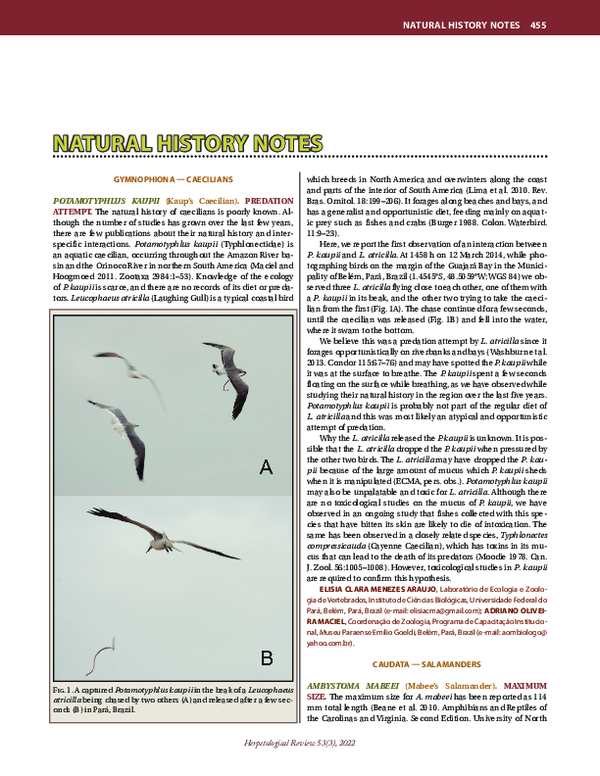
 Rodrigo Feitosa