Doklady Chemistry, Vol. 386, Nos. 4–6, 2002, pp. 251–254. Translated from Doklady Akademii Nauk, Vol. 386, No. 4, 2002, pp. 492–495.
Original Russian Text Copyright © 2002 by Bukalov, Mikhalitsyn, Leites.
CHEMISTRY
Unexpected Formation of an sp2-Carbon Needle from Dimethyl
Ether Vapor Under Mild Conditions on Exposure to Radiation
from a Low-Power Ar+ Laser
S. S. Bukalov, L. A. Mikhalitsyn, and L. A. Leites
Presented by Academician Yu.N. Bubnov June 13, 2002
Received June 13, 2002
The formation of carbon modifications from carbon-containing light molecules in the gas phase has
attracted considerable attention in recent years. In particular, processes of this type giving rise to diamond or
nondiamond carbon and proceeding at atmospheric
pressure but at high temperatures (1000–1500°ë) and
often in the presence of a catalyst are described in a
review [1].
We discovered the formation of a carbon needle
from dimethyl ether vapor, which took place at a pressure of ~1.5 atm and a temperature of 150°ë upon irradiation with a ~250-mW Ar+ laser at a wavelength of
514.5 nm during recording of the Raman spectrum.
Dimethyl ether was placed into a molybdate-glass
tube (wall thickness ~1 mm, diameter 25 mm) by freezing in a vacuum. The vapor pressure was about 1 atm.
Nitrogen, which was meant for measurement of the real
gas temperature in the laser beam (from the intensity
ratio of the Stokes and anti-Stokes lines in the rotational
Raman spectrum of nitrogen) was admitted into the
tube (at a pressure of 0.4 atm). The tube with this gas
mixture was sealed and placed in the chamber of the
Raman spectrometer. The tube with the gas was heated
by an electric furnace. The temperature was controlled
by a heating controller and measured by a thermocouple. The diameter of the laser beam at the inlet of the
tube was ~2.5 mm.
During recording of the Raman spectrum, periodic
bright flashes (approximately one flash per second)
started unexpectedly in the gas bulk exposed to the laser
beam. This process lasted for several minutes and then
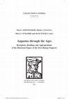
the experiment was stopped to avoid the possible explosion of the tube. Subsequent examination showed that a
thin black needle had formed inside the tube along the
laser beam. The needle was welded to the tube glass at
the point of laser beam entrance and was arranged
strictly at a right angle to the tube wall. It was several
mm shorter than the tube internal diameter, and its end
fell short of the opposite wall and remained free (see
Fig. 1). The needle had a circular cross section and gradually thinned along the length (from 0.20 to 0.12 mm
in diameter). Unfortunately, when the tube was taken
out of the spectrometer chamber, the needle broke off
from the tube wall and fell to the bottom, having split
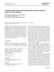
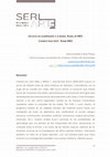
into several pieces. The photograph of the free tip of the
needle taken using an electron microscope is shown in
Fig. 2 with a magnification degree of 1500. It can be
seen that the tip surface is smooth and rounded. A fragment of the needle with the round tip (with a length of
6 mm and an average diameter of 0.15 mm) was studied
by X-ray powder diffraction using a Bruker Smart 1K
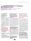
CCD diffractometer with an area detector (MoKα radiation, λ = 0.71073 Å). The X-ray diffraction pattern
exhibits three clear-cut maxima with different intensities (Fig. 3), which correspond to interplanar spacings
d ≈ 3.35, 2.08, and 1.70 Å. These distances are characteristic of the crystal lattice of hexagonal graphite. The
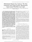
Vapors of
(CH3)2O
and N2
Laser beam
Lense
Needle
Nesmeyanov Institute of Organoelement Compounds,
Russian Academy of Sciences, ul. Vavilova 28, Moscow,
GSP-1, 117813 Russia
Fig. 1. Experimental setup (the tube with dimethyl ether
vapor in the chamber of the Raman spectrometer).
0012-5008/02/0010-0251$27.00 © 2002 åÄIä “Nauka /Interperiodica”
252
BUKALOV et al.
~ 20 µm
Fig. 3. X-ray diffraction pattern of the needle.
Fig. 2. Photograph of the free tip of the needle (magnification 1500×).
value of 3.35 Å, equal to the distance between graphite
layers in the unit cell, is especially typical [2, 3].
Raman spectroscopy is known to be a diagnostic
method for identification of various carbon polymorphs
[4, 5]. We recorded the Raman spectra of the needle
using a T64000 Jobin Yvon Raman spectrometer
equipped with a highly sensitive CCD detector cooled
by liquid nitrogen and a microscope with a TV camera.
The 514.5-nm line of a 1-mW Är+ laser was used for
excitation. The spectra were recorded both for the lateral surface of the needle and for the chip butt-end; neither of these contained a narrow line at 1332 cm–1 typical of diamond [4, 5].
The Raman spectrum of the needle butt-end
(Fig. 4a) exhibits a single first-order line at 1581 cm–1
(G line), which is typical of the spectrum of highly
ordered crystalline graphite [4–7]. This line corresponds to the Raman-active ν2 mode of the E2g class
[6]. The Raman spectrum of the lateral surface (Fig. 4b)
is markedly different and contains two broadened lines,
a line at 1588 cm–1 (G line) with a shoulder at 1619 cm–1
and a line at 1354 cm–1 (D line). The opinions concerning the origin of the latter line vary [5, 9]; however, it is
always attributed to defects or disorder in the crystal
structure of graphite. A Raman spectrum similar to that
shown in Fig. 4b is typical of synthetic diamond-like
carbon (DLC) films and of so-called glassy carbon [4,
6, 8, 9]. In particular, the spectrum of the needle surface
is very similar in line positions, intensities, and halfwidths to the Raman spectrum of the carbon part of natural schungite (glassy carbon), reported recently [8],
and also resembles the spectrum that we recorded for
the DLC films covering the crystalline germanium
whiskers formed from tetraalkylgermanes upon a
MOCVD procedure [10].
The micromap making performed by Raman spectroscopy indicates homogeneity of the needle surface.
These results provide the unambiguous conclusion
that the needle consists of sp2 carbon. It is ordered crystalline graphite coated by a film of so-called glassy carbon.
Unfortunately, we did not analyze the composition
of the gas formed due to a microcrack in the tube wall.
However, by analogy with [1], one can suggest that the
reaction proceeded as follows:
(CH3)2O
hν, 150°C
Cg + H2O + 2H2.
The nature of the periodic flashes in the laser beam
resulting in cleavage of strong CH and CO bonds in the
dimethyl ether molecule to give sp2 carbon (at a pressure not exceeding 1.5 atm, a temperature of 150°ë,
and relatively low power of laser radiation) has not yet
been elucidated and requires further investigation.
However, the results (confirmed by Raman spectroscopy and X-ray diffraction) demonstrate that this
decomposition can, in principle, take place under mild
conditions. It is also worth noting that we repeatedly
observed decomposition of various organic or organometallic compounds to give sp2 carbon (with appearance of intense Raman lines at ~1580 and ~1350 cm–1)
induced by radiation of an Ar+ or He–Ne laser during
DOKLADY CHEMISTRY
Vol. 386
Nos. 4–6
2002
UNEXPECTED FORMATION
253
1581
‡
1354
1588
b
1300
1400
1500
1600
∆ν, cm–1
Fig. 4. Raman spectra of (a) the butt-end and (b) the surface of the needle.
our extensive experience in recording Raman spectra at
the Science and Engineering Center for Raman Spectroscopy at the Russian Academy of Sciences.
This work was supported by the Russian Foundation
for Basic Research, project nos. 01–03–33057, 02–03–
06307, and 00–15–97307.
ACKNOWLEDGMENTS
REFERENCES
The authors are grateful to M.Yu. Antipin and
I.I. Vorontsov for performing X-ray diffraction analysis.
DOKLADY CHEMISTRY
Vol. 386
Nos. 4–6
2002
1. Rudenko, A.P., Kulakova, I.I., and Skvortsova, V.L.,
Usp. Khim., 1993, vol. 62, no. 2, pp. 99–102.
2. Kurdyumov, A.V. and Pilyankevich, A.N., Fazovye prevrashcheniya v uglerode i nitride bora (Phase Transi-
254
3.
4.
5.
6.
BUKALOV et al.
tions in Carbon and in Boron Nitride), Kiev: Naukova
Dumka, 1979, p. 235.
Ubbelode, A.R. and L’yuis, F.A., Grafit i ego kristallicheskie soedineniya (TRaNSl), Moscow: Mir, 1965,
p. 356.
Raman Microscopy. Development and Applications,
Turrell, G., Ed., London: Academic, 1996.
Lauer, J.L., Handbook of Raman Spectroscopy, New
York: Marcel Dekker, 2001, ch. 22, pp. 863–917.
Huong, P.V., Diamond Relat. Mater., 1991, no. 1,
pp. 33–41.
7. Tuinstra, F. and Koenig, J.L., J. Chem. Phys., 1970,
vol. 53, no. 3, pp. 1126–1130.
8. Kholodkevich, S.V., Berezkin, V.I., and Davydov, V.Yu.,
Fiz. Tverd. Tela, 1999, vol. 41, no. 8, pp. 1412–1415.
9. Knight, D.S. and White, W.B., J. Mater. Res., 1989,
vol. 4, pp. 385–393.
10. Bukalov, S.S., Mikhalitsyn, L.A., Leites, L.A., et al.,
Proc. Int. Conf. “New Approaches in Coordination and
Organometallic Chemistry. Look from XXI Century,”
Nizhny Novgorod, 2002, p. 115.
DOKLADY CHEMISTRY
Vol. 386
Nos. 4–6
2002
Keep reading this paper — and 50 million others — with a free Academia account
Used by leading Academics

Richard Gauthier
Santa Rosa Junior College

Duane Jaecks
University of Nebraska Lincoln

Sergey A Khaibrakhmanov
Saint-Petersburg State University

Daniel Condurache
Technical University of Iasi