Early epidural blood patch in
spontaneous intracranial hypotension
S. Berroir, MD; B. Loisel, MD; A. Ducros, MD; M. Boukobza, MD; C. Tzourio, MD;
D. Valade, MD; and M-G. Bousser, MD
Abstract—Thirty patients with a typical orthostatic headache were treated by early lumbar epidural blood patch (EBP)
without previously performing lumbar puncture or identifying a CSF leak and with or without typical MRI changes. A
complete cure was obtained in 77% of patients after one (57%) or two (20%) EBPs. Spontaneous intracranial hypotension
with typical orthostatic headache can be diagnosed without lumbar puncture and can be cured by early EBP in a majority
of patients.
NEUROLOGY 2004;63:1950 –1951
Spontaneous intracranial hypotension (SIH) is an
uncommon disabling condition occurring in the absence of an obvious dural tear. Its prominent clinical
feature is orthostatic headache, frequently associated with neck pain, nausea, vomiting, diplopia,
blurred vision, and distorted hearing.1-3 MRI abnormalities include diffuse pachymeningeal gadolinium
enhancement (PMGE), subdural hematomas or hygromas, and downward displacement of the cranial
contents.1-4
The role of lumbar puncture (LP) to demonstrate
low CSF pressure and the timing of further investigations to identify a leak are debated particularly
because they require a dural puncture that may
worsen the patient’s condition. Epidural blood patch
(EBP) is the most effective treatment,2,3,5 but its timing is also debated. We report a consecutive series of
30 patients with SIH and severe orthostatic headache treated with early lumbar EBP, even in the
absence of typical MRI changes, without previously
performing LP or looking for a leak.
Methods. Patients were included if they had a typical SIH defined as a severe purely orthostatic headache in the absence of
obvious causes of dural tear. Headache was defined as severe
when it interfered with daily activities and as purely orthostatic
when it occurred in ⬍15 minutes in the upright position and
disappeared in ⬍15 minutes with recumbency.
Brain MRI was performed using a 1.5-T system with unenhanced T1- and T2-weighted imaging and gadolinium-enhanced
T1-weighted imaging in the sagittal and coronal planes.
Once SIH diagnosis was established and after full informed
consent was obtained, a first EBP was done, followed by a second
in case of failure or relapse. After the failure of two to four EBPs,
CSF leak was looked for by MRI, CT myelography, and/or radioisotope cisternography.
The same anesthetist performed all EBPs under strict aseptic
conditions in an operating room. Up to 40 mL of the patients’ own
blood was slowly injected in L3-L4 or L4-L5 spaces and only was
stopped in case of severe lumbar pain. The patient remained supine for 2 hours and was asked to refrain from strenuous exercise
for 3 weeks. Follow-up evaluation was performed at 1 month and
yearly thereafter or more frequently if necessary. The duration of
the follow-up period was 1 to 4 years.
Results. Baseline characteristics. From July 1999 to
July 2002, 33 patients (21 women, 12 men; aged 15 to 68
years; mean, 40 years) were consecutively seen with SIH
and severe purely orthostatic headache; of these, 21 were
newly diagnosed in our Emergency Headache Center.
Mean time from onset to diagnosis was 20 ⫾ 15 days. Ten
patients reported physical effort as a triggering factor.
Four patients had headache exacerbation when coughing
or on exertion. Other symptoms included nausea and/or
vomiting in 23 patients (70%), neck pain in 16 (48%), hearing disturbances in 14 (42%), back pain in 3, and horizontal diplopia and drowsiness in 1.
Brain MRI (31 patients) showed diffuse PMGE in 19
patients (61%), a sagging brain and subdural collections in
11 (35%), and an isolated sagging brain in 1. MRI was
normal in 10 patients (32%).
Treatment and outcome.
Three patients did not receive EBP because their headache changed rapidly during
evaluation. One improved spontaneously in a few days,
and two others had sinus thrombosis and were treated
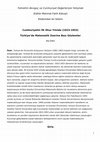
with heparin.6 Among the 30 patients who underwent a
lumbar EBP, 27 patients (90%) had immediate relief
(⬎90% on a verbal analog scale 0 to 10), and 3 did not
improve. No complication was observed. Among the 27
with immediate relief, 17 remained pain free at the end of
follow-up period, 1 was lost to follow-up evaluation, and 9
had a relapse within a few days to several weeks. In these
9 patients, the second EBP was followed with immediate
and sustained relief in 6 (20%); therefore, 23 (77%) patients were pain free after one or two EBPs (figure).
Three patients improved after the second EBP but relapsed. Investigations showed a leak at the T9, C7, and T8
levels. One patient was cured after surgery, and another
one was cured after five EBPs, including two at the site of
the leak. The third patient has a disc herniation at the T8
level for which surgery is still debated.
From the Service de Neurologie (Drs. Berroir, Tzourio, and Bousser), Département d’anesthésie reanimation (Dr. Loisel), Centre d’urgence céphalées
(Drs. Ducros and Valade), and Service de neuroradiologie (Dr. Boukobza), Lariboisière Hospital, Paris, France.
Received April 20, 2004. Accepted in final form July 2, 2004.
Address correspondence and reprint requests to Dr. Marie-Germaine Bousser, Service de Neurologie, Hôpital Lariboisière, 2 rue Ambroise Paré, 75475 Paris
cedex 10, France; e-mail: mg.bousser@lrb.ap-hop-paris.fr
1950 Copyright © 2004 by AAN Enterprises, Inc.
sentations of SIH, such as nonpositional,7 exertional,8 or even absent headache.9
Baseline characteristics of our patients are essentially similar to those reported in other large series:
1,9
female preponderance, mean age of ~40 years,
physical effort as triggering factor, and frequent associated nausea, neck pain, tinnitus, or hearing disturbances. The lower rate of typical MRI changes
(68%) compared with other series (⬎80%1,3,10) may be
because of the greater number of recent cases.
There is no consensus regarding the management
of SIH. In mild forms, conservative measures are
usually sufficient. In severe cases, such as ours,
there is little debate about the indication of EBP2,3,5
when PMGE is present on MRI,5 but when MRI is
normal, it is usually recommended to proceed with
additional diagnostic studies.1,5 However, because
these studies imply a dural puncture that may
worsen the patient’s condition, we choose to first perform one or two EBPs even in patients with normal
MRI and to postpone additional investigations.
The overall success rate after one or two lumbar
EBPs (77%) is less than the 90% observed in postlumbar puncture headache, probably because the
leaks, when present, are mostly thoracic and thus
distant from the EBP level. Our 77% success rate is
higher than the 56% observed in a Mayo Clinic series
of 25 patients, possibly because their patients were
more severe cases, had a documented CSF leak (implying a dural puncture), and received a smaller
quantity of blood (10 to 20 mL) in contrast to 20 to 40
mL in our series.
References
Figure. Flow chart of 30 patients with spontaneous intracranial hypotension treated using epidural blood patch.
Discussion. Thirty patients with SIH and severe
purely orthostatic headache received early lumbar
EBP, performed whenever the typical headache persisted after the end of the clinical and MRI evaluation period; 23 (77%) were cured after one (57%) or
two (20%) EBPs, with a follow-up period of 1 to 4
years.
The fact that these 33 patients were recruited
during a 3-year period suggests that SIH occurs
more frequently than classically thought, particularly because the present series, based on a severe
purely orthostatic headache, excluded unusual pre-
1. Mokri B, Piepgras DG, Miller GM. Syndrome of orthostatic headaches
and diffuse pachymeningeal gadolinium enhancement. Mayo Clin Proc
1997;72:400 – 413.
2. Mokri B. Spontaneous cerebrospinal fluid leaks: from intracranial hypotension to cerebrospinal fluid hypovolemia-evolution of a concept. Mayo
Clin Proc 1999;74:1113–1123.
3. Mokri B, Posner JB. Spontaneous intracranial hypotension. The broadening spectrum of CSF leaks. Neurology 2000;55:1771–1772.
4. Brightbill TC, Goodwin RS, Ford RG. Magnetic resonance imaging of
intracranial hypotension syndrome with pathophysiological correlation.
Headache 2000;40:292–299.
5. Sencakova D, Mokri B, McClelland RL. The efficacy of epidural blood
patch in CSF leaks. Neurology 2001;57:1921–1923.
6. Berroir S, Grabli D, Héran F, Bakouche P, Bousser MG. Cerebral sinus
venous thrombosis in two patients with spontaneous intracranial hypotension. Cerebrovasc Dis 2004;17:9 –12.
7. Schievink WI, Smith KA. Nonpositional headache caused by spontaneous intracranial hypotension. Neurology 1998;51:1768 –1769.
8. Mokri B. Spontaneous CSF leaks mimicking benign exertional headaches. Cephalalgia 2002;22:780 –783.
9. Mokri B, Atkinson JLD, Piepgras DG. Absent headache despite CSF
volume depletion (intracranial hypotension). Neurology 2000;55:1722–
1724.
10. Chung SJ, Kim JS, Lee M. Syndrome of cerebral fluid hypovolemia.
Clinical and imaging features and outcome. Neurology 2000;55:1321–
1327.
November (2 of 2) 2004
NEUROLOGY 63
1951
Early epidural blood patch in spontaneous intracranial hypotension
S. Berroir, B. Loisel, A. Ducros, et al.
Neurology 2004;63;1950-1951
DOI 10.1212/01.WNL.0000144339.34733.E9
This information is current as of November 22, 2004
Updated Information &
Services
including high resolution figures, can be found at:
http://www.neurology.org/content/63/10/1950.full.html
References
This article cites 10 articles, 6 of which you can access for free at:
http://www.neurology.org/content/63/10/1950.full.html##ref-list-1
Citations
This article has been cited by 13 HighWire-hosted articles:
http://www.neurology.org/content/63/10/1950.full.html##otherarticles
Subspecialty Collections
This article, along with others on similar topics, appears in the
following collection(s):
All Clinical trials
http://www.neurology.org//cgi/collection/all_clinical_trials
Clinical trials Observational study (Cohort, Case control)
http://www.neurology.org//cgi/collection/clinical_trials_observational_
study_cohort_case_control
Low pressure syndrome
http://www.neurology.org//cgi/collection/low_pressure_syndrome
Permissions & Licensing
Information about reproducing this article in parts (figures,tables) or in
its entirety can be found online at:
http://www.neurology.org/misc/about.xhtml#permissions
Reprints
Information about ordering reprints can be found online:
http://www.neurology.org/misc/addir.xhtml#reprintsus
Neurology ® is the official journal of the American Academy of Neurology. Published continuously since
1951, it is now a weekly with 48 issues per year. Copyright . All rights reserved. Print ISSN: 0028-3878.
Online ISSN: 1526-632X.
Keep reading this paper — and 50 million others — with a free Academia account
Used by leading Academics

Carlo Semenza
Università degli Studi di Padova

Ludwig Kappos
University of Basel, University Hospital

Arif Celebi
Bezmialem Vakif University

Rommy von Bernhardi
Pontificia Universidad Catolica de Chile