Location via proxy:   [ UP ]  
[Report a bug]   [Manage cookies]                
SlideShare a Scribd company logo
CARIES DIAGNOSIS
KALIISA EDWARD
BDS 4
Lecture objectives
 Detection and diagnosis on individual surfaces; smooth surfaces, pits and fissures,
approximal, secondary or recurrent caries
 Diagnosis of caries risk
 Caries activity tests
 Caries detection dyes
 Infected and affected dentine
 Latest advances in dental caries diagnosis
 Diagnosis is the art or act of inferring from signs and symptoms/ manifestations the nature and cause of
illness/ condition
 Caries detection is the process of identifying an area of demineralization.
 To identify caries lesion, tooth must be clean and dry using compressed air
STAGES IN CARIES DIAGNOSIS
 Detect the presence demineralization
 Establish the severity the depth of the lesions( Done using radiology)
 Determine whether the lesion is active or inactive
Requirements for diagnosis of caries:
history taking, clinical examination, lighting source, clean teeth calculus/plaque free ,three in one syringe,
sharp eyes with vision aided equipment, Dental mirror, Caries probe/explorer
Conventional methods of caries detection
include:1.Visual Method-we can see a white or brown spot
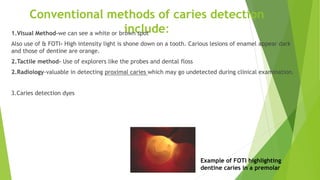
Also use of & FOTI- High intensity light is shone down on a tooth. Carious lesions of enamel appear dark
and those of dentine are orange.
2.Tactile method- Use of explorers like the probes and dental floss
2.Radiology-valuable in detecting proximal caries which may go undetected during clinical examination.
3.Caries detection dyes
Example of FOTI highlighting
dentine caries in a premolar
Smooth surface caries
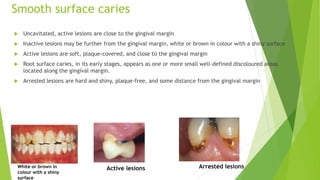
 Uncavitated, active lesions are close to the gingival margin
 Inactive lesions may be further from the gingival margin, white or brown in colour with a shiny surface
 Active lesions are soft, plaque-covered, and close to the gingival margin
 Root surface caries, in its early stages, appears as one or more small well-defined discoloured areas
located along the gingival margin.
 Arrested lesions are hard and shiny, plaque-free, and some distance from the gingival margin
White or brown in
colour with a shiny
surface
Active lesions Arrested lesions
Pits and fissures caries diagnosis
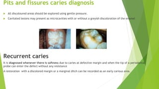
 All discoloured areas should be explored using gentle pressure.
 Cavitated lesions may present as microcavities with or without a greyish discoloration of the enamel
Recurrent caries
It is diagnosed whenever there is softness due to caries at defective margin and when the tip of a periodontal
probe can enter the defect without any resistance
A restoration with a discolored margin or a marginal ditch can be recorded as an early carious area.
APPROXIMAL SURFACES
• Difficult to detect enamel lesions because lesions formed just cervical to the
contact areas and vision is obscured by the adjacent tooth.
• A late stage lesion can be seen as a pinkish grey area shining through the marginal
ridge.
• Bite wing radiograph which appear as a dark triangular area in the enamel and
dentine. But cant detect cavities and caries activity
• Use of dental floss to detect rough areas, use an angled probe to detect cavities
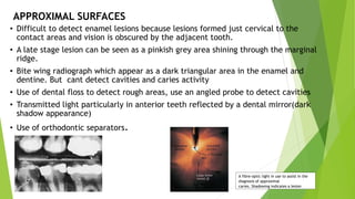
• Transmitted light particularly in anterior teeth reflected by a dental mirror(dark
shadow appearance)
• Use of orthodontic separators.
A fibre-optic light in use to assist in the
diagnosis of approximal
caries. Shadowing indicates a lesion
INFECTED AND AFFECTED DENTINE
INFECTED DENTINE AFFECTED DENTINE
Contaminated and contains the micro
organism with their toxins and demineralized
dentine
Not occupied by a micro organism
Contains the toxins and acids produced by
micro organisms of the infected dentine and
there is also demineralization.
Dentine is soft in texture Dentine is hard in texture
Contains irreversibly denatured collagen
stained by caries detection dye
Contains reversibly denatured collagen
Requires removal Does not require removal
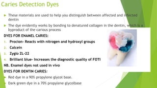
Caries Detection Dyes
 These materials are used to help you distinguish between affected and infected
dentin
 The dye evidently works by bonding to denatured collagen in the dentin, which is a
byproduct of the carious process
DYES FOR ENAMEL CARIES:
1. Procion- Reacts with nitrogen and hydroxyl groups
2. Calcein
3. Zyglo ZL-22
4. Brilliant blue- Increases the diagnostic quality of FOTI
NB. Enamel dyes not used in vivo
DYES FOR DENTIN CARIES:
 Red dye in a 90% propylene glycol base.
 Dark green dye in a 70% propylene glycolbase
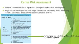
Caries Risk Assessment
 Involves determination of a patient’s susceptibility to caries development.
 A system was developed with 16 major risk factors. 11primary and 5 modifying
factors. Each has a direct or indirect influence on biofilm
1. Young age but with history of repeated restorat
2. Patients dentition
Caries Activity Tests.
Tests done to determine increment of active lesions over time
A. Microbiology tests
1. Lactobacilli colony count test: Saliva is cultured number of microorganisms formed determine the degree of
caries activity.
2. Calorimetric Snyder test which measure the ability of microorganisms to form organic acid in
carbohydrates.
3. Swab test based on the principle of Snyder test swab is taken from the teeth & incubated After 48 hrs is
read on a pH meter
4. Alban test. Patient expectorates saliva in test tube containing Alban test
medium. Incubated at 37C upto 4 days. colour change from green to
yellow is observed (YELLOW=caries activity, GREEN=INACTIVE)
5. S.Mutans in saliva
Tests for evaluating salivary defence
1. Salivary reductase test: measures the activity of this enzyme in salivary bacteria involved in formation of
dangerous products.
2. Salivary viscosity test
3. Salivary flow rate test
4. Salivary pH
5. Salivary buffer capacity
6. Fluoride level
7. Salivary amylase test
e..t.c
Latest advances in dental caries diagnosis.
 Laser fluorescence (DIAGNODent)-Uses the fluorescence of bacteria or metabolic products to detect a lesion.
 Fiberoptic transillumination (FOTI)
 Light Flourescence (QLF)-uses an intraoral camera to capture fluorescence. Enhances contrast btn sound &
demineralized enamel. We use blue light
 Digital Fibre optic transillumination (DiFOTI)- involves capturing of images.
 Ultrasound
 Electronic Caries Monitor
 Thermal imaging to view images as a result of heat production by organisms
 CarieScan PRO- pass current through a tooth to detect decay
 Caries ID- Detects optical behavior inside a tooth (uses infrared and red light)
 Bacteria detection dyes still under development
THE END

More Related Content

Caries diagnosis

  • 2. Lecture objectives  Detection and diagnosis on individual surfaces; smooth surfaces, pits and fissures, approximal, secondary or recurrent caries  Diagnosis of caries risk  Caries activity tests  Caries detection dyes  Infected and affected dentine  Latest advances in dental caries diagnosis
  • 3.  Diagnosis is the art or act of inferring from signs and symptoms/ manifestations the nature and cause of illness/ condition  Caries detection is the process of identifying an area of demineralization.  To identify caries lesion, tooth must be clean and dry using compressed air STAGES IN CARIES DIAGNOSIS  Detect the presence demineralization  Establish the severity the depth of the lesions( Done using radiology)  Determine whether the lesion is active or inactive Requirements for diagnosis of caries: history taking, clinical examination, lighting source, clean teeth calculus/plaque free ,three in one syringe, sharp eyes with vision aided equipment, Dental mirror, Caries probe/explorer
  • 4. Conventional methods of caries detection include:1.Visual Method-we can see a white or brown spot Also use of & FOTI- High intensity light is shone down on a tooth. Carious lesions of enamel appear dark and those of dentine are orange. 2.Tactile method- Use of explorers like the probes and dental floss 2.Radiology-valuable in detecting proximal caries which may go undetected during clinical examination. 3.Caries detection dyes Example of FOTI highlighting dentine caries in a premolar
  • 5. Smooth surface caries  Uncavitated, active lesions are close to the gingival margin  Inactive lesions may be further from the gingival margin, white or brown in colour with a shiny surface  Active lesions are soft, plaque-covered, and close to the gingival margin  Root surface caries, in its early stages, appears as one or more small well-defined discoloured areas located along the gingival margin.  Arrested lesions are hard and shiny, plaque-free, and some distance from the gingival margin White or brown in colour with a shiny surface Active lesions Arrested lesions
  • 6. Pits and fissures caries diagnosis  All discoloured areas should be explored using gentle pressure.  Cavitated lesions may present as microcavities with or without a greyish discoloration of the enamel Recurrent caries It is diagnosed whenever there is softness due to caries at defective margin and when the tip of a periodontal probe can enter the defect without any resistance A restoration with a discolored margin or a marginal ditch can be recorded as an early carious area.
  • 7. APPROXIMAL SURFACES • Difficult to detect enamel lesions because lesions formed just cervical to the contact areas and vision is obscured by the adjacent tooth. • A late stage lesion can be seen as a pinkish grey area shining through the marginal ridge. • Bite wing radiograph which appear as a dark triangular area in the enamel and dentine. But cant detect cavities and caries activity • Use of dental floss to detect rough areas, use an angled probe to detect cavities • Transmitted light particularly in anterior teeth reflected by a dental mirror(dark shadow appearance) • Use of orthodontic separators. A fibre-optic light in use to assist in the diagnosis of approximal caries. Shadowing indicates a lesion
  • 8. INFECTED AND AFFECTED DENTINE INFECTED DENTINE AFFECTED DENTINE Contaminated and contains the micro organism with their toxins and demineralized dentine Not occupied by a micro organism Contains the toxins and acids produced by micro organisms of the infected dentine and there is also demineralization. Dentine is soft in texture Dentine is hard in texture Contains irreversibly denatured collagen stained by caries detection dye Contains reversibly denatured collagen Requires removal Does not require removal
  • 9. Caries Detection Dyes  These materials are used to help you distinguish between affected and infected dentin  The dye evidently works by bonding to denatured collagen in the dentin, which is a byproduct of the carious process DYES FOR ENAMEL CARIES: 1. Procion- Reacts with nitrogen and hydroxyl groups 2. Calcein 3. Zyglo ZL-22 4. Brilliant blue- Increases the diagnostic quality of FOTI NB. Enamel dyes not used in vivo DYES FOR DENTIN CARIES:  Red dye in a 90% propylene glycol base.  Dark green dye in a 70% propylene glycolbase
  • 10. Caries Risk Assessment  Involves determination of a patient’s susceptibility to caries development.  A system was developed with 16 major risk factors. 11primary and 5 modifying factors. Each has a direct or indirect influence on biofilm 1. Young age but with history of repeated restorat 2. Patients dentition
  • 11. Caries Activity Tests. Tests done to determine increment of active lesions over time A. Microbiology tests 1. Lactobacilli colony count test: Saliva is cultured number of microorganisms formed determine the degree of caries activity. 2. Calorimetric Snyder test which measure the ability of microorganisms to form organic acid in carbohydrates. 3. Swab test based on the principle of Snyder test swab is taken from the teeth & incubated After 48 hrs is read on a pH meter 4. Alban test. Patient expectorates saliva in test tube containing Alban test medium. Incubated at 37C upto 4 days. colour change from green to yellow is observed (YELLOW=caries activity, GREEN=INACTIVE) 5. S.Mutans in saliva
  • 12. Tests for evaluating salivary defence 1. Salivary reductase test: measures the activity of this enzyme in salivary bacteria involved in formation of dangerous products. 2. Salivary viscosity test 3. Salivary flow rate test 4. Salivary pH 5. Salivary buffer capacity 6. Fluoride level 7. Salivary amylase test e..t.c
  • 13. Latest advances in dental caries diagnosis.  Laser fluorescence (DIAGNODent)-Uses the fluorescence of bacteria or metabolic products to detect a lesion.  Fiberoptic transillumination (FOTI)  Light Flourescence (QLF)-uses an intraoral camera to capture fluorescence. Enhances contrast btn sound & demineralized enamel. We use blue light  Digital Fibre optic transillumination (DiFOTI)- involves capturing of images.  Ultrasound  Electronic Caries Monitor  Thermal imaging to view images as a result of heat production by organisms  CarieScan PRO- pass current through a tooth to detect decay  Caries ID- Detects optical behavior inside a tooth (uses infrared and red light)  Bacteria detection dyes still under development