Location via proxy:   [ UP ]  
[Report a bug]   [Manage cookies]                
SlideShare a Scribd company logo
NEW TECHNOLOGIES IN
CARIES DIAGNOSIS
The Canary System in
Pediatric Practice


Dr. Ian McConnachie
AAPD Annual Session May 24-27, 2012
A couple of Gifts from the North
Disclosures


         • No Commercial Interest or Reimbursement
              from Quantum Dental Technologies

         • Member of Unpaid Dentist Advisory Panel to
              Quantum Dental Technologies

         • Some slides provided by QDT
Acknowledgements                                   Page 4


       • DR. STEPHEN ABRAMS
       President and CEO of Quantum Dental
       Technologies

       • DR. MARIELLE PARISEAU
       Creator of www.ShapingTheFutureofDentistry.org

       Note:
       The current final version of this presentation
       will be available on the Shaping the Future of
       Dentistry website later this week. Look in Menu
       under Resources
New Technologies in Caries Diagnosis


        • Diagnodent by KaVo
        • Caries ID by Dentsply
        • Spectra by Air Techniques
        • CarieScan by CarieScan Ltd
        • The Canary System by Quantum Dental
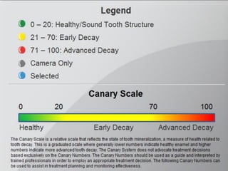
          Technologies
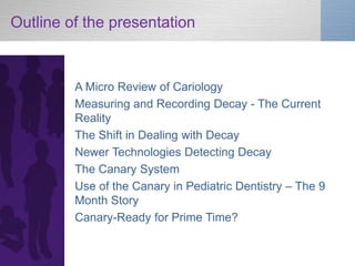
Outline of the presentation


         A Micro Review of Cariology
         Measuring and Recording Decay - The Current
         Reality
         The Shift in Dealing with Decay
         Newer Technologies Detecting Decay
         The Canary System
         Use of the Canary in Pediatric Dentistry – The 9
         Month Story
         Canary-Ready for Prime Time?
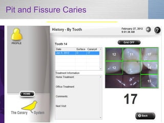
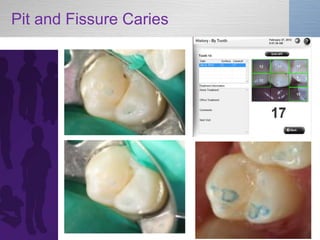
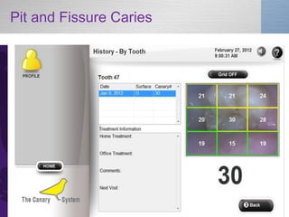
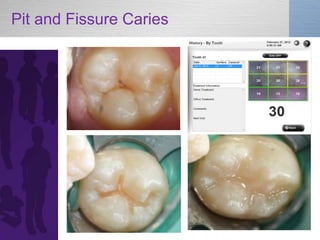
New Technologies in Caries Diagnosis: The Canary System in Pediatric Practice
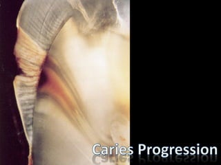
Early Carious Lesion in Enamel
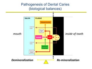
Pathogenesis of Dental Caries
           (biological balances)

         SALIVA              PLAQUE
                         PLAQUE                      ENAMEL
                                                      ENAMEL

                               Polysaccharides

              Calcium
              Salts
                             Plaque
                             buffers
 mouth                                                         inside of tooth
                         SUGARS               ACID


              Calcium
              Salts

                                  Bacterial
                                  Enzymes
              Salivary
              buffers


Demineralization                                        Re-mineralization
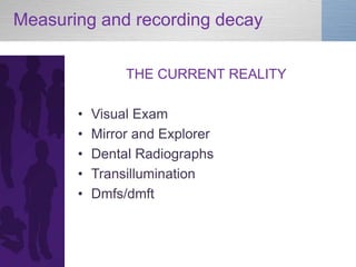
Measuring and recording decay

                THE CURRENT REALITY

       •   Visual Exam
       •   Mirror and Explorer
       •   Dental Radiographs
       •   Transillumination
       •   Dmfs/dmft
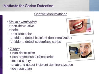
Methods for Caries Detection

                     Conventional methods

 • Visual examination:
   + non-destructive
   + safe
   - poor resolution
   - unable to detect incipient demineralization
   - unable to detect subsurface caries

  • X-rays:
    + non-destructive
    + can detect subsurface caries
    - limited safety
    - unable to detect incipient demineralization
    - low resolution
Radiographs
        • Radiographic imaging of pits and fissures is of minimal
          diagnostic value because of the large amounts of
          surrounding enamel .
        • Literature review by Dove:
        • “overall the strength of the evidence for radiographic
          methods for the detection of dental caries is poor for all
          types of lesions on proximal and occlusal surfaces”.
        • “it is beneficial only if the intervention is the surgical
          removal of tooth structure and detrimental if it is used
          for non-invasive remineralization methods.”
      McKnight-Hanes C, Myers DR, Dushku JC, Thompson WO, Durham LC. Radiographic
      recommendations for the primary dentition: comparison of general dentists and pediatric
      dentists. Pediatr Dent. 1990 Jul-Aug;12(4):212-216
       Flaitz CM, Hicks MJ, Silverston LM. Radiographic, histologic, and electronic comparison
      of basic mode videoprints with bitewing radiography. Caries Res. 1993; 27(1): 65-70.
      Lussi A, Comparison of different methods for the diagnosis of fissure caries without
      cavitation. Caries Res 27:409-16, 1993
      Dove,  S.  B.,  “Radiographic  Diagnosis  of  Dental  Caries  in  Consensus  Conference  on  
      Dental Caries Management Throughout Life, March 2001, Journal of Dental Education,
      2001; 65 (10): 985 – 990
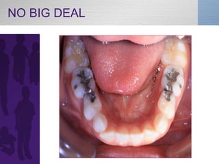
NO BIG DEAL
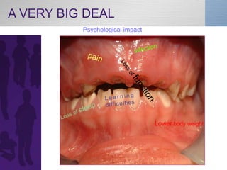
A VERY BIG DEAL
          Psychological impact


                                 Lower body weight
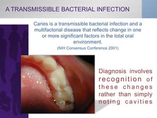
A TRANSMISSIBLE BACTERIAL INFECTION

       Caries is a transmissible bacterial infection and a
       multifactorial disease that reflects change in one
          or more significant factors in the total oral
                          environment.
                  (NIH Consensus Conference 2001)


                                      Diagnosis involves
                                      recognition of
                                      these changes
                                      rather than simply
                                      noting cavities
TOOTH DECAY
is   PREVENTABLE
BECAUSE FILLINGS


              Don’t          treat          underlying                   disease
              Don’t address plaque biofilm issues

              Don’t               change                    risk           level


 We need to   from a surgical approach to a RISK management & preventive approach.
The SHIFT in Dealing with Decay


       • Growing awareness of social
         determinants
       • Newer recording of caries levels-
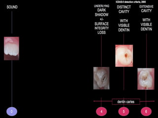
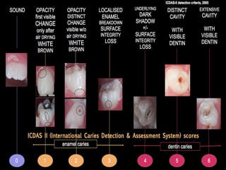
         ICDAS
       • Risk-based care
       • A myriad of new products
       • New diagnostic devices
“ It is change, continuing
change, inevitable change,
that is the dominant factor in
society today. No sensible
decision can be made any
longer without taking into
account not only the world
as it is, but the world as it
will be”
                 Isaac Asimov
New Technologies in Caries Diagnosis: The Canary System in Pediatric Practice
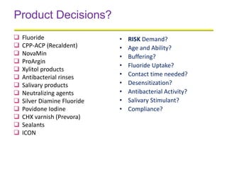
Product Decisions?
   Fluoride                  •   RISK Demand?
   CPP-ACP (Recaldent)       •   Age and Ability?
   NovaMin                   •   Buffering?
   ProArgin
                              •   Fluoride Uptake?
   Xylitol products
   Antibacterial rinses      •   Contact time needed?
   Salivary products         •   Desensitization?
   Neutralizing agents       •   Antibacterial Activity?
   Silver Diamine Fluoride   •   Salivary Stimulant?
   Povidone Iodine           •   Compliance?
   CHX varnish (Prevora)
   Sealants
   ICON
Newer Technologies
Detecting
Decay
The Value of Early Detection

        1. Is the ability to control the disease process in
           order

        1. To contain, arrest or remineralize lesions, in
         order

        2. To avoid or delay the burdens or costs
         associated with a spiral of restoration and re-
         restoration

               If a lesion is left to extend until a filling
              is needed, the clinical opportunity for
                     effective prevention is lost
New Technology Methods
           For Caries Detection

         Fluorescence-based Technology
         • Diagnodent
         • Caries ID
         • Quantitative Light-Induced Fluorescence
         • Spectra (QLF Technology)

         Digital Fibreoptic Transillumination (DIFOTI)

         Electrical Impedance Measurement
         • Caries Scan
The Characteristics of an Ideal
                       Caries Detection System

           Primary
           •   High sensitivity & specificity for caries detection
           •   Detects & monitors de & re-mineralization
           •   Detects smooth surface, root surface, occlusal
               surface & interproximal lesions
           •   Detects caries around restoration margins
           •   Non-invasive & safe
           •   Repeatable measurements
The Characteristics of an Ideal
                          Caries Detection System

           Secondary
           •   Imaging and or image capture
           •   System for recording & storing
               measurements
           •   Patient Education and Motivation
           •   In-vitro and in-vivo data & publications
           •   Minimal or no preparation of the tooth surface
               prior to taking a reading
           •   Ability to detect and monitor erosion lesions
The Characteristics of an Ideal
                          Caries Detection System


                  The key is to understand
                  what the device is measuring
Sensitivity and Specificity

         Sensitivity
         • The proportion of true positives correctly identified by the
           test
         Specificity
         • The proportion of true negatives correctly identified by
           the test


             Therefore, an experimental test aims to achieve
             100% sensitivity (no false positives) and 100%
             specificity (no false negatives)


             Because these are proportions, can calculate confidence
             intervals
             Closer the C.I. is to 1.0 the better
The Canary System
by Quantum Dental Technologies


                                 Canary interactive software
                                 and printed patient reports

     The Canary Console
Science Behind The Canary System

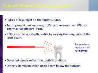
•Pulses of laser light hit the tooth surface.
•Tooth glows (Luminescence, LUM) and releases heat (Photo-
 Thermal Radiometry, PTR).
•PTR can provide a depth profile by varying the frequency of the
 laser beam.
                                                          Temperature
                                                          increase < 1oC
                                                          not harmful


•Detected  signals  reflect  the  tooth’s  condition.  
•Detects 50 micron lesion up to 5 mm below the surface.
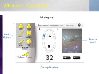
What it is - Screenshot

                  Odontogram


 Menu
Options
                                  Camera
                                   Image


                  Canary Number
Caries Mapping


                                   Camera
                                 Image with
                                    Grid


                 Canary Number
New Technologies in Caries Diagnosis: The Canary System in Pediatric Practice
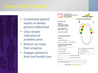
Canary Patient Report

       • Customized patient
         report on dental
         practice letterhead
       • Clear simple
         indication of
         problem areas
       • Patient can track
         their progress
       • Engages patient in
         their oral health care
Internet Connectivity


   • The Canary System equipped with wireless connect to
     the  ‘Canary  Cloud’.
   • Benefits for internet connectivity include:
      – Back-up and storage of data
      – Seamless software updates
      – Data and risk analysis for report generation
      – Online access of patient reports via Canary Web Portal
      – Access to data even when the system is being serviced or
        upgraded
      – Enables dentists to access all patient data among all dental
        operatories
      – HIPPA and PIPEDA Compliant
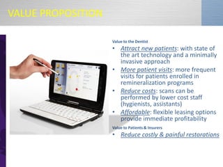
VALUE PROPOSITION

                    Value to the Dentist
                    • Attract new patients: with state of
                      the art technology and a minimally
                      invasive approach
                    • More patient visits: more frequent
                      visits for patients enrolled in
                      remineralization programs
                    • Reduce costs: scans can be
                      performed by lower cost staff
                      (hygienists, assistants)
                    • Affordable: flexible leasing options
                      provide immediate profitability
                    Value to Patients & Insurers
                    • Reduce costly & painful restorations
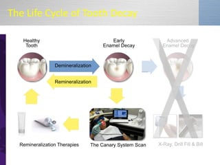
The Life Cycle of Tooth Decay

   Healthy                              Early               Advanced
    Tooth                            Enamel Decay          Enamel Decay


                 Demineralization


                 Remineralization


  Remineralization Therapies    The Canary System Scan   X-Ray, Drill Fill & Bill
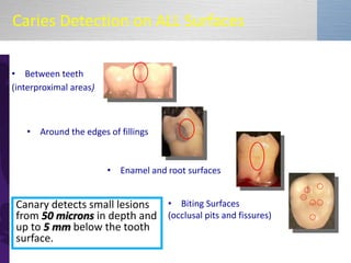
Caries Detection on ALL Surfaces

• Between teeth
(interproximal areas)


   • Around the edges of fillings


                        • Enamel and root surfaces


 Canary detects small lesions • Biting Surfaces
 from 50 microns in depth and (occlusal pits and fissures)
 up to 5 mm below the tooth
 surface.
Sensitivity and Specificity

         Sensitivity
         • Overall measurement in vitro 97%

         Specificity
         • Overall measurement in vitro 82%


            *Bench study Dr. B Amaechi UTSA
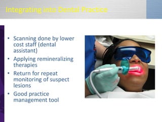
Integrating into Dental Practice


• Scanning done by lower
  cost staff (dental
  assistant)
• Applying remineralizing
  therapies
• Return for repeat
  monitoring of suspect
  lesions
• Good practice
  management tool
The Canary in Pediatric Practice –

         THE 9 MONTH STORY

         Our questions at the start

         •   How easy to measure
         •   How reproducible
         •   How accurate
         •   Canary Scale 21-70
         •   Specificity and Sensitivity
The Canary in Pediatric Practice –

         THE 9 MONTH STORY

         Lesions evaluated clinically

         Pit and fissure
         Interproximal
         Facial
         Under sealants
         ICON
         Under and around restorations
Pit and Fissure Caries
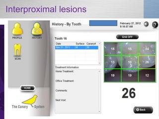
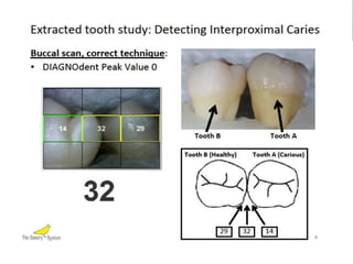
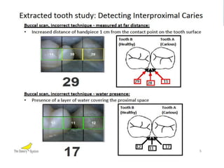
Interproximal lesions
                          Issues at Outset

                •   How easy to learn
                •   How reproducible the numbers
                •   Canary Scale 21-70
                •   Sensitivity and specificity
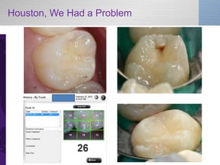
Houston, We Had a Problem
New Technologies in Caries Diagnosis: The Canary System in Pediatric Practice
Range of Measurements
                 Verified by Treatment

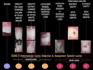
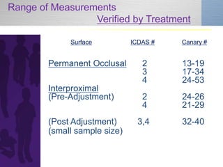
             Surface          ICDAS #   Canary #


        Permanent Occlusal       2      13-19
                                 3      17-34
                                 4      24-53
        Interproximal
        (Pre-Adjustment)         2      24-26
                                 4      21-29
        (Post Adjustment)       3,4     32-40
        (small sample size)
Interproximal lesions

              The key is to understand
               what the   device is measuring


            • Canary number will under
              report lesion size

            • Intervention decisions
              remain based on CAMBRA
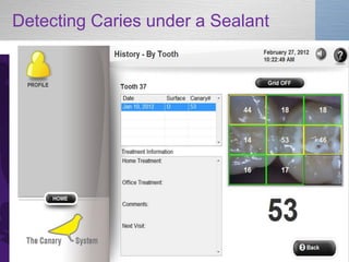
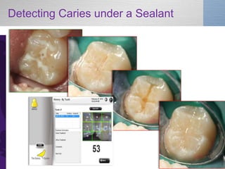
Detecting Caries under a Sealant

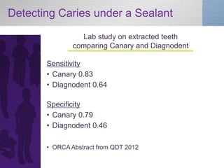
                 Lab study on extracted teeth
               comparing Canary and Diagnodent

       Sensitivity
       • Canary 0.83
       • Diagnodent 0.64

       Specificity
       • Canary 0.79
       • Diagnodent 0.46

       • ORCA Abstract from QDT 2012
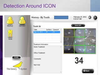
Detection Around ICON

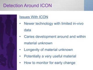
      Issues With ICON
      • Newer technology with limited in-vivo
        data
      • Caries development around and within
        material unknown
      • Longevity of material unknown
      • Potentially a very useful material
      • How to monitor for early change
Detection Around ICON
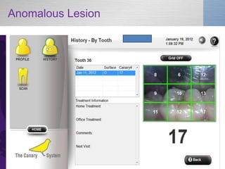
Anomalous Lesion
Anomalous Lesion

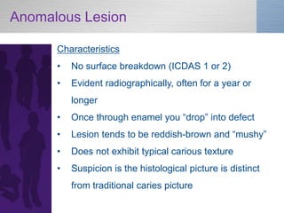
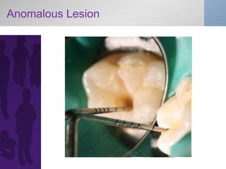
      Characteristics
      •   No surface breakdown (ICDAS 1 or 2)
      •   Evident radiographically, often for a year or
          longer
      •   Once  through  enamel  you  “drop”  into  defect
      •   Lesion tends to be reddish-brown  and  “mushy”
      •   Does not exhibit typical carious texture
      •   Suspicion is the histological picture is distinct
          from traditional caries picture
Anomalous Lesion
Anomalous Lesion – Preliminary                    Page
                                                   76
Observations

        • Canary does not accurately measure
         defect
        • Lesion appears to have a different
        pathology and suspected       different
         histological picture
        • More information required
Preliminary Observations on Canary
Overall

         Occlusal Surface
         • Highest ease of measurement and
            predictability
         Facial surface
         • Highest ease of measurement and
            predictability
         Interproximal Permanent Teeth
         • Significant learning curve
         • Lesion will be at least as deep as
            measurement indicates
Preliminary Observations on Canary
(cont’d)

         Interproximal Primary Teeth
         • Greater accuracy of Canary number
            compared to permanent teeth
         Under Sealants
         • Very good indicator of what lies beneath
         ICON
         • Likely good indicator of change with
            repeat scans
         Under and Around Restorations
         • Good indicator of restoration failure
Orthodontic Treatment –
              A Unique Opportunity
      Facts on Orthodontic-Related Decalcification and
      Caries
      •   Approximately 50% of ortho cases  end  with  “white  
          spot  lesions”
      •   Lesions can commence within 1 month of bracketing
      •   Braces can preclude bitewing radiographs for 3
          years or longer
      •   Risk-based preventive strategies exist
      •   Canary System an excellent diagnostic tool
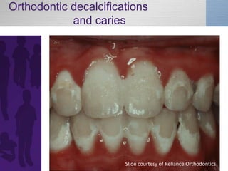
Orthodontic decalcifications
            and caries


                       Slide courtesy of Reliance Orthodontics
Orthodontic Treatment – Recommended
Canary Protocol
       •   Collaboration and communication triad established
           pre-orthodontics
       •   Baseline risk assessment
       •   Baseline bitewing radiographs
       •   If moderate or high risk then full Canary scan at
           baseline
       •   Rescan of interproximal and facial surfaces at 3-6
           months based on risk
       •   Customized preventive strategy for home, primary
           care office and orthodontic office
Dear Dr.                                                           Re: Patient

Our mutual patient was in recently for regular care. You will recall that he/she demonstrates a
higher risk for dental caries. As a result, we have initiated a customized preventive programme
for him/her while undergoing the orthodontic care under your supervision. Specific components
of this preventive programme include:

___        Higher fluoride toothpaste used at bedtime
___        More frequent dental hygiene visits for scaling, prophylaxis
___        More frequent dental hygiene visits for additional fluoride varnish application
___        Review of home hygiene techniques including use of floss and proxybrush
___        Scanning of at risk sites on teeth with the Canary System

The  current  review  of  ________’s  oral  hygiene  and  caries  status  reveals:

___        Oral hygiene is under control
___        Adjustments to the preventive programme are required and involve the following:

___        A rescan of the at risk sites is planned for ___ months

We appreciate your collaboration in the oral care for _______. Please contact our office if you
have concerns about anything for him/her.


Sincerely yours,
The Canary: Clinical Indications

       •   Complementary to bitewing radiographs – may
           reduce frequency need
       •   Monitor of remineralization therapy
       •   Monitor of margins of existing restorations
       •   Monitor of pre-cavitated lesions on all surfaces
       •   Complement to Caries Management by Risk
           Assessment
       •   Part of collaboration triad in orthodontic care
Office Integration
                                              Recall or Specific Exam
           Reassess 6 Months                  •Identify White Spots
           •Assess Lesion                     •ICDAS or Measure
           •ICDAS or Measure                  •Risk Assessment
                                              •Apply Remineralization
           •Apply
                                              Therapy
           Remineralization                   •Oral Hygiene
           Therapy                            Instruction
           •Dispense Home-                    •Provide Home-based
           Based Therapy                      Therapy


                               Reassess 3 Months
                               •Assess lesion
                               •ICDAS or Measure
                               •Apply
                               Remineralization
                               therapy
                               •Dispense Home-
                               based therapy
The Characteristics of an Ideal Caries Detection
System - How Does Canary Rate?


      Primary
      ?   - High sensitivity & specificity for caries detection
      YES - Detects & monitors de & re-mineralization
      YES BUT- Detects smooth surface, root surface, occlusal surface
             & interproximal lesions
      YES - Detects caries around restoration margins
      YES - Non-invasive & safe
      YES BUT - Repeatable measurements

          The key is to understand what the device is measuring.
The Characteristics of an Ideal Caries Detection System


      Secondary
      YES - Imaging and or image capture
      YES - System for recording & storing measurements
      YES - Patient Education and Motivation
      YES but More Needed - In-vitro and in-vivo data & publications
      YES BUT - Minimal or no preparation of the tooth surface prior
             to taking a reading
      ?   - Ability to detect and monitor erosion lesions


          The key is to understand what the device is measuring.
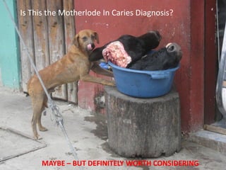
Is This the Motherlode In Caries Diagnosis?


      MAYBE – BUT DEFINITELY WORTH CONSIDERING
New Technologies in Caries Diagnosis: The Canary System in Pediatric Practice

More Related Content

New Technologies in Caries Diagnosis: The Canary System in Pediatric Practice

  • 1. NEW TECHNOLOGIES IN CARIES DIAGNOSIS The Canary System in Pediatric Practice Dr. Ian McConnachie AAPD Annual Session May 24-27, 2012
  • 2. A couple of Gifts from the North
  • 3. Disclosures • No Commercial Interest or Reimbursement from Quantum Dental Technologies • Member of Unpaid Dentist Advisory Panel to Quantum Dental Technologies • Some slides provided by QDT
  • 4. Acknowledgements Page 4 • DR. STEPHEN ABRAMS President and CEO of Quantum Dental Technologies • DR. MARIELLE PARISEAU Creator of www.ShapingTheFutureofDentistry.org Note: The current final version of this presentation will be available on the Shaping the Future of Dentistry website later this week. Look in Menu under Resources
  • 5. New Technologies in Caries Diagnosis • Diagnodent by KaVo • Caries ID by Dentsply • Spectra by Air Techniques • CarieScan by CarieScan Ltd • The Canary System by Quantum Dental Technologies
  • 6. Outline of the presentation A Micro Review of Cariology Measuring and Recording Decay - The Current Reality The Shift in Dealing with Decay Newer Technologies Detecting Decay The Canary System Use of the Canary in Pediatric Dentistry – The 9 Month Story Canary-Ready for Prime Time?
  • 9. Pathogenesis of Dental Caries (biological balances) SALIVA PLAQUE PLAQUE ENAMEL ENAMEL Polysaccharides Calcium Salts Plaque buffers mouth inside of tooth SUGARS ACID Calcium Salts Bacterial Enzymes Salivary buffers Demineralization Re-mineralization
  • 10. Measuring and recording decay THE CURRENT REALITY • Visual Exam • Mirror and Explorer • Dental Radiographs • Transillumination • Dmfs/dmft
  • 11. Methods for Caries Detection Conventional methods • Visual examination: + non-destructive + safe - poor resolution - unable to detect incipient demineralization - unable to detect subsurface caries • X-rays: + non-destructive + can detect subsurface caries - limited safety - unable to detect incipient demineralization - low resolution
  • 12. Radiographs • Radiographic imaging of pits and fissures is of minimal diagnostic value because of the large amounts of surrounding enamel . • Literature review by Dove: • “overall the strength of the evidence for radiographic methods for the detection of dental caries is poor for all types of lesions on proximal and occlusal surfaces”. • “it is beneficial only if the intervention is the surgical removal of tooth structure and detrimental if it is used for non-invasive remineralization methods.” McKnight-Hanes C, Myers DR, Dushku JC, Thompson WO, Durham LC. Radiographic recommendations for the primary dentition: comparison of general dentists and pediatric dentists. Pediatr Dent. 1990 Jul-Aug;12(4):212-216 Flaitz CM, Hicks MJ, Silverston LM. Radiographic, histologic, and electronic comparison of basic mode videoprints with bitewing radiography. Caries Res. 1993; 27(1): 65-70. Lussi A, Comparison of different methods for the diagnosis of fissure caries without cavitation. Caries Res 27:409-16, 1993 Dove,  S.  B.,  “Radiographic  Diagnosis  of  Dental  Caries  in  Consensus  Conference  on   Dental Caries Management Throughout Life, March 2001, Journal of Dental Education, 2001; 65 (10): 985 – 990
  • 14. A VERY BIG DEAL Psychological impact Lower body weight
  • 15. A TRANSMISSIBLE BACTERIAL INFECTION Caries is a transmissible bacterial infection and a multifactorial disease that reflects change in one or more significant factors in the total oral environment. (NIH Consensus Conference 2001) Diagnosis involves recognition of these changes rather than simply noting cavities
  • 16. TOOTH DECAY is PREVENTABLE
  • 17. BECAUSE FILLINGS Don’t treat underlying disease Don’t address plaque biofilm issues Don’t change risk level We need to from a surgical approach to a RISK management & preventive approach.
  • 18. The SHIFT in Dealing with Decay • Growing awareness of social determinants • Newer recording of caries levels- ICDAS • Risk-based care • A myriad of new products • New diagnostic devices
  • 19. “ It is change, continuing change, inevitable change, that is the dominant factor in society today. No sensible decision can be made any longer without taking into account not only the world as it is, but the world as it will be” Isaac Asimov
  • 22. Product Decisions?  Fluoride • RISK Demand?  CPP-ACP (Recaldent) • Age and Ability?  NovaMin • Buffering?  ProArgin • Fluoride Uptake?  Xylitol products  Antibacterial rinses • Contact time needed?  Salivary products • Desensitization?  Neutralizing agents • Antibacterial Activity?  Silver Diamine Fluoride • Salivary Stimulant?  Povidone Iodine • Compliance?  CHX varnish (Prevora)  Sealants  ICON
  • 24. The Value of Early Detection 1. Is the ability to control the disease process in order 1. To contain, arrest or remineralize lesions, in order 2. To avoid or delay the burdens or costs associated with a spiral of restoration and re- restoration If a lesion is left to extend until a filling is needed, the clinical opportunity for effective prevention is lost
  • 25. New Technology Methods For Caries Detection Fluorescence-based Technology • Diagnodent • Caries ID • Quantitative Light-Induced Fluorescence • Spectra (QLF Technology) Digital Fibreoptic Transillumination (DIFOTI) Electrical Impedance Measurement • Caries Scan
  • 26. The Characteristics of an Ideal Caries Detection System Primary • High sensitivity & specificity for caries detection • Detects & monitors de & re-mineralization • Detects smooth surface, root surface, occlusal surface & interproximal lesions • Detects caries around restoration margins • Non-invasive & safe • Repeatable measurements
  • 27. The Characteristics of an Ideal Caries Detection System Secondary • Imaging and or image capture • System for recording & storing measurements • Patient Education and Motivation • In-vitro and in-vivo data & publications • Minimal or no preparation of the tooth surface prior to taking a reading • Ability to detect and monitor erosion lesions
  • 28. The Characteristics of an Ideal Caries Detection System The key is to understand what the device is measuring
  • 29. Sensitivity and Specificity Sensitivity • The proportion of true positives correctly identified by the test Specificity • The proportion of true negatives correctly identified by the test Therefore, an experimental test aims to achieve 100% sensitivity (no false positives) and 100% specificity (no false negatives) Because these are proportions, can calculate confidence intervals Closer the C.I. is to 1.0 the better
  • 31. by Quantum Dental Technologies Canary interactive software and printed patient reports The Canary Console
  • 32. Science Behind The Canary System •Pulses of laser light hit the tooth surface. •Tooth glows (Luminescence, LUM) and releases heat (Photo- Thermal Radiometry, PTR). •PTR can provide a depth profile by varying the frequency of the laser beam. Temperature increase < 1oC not harmful •Detected  signals  reflect  the  tooth’s  condition.   •Detects 50 micron lesion up to 5 mm below the surface.
  • 33. What it is - Screenshot Odontogram Menu Options Camera Image Canary Number
  • 34. Caries Mapping Camera Image with Grid Canary Number
  • 36. Canary Patient Report • Customized patient report on dental practice letterhead • Clear simple indication of problem areas • Patient can track their progress • Engages patient in their oral health care
  • 37. Internet Connectivity • The Canary System equipped with wireless connect to the  ‘Canary  Cloud’. • Benefits for internet connectivity include: – Back-up and storage of data – Seamless software updates – Data and risk analysis for report generation – Online access of patient reports via Canary Web Portal – Access to data even when the system is being serviced or upgraded – Enables dentists to access all patient data among all dental operatories – HIPPA and PIPEDA Compliant
  • 38. VALUE PROPOSITION Value to the Dentist • Attract new patients: with state of the art technology and a minimally invasive approach • More patient visits: more frequent visits for patients enrolled in remineralization programs • Reduce costs: scans can be performed by lower cost staff (hygienists, assistants) • Affordable: flexible leasing options provide immediate profitability Value to Patients & Insurers • Reduce costly & painful restorations
  • 39. The Life Cycle of Tooth Decay Healthy Early Advanced Tooth Enamel Decay Enamel Decay Demineralization Remineralization Remineralization Therapies The Canary System Scan X-Ray, Drill Fill & Bill
  • 40. Caries Detection on ALL Surfaces • Between teeth (interproximal areas) • Around the edges of fillings • Enamel and root surfaces Canary detects small lesions • Biting Surfaces from 50 microns in depth and (occlusal pits and fissures) up to 5 mm below the tooth surface.
  • 41. Sensitivity and Specificity Sensitivity • Overall measurement in vitro 97% Specificity • Overall measurement in vitro 82% *Bench study Dr. B Amaechi UTSA
  • 42. Integrating into Dental Practice • Scanning done by lower cost staff (dental assistant) • Applying remineralizing therapies • Return for repeat monitoring of suspect lesions • Good practice management tool
  • 43. The Canary in Pediatric Practice – THE 9 MONTH STORY Our questions at the start • How easy to measure • How reproducible • How accurate • Canary Scale 21-70 • Specificity and Sensitivity
  • 44. The Canary in Pediatric Practice – THE 9 MONTH STORY Lesions evaluated clinically Pit and fissure Interproximal Facial Under sealants ICON Under and around restorations
  • 45. Pit and Fissure Caries
  • 46. Pit and Fissure Caries
  • 47. Pit and Fissure Caries
  • 48. Pit and Fissure Caries
  • 49. Interproximal lesions Issues at Outset • How easy to learn • How reproducible the numbers • Canary Scale 21-70 • Sensitivity and specificity
  • 50. Houston, We Had a Problem
  • 65. Range of Measurements Verified by Treatment Surface ICDAS # Canary # Permanent Occlusal 2 13-19 3 17-34 4 24-53 Interproximal (Pre-Adjustment) 2 24-26 4 21-29 (Post Adjustment) 3,4 32-40 (small sample size)
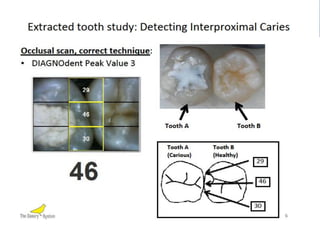
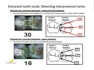
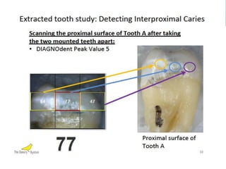
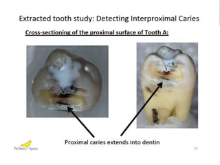
  • 66. Interproximal lesions The key is to understand what the device is measuring • Canary number will under report lesion size • Intervention decisions remain based on CAMBRA
  • 69. Detecting Caries under a Sealant Lab study on extracted teeth comparing Canary and Diagnodent Sensitivity • Canary 0.83 • Diagnodent 0.64 Specificity • Canary 0.79 • Diagnodent 0.46 • ORCA Abstract from QDT 2012
  • 70. Detection Around ICON Issues With ICON • Newer technology with limited in-vivo data • Caries development around and within material unknown • Longevity of material unknown • Potentially a very useful material • How to monitor for early change
  • 74. Anomalous Lesion Characteristics • No surface breakdown (ICDAS 1 or 2) • Evident radiographically, often for a year or longer • Once  through  enamel  you  “drop”  into  defect • Lesion tends to be reddish-brown  and  “mushy” • Does not exhibit typical carious texture • Suspicion is the histological picture is distinct from traditional caries picture
  • 76. Anomalous Lesion – Preliminary Page 76 Observations • Canary does not accurately measure defect • Lesion appears to have a different pathology and suspected different histological picture • More information required
  • 77. Preliminary Observations on Canary Overall Occlusal Surface • Highest ease of measurement and predictability Facial surface • Highest ease of measurement and predictability Interproximal Permanent Teeth • Significant learning curve • Lesion will be at least as deep as measurement indicates
  • 78. Preliminary Observations on Canary (cont’d) Interproximal Primary Teeth • Greater accuracy of Canary number compared to permanent teeth Under Sealants • Very good indicator of what lies beneath ICON • Likely good indicator of change with repeat scans Under and Around Restorations • Good indicator of restoration failure
  • 79. Orthodontic Treatment – A Unique Opportunity Facts on Orthodontic-Related Decalcification and Caries • Approximately 50% of ortho cases  end  with  “white   spot  lesions” • Lesions can commence within 1 month of bracketing • Braces can preclude bitewing radiographs for 3 years or longer • Risk-based preventive strategies exist • Canary System an excellent diagnostic tool
  • 80. Orthodontic decalcifications and caries Slide courtesy of Reliance Orthodontics
  • 81. Orthodontic Treatment – Recommended Canary Protocol • Collaboration and communication triad established pre-orthodontics • Baseline risk assessment • Baseline bitewing radiographs • If moderate or high risk then full Canary scan at baseline • Rescan of interproximal and facial surfaces at 3-6 months based on risk • Customized preventive strategy for home, primary care office and orthodontic office
  • 82. Dear Dr. Re: Patient Our mutual patient was in recently for regular care. You will recall that he/she demonstrates a higher risk for dental caries. As a result, we have initiated a customized preventive programme for him/her while undergoing the orthodontic care under your supervision. Specific components of this preventive programme include: ___ Higher fluoride toothpaste used at bedtime ___ More frequent dental hygiene visits for scaling, prophylaxis ___ More frequent dental hygiene visits for additional fluoride varnish application ___ Review of home hygiene techniques including use of floss and proxybrush ___ Scanning of at risk sites on teeth with the Canary System The  current  review  of  ________’s  oral  hygiene  and  caries  status  reveals: ___ Oral hygiene is under control ___ Adjustments to the preventive programme are required and involve the following: ___ A rescan of the at risk sites is planned for ___ months We appreciate your collaboration in the oral care for _______. Please contact our office if you have concerns about anything for him/her. Sincerely yours,
  • 83. The Canary: Clinical Indications • Complementary to bitewing radiographs – may reduce frequency need • Monitor of remineralization therapy • Monitor of margins of existing restorations • Monitor of pre-cavitated lesions on all surfaces • Complement to Caries Management by Risk Assessment • Part of collaboration triad in orthodontic care
  • 84. Office Integration Recall or Specific Exam Reassess 6 Months •Identify White Spots •Assess Lesion •ICDAS or Measure •ICDAS or Measure •Risk Assessment •Apply Remineralization •Apply Therapy Remineralization •Oral Hygiene Therapy Instruction •Dispense Home- •Provide Home-based Based Therapy Therapy Reassess 3 Months •Assess lesion •ICDAS or Measure •Apply Remineralization therapy •Dispense Home- based therapy
  • 85. The Characteristics of an Ideal Caries Detection System - How Does Canary Rate? Primary ? - High sensitivity & specificity for caries detection YES - Detects & monitors de & re-mineralization YES BUT- Detects smooth surface, root surface, occlusal surface & interproximal lesions YES - Detects caries around restoration margins YES - Non-invasive & safe YES BUT - Repeatable measurements The key is to understand what the device is measuring.
  • 86. The Characteristics of an Ideal Caries Detection System Secondary YES - Imaging and or image capture YES - System for recording & storing measurements YES - Patient Education and Motivation YES but More Needed - In-vitro and in-vivo data & publications YES BUT - Minimal or no preparation of the tooth surface prior to taking a reading ? - Ability to detect and monitor erosion lesions The key is to understand what the device is measuring.
  • 87. Is This the Motherlode In Caries Diagnosis? MAYBE – BUT DEFINITELY WORTH CONSIDERING