Location via proxy:   [ UP ]  
[Report a bug]   [Manage cookies]                
Next Issue
Volume 12, April
Previous Issue
Volume 12, February


Viruses, Volume 12, Issue 3 (March 2020) – 107 articles

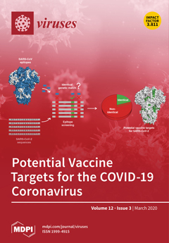
Cover Story (view full-size image): The COVID-19 outbreak, caused by severe acute respiratory syndrome coronavirus 2 (SARS-CoV-2), has rapidly become a global threat. Vaccines are widely recognized as important for controlling and preventing this pandemic. This work identifies potential vaccine targets against SARS-CoV-2. These targets are found by exploiting the genetic similarity between SARS-CoV-2 and SARS-CoV (the virus causing the 2003 SARS outbreak), together with existing immunological studies of SARS-CoV. It is shown that about 20% of the known SARS-derived T cell and B cell epitopes have an identical genetic match in SARS-CoV-2, comprising no mutation in the SARS-CoV-2 sequences available at the time of the study. These epitopes are promising candidates for triggering an effective cross-reactive immune response against SARS-CoV-2. View this paper.
  • Issues are regarded as officially published after their release is announced to the table of contents alert mailing list.
  • You may sign up for e-mail alerts to receive table of contents of newly released issues.
  • PDF is the official format for papers published in both, html and pdf forms. To view the papers in pdf format, click on the "PDF Full-text" link, and use the free Adobe Reader to open them.
Order results
Result details
Section
Select all
Export citation of selected articles as:
18 pages, 2719 KiB  
Article
Genetic Strain Diversity of Multi-Host RNA Viruses that Infect a Wide Range of Pollinators and Associates is Shaped by Geographic Origins
by Jana Dobelmann, Antoine Felden and Philip J. Lester
Viruses 2020, 12(3), 358; https://doi.org/10.3390/v12030358 - 24 Mar 2020
Cited by 22 | Viewed by 4827
Abstract
Emerging viruses have caused concerns about pollinator population declines, as multi-host RNA viruses may pose a health threat to pollinators and associated arthropods. In order to understand the ecology and impact these viruses have, we studied their host range and determined to what [...] Read more.
Emerging viruses have caused concerns about pollinator population declines, as multi-host RNA viruses may pose a health threat to pollinators and associated arthropods. In order to understand the ecology and impact these viruses have, we studied their host range and determined to what extent host and spatial variation affect strain diversity. Firstly, we used RT-PCR to screen pollinators and associates, including honey bees (Apis mellifera) and invasive Argentine ants (Linepithema humile), for virus presence and replication. We tested for the black queen cell virus (BQCV), deformed wing virus (DWV), and Kashmir bee virus (KBV) that were initially detected in bees, and the two recently discovered Linepithema humile bunya-like virus 1 (LhuBLV1) and Moku virus (MKV). DWV, KBV, and MKV were detected and replicated in a wide range of hosts and commonly co-infected hymenopterans. Secondly, we placed KBV and DWV in a global phylogeny with sequences from various countries and hosts to determine the association of geographic origin and host with shared ancestry. Both phylogenies showed strong geographic rather than host-specific clustering, suggesting frequent inter-species virus transmission. Transmission routes between hosts are largely unknown. Nonetheless, avoiding the introduction of non-native species and diseased pollinators appears important to limit spill overs and disease emergence. Full article
(This article belongs to the Section Invertebrate Viruses)
Show Figures

Graphical abstract

10 pages, 2387 KiB  
Article
Molecular Characterization of a Novel Strain of Fusarium graminearum Virus 1 Infecting Fusarium graminearum
by Lihang Zhang, Xiaoguang Chen, Pallab Bhattacharjee, Yue Shi, Lihua Guo and Shuangchao Wang
Viruses 2020, 12(3), 357; https://doi.org/10.3390/v12030357 - 24 Mar 2020
Cited by 13 | Viewed by 4497
Abstract
Fungal viruses (mycoviruses) have attracted more attention for their possible hypovirulence (attenuation of fungal virulence) trait, which may be developed as a biocontrol agent of plant pathogenic fungi. However, most discovered mycoviruses are asymptomatic in their hosts. In most cases, mycovirus hypovirulent factors [...] Read more.
Fungal viruses (mycoviruses) have attracted more attention for their possible hypovirulence (attenuation of fungal virulence) trait, which may be developed as a biocontrol agent of plant pathogenic fungi. However, most discovered mycoviruses are asymptomatic in their hosts. In most cases, mycovirus hypovirulent factors have not been explored clearly. In this study, we characterized a ssRNA mycovirus in Fusarium graminearum strain HB56-9. The complete nucleotide genome was obtained by combining random sequencing and rapid amplification of cDNA ends (RACE). The full genome was 6621-nucleotides long, excluding the poly(A) tail. The mycovirus was quite interesting because it shared 95.91% nucleotide identities with previously reported Fusarium graminearum virus 1 strain DK21 (FgV1-DK21), while the colony morphology of their fungal hosts on PDA plates were very different. The novel virus was named Fusarium graminearum virus 1 Chinese isolate (FgV1-ch). Like FgV1-DK21, FgV1-ch also contains four putative open reading frames (ORFs), including one long and three short ORFs. A phylogenetic analysis indicated that FgV1-ch is clustered into a proposed family Fusariviridae. FgV1-ch, unlike FgV1-DK21, had mild or no effects on host mycelial growth, spore production and virulence. The nucleotide differences between FgV1-ch and FgV1-DK21 will help to elucidate the hypovirulence determinants during mycovirus–host interaction. Full article
(This article belongs to the Collection Mycoviruses)
Show Figures

Figure 1

19 pages, 5647 KiB  
Article
Transcriptomic Analysis Suggests the M1 Polarization and Launch of Diverse Programmed Cell Death Pathways in Japanese Encephalitis Virus-Infected Macrophages
by Zhao-Yang Wang, Zi-Da Zhen, Dong-Ying Fan, Pei-Gang Wang and Jing An
Viruses 2020, 12(3), 356; https://doi.org/10.3390/v12030356 - 24 Mar 2020
Cited by 19 | Viewed by 3867
Abstract
The Japanese encephalitis virus (JEV) is a Culex mosquito-borne flavivirus and is the pathogenic agent of Japanese encephalitis, which is the most important type of viral encephalitis in the world. Macrophages are a type of pivotal innate immunocyte that serve as sentinels and [...] Read more.
The Japanese encephalitis virus (JEV) is a Culex mosquito-borne flavivirus and is the pathogenic agent of Japanese encephalitis, which is the most important type of viral encephalitis in the world. Macrophages are a type of pivotal innate immunocyte that serve as sentinels and respond quickly to pathogen invasions. However, some viruses like JEV can hijack macrophages as a refuge for viral replication and immune escape. Despite their crucial involvement in early JEV infection, the transcriptomic landscapes of JEV-infected macrophages are void. Here, by using an in situ JEV infection model, we investigate the transcriptomic alteration of JEV-infected peritoneal macrophages. We found that, upon JEV infection, the macrophages underwent M1 polarization and showed the drastic activation of innate immune and inflammatory pathways. Interestingly, almost all the programmed cell death (PCD) pathways were activated, especially the apoptosis, pyroptosis, and necroptosis pathways, which were verified by the immunofluorescent staining of specific markers. Further transcriptomic analysis and TUNEL staining revealed that JEV infection caused apparent DNA damage. The transcriptomic analysis also revealed that JEV infection promoted ROS and RNS generation and caused oxidative stress, which activated multiple cell death pathways. Our work uncovers the pivotal pathogenic roles of oxidative stress and multiple PCD pathways in JEV infection, providing a novel perspective on JEV–host interactions. Full article
Show Figures

Graphical abstract

14 pages, 2702 KiB  
Article
Protein Arginine N-methyltransferases 5 and 7 Promote HIV-1 Production
by Hironobu Murakami, Takehiro Suzuki, Kiyoto Tsuchiya, Hiroyuki Gatanaga, Manabu Taura, Eriko Kudo, Seiji Okada, Masami Takei, Kazumichi Kuroda, Tatsuo Yamamoto, Kyoji Hagiwara, Naoshi Dohmae and Yoko Aida
Viruses 2020, 12(3), 355; https://doi.org/10.3390/v12030355 - 23 Mar 2020
Cited by 10 | Viewed by 3321
Abstract
Current therapies for human immunodeficiency virus type 1 (HIV-1) do not completely eliminate viral reservoirs in cells, such as macrophages. The HIV-1 accessory protein viral protein R (Vpr) promotes virus production in macrophages, and the maintenance of Vpr is essential for HIV-1 replication [...] Read more.
Current therapies for human immunodeficiency virus type 1 (HIV-1) do not completely eliminate viral reservoirs in cells, such as macrophages. The HIV-1 accessory protein viral protein R (Vpr) promotes virus production in macrophages, and the maintenance of Vpr is essential for HIV-1 replication in these reservoir cells. We identified two novel Vpr-binding proteins, i.e., protein arginine N-methyltransferases (PRMTs) 5 and 7, using human monocyte-derived macrophages (MDMs). Both proteins found to be important for prevention of Vpr degradation by the proteasome; in the context of PRMT5 and PRMT7 knockdowns, degradation of Vpr could be prevented using a proteasome inhibitor. In MDMs infected with a wild-type strain, knockdown of PRMT5/PRMT7 and low expression of PRMT5 resulted in inefficient virus production like Vpr-deficient strain infections. Thus, our findings suggest that PRMT5 and PRMT7 support HIV-1 replication via maintenance of Vpr protein stability. Full article
(This article belongs to the Section Animal Viruses)
Show Figures

Figure 1

19 pages, 7917 KiB  
Article
Transcriptional Profiles Associated with Marek’s Disease Virus in Bursa and Spleen Lymphocytes Reveal Contrasting Immune Responses during Early Cytolytic Infection
by Huan Jin, Zimeng Kong, Arslan Mehboob, Bo Jiang, Jian Xu, Yunhong Cai, Wenxiao Liu, Jiabing Hong and Yongqing Li
Viruses 2020, 12(3), 354; https://doi.org/10.3390/v12030354 - 23 Mar 2020
Cited by 9 | Viewed by 3373
Abstract
Marek’s disease virus (MDV), an alpha herpes virus, causes a lymphoproliferative state in chickens known as Marek’s disease (MD), resulting in severe monetary losses to the poultry industry. Because lymphocytes of bursa of Fabricius and spleen are prime targets of MDV replication during [...] Read more.
Marek’s disease virus (MDV), an alpha herpes virus, causes a lymphoproliferative state in chickens known as Marek’s disease (MD), resulting in severe monetary losses to the poultry industry. Because lymphocytes of bursa of Fabricius and spleen are prime targets of MDV replication during the early cytolytic phase of infection, the immune response in bursa and spleen should be the foundation of late immunity induced by MDV. However, the mechanism of the MDV-mediated host immune response in lymphocytes in the early stage is poorly understood. The present study is primarily aimed at identifying the crucial genes and significant pathways involved in the immune response of chickens infected with MDV CVI988 and the very virulent RB1B (vvRB1B) strains. Using the RNA sequencing approach, we analyzed the generated transcriptomes from lymphocytes isolated from chicken bursa and spleen. Our findings validated the expression of previously characterized genes; however, they also revealed the expression of novel genes during the MDV-mediated immune response. The results showed that after challenge with CVI988 or vvRB1B strains, 634 and 313 differentially expressed genes (DEGs) were identified in splenic lymphocytes, respectively. However, 58 and 47 DEGs were observed in bursal lymphocytes infected with CVI988 and vvRB1B strains, respectively. Following MDV CVI988 or vvRB1B challenge, the bursal lymphocytes displayed changes in IL-6 and IL-4 gene expression. Surprisingly, splenic lymphocytes exhibited an overwhelming alteration in the expression of cytokines and cytokine receptors involved in immune response signaling. On the other hand, there was no distinct trend between infection with CVI988 and vvRB1B and the expression of cytokines and chemokines, such as IL-10, IFN-γ, STAT1, IRF1, CCL19, and CCL26. However, the expression profiles of IL-1β, IL-6, IL8L1, CCL4 (GGCL1), and CCL5 were significantly upregulated in splenic lymphocytes from chickens infected with CVI988 compared with those of chickens infected with vvRB1B. Because these cytokines and chemokines are considered to be associated with B cell activation and antigenic signal transduction to T cells, they may indicate differences of immune responses initiated by vaccinal and virulent strains during the early phase of infection. Collectively, our study provides valuable data on the transcriptional landscape using high-throughput sequencing to understand the different mechanism between vaccine-mediated protection and pathogenesis of virulent MDV in vivo. Full article
(This article belongs to the Section Animal Viruses)
Show Figures

Figure 1

23 pages, 1137 KiB  
Review
In Vitro Systems for Studying Different Genotypes/Sub-Genotypes of Hepatitis B Virus: Strengths and Limitations
by Constance N. Wose Kinge, Nimisha H. Bhoola and Anna Kramvis
Viruses 2020, 12(3), 353; https://doi.org/10.3390/v12030353 - 23 Mar 2020
Cited by 15 | Viewed by 6227
Abstract
Hepatitis B virus (HBV) infects the liver resulting in end stage liver disease, cirrhosis, and hepatocellular carcinoma. Despite an effective vaccine, HBV poses a serious health problem globally, accounting for 257 million chronic carriers. Unique features of HBV, including its narrow virus–host range [...] Read more.
Hepatitis B virus (HBV) infects the liver resulting in end stage liver disease, cirrhosis, and hepatocellular carcinoma. Despite an effective vaccine, HBV poses a serious health problem globally, accounting for 257 million chronic carriers. Unique features of HBV, including its narrow virus–host range and its hepatocyte tropism, have led to major challenges in the development of suitable in vivo and in vitro model systems to recapitulate the HBV replication cycle and to test various antiviral strategies. Moreover, HBV is classified into at least nine genotypes and 35 sub-genotypes with distinct geographical distributions and prevalence, which have different natural histories of infection, clinical manifestation, and response to current antiviral agents. Here, we review various in vitro systems used to study the molecular biology of the different (sub)genotypes of HBV and their response to antiviral agents, and we discuss their strengths and limitations. Despite the advances made, no system is ideal for pan-genotypic HBV research or drug development and therefore further improvement is required. It is necessary to establish a centralized repository of HBV-related generated materials, which are readily accessible to HBV researchers, with international collaboration toward advancement and development of in vitro model systems for testing new HBV antivirals to ensure their pan-genotypic and/or customized activity. Full article
(This article belongs to the Special Issue Hepatitis B Virus: From Diagnostics to Treatments)
Show Figures

Figure 1

15 pages, 1592 KiB  
Article
BoLA-DRB3 Polymorphism is Associated with Differential Susceptibility to Bovine Leukemia Virus-Induced Lymphoma and Proviral Load
by Chieh-Wen Lo, Liushiqi Borjigin, Susumu Saito, Koya Fukunaga, Etsuko Saitou, Katsunori Okazaki, Tetsuya Mizutani, Satoshi Wada, Shin-nosuke Takeshima and Yoko Aida
Viruses 2020, 12(3), 352; https://doi.org/10.3390/v12030352 - 22 Mar 2020
Cited by 56 | Viewed by 4135
Abstract
Bovine leukemia virus (BLV) is the causative agent of enzootic bovine leucosis. However, less than 5% of BLV-infected cattle will develop lymphoma, suggesting that, in addition to viral infection, host genetic polymorphisms might play a role in disease susceptibility. Bovine leukocyte antigen (BoLA) [...] Read more.
Bovine leukemia virus (BLV) is the causative agent of enzootic bovine leucosis. However, less than 5% of BLV-infected cattle will develop lymphoma, suggesting that, in addition to viral infection, host genetic polymorphisms might play a role in disease susceptibility. Bovine leukocyte antigen (BoLA)-DRB3 is a highly polymorphic gene associated with BLV proviral load (PVL) susceptibility. Due to the fact that PVL is positively associated with disease progression, it is believed that controlling PVL can prevent lymphoma development. Thus, many studies have focused on the relationship between PVL and BoLA-DRB3. Despite this, there is little information regarding the relationship between lymphoma and BoLA-DRB3. Furthermore, whether or not PVL-associated BoLA-DRB3 is linked to lymphoma-associated BoLA-DRB3 has not been clarified. Here, we investigated whether or not lymphoma-associated BoLA-DRB3 is correlated with PVL-associated BoLA-DRB3. We demonstrate that two BoLA-DRB3 alleles were specifically associated with lymphoma resistance (*010:01 and *011:01), but no lymphoma-specific susceptibility alleles were found; furthermore, two other alleles, *002:01 and *012:01, were associated with PVL resistance and susceptibility, respectively. In contrast, lymphoma and PVL shared two resistance-associated (DRB3*014:01:01 and *009:02) BoLA-DRB3 alleles. Interestingly, we found that PVL associated alleles, but not lymphoma associated alleles, are related with the anti-BLV gp51 antibody production level in cows. Overall, our study is the first to demonstrate that the BoLA-DRB3 polymorphism confers differential susceptibility to BLV-induced lymphoma and PVL. Full article
(This article belongs to the Section Animal Viruses)
Show Figures

Figure 1

17 pages, 4670 KiB  
Article
Antiviral Activity of Benzavir-2 against Emerging Flaviviruses
by Yong-Dae Gwon, Mårten Strand, Richard Lindqvist, Emma Nilsson, Michael Saleeb, Mikael Elofsson, Anna K. Överby and Magnus Evander
Viruses 2020, 12(3), 351; https://doi.org/10.3390/v12030351 - 22 Mar 2020
Cited by 12 | Viewed by 3911
Abstract
Most flaviviruses are arthropod-borne viruses, transmitted by either ticks or mosquitoes, and cause morbidity and mortality worldwide. They are endemic in many countries and have recently emerged in new regions, such as the Zika virus (ZIKV) in South-and Central America, the West Nile [...] Read more.
Most flaviviruses are arthropod-borne viruses, transmitted by either ticks or mosquitoes, and cause morbidity and mortality worldwide. They are endemic in many countries and have recently emerged in new regions, such as the Zika virus (ZIKV) in South-and Central America, the West Nile virus (WNV) in North America, and the Yellow fever virus (YFV) in Brazil and many African countries, highlighting the need for preparedness. Currently, there are no antiviral drugs available to treat flavivirus infections. We have previously discovered a broad-spectrum antiviral compound, benzavir-2, with potent antiviral activity against both DNA- and RNA-viruses. Our purpose was to investigate the inhibitory activity of benzavir-2 against flaviviruses. We used a ZIKV ZsGreen-expressing vector, two lineages of wild-type ZIKV, and other medically important flaviviruses. Benzavir-2 inhibited ZIKV derived reporter gene expression with an EC50 value of 0.8 ± 0.1 µM. Furthermore, ZIKV plaque formation, progeny virus production, and viral RNA expression were strongly inhibited. In addition, 2.5 µM of benzavir-2 reduced infection in vitro in three to five orders of magnitude for five other flaviviruses: WNV, YFV, the tick-borne encephalitis virus, Japanese encephalitis virus, and dengue virus. In conclusion, benzavir-2 was a potent inhibitor of flavivirus infection, which supported the broad-spectrum antiviral activity of benzavir-2. Full article
(This article belongs to the Section Viral Immunology, Vaccines, and Antivirals)
Show Figures

Figure 1

12 pages, 2216 KiB  
Article
Translation-Independent Roles of RNA Secondary Structures within the Replication Protein Coding Region of Turnip Crinkle Virus
by Rong Sun, Shaoyan Zhang, Limin Zheng and Feng Qu
Viruses 2020, 12(3), 350; https://doi.org/10.3390/v12030350 - 22 Mar 2020
Cited by 4 | Viewed by 2599
Abstract
RNA secondary structures play diverse roles in positive-sense (+) RNA virus infections, but those located with the replication protein coding sequence can be difficult to investigate. Structures that regulate the translation of replication proteins pose particular challenges, as their potential involvement in post-translational [...] Read more.
RNA secondary structures play diverse roles in positive-sense (+) RNA virus infections, but those located with the replication protein coding sequence can be difficult to investigate. Structures that regulate the translation of replication proteins pose particular challenges, as their potential involvement in post-translational steps cannot be easily discerned independent of their roles in regulating translation. In the current study, we attempted to overcome these difficulties by providing viral replication proteins in trans. Specifically, we modified the plant-infecting turnip crinkle virus (TCV) into variants that are unable to translate one (p88) or both (p28 and p88) replication proteins, and complemented their replication with the corresponding replication protein(s) produced from separate, non-replicating constructs. This approach permitted us to re-examine the p28/p88 coding region for potential RNA elements needed for TCV replication. We found that, while more than a third of the p88 coding sequence could be deleted without substantially affecting viral RNA levels, two relatively small regions, known as RSE and IRE, were essential for robust accumulation of TCV genomic RNA, but not subgenomic RNAs. In particular, the RSE element, found previously to be required for regulating the translational read-through of p28 stop codon to produce p88, contained sub-elements needed for efficient replication of the TCV genome. Application of this new approach in other viruses could reveal novel RNA secondary structures vital for viral multiplication. Full article
(This article belongs to the Section Viruses of Plants, Fungi and Protozoa)
Show Figures

Figure 1

9 pages, 2136 KiB  
Case Report
Human Papillomavirus (HPV69/HPV73) Coinfection associated with Simultaneous Squamous Cell Carcinoma of the Anus and Presumed Lung Metastasis
by Stephanie Shea, Marina Muñoz, Stephen C. Ward, Mary B. Beasley, Melissa R Gitman, Michael D Nowak, Jane Houldsworth, Emilia Mia Sordillo, Juan David Ramirez and Alberto E. Paniz Mondolfi
Viruses 2020, 12(3), 349; https://doi.org/10.3390/v12030349 - 22 Mar 2020
Cited by 3 | Viewed by 3698
Abstract
Background: Human papillomaviruses (HPVs) have been linked to a variety of human cancers. As the landscape of HPV-related neoplasia continues to expand, uncommon and rare HPV genotypes have also started to emerge. Host-virus interplay is recognized as a key driver in HPV carcinogenesis, [...] Read more.
Background: Human papillomaviruses (HPVs) have been linked to a variety of human cancers. As the landscape of HPV-related neoplasia continues to expand, uncommon and rare HPV genotypes have also started to emerge. Host-virus interplay is recognized as a key driver in HPV carcinogenesis, with host immune status, virus genetic variants and coinfection highly influencing the dynamics of malignant transformation. Immunosuppression and tissue tropism are also known to influence HPV pathogenesis. Methods: Herein, we present a case of a patient who, in the setting of HIV positivity, developed anal squamous cell carcinoma associated with HPV69 and later developed squamous cell carcinoma in the lungs, clinically presumed to be metastatic disease, associated with HPV73. Consensus PCR screening for HPV was performed by real-time PCR amplification of the L1 gene region, amplification of the E6 regions with High-Resolution Melting Curve Analysis followed by Sanger sequencing confirmation and phylogenetic analysis. Results: Sanger sequencing of the consensus PCR amplification product determined that the anal tissue sample was positive for HPV 69, and the lung tissue sample was positive for HPV 73. Conclusions: This case underscores the importance of recognizing the emerging role of these rare “possibly carcinogenic” HPV types in human carcinogenesis. Full article
Show Figures

Figure 1

12 pages, 928 KiB  
Review
Fluorescence Microscopy of the HIV-1 Envelope
by Pablo Carravilla, José L. Nieva and Christian Eggeling
Viruses 2020, 12(3), 348; https://doi.org/10.3390/v12030348 - 21 Mar 2020
Cited by 7 | Viewed by 5225
Abstract
Human immunodeficiency virus (HIV) infection constitutes a major health and social issue worldwide. HIV infects cells by fusing its envelope with the target cell plasma membrane. This process is mediated by the viral Env glycoprotein and depends on the envelope lipid composition. Fluorescent [...] Read more.
Human immunodeficiency virus (HIV) infection constitutes a major health and social issue worldwide. HIV infects cells by fusing its envelope with the target cell plasma membrane. This process is mediated by the viral Env glycoprotein and depends on the envelope lipid composition. Fluorescent microscopy has been employed to investigate the envelope properties, and the processes of viral assembly and fusion, but the application of this technique to the study of HIV is still limited by a number of factors, such as the small size of HIV virions or the difficulty to label the envelope components. Here, we review fluorescence imaging studies of the envelope lipids and proteins, focusing on labelling strategies and model systems. Full article
(This article belongs to the Special Issue Mechanisms of Viral Fusion and Applications in Antivirals)
Show Figures

Figure 1

17 pages, 3309 KiB  
Article
Identification of Immunogenic Epitopes That Permit the Detection of Antigen-Specific T Cell Responses in Multiple Serotypes of Group B Coxsackievirus Infections
by Ninaad Lasrado, Arunakumar Gangaplara, Rajkumar Arumugam, Chandirasegaran Massilamany, Sayli Pokal, Yuzhen Zhou, Shi-Hua Xiang, David Steffen and Jay Reddy
Viruses 2020, 12(3), 347; https://doi.org/10.3390/v12030347 - 21 Mar 2020
Cited by 13 | Viewed by 4394
Abstract
Coxsackievirus group B (CVB) contains six serotypes that can affect various organs. Some of these organ-specific diseases such as myocarditis and pancreatitis can be caused by more than one serotype. Thus, development of immunological tools common to multiple serotypes is desired. This is [...] Read more.
Coxsackievirus group B (CVB) contains six serotypes that can affect various organs. Some of these organ-specific diseases such as myocarditis and pancreatitis can be caused by more than one serotype. Thus, development of immunological tools common to multiple serotypes is desired. This is especially critical for analyzing antigen-specific T cell responses at a single cell level. To this end, we made efforts to identify the immunogenic epitopes of CVB3 leading us to localize three T cell epitopes within the viral protein 1 (VP1) namely, VP1 681–700, VP1 721–740 and VP1 771–790. First, we confirmed their immunogenicity in the immunization settings. Second, we sought to verify the ability of VP1 epitopes to bind major histocompatibility complex (MHC) class II (IAk) molecules. Third, we created MHC class II (IAk) dextramers and tetramers and ascertained the T cell responses to be antigen-specific. Fourth, we analyzed the T cell responses in animals infected with CVB3 and noted the magnitude of antigen-specific T cell responses occurring in the order of VP1 721–740 and VP1 681–700 followed by VP1 771–790 as verified by proliferation assay and IAk tetramer staining. All epitopes induced interferon (IFN)-γ as a major cytokine. Finally, we investigated whether the VP1 tools generated for CVB3 can also be used to verify T cell responses in infections caused by other serotypes. To this end, we established the CVB4 infection model in A/J mice and found that the CVB4 infection led to the induction of IFN-γ-producing T cell responses primarily for VP1 721–740 and VP1 681–700. Thus, the VP1-specific tools, particularly IAk tetramers can be used to monitor anti-viral T cell responses in multiple CVB serotypes. Full article
(This article belongs to the Section Animal Viruses)
Show Figures

Figure 1

21 pages, 3916 KiB  
Article
The E3 Ubiquitin Ligase TRIM21 Promotes HBV DNA Polymerase Degradation
by Ting Mu, Xiaoqing Zhao, Yanan Zhu, Hongxia Fan and Hua Tang
Viruses 2020, 12(3), 346; https://doi.org/10.3390/v12030346 - 21 Mar 2020
Cited by 44 | Viewed by 5508
Abstract
The tripartite motif (TRIM) protein family is an E3 ubiquitin ligase family. Recent reports have indicated that some TRIM proteins have antiviral functions, especially against retroviruses. However, most studies mainly focus on the relationship between TRIM21 and interferon or other antiviral effectors. The [...] Read more.
The tripartite motif (TRIM) protein family is an E3 ubiquitin ligase family. Recent reports have indicated that some TRIM proteins have antiviral functions, especially against retroviruses. However, most studies mainly focus on the relationship between TRIM21 and interferon or other antiviral effectors. The effect of TRIM21 on virus-encoded proteins remains unclear. In this study, we screened candidate interacting proteins of HBV DNA polymerase (Pol) by FLAG affinity purification and mass spectrometry assay and identified TRIM21 as its regulator. We used a coimmunoprecipitation (co-IP) assay to demonstrate that TRIM21 interacted with the TP domain of HBV DNA Pol. In addition, TRIM21 promoted the ubiquitination and degradation of HBV DNA Pol using its RING domain, which has E3 ubiquitin ligase activity. Lys260 and Lys283 of HBV DNA Pol were identified as targets for ubiquitination mediated by TRIM21. Finally, we uncovered that TRIM21 degrades HBV DNA Pol to restrict HBV DNA replication, and its SPRY domain is critical for this activity. Taken together, our results indicate that TRIM21 suppresses HBV DNA replication mainly by promoting the ubiquitination of HBV DNA Pol, which may provide a new potential target for the treatment of HBV. Full article
(This article belongs to the Section Animal Viruses)
Show Figures

Figure 1

18 pages, 1780 KiB  
Review
Non-Coding RNAs and Their Role in Respiratory Syncytial Virus (RSV) and Human Metapneumovirus (hMPV) Infections
by Wenzhe Wu, Eun-Jin Choi, Inhan Lee, Yong Sun Lee and Xiaoyong Bao
Viruses 2020, 12(3), 345; https://doi.org/10.3390/v12030345 - 21 Mar 2020
Cited by 31 | Viewed by 6208
Abstract
Recent high-throughput sequencing revealed that only 2% of the transcribed human genome codes for proteins, while the majority of transcriptional products are non-coding RNAs (ncRNAs). Herein, we review the current knowledge regarding ncRNAs, both host- and virus-derived, and their role in respiratory syncytial [...] Read more.
Recent high-throughput sequencing revealed that only 2% of the transcribed human genome codes for proteins, while the majority of transcriptional products are non-coding RNAs (ncRNAs). Herein, we review the current knowledge regarding ncRNAs, both host- and virus-derived, and their role in respiratory syncytial virus (RSV) and human metapneumovirus (hMPV) infections. RSV is known as the most common cause of lower respiratory tract infection (LRTI) in children, while hMPV is also a significant contributor to LRTI in the pediatrics population. Although RSV and hMPV are close members, belonging to the Pneumoviridae family, they induce distinct changes in the ncRNA profile. Several types of host ncRNAs, including long ncRNA (lncRNA), microRNAs (miRNAs), and transfer RNA (tRNA)-derived RNA fragments (tRFs), are involved as playing roles in RSV and/or hMPV infection. Given the importance of ncRNAs in regulating the expression and functions of genes and proteins, comprehensively understanding the roles of ncRNAs in RSV/hMPV infection could shed light upon the disease mechanisms of RSV and hMPV, potentially providing insights into the development of prevention strategies and antiviral therapy. The presence of viral-derived RNAs and the potential of using ncRNAs as diagnostic biomarkers are also discussed in this review. Full article
(This article belongs to the Special Issue From RSV to hMPV: Role of Innate Immunity in Pathogenesis)
Show Figures

Figure 1

16 pages, 2781 KiB  
Article
Berberine Hampers Influenza A Replication through Inhibition of MAPK/ERK Pathway
by Paweł Botwina, Katarzyna Owczarek, Zenon Rajfur, Marek Ochman, Maciej Urlik, Maria Nowakowska, Krzysztof Szczubiałka and Krzysztof Pyrc
Viruses 2020, 12(3), 344; https://doi.org/10.3390/v12030344 - 21 Mar 2020
Cited by 23 | Viewed by 5174
Abstract
Background: Berberine (BBR) is an isoquinoline alkaloid which exhibits a variety of biological and therapeutic properties, and has been reported by some to block replication of the influenza virus. However, contradictory results have also been presented, and the mechanistic explanation is lacking. Methods: [...] Read more.
Background: Berberine (BBR) is an isoquinoline alkaloid which exhibits a variety of biological and therapeutic properties, and has been reported by some to block replication of the influenza virus. However, contradictory results have also been presented, and the mechanistic explanation is lacking. Methods: A panel of cell lines (Madin–Darby canine kidney (MDCK), adenocarcinoma human alveolar basal epithelial cells (A549), lung epithelial type I (LET1)) and primary human airway epithelial cells (HAE) susceptible to influenza virus infection were infected with a seasonal influenza A virus in the presence or absence of BBR. Cytotoxicity towards cell lines was measured using XTT assay. The yield of the virus was analyzed using RT-qPCR. To study the molecular mechanism of BBR, confocal microscopy and Western blot analyses of cellular fractions were applied. Results and conclusions: Our results show cell-type-dependent anti-influenza properties of BBR in vitro which suggests that the compound acts on the cell and not the virus. Importantly, BBR hampers influenza replication in primary human airway epithelium 3D cultures that mimic the natural replication site of the virus. Studies show that the influenza A virus upregulates the mitogen-activated protein kinase/extracellular signal-related kinase (MAPK/ERK) pathway and hijacks this pathway for nucleolar export of the viral ribonucleoprotein. Our results suggest that BBR interferes with this process and hampers influenza A replication. Full article
(This article belongs to the Section Viral Immunology, Vaccines, and Antivirals)
Show Figures

Graphical abstract

14 pages, 2199 KiB  
Communication
Differential Localization of Structural and Non-Structural Proteins during the Bluetongue Virus Replication Cycle
by Bjorn-Patrick Mohl, Adeline Kerviel, Thomas Labadie, Eiko Matsuo and Polly Roy
Viruses 2020, 12(3), 343; https://doi.org/10.3390/v12030343 - 20 Mar 2020
Cited by 9 | Viewed by 3393
Abstract
Members of the Reoviridae family assemble virus factories within the cytoplasm of infected cells to replicate and assemble virus particles. Bluetongue virus (BTV) forms virus inclusion bodies (VIBs) that are aggregates of viral RNA, certain viral proteins, and host factors, and have been [...] Read more.
Members of the Reoviridae family assemble virus factories within the cytoplasm of infected cells to replicate and assemble virus particles. Bluetongue virus (BTV) forms virus inclusion bodies (VIBs) that are aggregates of viral RNA, certain viral proteins, and host factors, and have been shown to be sites of the initial assembly of transcriptionally active virus-like particles. This study sought to characterize the formation, composition, and ultrastructure of VIBs, particularly in relation to virus replication. In this study we have utilized various microscopic techniques, including structured illumination microscopy, and virological assays to show for the first time that the outer capsid protein VP5, which is essential for virus maturation, is also associated with VIBs. The addition of VP5 to assembled virus cores exiting VIBs is required to arrest transcriptionally active core particles, facilitating virus maturation. Furthermore, we observed a time-dependent association of the glycosylated non-structural protein 3 (NS3) with VIBs, and report on the importance of the two polybasic motifs within NS3 that facilitate virus trafficking and egress from infected cells at the plasma membrane. Thus, the presence of VP5 and the dynamic nature of NS3 association with VIBs that we report here provide novel insight into these previously less well-characterized processes. Full article
(This article belongs to the Section Animal Viruses)
Show Figures

Figure 1

16 pages, 2002 KiB  
Review
Structural Insight into Paramyxovirus and Pneumovirus Entry Inhibition
by Megha Aggarwal and Richard K Plemper
Viruses 2020, 12(3), 342; https://doi.org/10.3390/v12030342 - 20 Mar 2020
Cited by 14 | Viewed by 5300
Abstract
Paramyxoviruses and pneumoviruses infect cells through fusion (F) protein-mediated merger of the viral envelope with target membranes. Members of these families include a range of major human and animal pathogens, such as respiratory syncytial virus (RSV), measles virus (MeV), human parainfluenza viruses (HPIVs), [...] Read more.
Paramyxoviruses and pneumoviruses infect cells through fusion (F) protein-mediated merger of the viral envelope with target membranes. Members of these families include a range of major human and animal pathogens, such as respiratory syncytial virus (RSV), measles virus (MeV), human parainfluenza viruses (HPIVs), and highly pathogenic Nipah virus (NiV). High-resolution F protein structures in both the metastable pre- and the postfusion conformation have been solved for several members of the families and a number of F-targeting entry inhibitors have progressed to advanced development or clinical testing. However, small-molecule RSV entry inhibitors have overall disappointed in clinical trials and viral resistance developed rapidly in experimental settings and patients, raising the question of whether the available structural information may provide a path to counteract viral escape through proactive inhibitor engineering. This article will summarize current mechanistic insight into F-mediated membrane fusion and examine the contribution of structural information to the development of small-molecule F inhibitors. Implications are outlined for future drug target selection and rational drug engineering strategies. Full article
(This article belongs to the Special Issue Mechanisms of Viral Fusion and Applications in Antivirals)
Show Figures

Figure 1

2 pages, 152 KiB  
Editorial
Special Issue “Recent Advances in Morbillivirus Vaccine Development and Oncolytic Virotherapy”
by Liang-Tzung Lin
Viruses 2020, 12(3), 341; https://doi.org/10.3390/v12030341 - 20 Mar 2020
Viewed by 2174
Abstract
Members of the Morbillivirus genus are enveloped, negative-strand RNA viruses that include a number of highly contagious pathogens important to humans and animals [...] Full article
11 pages, 1179 KiB  
Brief Report
Coinfections of Novel Polyomavirus, Anelloviruses and a Recombinant Strain of Myxoma Virus-MYXV-Tol Identified in Iberian Hares
by Ana Águeda-Pinto, Simona Kraberger, Michael C. Lund, Christian Gortázar, Grant McFadden, Arvind Varsani and Pedro J. Esteves
Viruses 2020, 12(3), 340; https://doi.org/10.3390/v12030340 - 20 Mar 2020
Cited by 7 | Viewed by 3277
Abstract
Viruses are ubiquitous in nature; however, very few have been identified in the Leporid species. In the fall of 2018, an outbreak of myxomatosis in Iberian hares (Lepus granatensis) was reported in Spain and a novel recombinant myxoma virus strain (MYXV-Tol) [...] Read more.
Viruses are ubiquitous in nature; however, very few have been identified in the Leporid species. In the fall of 2018, an outbreak of myxomatosis in Iberian hares (Lepus granatensis) was reported in Spain and a novel recombinant myxoma virus strain (MYXV-Tol) was identified. To investigate variability within the recombinant region of the MYXV-Tol and identify any potential viral coinfections, samples (ear, eyelid or vaginal) of Iberian hares were collected from Spain and analyzed. The presence of the recombinant region of the MYXV-Tol was confirmed in six out of eleven samples analyzed. Additionally, a polyomavirus (family Polyomaviridae), representing a putative new species, and anelloviruses (family Anelloviridae) belonging to two putative species were identified, some as coinfection with the recombinant MYXV-Tol. The two polyomavirus genomes were identified in two hares and share >99% genome-wide identity. Based on the analysis of their large T-antigen, the new polyomavirus clusters in a distant clade from other mammals sharing <64% amino acid identity. A total of 14 anelloviruses were identified, which share 63–99% genome-wide identity. Overall, our results show a coinfection of different DNA viruses in the studied samples and raise awareness regarding the extensive unsampled diversity of viruses in hares. Full article
(This article belongs to the Section Animal Viruses)
Show Figures

Figure 1

20 pages, 5190 KiB  
Review
Viral Pathogenesis, Recombinant Vaccines, and Oncolytic Virotherapy: Applications of the Canine Distemper Virus Reverse Genetics System
by Jianjun Zhao, Yanrong Ren, Jie Chen, Jiasan Zheng and Dongbo Sun
Viruses 2020, 12(3), 339; https://doi.org/10.3390/v12030339 - 20 Mar 2020
Cited by 12 | Viewed by 7609
Abstract
Canine distemper virus (CDV) is a highly contagious pathogen transmissible to a broad range of terrestrial and aquatic carnivores. Despite the availability of attenuated vaccines against CDV, the virus remains responsible for outbreaks of canine distemper (CD) with significant morbidity and mortality in [...] Read more.
Canine distemper virus (CDV) is a highly contagious pathogen transmissible to a broad range of terrestrial and aquatic carnivores. Despite the availability of attenuated vaccines against CDV, the virus remains responsible for outbreaks of canine distemper (CD) with significant morbidity and mortality in domesticated and wild carnivores worldwide. CDV uses the signaling lymphocytic activation molecule (SLAM, or CD150) and nectin-4 (PVRL4) as entry receptors, well-known tumor-associated markers for several lymphadenomas and adenocarcinomas, which are also responsible for the lysis of tumor cells and apparent tumor regression. Thus, CDV vaccine strains have emerged as a promising platform of oncolytic viruses for use in animal cancer therapy. Recent advances have revealed that use of the CDV reverse genetic system (RGS) has helped increase the understanding of viral pathogenesis and explore the development of recombinant CDV vaccines. In addition, genetic engineering of CDV based on RGS approaches also has the potential of enhancing oncolytic activity and selectively targeting tumors. Here, we reviewed the host tropism and pathogenesis of CDV, and current development of recombinant CDV-based vaccines as well as their use as oncolytic viruses against cancers. Full article
Show Figures

Figure 1

10 pages, 1861 KiB  
Article
Molecular Basis of the Ternary Interaction between NS1 of the 1918 Influenza A Virus, PI3K, and CRK
by Alyssa Dubrow, Sirong Lin, Nowlan Savage, Qingliang Shen and Jae-Hyun Cho
Viruses 2020, 12(3), 338; https://doi.org/10.3390/v12030338 - 20 Mar 2020
Cited by 7 | Viewed by 3204
Abstract
The 1918 influenza A virus (IAV) caused the worst flu pandemic in human history. Non-structural protein 1 (NS1) is an important virulence factor of the 1918 IAV and antagonizes host antiviral immune responses. NS1 increases virulence by activating phosphoinositide 3-kinase (PI3K) via binding [...] Read more.
The 1918 influenza A virus (IAV) caused the worst flu pandemic in human history. Non-structural protein 1 (NS1) is an important virulence factor of the 1918 IAV and antagonizes host antiviral immune responses. NS1 increases virulence by activating phosphoinositide 3-kinase (PI3K) via binding to the p85β subunit of PI3K. Intriguingly, unlike the NS1 of other human IAV strains, 1918 NS1 hijacks another host protein, CRK, to form a ternary complex with p85β, resulting in hyperactivation of PI3K. However, the molecular basis of the ternary interaction between 1918 NS1, CRK, and PI3K remains elusive. Here, we report the structural and thermodynamic bases of the ternary interaction. We find that the C-terminal tail (CTT) of 1918 NS1 remains highly flexible in the complex with p85β. Thus, the CTT of 1918 NS1 in the complex with PI3K can efficiently hijack CRK. Notably, our study indicates that 1918 NS1 enhances its affinity to p85β in the presence of CRK, which might result in enhanced activation of PI3K. Our results provide structural insight into how 1918 NS1 hijacks two host proteins simultaneously. Full article
(This article belongs to the Special Issue Viral Evasion or Suppression of Host Immunity)
Show Figures

Figure 1

14 pages, 2653 KiB  
Article
A Quinolinone Compound Inhibiting the Oligomerization of Nucleoprotein of Influenza A Virus Prevents the Selection of Escape Mutants
by Juliann Nzembi Makau, Ken Watanabe, Hiroki Otaki, Satoshi Mizuta, Takeshi Ishikawa, Yuji O. Kamatari and Noriyuki Nishida
Viruses 2020, 12(3), 337; https://doi.org/10.3390/v12030337 - 19 Mar 2020
Cited by 3 | Viewed by 4080
Abstract
The emergence of resistance to currently available anti-influenza drugs has heightened the need for antivirals with novel mechanisms of action. The influenza A virus (IAV) nucleoprotein (NP) is highly conserved and essential for the formation of viral ribonucleoprotein (vRNP), which serves as the [...] Read more.
The emergence of resistance to currently available anti-influenza drugs has heightened the need for antivirals with novel mechanisms of action. The influenza A virus (IAV) nucleoprotein (NP) is highly conserved and essential for the formation of viral ribonucleoprotein (vRNP), which serves as the template for replication and transcription. Recently, using in silico screening, we identified an antiviral compound designated NUD-1 (a 4-hydroxyquinolinone derivative) as a potential inhibitor of NP. In this study, we further analyzed the interaction between NUD-1 and NP and found that the compound interferes with the oligomerization of NP, which is required for vRNP formation, leading to the suppression of viral transcription, protein synthesis, and nuclear export of NP. We further assessed the selection of resistant variants by serially passaging a clinical isolate of the 2009 H1N1 pandemic influenza virus in the presence of NUD-1 or oseltamivir. NUD-1 did not select for resistant variants after nine passages, whereas oseltamivir selected for resistant variants after five passages. Our data demonstrate that NUD-1 interferes with the oligomerization of NP and less likely induces drug-resistant variants than oseltamivir; hence, it is a potential lead compound for the development of novel anti-influenza drugs. Full article
(This article belongs to the Section Viral Immunology, Vaccines, and Antivirals)
Show Figures

Figure 1

17 pages, 6716 KiB  
Article
Mosquito Cell-Derived Japanese Encephalitis Virus-Like Particles Induce Specific Humoral and Cellular Immune Responses in Mice
by Yu-Hsiu Chang, Der-Jiang Chiao, Yu-Lin Hsu, Chang-Chi Lin, Hsueh-Ling Wu, Pei-Yun Shu, Shu-Fen Chang, Jui-Huan Chang and Szu-Cheng Kuo
Viruses 2020, 12(3), 336; https://doi.org/10.3390/v12030336 - 19 Mar 2020
Cited by 13 | Viewed by 5158
Abstract
The Japanese encephalitis virus (JEV) is the major cause of an acute encephalitis syndrome in many Asian countries, despite the fact that an effective vaccine has been developed. Virus-like particles (VLPs) are self-assembled multi-subunit protein structures which possess specific epitope antigenicities related to [...] Read more.
The Japanese encephalitis virus (JEV) is the major cause of an acute encephalitis syndrome in many Asian countries, despite the fact that an effective vaccine has been developed. Virus-like particles (VLPs) are self-assembled multi-subunit protein structures which possess specific epitope antigenicities related to corresponding native viruses. These properties mean that VLPs are considered safe antigens that can be used in clinical applications. In this study, we developed a novel baculovirus/mosquito (BacMos) expression system which potentially enables the scalable production of JEV genotype III (GIII) VLPs (which are secreted from mosquito cells). The mosquito-cell-derived JEV VLPs comprised 30-nm spherical particles as well as precursor membrane protein (prM) and envelope (E) proteins with densities that ranged from 30% to 55% across a sucrose gradient. We used IgM antibody-capture enzyme-linked immunosorbent assays to assess the resemblance between VLPs and authentic virions and thereby characterized the epitope specific antigenicity of VLPs. VLP immunization was found to elicit a specific immune response toward a balanced IgG2a/IgG1 ratio. This response effectively neutralized both JEV GI and GIII and elicited a mixed Th1/Th2 response in mice. This study supports the development of mosquito cell-derived JEV VLPs to serve as candidate vaccines against JEV. Full article
(This article belongs to the Section Viral Immunology, Vaccines, and Antivirals)
Show Figures

Figure 1

16 pages, 5341 KiB  
Article
Cloning, Prokaryotic Soluble Expression, and Analysis of Antiviral Activity of Two Novel Feline IFN-ω Proteins
by Xiaona Wang, Fengsai Li, Meijing Han, Shuo Jia, Li Wang, Xinyuan Qiao, Yanping Jiang, Wen Cui, Lijie Tang, Yijing Li and Yi-Gang Xu
Viruses 2020, 12(3), 335; https://doi.org/10.3390/v12030335 - 19 Mar 2020
Cited by 11 | Viewed by 4682
Abstract
Cats are becoming more popular as household companions and pets, forming close relationships with humans. Although feline viral diseases can pose serious health hazards to pet cats, commercialized preventative vaccines are lacking. Interferons (IFNs), especially type I IFNs (IFN-α, IFN-β, and interferon omega [...] Read more.
Cats are becoming more popular as household companions and pets, forming close relationships with humans. Although feline viral diseases can pose serious health hazards to pet cats, commercialized preventative vaccines are lacking. Interferons (IFNs), especially type I IFNs (IFN-α, IFN-β, and interferon omega (IFN-ω)), have been explored as effective therapeutic drugs against viral diseases in cats. Nevertheless, there is limited knowledge regarding feline IFN-ω (feIFN-ω), compared to IFN-α and IFN-β. In this study, we cloned the genes encoding feIFN-ωa and feIFN-ωb from cat spleen lymphocytes. Homology and phylogenetic tree analysis revealed that these two genes belonged to new subtypes of feIFN-ω. The recombinant feIFN-ωa and feIFN-ωb proteins were expressed in their soluble forms in Escherichia coli, followed by purification. Both proteins exhibited effective anti-vesicular stomatitis virus (VSV) activity in Vero, F81 (feline kidney cell), Madin–Darby bovine kidney (MDBK), Madin–Darby canine kidney (MDCK), and porcine kidney (PK-15) cells, showing broader cross-species antiviral activity than the INTERCAT IFN antiviral drug. Furthermore, the recombinant feIFN-ωa and feIFN-ωb proteins demonstrated antiviral activity against VSV, feline coronavirus (FCoV), canine parvovirus (CPV), bovine viral diarrhea virus (BVDV), and porcine epidemic diarrhea virus (PEDV), indicating better broad-spectrum antiviral activity than the INTERCAT IFN. The two novel feIFN-ω proteins (feIFN-ωa and feIFN-ωb) described in this study show promising potential to serve as effective therapeutic agents for treating viral infections in pet cats. Full article
(This article belongs to the Section Viral Immunology, Vaccines, and Antivirals)
Show Figures

Figure 1

9 pages, 2098 KiB  
Article
Whole Genome Characterization and Genetic Evolution Analysis of a New Ostrich Parvovirus
by Kunpeng Yuan, Dongdong Wang, Qingdong Luan, Ju Sun, Qianwen Gao, Zhiyao Jiang, Shouchun Wang, Yijun Han, Xueting Qu, Yueying Cui, Shimei Qiu, Youxia Di, Xiaoyi Wang, Shige Song, Peiheng Wang, Shilong Xia, Yongle Yu, Weiquan Liu and Yanbo Yin
Viruses 2020, 12(3), 334; https://doi.org/10.3390/v12030334 - 19 Mar 2020
Cited by 3 | Viewed by 2487
Abstract
Ostrich diseases characterized by paralysis have been breaking out in broad areas of China since 2015, causing major damage to the ostrich breeding industry in China. This report describes a parvovirus detected in ostriches from four different regions. The entire genomes of four [...] Read more.
Ostrich diseases characterized by paralysis have been breaking out in broad areas of China since 2015, causing major damage to the ostrich breeding industry in China. This report describes a parvovirus detected in ostriches from four different regions. The entire genomes of four parvovirus strains were sequenced following amplification by PCR, and we conducted comprehensive analysis of the ostrich parvovirus genome. Results showed that the length genomes of the parvovirus contained two open reading frames. Ostrich parvovirus (OsPV) is a branch of goose parvovirus (GPV). Genetic distance analysis revealed a close relationship between the parvovirus and goose parvovirus strains from China, with the closest being the 2016 goose parvovirus RC16 strain from Chongqing. This is the first report of a parvovirus in ostriches. However, whether OsPV is the pathogen of ostrich paralysis remains uncertain. This study contributes new information about the evolution and epidemiology of parvovirus in China, which provides a new way for the study of paralysis in ostriches. Full article
(This article belongs to the Section Animal Viruses)
Show Figures

Figure 1

23 pages, 6004 KiB  
Article
Preparation, Characterization and Diagnostic Valuation of Two Novel Anti-HPV16 E7 Oncoprotein Monoclonal Antibodies
by Renjian Hu, Zhen Dong, Kui Zhang, Guangzhao Pan, Chongyang Li and Hongjuan Cui
Viruses 2020, 12(3), 333; https://doi.org/10.3390/v12030333 - 19 Mar 2020
Cited by 4 | Viewed by 3926
Abstract
At present, the clinical detection method of human papillomavirus (HPV) is mainly based on the PCR method. However, this method can only be used to detect HPV DNA and HPV types, and cannot be used to accurately predict cervical cancer. HPV16 E7 is [...] Read more.
At present, the clinical detection method of human papillomavirus (HPV) is mainly based on the PCR method. However, this method can only be used to detect HPV DNA and HPV types, and cannot be used to accurately predict cervical cancer. HPV16 E7 is an oncoprotein selectively expressed in cervical cancers. In this study, we prepared an HPV16 E7-histidine (HIS) fusion oncoprotein by using a prokaryotic expression and gained several mouse anti-HPV16 E7-HIS fusion oncoprotein monoclonal antibodies (mAbs) by using hybridoma technology. Two mAbs, 69E2 (IgG2a) and 79A11 (IgM), were identified. Immunocytochemistry, immunofluorescence, immunohistochemistry, and Western blot were used to characterize the specificity of these mAbs. The sequences of the nucleotide bases and predicted amino acids of the 69E2 and 79A11 antibodies showed that they were novel antibodies. Indirect enzyme-linked immunosorbent assay (ELISA) with overlapping peptides, indirect competitive ELISA, and 3D structural modeling showed that mAbs 69E2 and 79A11 specifically bound to the three exposed peptides of the HPV16 E7 (HPV16 E749–66, HPV16 E773–85, and HPV16 E791–97). We used these two antibodies (79A11 as a capture antibody and 69E2 as a detection antibody) to establish a double-antibody sandwich ELISA based on a horseradish peroxidase (HRP)-labeled mAb and tetramethylbenzidine (TMB) detection system for quantitative detection of the HPV16 E7-HIS fusion oncoprotein, however, it was not ideal. Then we established a chemiluminescence immunoassay based on a labeled streptavidin-biotin (LSAB)-ELISA method and luminol detection system—this was sufficient for quantitative detection of the HPV16 E7-HIS fusion oncogenic protein in ng levels and was suitable for the detection of HPV16-positive cervical carcinoma tissues. Collectively, we obtained two novel mouse anti-HPV16 E7 oncoprotein mAbs and established an LSAB-lumino-dual-antibody sandwich ELISA method for the detection of the HPV16 E7-HIS fusion oncogenic protein, which might be a promising method for the diagnosis of HPV16-type cervical cancers in the early stage. Full article
(This article belongs to the Section Viral Immunology, Vaccines, and Antivirals)
Show Figures

Figure 1

13 pages, 435 KiB  
Review
FV Vectors as Alternative Gene Vehicles for Gene Transfer in HSCs
by Emmanouil Simantirakis, Ioannis Tsironis and George Vassilopoulos
Viruses 2020, 12(3), 332; https://doi.org/10.3390/v12030332 - 19 Mar 2020
Cited by 5 | Viewed by 3368
Abstract
Hematopoietic Stem Cells (HSCs) are a unique population of cells, capable of reconstituting the blood system of an organism through orchestrated self-renewal and differentiation. They play a pivotal role in stem cell therapies, both autologous and allogeneic. In the field of gene and [...] Read more.
Hematopoietic Stem Cells (HSCs) are a unique population of cells, capable of reconstituting the blood system of an organism through orchestrated self-renewal and differentiation. They play a pivotal role in stem cell therapies, both autologous and allogeneic. In the field of gene and cell therapy, HSCs, genetically modified or otherwise, are used to alleviate or correct a genetic defect. In this concise review, we discuss the use of SFVpsc_huHSRV.13, formerly known as Prototype Foamy Viral (PFV or FV) vectors, as vehicles for gene delivery in HSCs. We present the properties of the FV vectors that make them ideal for HSC delivery vehicles, we review their record in HSC gene marking studies and their potential as therapeutic vectors for monogenic disorders in preclinical animal models. FVs are a safe and efficient tool for delivering genes in HSCs compared to other retroviral gene delivery systems. Novel technological advancements in their production and purification in closed systems, have allowed their production under cGMP compliant conditions. It may only be a matter of time before they find their way into the clinic. Full article
(This article belongs to the Special Issue Spumaretroviruses)
Show Figures

Figure 1

21 pages, 4009 KiB  
Article
Phylogenetic and Demographic Characterization of Directed HIV-1 Transmission Using Deep Sequences from High-Risk and General Population Cohorts/Groups in Uganda
by Nicholas Bbosa, Deogratius Ssemwanga, Alfred Ssekagiri, Xiaoyue Xi, Yunia Mayanja, Ubaldo Bahemuka, Janet Seeley, Deenan Pillay, Lucie Abeler-Dörner, Tanya Golubchik, Christophe Fraser, Pontiano Kaleebu, Oliver Ratmann and on behalf of the MRC/UVRI & LSHTM Uganda Research Unit and The PANGEA Consortium
Viruses 2020, 12(3), 331; https://doi.org/10.3390/v12030331 - 18 Mar 2020
Cited by 13 | Viewed by 5102
Abstract
Across sub-Saharan Africa, key populations with elevated HIV-1 incidence and/or prevalence have been identified, but their contribution to disease spread remains unclear. We performed viral deep-sequence phylogenetic analyses to quantify transmission dynamics between the general population (GP), fisherfolk communities (FF), and women at [...] Read more.
Across sub-Saharan Africa, key populations with elevated HIV-1 incidence and/or prevalence have been identified, but their contribution to disease spread remains unclear. We performed viral deep-sequence phylogenetic analyses to quantify transmission dynamics between the general population (GP), fisherfolk communities (FF), and women at high risk of infection and their clients (WHR) in central and southwestern Uganda. Between August 2014 and August 2017, 6185 HIV-1 positive individuals were enrolled in 3 GP and 10 FF communities, 3 WHR enrollment sites. A total of 2531 antiretroviral therapy (ART) naïve participants with plasma viral load >1000 copies/mL were deep-sequenced. One hundred and twenty-three transmission networks were reconstructed, including 105 phylogenetically highly supported source–recipient pairs. Only one pair involved a WHR and male participant, suggesting that improved population sampling is needed to assess empirically the role of WHR to the transmission dynamics. More transmissions were observed from the GP communities to FF communities than vice versa, with an estimated flow ratio of 1.56 (95% CrI 0.68–3.72), indicating that fishing communities on Lake Victoria are not a net source of transmission flow to neighboring communities further inland. Men contributed disproportionally to HIV-1 transmission flow regardless of age, suggesting that prevention efforts need to better aid men to engage with and stay in care. Full article
(This article belongs to the Special Issue HIV Molecular Epidemiology for Prevention)
Show Figures

Figure 1

12 pages, 2432 KiB  
Article
CRISPR-Cas9 Dual-gRNA Attack Causes Mutation, Excision and Inversion of the HIV-1 Proviral DNA
by Caroline S. Binda, Bep Klaver, Ben Berkhout and Atze T. Das
Viruses 2020, 12(3), 330; https://doi.org/10.3390/v12030330 - 18 Mar 2020
Cited by 19 | Viewed by 6012
Abstract
Although several studies demonstrated that the HIV proviral DNA can be effectively targeted and inactivated by the CRISPR-Cas9 system, the precise inactivation mechanism has not yet been analyzed. Whereas some studies suggested efficient proviral DNA excision upon dual-gRNA/Cas9 treatment, we previously demonstrated that [...] Read more.
Although several studies demonstrated that the HIV proviral DNA can be effectively targeted and inactivated by the CRISPR-Cas9 system, the precise inactivation mechanism has not yet been analyzed. Whereas some studies suggested efficient proviral DNA excision upon dual-gRNA/Cas9 treatment, we previously demonstrated that hypermutation of the target sites correlated with permanent virus inactivation. To better understand the mechanism underlying HIV inactivation, we analyzed the proviral DNA upon Cas9 attack with gRNA pairs. We observed that dual-gRNA targeting resulted more frequently in target site mutation than fragment excision, while fragment inversion was rarely observed. The frequencies varied for different gRNA combinations without an obvious relationship with the distance between the target sites, indicating that other gRNA and target DNA characteristics influence the DNA cleavage and repair processes. Full article
(This article belongs to the Special Issue Antiretroviral Drug Development and HIV Cure Research)
Show Figures

Figure 1

20 pages, 1897 KiB  
Article
Pervasive Differential Splicing in Marek’s Disease Virus Can Discriminate CVI-988 Vaccine Strain from RB-1B Very Virulent Strain in Chicken Embryonic Fibroblasts
by Yashar Sadigh, Abdessamad Tahiri-Alaoui, Stephen Spatz, Venugopal Nair and Paolo Ribeca
Viruses 2020, 12(3), 329; https://doi.org/10.3390/v12030329 - 18 Mar 2020
Cited by 5 | Viewed by 3905
Abstract
Marek’s disease is a major scourge challenging poultry health worldwide. It is caused by the highly contagious Marek’s disease virus (MDV), an alphaherpesvirus. Here, we showed that, similar to other members of its Herpesviridae family, MDV also presents a complex landscape of splicing [...] Read more.
Marek’s disease is a major scourge challenging poultry health worldwide. It is caused by the highly contagious Marek’s disease virus (MDV), an alphaherpesvirus. Here, we showed that, similar to other members of its Herpesviridae family, MDV also presents a complex landscape of splicing events, most of which are uncharacterised and/or not annotated. Quite strikingly, and although the biological relevance of this fact is unknown, we found that a number of viral splicing isoforms are strain-specific, despite the close sequence similarity of the strains considered: very virulent RB-1B and vaccine CVI-988. We validated our findings by devising an assay that discriminated infections caused by the two strains in chicken embryonic fibroblasts on the basis of the presence of some RNA species. To our knowledge, this study is the first to accomplish such a result, emphasizing how relevant a comprehensive picture of the viral transcriptome is to fully understand viral pathogenesis. Full article
(This article belongs to the Section Animal Viruses)
Show Figures

Figure 1

Previous Issue
Next Issue
Back to TopTop