Location via proxy:   [ UP ]  
[Report a bug]   [Manage cookies]                
Article

Ligand-Independent Spontaneous Activation of Purinergic P2Y6 Receptor Under Cell Culture Soft Substrate

1
Division of Cardiocirculatory Signaling, National Institute for Physiological Sciences (NIPS), National Institutes of Natural Sciences, Okazaki 444-8787, Japan
2
Exploratory Research Center on Life and Living Systems (ExCELLS), National Institutes of Natural Sciences, Okazaki 444-8787, Japan
3
Department of Physiological Sciences, SOKENDAI (School of Life Science, The Graduate University for Advanced Studies), Okazaki 444-8787, Japan
4
Department of Physiology, Graduate School of Pharmaceutical Sciences, Kyushu University, Fukuoka 812-8582, Japan
5
Laboratory of Prophylactic Pharmacology, Graduate School of Veterinary Science, Osaka Metropolitan University, Osaka 598-8531, Japan
6
Graduate School of Pharmaceutical Sciences, Tohoku University, Sendai 980-8578, Japan
7
Graduate School of Pharmaceutical Sciences, Kyoto University, Kyoto 606-8501, Japan
8
Department of Health Chemistry, Graduate School of Pharmaceutical Sciences, The University of Tokyo, Tokyo 113-0033, Japan
*
Authors to whom correspondence should be addressed.
Cells 2025, 14(3), 216; https://doi.org/10.3390/cells14030216
Submission received: 4 January 2025 / Revised: 30 January 2025 / Accepted: 31 January 2025 / Published: 3 February 2025
(This article belongs to the Section Cell Signaling)

Abstract

:
G protein-coupled receptors (GPCRs) exist in the conformational equilibrium between inactive state and active state, where the proportion of active state in the absence of a ligand determines the basal activity of GPCRs. Although many GPCRs have different basal activity, it is still unclear whether physiological stresses such as substrate stiffness affect the basal activity of GPCRs. In this study, we identified that purinergic P2Y6 receptor (P2Y6R) induced spontaneous Ca2+ oscillation without a nucleotide ligand when cells were cultured in a silicon chamber. This P2Y6R-dependent Ca2+ oscillation was absent in cells cultured in glass dishes. Coating substrates, including collagen, laminin, and fibronectin, did not affect the P2Y6R spontaneous activity. Mutation of the extracellular Arg-Gly-Asp (RGD) motif of P2Y6R inhibited spontaneous activity. Additionally, extracellular Ca2+ was required for P2Y6R-dependent spontaneous Ca2+ oscillation. The GPCR screening assay identified cells expressing 10 GPCRs, including purinergic P2Y1R, P2Y2R, and P2Y6R, that exhibited spontaneous Ca2+ oscillation under cell culture soft substrate. Our results suggest that stiffness of the cell adhesion surface modulates spontaneous activities of several GPCRs, including P2Y6R, through a ligand-independent mechanism.

1. Introduction

G protein-coupled receptors (GPCRs) are the largest family of transmembrane proteins. They are encoded by more than 800 genes in the human genome, and GPCR signaling mediates a wide variety of physiological and pathological events [1]. Therefore, GPCRs are the most commonly addressed drug targets, being the targets of 30–40% of approved drugs [2]. GPCRs precouple to heterotrimeric G protein, and ligand binding leads to conformational changes of GPCRs that catalyze the GDP/GTP exchange on the G protein α (Gα) subunit. Gα-GTP is dissociated from the Gβγ dimer, and Gα-GTP and Gβγ interact with downstream effectors for signal transduction.
Purinergic receptors are a type of receptor that respond to extracellular purines or pyrimidines [3]. Purinergic receptors are divided into two groups: P1 and P2 receptors; the P2 receptors are subdivided into P2X and P2Y subfamilies. Among these categories, P1 and P2Y receptors are GPCRs. P1 receptors are adenosine receptors that are subcategorized into A1, A2A, A2B, and A3 receptors. Meanwhile, mammalian P2Y receptors contain eight subtypes (P2Y1R, P2Y2R, P2Y4R, P2Y6R, P2Y11R, P2Y12R, P2Y13R, and P2Y14R) with different G-protein coupling and ligand selectivity. Among these P2Y receptors, P2Y6R couples with the Gq and G12 family and is mainly activated by uridine diphosphate (UDP). P2Y6R plays pivotal roles in physiological and pathological events in cardiovascular fields [4,5]. P2Y6R is upregulated in cardiomyocytes by mechanical stress and can be involved in progression of heart failure included by pressure overload [6]. P2Y6R contributes to hypertensive vascular remodeling through GPCR heterodimerization with angiotensin II receptor type 1 (AT1R) [7,8]. Experiments using P2Y6R-deficient mice also suggest the deleterious role of this receptor in atherosclerosis by promoting inflammation [9,10,11].
GPCRs can be activated by various types of ligands, not only chemical molecules but also physiological stresses such as light, mechanical force, and temperature [12,13]. AT1R was first identified as a mechano-sensitive GPCR directly activated by mechanical force in an angiotensin II-independent manner [14]. Recently, several Gq-coupled GPCRs such as endothelin ETA receptor, muscarinic M5 receptor, vasopressin V1A receptor, histamine H1 receptor, bradykinin B2 receptor, sphingosine 1-phosphate receptor, dopamine D5 receptor, and GPR68 receptor have been reported as mechano-sensitive GPCRs [15,16,17,18,19,20]. These mechano-sensitive GPCRs have critical roles in vascular function. Shear stress is the tangential force from blood flow on the endothelial surface of the blood vessel, and several mechano-sensitive GPCRs sense shear stress to control vasodilation [21]. Mechano-sensitive GPCRs are also involved in hemodynamic load-induced cardiac hypertrophy [14] and preeclampsia [22].
Because P2Y6R forms a heterodimer with mechano-sensitive AT1R [7,8] and mediates mechanical stress-induced fibrogenic factor formation in cardiomyocytes [6], we speculated mechano-sensitivity of P2Y6R. However, when P2Y6R-expressing HeLa cells were seeded onto a silicon/polydimethylsiloxane (PDMS)-based stretch chamber, spontaneous Ca2+ oscillation was observed without mechanical stimulation. In this study, we investigated the characterization of GPCR-mediated Ca2+ oscillation under cell culture soft substrates.

2. Materials and Methods

2.1. Plasmid Construction

The mouse P2Y receptor (P2Y1R, P2Y2R, P2Y4R, P2Y6R, P2Y12R, P2Y13R, and P2Y14R) genes were amplified by PCR and cloned in pcDNA3 vector. P2Y6R mutants were generated by site-directed PCR mutagenesis. For inactive P2Y6R AAY mutants, Gln123 and Arg124 were replaced with Ala. The extracellular RGD motif of P2Y6R was mutated with RGE by replacing Asp90 with Glu. For GPCR library plasmids, full-length human GPCR genes were cloned into the pCAGGS or pcDNA3.1 vectors [23].

2.2. Cell Culture and Transfection

HeLa (ATCC Cat# CCL-2) cells were cultured in DMEM (high glucose) supplemented with 10% fetal bovine serum (FBS) and 1% penicillin/streptomycin (P/S). Plasmid DNA was transfected using Viafect (Promega, Madison, WI, USA). pcDNA3 vector was used as a control plasmid.

2.3. Intracellular Ca2+ Imaging Using Fura-2

HeLa cells were used for Ca2+ oscillation analysis, because treatment of histamine induces prolonged Ca2+ oscillation in HeLa cells; its mechanism includes the involvement of IP3 receptor and Ca2+-induced Ca2+ release (CICR) [24,25]. The silicon chambers (stretch chamber, 4 cm2, Menicon Life Science, Aichi, Japan) and glass-bottom dishes were coated with collagen (Cellmatrix Type I-C, Nitta Gelatin, Osaka, Japan), laminin (Fujifilm Medical, Tokyo, Japan), or fibronectin (Fujifilm Medical) for 3 h at 37 °C. HeLa cells transfected with P2Y6R or its mutant were trypsinized and seeded onto a substrate-coated silicon chamber or glass-bottom dish. The next day, cells were loaded with 5 µM Fura-2 AM (Dojindo, Kumamoto, Japan) for 30 min at 37 °C in DMEM. The dye solution was then replaced with HEPES-buffered saline solution (HBSS) containing 10 mM HEPES pH 7.4, 118 mM NaCl, 5.4 mM KCl, 1.2 mM MgCl2, 2 mM CaCl2, and 10 mM glucose). The silicon chamber or dish was mounted on the stage of an upright fluorescence microscope (Olympus, Tokyo, Japan). Fura-2 signals were recorded and analyzed using a video image analysis system (Aquacosmos, Hamamatsu Photonics, Shizuoka, Japan) (Figure 1A). P2Y6R-specific agonist 10 µM 3-pUDP was added for the indicated timing. The 3-pUDP-responsible cells were used for Ca2+ oscillation analysis as P2Y6R-positive cells, and the population of Ca2+ oscillating cells among the P2Y6R-positive cells was calculated. For the P2Y6R-specific antagonist MRS2578 experiment, cells co-transfected with P2Y6R and GFP were incubated with 10 µM MRS2578 in HBSS for 30 min before imaging. Fura-2 signals of GFP-positive cells were recorded. For P2Y6R AAY and other P2Y receptor experiments, HeLa cells co-transfected with each P2Y receptor and GFP were cultured in a collagen-coated silicon chamber. To reduce the proportion of cells expressing only GFP, a 10-fold higher amount of receptor plasmid was transfected compared with GFP plasmid. Fura-2 signals of GFP-positive cells were recorded, and the population of Ca2+ oscillating cells among the GFP-positive cells was calculated. About 50 successfully transfected cells in the field of view of the microscope were analyzed per experiment.

2.4. Intracellular Ca2+ Imaging Using GCaMP6

GCaMP is a calmodulin-based genetically encoded fluorescent calcium indicator, and GCaMP6 shows superior brightness with high calcium sensitivity, detecting rapid Ca2+ transients and oscillation [26]. HeLa cells co-transfected with the GPCR library and GCaMP6 were seeded onto a collagen-coated silicon chamber. To reduce cells expressing only GCaMP6, a 10-fold higher amount of GPCR library plasmid was transfected compared with the GCaMP6 plasmid. The next day, the medium was replaced with HBSS, and the silicon chamber was mounted on the stage of an upright fluorescence microscope. GCaMP fluorescence intensity was measured using the GFP filter set and analyzed.

2.5. Western Blotting

Cells were lysed with a lysis buffer (20 mM HEPES pH 7.4, 100 mM NaCl, 1 mM EDTA, 1% Triton X-100, and 0.1% SDS) with protease inhibitor cocktail. The lysate was centrifuged (16,000× g for 15 min at 4 °C), and an aliquot of the supernatant (10 µg of protein) was mixed with 2× Laemmli buffer with DTT. Samples were subjected to SDS–polyacrylamide gel electrophoresis and transferred onto PVDF membrane. The membrane was blocked in 2% BSA in Tris-buffered saline with Tween-20 (TBS-T) and then incubated with anti-Gqα (sc-136181, Santa Cruz, Dallas, TX, USA) or anti-GAPDH (016-25523, Fujifilm Medical) antibodies overnight at 4 °C, followed by HRP-conjugated mouse IgG (7076, Cell Signaling Technology, Danvers, MA, USA). Blots were developed with Clarity Max Western ECL Substrate (Bio-rad, Hercules, CA, USA) and detected using ImageQuant LAS4000 Ver1.2 (Cytiva, Marlborough, MA, USA). Protein band intensity was measured using ImageQuant TL software Ver8.1.

2.6. Statistical Analysis

We performed statistical analysis using GraphPad Prism 9.0 (GraphPad Software, LaJolla, CA, USA). Results are presented as mean ± SEM from at least 3 independent experiments. Statistical comparisons were determined using a two-tailed Student’s t-test (for two groups) or one-way ANOVA with Tukey’s post hoc test (for three or more groups).

3. Results

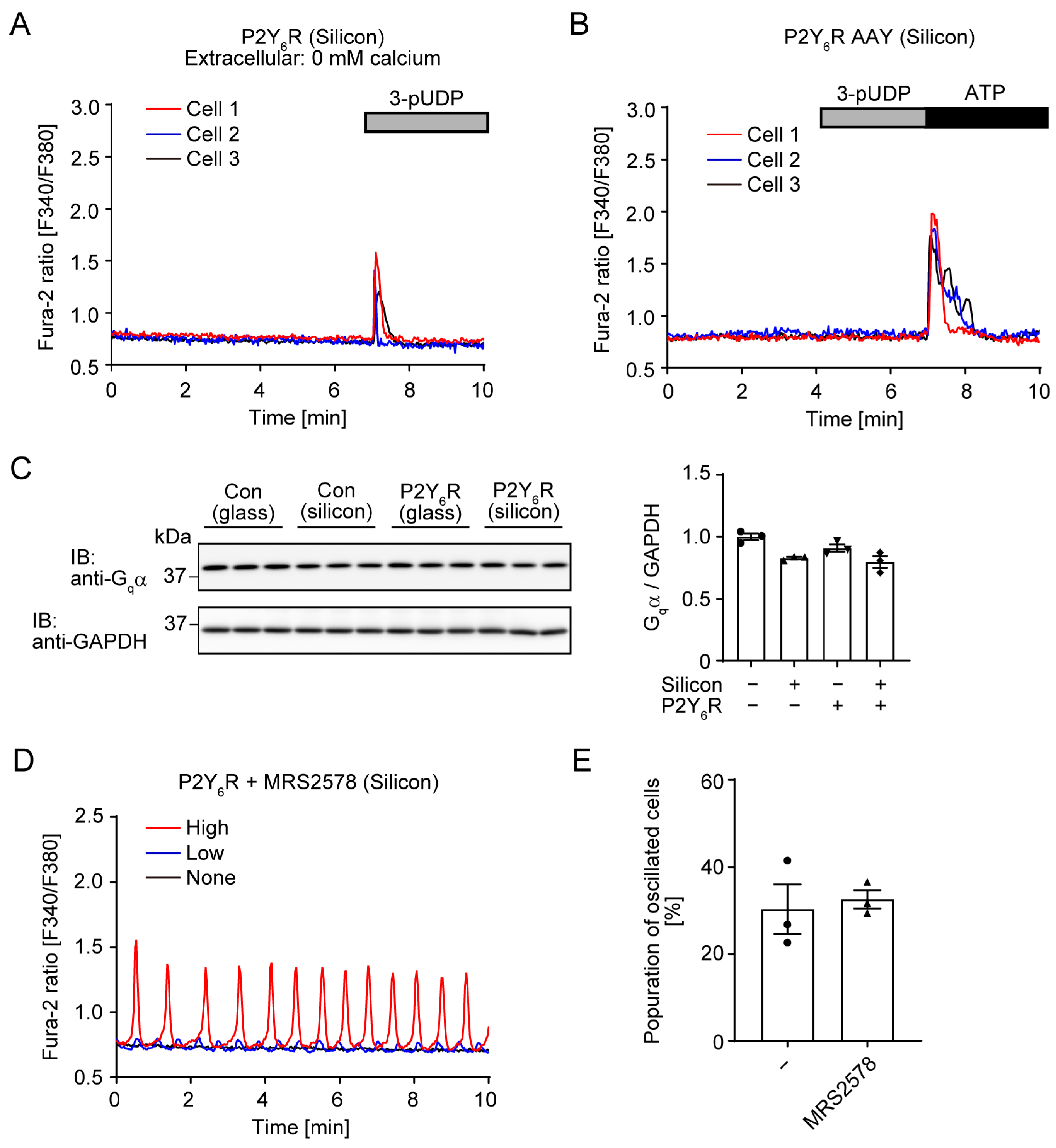
3.1. P2Y6R-Mediated Spontaneous Ca2+ Oscillation Under Silicon Chamber Culture Conditions

To analyze P2Y6R activity, HeLa cells were transiently transfected with P2Y6R-expressing plasmid, and intracellular Ca2+ dynamics were then measured using a ratiometric fluorescence dye, Fura-2 AM. When P2Y6R-expressing HeLa cells were cultured in a collagen-coated silicon/PDMS chamber, Ca2+ oscillation at a constant rhythm was observed without nucleotide ligand administration (Figure 1B, Video S1). Treatment with the P2Y6R-specific agonist 3-phenacyl-UDP (3-pUDP) rapidly and transiently increased intracellular Ca2+ in oscillating and non-oscillating cells (Figure 1B). When P2Y6R-expressing HeLa cells were cultured in a collagen-coated glass bottom dish, spontaneous Ca2+ oscillation was not observed, whereas 3-pUDP treatment induced a transient Ca2+ increase (Figure 1C). Moreover, when HeLa cells transfecting the control plasmid were cultured in a collagen-coated silicon chamber, Ca2+ oscillation was not observed (Figure 1D). These results suggest that P2Y6R is spontaneously activated without agonist administration when cells are cultured onto a cell culture soft substrate such as PDMS. About 30% of cells showed Ca2+ oscillation at various amplitudes (Figure 1B,E).

3.2. Effet of Extracellular Matrix Proteins on P2Y6R-Mediated Ca2+ Oscillation

The extracellular matrix is important for mechanotransduction and cellular responses [27,28]. Collagens are the most abundant feature of the extracellular matrix, whereas fibronectin and laminin, which are noncollagenous glycoproteins, are also known as major components of the basal membrane. To investigate whether different types of extracellular matrix proteins would affect spontaneous Ca2+ oscillation, we tested not only collagen (Figure 1) but also laminin and fibronectin. P2Y6R-expressing HeLa cells were cultured in a laminin or fibronectin-coated silicon chamber. The population of oscillating cells was almost 20–30% in both the laminin or fibronectin-coated silicon chamber (Figure 2A,B), which was almost the same as with the collagen coating (Figure 1E). These results suggest that type of substrate does not affect P2Y6R-mediated spontaneous Ca2+ oscillation.

3.3. G Protein-Mediated Signaling and Ca2+ Entry from Extracellular Space Is Required for Spontaneous Ca2+ Oscillation of P2Y6R

Intracellular Ca2+ increase is mainly triggered by Ca2+ release from the ER or extracellular space. To investigate whether Ca2+ entry from the extracellular space through Ca2+ channels is required for spontaneous Ca2+ oscillation, Ca2+ imaging was performed in HBSS buffer without Ca2+. The 3-pUDP treatment still increased the intracellular Ca2+ because P2Y6R-mediated Gqα activation induced Ca2+ release from the ER through inositol 1,4,5-triphosphate (IP3) receptors (Figure 3A). However, P2Y6R-mediated Ca2+ oscillation under a collagen-coated silicon chamber completely disappeared using HBSS without Ca2+ (Figure 3A), suggesting that Ca2+ entry from the extracellular space is required for spontaneous Ca2+ oscillation.
G protein is the main signal transducer of GPCR signaling pathways. The DRY motif of GPCR is important for coupling to G proteins and receptor activation [29]. The AAY mutant of AT1R does not couple to G protein but still can transduce β-arrestin signaling [30]. HeLa cells expressing the P2Y6R AAY mutant were cultured in a collagen-coated silicon chamber. P2Y6R AAY mutants lost spontaneous Ca2+ oscillation (Figure 3B). As a control experiment, Ca2+ transients induced by 3-pUDP disappeared and ATP-mediated Ca2+ transients through other P2Y receptors were observed (Figure 3B). This result indicates that G protein-mediated signaling is required for spontaneous Ca2+ oscillation. The amount of G protein is important for the intensity of downstream signaling. We checked Gqα expression levels and found that Gqα expression was not increased in Ca2+ oscillating cells (Figure 3C).
Locally released nucleotides activate P2Y receptors in an autocrine/paracrine manner [31]. To investigate the possibility that cells cultured in a collagen-coated silicon chamber would spontaneously release UDP for P2Y6R activation, we ran the antagonist experiment. MRS2578 is a selective antagonist of P2Y6R and blocks UDP-induced Ca2+ transients [32]. To test the effect of MRS2578, cells were treated with MRS2578 for 30 min before Ca2+ imaging. P2Y6R-mediated Ca2+ oscillation was observed under MRS2578 treatment (Figure 3D). Similar to untreated conditions, about 30% of cells expressing P2Y6R still have spontaneous Ca2+ oscillation under MRS2578 treatment (Figure 3E). This result would exclude the possibility that P2Y6R was activated by spontaneously released nucleotides from cells.

3.4. The Extracellular RGD Motif of P2Y6R Is Required for Spontaneous Ca2+ Oscillation

The RGD motif is a cell adhesion sequence that binds to integrin [33]. P2Y6R has the RGD motif in the first extracellular loop (Figure 4A). The mutation of the RGD sequence to RGE causes the loss of the ability to bind to integrin [34]. P2Y2R associates with αvβ3/β5 integrin and P2Y2R RGE mutant reduces its binding [35]. P2Y6R RGE-expressing cells under a collagen-coated silicon chamber showed Ca2+ transients after 3-pUDP administration (Figure 4B) and their responsiveness was almost the same as that of P2Y6R WT (Figure 4C), indicating that the P2Y6R RGE mutant is functional. On the other hand, spontaneous Ca2+ oscillation was not observed in the P2Y6R RGE mutant, suggesting the critical role of the RGD motif of P2Y6R in relation to Ca2+ oscillation (Figure 4B).

3.5. Ligand-Independent Ca2+ Oscillation Is Observed in Several GPCRs

There are eight mammalian P2Y receptors; among these, P2Y2R has the RGD motif in the extracellular loop region, like P2Y6R. To investigate whether spontaneous Ca2+ oscillation would be observed in other P2Y receptors, HeLa cells were transfected with a plasmid expressing each P2Y receptor and cultured in a collagen-coated silicon chamber. The RGD motif containing P2Y2R induced spontaneous Ca2+ oscillation, albeit in a slightly irregular rhythm (Figure 5A). Moreover, P2Y1R that did not have the RGD motif also showed obvious Ca2+ oscillation (Figure 5A). About 10% of cells expressing P2Y1R showed spontaneous Ca2+ oscillation (Figure 5B). On the other hand, other P2Y receptors including P2Y4R, P2Y12R, P2Y13R, and P2Y14R did not induce Ca2+ oscillation.
To verify how many GPCRs showed spontaneous Ca2+ oscillation, HeLa cells were transfected with GPCR plasmid library and Ca2+ indicator GCaMP6 and cultured in a collagen-coated silicon chamber. Using 259 GPCR-expressing plasmids, we monitored GCaMP6 intensity for 5 min and oscillation frequency was counted. Among 259 GPCRs, spontaneous Ca2+ oscillation was observed in 10 GPCRs, including P2Y6R, P2Y1R, P2Y2R, prostaglandin E2 receptor 1 (EP1), growth hormone secretagogue receptor (GHSR), histamine H1 receptor (H1R), platelet-activating factor receptor (PAFR), Ca2+-sensing receptor (CaSR), gastrin-releasing peptide receptor (GRPR), and Mas-related GPCR, member H (MRGH) (Figure 5C). It was observed that oscillation patterns were different for each GPCR (Figure 5D).

4. Discussion

In this study, we investigated the nucleotide ligand-independent activity of P2Y6R and found that P2Y6R induced spontaneous Ca2+ oscillation when cells were cultured in a silicon chamber (Figure 1). Nucleotides can be released from cells by two different pathways: exocytotic release from secretory pathways and conductive/transport pathways [36]. Some central and peripheral neurons release ATP via neuronal synaptic vesicles via purinergic neurotransmission. Multiple channels including connexin (Cx) hemichannels, pannexin 1 (PANX1), volume-regulated anion channels (VRACs), calcium homeostasis modulator 1 (CALHM1), and maxi-anion channels (MACs) have been reported to regulate ATP release [37]. Some Cx hemichannels [38], PANX1 [6], and CALHM1 [39] mediate mechanical stress-induced ATP release. MACs mediate hypoosmotic stress-induced ATP release in astrocytes [40]. In addition, we previously found that mechanical stress induced ATP and UDP release from cardiomyocytes through PANX1, and released nucleotides activated P2Y6R to induce fibrotic responses in an autocrine or paracrine manner [6]. This evidence leads to speculation on the possibility that locally released nucleotides continuously activate P2Y6R for Ca2+ oscillation in HeLa cells cultured in a silicon chamber, although nucleotide release in response to substrate stiffness has not been reported. The P2Y6R antagonist MRS2578, which inhibits UDP-induced Ca2+ increases, did not prevent Ca2+ oscillation under a silicon chamber (Figure 3D). This result would exclude the possibility that P2Y6R is continuously activated by released nucleotides from cells in response to substrate stiffness.
The GPCR screening assay revealed that spontaneous activity under a silicon chamber was observed in 10 GPCRs of 259 GPCRs we examined (Figure 5). Because we used the screening system to detect intracellular Ca2+ dynamics, all hit GPCRs were Gq-coupled receptors. cAMP oscillation has been observed in several biological events [41], and extracellular matrix such as laminin can regulate cAMP signaling of Gs-coupled GPCRs [42,43]. Novel screening systems detecting other second messengers such as cAMP or individual G-protein activity using fluorescence resonance energy transfer (FRET) or bioluminescence resonance energy transfer (BRET)-based biosensors [44,45] may additionally identify GPCRs activated under a silicon chamber. Substrate stiffness is one of the important physical factors that determine cellular response and function [46,47]. PDMS has frequently been used to analyze cellular response in relation to substrate stiffness. P2Y6R and others may respond to substrate stiffness, especially soft substrate. It has been reported that mechano-sensors such as integrins, Piezo1, Notch, and TRPV4 recognize substrate stiffness [48,49,50]. GPCRs activated by mechanical force have been well investigated, and among the 10 hit GPCRs, the H1 receptor is known as a mechanosensitive GPCR [16]. However, little is known about the role of GPCRs in cellular response to substrate stiffness. Isoproterenol-induced β2 adrenergic receptor-PKA signaling decreased under cell culture soft substrate [51]. In the current study, we observed that the basal activity of GPCRs in the absence of ligands was affected by substrate stiffness. As far as we know, the relationship between the basal activity of GPCR and substrate stiffness has not been mentioned before.
Recent biochemical and structural studies of GPCRs reveal the conformational dynamics of GPCRs that contribute to their activation. In the absence of ligands, GPCRs are considered to exist in dynamic equilibrium between the inactive and active conformation, and agonist binding shifts the equilibrium toward the active conformation [52]. The proportion of the active conformation that exists in the ligand-absent condition determines the basal activity of GPCRs [53,54]. Many GPCRs show varying degrees of basal activity. Binding with agonists or inverse agonists, receptor dimerization, and some mutations can affect the basal activity of GPCRs [55,56]. Some adhesion GPCRs can associate with extracellular matrix components and regulate their activity [57]. It is quite reasonable that the mutation of the RGD motif of P2Y6R led to the loss of spontaneous Ca2+ oscillation, because the RGD motif is important for the interaction with integrin [34] which is a well-known stiffness sensor [48]. However, the fact that only P2Y6R and P2Y2R from among the 10 GPCRs share the RGD motif suggests that the molecular mechanism of spontaneous Ca2+ oscillation under a silicon chamber is more complex. We compared the amino acid sequences of 10 GPCRs, but there was no obvious consensus feature among the amino acid sequence alignments. More detailed structural and functional analysis will be required for the characterization of these 10 GPCRs.
Ca2+ is the most fundamental regulator of various cellular responses. Signaling patterns of Ca2+ include spike, wave, and oscillation. Oscillation is the most complex of these, and Ca2+ entry and release are continuously repeated within a suitable balance [58,59]. Ca2+ enters from the ER and extracellular space. All 10 GPCRs that we identified in this study are Gq-coupled GPCRs. Gq signaling induces Ca2+ release from the ER through IP3 receptors. Activation of Gqα also induces receptor-operated Ca2+ entry from extracellular space through Ca2+ channels such as transient receptor potential canonical (TRPC) [60]. Treatment of Gq-coupled GPCR agonists such as histamine and UTP induced ligand-triggered prolonged Ca2+ oscillation in HeLa cells [61,62], its mechanism in HeLa cells has been discussed to some extent. Ca2+ oscillation is initially dependent on the IP3 receptor and not external Ca2+, whereas transmembrane Ca2+ flux is critical for Ca2+ oscillation during the latter phase [24,25]. Because external Ca2+ and Gq signaling were required for spontaneous Ca2+ oscillation under a silicon chamber (Figure 3A,B), it is expected that there are many similarities in the mechanisms of ligand-triggered and ligand-independent Ca2+ oscillations.
In this study, we first found that several GPCRs, including P2Y6R, spontaneously activated under cell culture soft substrate. Representing a limitation of this study, the molecular mechanism of spontaneous Ca2+ oscillation of GPCRs under cell culture soft substrate is still mostly unclear, and its elucidation is a major issue for the future. About 30% of P2Y6R-expressing HeLa cells showed spontaneous Ca2+ oscillation (Figure 1D). Because HeLa cells are from heterogenous cellular origin and have a variable genome and transcriptome at single-cell level [63], only some HeLa cell populations have complete components for spontaneous Ca2+ oscillation. Comparison of gene expression patterns between oscillated and non-oscillated HeLa cell clones would help to identify the molecular mechanism of spontaneous Ca2+ oscillation. Because the frequency of Ca2+ oscillation depends on cell type [64], validation of spontaneous activation of P2Y6R within various cell lines would also contribute to identification of the mechanism. Additionally, the surrounding environment of oscillating cells, including cell–cell interaction, may be involved in spontaneous activity, because the P2Y6R RGD motif integrin-binding site was required for Ca2+ oscillation (Figure 4). Detailed analysis of the frequency and pattern of Ca2+ oscillation at different cell densities would also contribute to identifying the molecular mechanism.
Confirmation of spontaneous Ca2+ oscillation of endogenous P2Y6R and other examples in primary cells is important for proving the physiological and pathophysiological meaning of GPCR spontaneous activity. The extracellular matrix has important roles in various aspects of cardiovascular physiology, such as heart development, and pathophysiology, such as heart failure progression [65,66]. Cardiac fibrosis, which refers to excess deposition of the extracellular matrix, is considered a common feature in various types of heart failure, and increased stiffness associated with cardiac fibrosis contributes to cardiac dysfunction and heart failure progression. P2Y6R expression is upregulated in the pressure-overloaded heart, which contributes to cardiac fibrosis formation [6]. Additionally, cardiomyocyte-specific overexpression of P2Y6R exacerbates pressure overload-induced heart failure [67]. In addition to P2Y6R, the mRNA of P2Y2R is upregulated in congestive heart failure and P2Y2R-mediated signaling contributes to cardiac fibrosis [68]. P2Y1R is involved in shear stress-mediated mechanotransduction in atrial myocytes and endothelial cells [69,70]. Additionally, it has been reported that P2Y6R promotes cancer progression and metastasis [71]. Since matrix stiffness is critical for the progression of various types of cancer [72], the spontaneous activity of GPCRs may contribute to cancer formation. To investigate the physiological and pathophysiological meaning of spontaneous GPCR activation under a silicon chamber, it will be important to analyze whether spontaneous GPCR activation is observed in more physiological conditions such as tissue slices and 3D hydrogel-based organ-like culture.

5. Conclusions

In this study, we found that some GPCRs, including purinergic P2Y6R, showed spontaneous Ca2+ oscillation without ligand stimulation when cells were cultured in a silicon chamber. However, its molecular mechanism is still largely unknown. Because various GPCRs are directly activated by mechanical forces such as shear stress, the similarities and differences in the their stiffness and shear stress sensing mechanisms, and the relationships between these, are important issues. Basal activity of GPCRs is important for various biological events, and disease-causing mutations of GPCRs with increased basal activity have been reported [54]. Future analysis to identify the molecular mechanism of spontaneous GPCR activity under cell culture soft substrate would provide novel insight into GPCR-mediated stiffness regulation and its physiological role.

Supplementary Materials

The following supporting information can be downloaded at https://www.mdpi.com/article/10.3390/cells14030216/s1: Video S1: Spontaneous Ca2+ oscillation of P2Y6R-expressing cells cultured in silicon chamber.

Author Contributions

Conceptualization, A.N. and M.N.; experiment, A.N., K.N., T.I., X.M. and Y.K.; resources, A.I. and J.A.; writing—original draft preparation, A.N.; writing—review and editing, K.N., T.I., X.M., Y.K., A.I., J.A. and M.N.; funding acquisition, A.N. and M.N. All authors have read and agreed to the published version of the manuscript.

Funding

This work was supported by JST CREST Grant Number JPMJCR2024 (20348438 to M.N. and A.N.), JSPS KAKENHI (24K02869 to A.N., 22H02772 and 22K19395 to M.N.), Grant-in-Aid for Scientific Research on Innovative Areas (A) “Sulfur biology” (21H05269 and 21H05258 to M.N.) from the Ministry of Education, Culture, Sports, Science and Technology of Japan, Naito Foundation (50636-2 to M.N.) and Smoking Research Foundation (2024G007 to M.N.). A.I. was funded by JSPS KAKENHI (JP21H04791, JP21H05113, JP21H05037, and JP24K21281); JST (JPMJFR215T and JPMJMS2023); AMED (JP22ama121038 and JP22zf0127007).

Institutional Review Board Statement

This study was approved by the ethics committee at the National Institutes of Natural Sciences (protocol code: P13-051-A).

Informed Consent Statement

Not applicable.

Data Availability Statement

The original contributions presented in this study are included in the article/Supplementary Materials. Further inquiries can be directed to the corresponding authors.

Acknowledgments

We thank Masamichi Ohkura for the kind gift of GCaMP6 plasmid. We also thank Naoyuki Kitajima, Kosuke Sakata, and Tomohiro Shigematsu for technical support.

Conflicts of Interest

The authors declare no conflicts of interest.

References

  1. Fredriksson, R.; Lagerstrom, M.C.; Lundin, L.G.; Schioth, H.B. The G-protein-coupled receptors in the human genome form five main families. Phylogenetic analysis, paralogon groups, and fingerprints. Mol. Pharmacol. 2003, 63, 1256–1272. [Google Scholar] [CrossRef]
  2. Hauser, A.S.; Attwood, M.M.; Rask-Andersen, M.; Schioth, H.B.; Gloriam, D.E. Trends in GPCR drug discovery: New agents, targets and indications. Nat. Rev. Drug Discov. 2017, 16, 829–842. [Google Scholar] [CrossRef] [PubMed]
  3. Nishimura, A.; Sunggip, C.; Oda, S.; Numaga-Tomita, T.; Tsuda, M.; Nishida, M. Purinergic P2Y receptors: Molecular diversity and implications for treatment of cardiovascular diseases. Pharmacol. Ther. 2017, 180, 113–128. [Google Scholar] [CrossRef]
  4. Nishiyama, K. The role of P2Y6 receptor in the pathogenesis of cardiovascular and inflammatory diseases. J. Pharmacol. Sci. 2024, 154, 108–112. [Google Scholar] [CrossRef] [PubMed]
  5. Zhou, M.; Wang, W.; Li, Y.; Zhang, Q.; Ji, H.; Li, H.; Hu, Q. The role of P2Y6R in cardiovascular diseases and recent development of P2Y6R antagonists. Drug Discov. Today 2020, 25, 568–573. [Google Scholar] [CrossRef] [PubMed]
  6. Nishida, M.; Sato, Y.; Uemura, A.; Narita, Y.; Tozaki-Saitoh, H.; Nakaya, M.; Ide, T.; Suzuki, K.; Inoue, K.; Nagao, T.; et al. P2Y6 receptor-Galpha12/13 signalling in cardiomyocytes triggers pressure overload-induced cardiac fibrosis. EMBO J. 2008, 27, 3104–3115. [Google Scholar] [CrossRef] [PubMed]
  7. Daghbouche-Rubio, N.; Alvarez-Miguel, I.; Flores, V.A.; Rojo-Mencia, J.; Navedo, M.; Nieves-Citron, M.; Cidad, P.; Perez-Garcia, M.T.; Lopez-Lopez, J.R. The P2Y6 Receptor as a Potential Keystone in Essential Hypertension. Function 2024, 5, zqae045. [Google Scholar] [CrossRef] [PubMed]
  8. Nishimura, A.; Sunggip, C.; Tozaki-Saitoh, H.; Shimauchi, T.; Numaga-Tomita, T.; Hirano, K.; Ide, T.; Boeynaems, J.M.; Kurose, H.; Tsuda, M.; et al. Purinergic P2Y6 receptors heterodimerize with angiotensin AT1 receptors to promote angiotensin II-induced hypertension. Sci. Signal 2016, 9, ra7. [Google Scholar] [CrossRef] [PubMed]
  9. Stachon, P.; Peikert, A.; Michel, N.A.; Hergeth, S.; Marchini, T.; Wolf, D.; Dufner, B.; Hoppe, N.; Ayata, C.K.; Grimm, M.; et al. P2Y6 deficiency limits vascular inflammation and atherosclerosis in mice. Arterioscler. Thromb. Vasc. Biol. 2014, 34, 2237–2245. [Google Scholar] [CrossRef] [PubMed]
  10. Garcia, R.A.; Yan, M.; Search, D.; Zhang, R.; Carson, N.L.; Ryan, C.S.; Smith-Monroy, C.; Zheng, J.; Chen, J.; Kong, Y.; et al. P2Y6 receptor potentiates pro-inflammatory responses in macrophages and exhibits differential roles in atherosclerotic lesion development. PLoS ONE 2014, 9, e111385. [Google Scholar] [CrossRef]
  11. Li, Y.; Zhou, M.; Li, H.; Dai, C.; Yin, L.; Liu, C.; Li, Y.; Zhang, E.; Dong, X.; Ji, H.; et al. Macrophage P2Y6 receptor deletion attenuates atherosclerosis by limiting foam cell formation through phospholipase Cbeta/store-operated calcium entry/calreticulin/scavenger receptor A pathways. Eur. Heart J. 2024, 45, 268–283. [Google Scholar] [CrossRef] [PubMed]
  12. Hofmann, L.; Palczewski, K. The G protein-coupled receptor rhodopsin: A historical perspective. Methods Mol. Biol. 2015, 1271, 3–18. [Google Scholar] [PubMed]
  13. Ohnishi, K.; Sokabe, T.; Miura, T.; Tominaga, M.; Ohta, A.; Kuhara, A. G protein-coupled receptor-based thermosensation determines temperature acclimatization of Caenorhabditis elegans. Nat. Commun. 2024, 15, 1660. [Google Scholar] [CrossRef]
  14. Zou, Y.; Akazawa, H.; Qin, Y.; Sano, M.; Takano, H.; Minamino, T.; Makita, N.; Iwanaga, K.; Zhu, W.; Kudoh, S.; et al. Mechanical stress activates angiotensin II type 1 receptor without the involvement of angiotensin II. Nat. Cell Biol. 2004, 6, 499–506. [Google Scholar] [CrossRef]
  15. Xu, J.; Mathur, J.; Vessieres, E.; Hammack, S.; Nonomura, K.; Favre, J.; Grimaud, L.; Petrus, M.; Francisco, A.; Li, J.; et al. GPR68 Senses Flow and Is Essential for Vascular Physiology. Cell 2018, 173, 762–775.e716. [Google Scholar] [CrossRef]
  16. Erdogmus, S.; Storch, U.; Danner, L.; Becker, J.; Winter, M.; Ziegler, N.; Wirth, A.; Offermanns, S.; Hoffmann, C.; Gudermann, T.; et al. Helix 8 is the essential structural motif of mechanosensitive GPCRs. Nat. Commun. 2019, 10, 5784. [Google Scholar] [CrossRef] [PubMed]
  17. Mederos y Schnitzler, M.; Storch, U.; Meibers, S.; Nurwakagari, P.; Breit, A.; Essin, K.; Gollasch, M.; Gudermann, T. Gq-coupled receptors as mechanosensors mediating myogenic vasoconstriction. EMBO J. 2008, 27, 3092–3103. [Google Scholar] [CrossRef] [PubMed]
  18. Chachisvilis, M.; Zhang, Y.L.; Frangos, J.A. G protein-coupled receptors sense fluid shear stress in endothelial cells. Proc. Natl. Acad. Sci. USA 2006, 103, 15463–15468. [Google Scholar] [CrossRef]
  19. Jung, B.; Obinata, H.; Galvani, S.; Mendelson, K.; Ding, B.S.; Skoura, A.; Kinzel, B.; Brinkmann, V.; Rafii, S.; Evans, T.; et al. Flow-regulated endothelial S1P receptor-1 signaling sustains vascular development. Dev. Cell 2012, 23, 600–610. [Google Scholar] [CrossRef] [PubMed]
  20. Abdul-Majeed, S.; Nauli, S.M. Dopamine receptor type 5 in the primary cilia has dual chemo- and mechano-sensory roles. Hypertension 2011, 58, 325–331. [Google Scholar] [CrossRef] [PubMed]
  21. Xiao, R.; Liu, J.; Xu, X.Z.S. Mechanosensitive GPCRs and ion channels in shear stress sensing. Curr. Opin. Cell Biol. 2023, 84, 102216. [Google Scholar] [CrossRef] [PubMed]
  22. Quitterer, U.; Fu, X.; Pohl, A.; Bayoumy, K.M.; Langer, A.; AbdAlla, S. Beta-Arrestin1 Prevents Preeclampsia by Downregulation of Mechanosensitive AT1-B2 Receptor Heteromers. Cell 2019, 176, 318–333.e319. [Google Scholar] [CrossRef] [PubMed]
  23. Janetzko, J.; Kise, R.; Barsi-Rhyne, B.; Siepe, D.H.; Heydenreich, F.M.; Kawakami, K.; Masureel, M.; Maeda, S.; Garcia, K.C.; von Zastrow, M.; et al. Membrane phosphoinositides regulate GPCR-beta-arrestin complex assembly and dynamics. Cell 2022, 185, 4560–4573.e4519. [Google Scholar] [CrossRef]
  24. Sauve, R.; Diarra, A.; Chahine, M.; Simoneau, C.; Morier, N.; Roy, G. Ca2+ oscillations induced by histamine H1 receptor stimulation in HeLa cells: Fura-2 and patch clamp analysis. Cell Calcium 1991, 12, 165–176. [Google Scholar] [CrossRef] [PubMed]
  25. Thorn, P. Ca2+ influx during agonist and Ins(2,4,5)P3-evoked Ca2+ oscillations in HeLa epithelial cells. J. Physiol. 1995, 482 Pt 2, 275–281. [Google Scholar] [CrossRef]
  26. Chen, T.W.; Wardill, T.J.; Sun, Y.; Pulver, S.R.; Renninger, S.L.; Baohan, A.; Schreiter, E.R.; Kerr, R.A.; Orger, M.B.; Jayaraman, V.; et al. Ultrasensitive fluorescent proteins for imaging neuronal activity. Nature 2013, 499, 295–300. [Google Scholar] [CrossRef]
  27. Burgess, J.K.; Gosens, R. Mechanotransduction and the extracellular matrix: Key drivers of lung pathologies and drug responsiveness. Biochem. Pharmacol. 2024, 228, 116255. [Google Scholar] [CrossRef]
  28. Stanton, A.E.; Tong, X.; Yang, F. Extracellular matrix type modulates mechanotransduction of stem cells. Acta Biomater. 2019, 96, 310–320. [Google Scholar] [CrossRef] [PubMed]
  29. Han, X.; Feng, Y.; Chen, X.; Gerard, C.; Boisvert, W.A. Characterization of G protein coupling mediated by the conserved D134(3.49) of DRY motif, M241(6.34), and F251(6.44) residues on human CXCR1. FEBS Open Bio 2015, 5, 182–190. [Google Scholar] [CrossRef] [PubMed]
  30. Wei, H.; Ahn, S.; Shenoy, S.K.; Karnik, S.S.; Hunyady, L.; Luttrell, L.M.; Lefkowitz, R.J. Independent beta-arrestin 2 and G protein-mediated pathways for angiotensin II activation of extracellular signal-regulated kinases 1 and 2. Proc. Natl. Acad. Sci. USA 2003, 100, 10782–10787. [Google Scholar] [CrossRef] [PubMed]
  31. Corriden, R.; Insel, P.A. Basal release of ATP: An autocrine-paracrine mechanism for cell regulation. Sci. Signal 2010, 3, re1. [Google Scholar] [CrossRef] [PubMed]
  32. Mamedova, L.K.; Joshi, B.V.; Gao, Z.G.; von Kugelgen, I.; Jacobson, K.A. Diisothiocyanate derivatives as potent, insurmountable antagonists of P2Y6 nucleotide receptors. Biochem. Pharmacol. 2004, 67, 1763–1770. [Google Scholar] [CrossRef] [PubMed]
  33. Ruoslahti, E. RGD and other recognition sequences for integrins. Annu. Rev. Cell Dev. Biol. 1996, 12, 697–715. [Google Scholar] [CrossRef] [PubMed]
  34. Greenspoon, N.; Hershkoviz, R.; Alon, R.; Varon, D.; Shenkman, B.; Marx, G.; Federman, S.; Kapustina, G.; Lider, O. Structural analysis of integrin recognition and the inhibition of integrin-mediated cell functions by novel nonpeptidic surrogates of the Arg-Gly-Asp sequence. Biochemistry 1993, 32, 1001–1008. [Google Scholar] [CrossRef] [PubMed]
  35. Erb, L.; Liu, J.; Ockerhausen, J.; Kong, Q.; Garrad, R.C.; Griffin, K.; Neal, C.; Krugh, B.; Santiago-Perez, L.I.; Gonzalez, F.A.; et al. An RGD sequence in the P2Y2 receptor interacts with αVβ3 integrins and is required for Go-mediated signal transduction. J. Cell Biol. 2001, 153, 491–501. [Google Scholar] [CrossRef]
  36. Lazarowski, E.R.; Sesma, J.I.; Seminario-Vidal, L.; Kreda, S.M. Molecular mechanisms of purine and pyrimidine nucleotide release. Adv. Pharmacol. 2011, 61, 221–261. [Google Scholar] [PubMed]
  37. Taruno, A. ATP Release Channels. Int. J. Mol. Sci. 2018, 19, 808. [Google Scholar] [CrossRef]
  38. Riquelme, M.A.; Cardenas, E.R.; Xu, H.; Jiang, J.X. The Role of Connexin Channels in the Response of Mechanical Loading and Unloading of Bone. Int. J. Mol. Sci. 2020, 21, 1146. [Google Scholar] [CrossRef] [PubMed]
  39. Workman, A.D.; Carey, R.M.; Chen, B.; Saunders, C.J.; Marambaud, P.; Mitchell, C.H.; Tordoff, M.G.; Lee, R.J.; Cohen, N.A. CALHM1-Mediated ATP Release and Ciliary Beat Frequency Modulation in Nasal Epithelial Cells. Sci. Rep. 2017, 7, 6687. [Google Scholar] [CrossRef]
  40. Liu, H.T.; Toychiev, A.H.; Takahashi, N.; Sabirov, R.Z.; Okada, Y. Maxi-anion channel as a candidate pathway for osmosensitive ATP release from mouse astrocytes in primary culture. Cell Res. 2008, 18, 558–565. [Google Scholar] [CrossRef] [PubMed]
  41. Singer, G.; Araki, T.; Weijer, C.J. Oscillatory cAMP cell-cell signalling persists during multicellular Dictyostelium development. Commun. Biol. 2019, 2, 139. [Google Scholar] [CrossRef] [PubMed]
  42. Petersen, S.C.; Luo, R.; Liebscher, I.; Giera, S.; Jeong, S.J.; Mogha, A.; Ghidinelli, M.; Feltri, M.L.; Schoneberg, T.; Piao, X.; et al. The adhesion GPCR GPR126 has distinct, domain-dependent functions in Schwann cell development mediated by interaction with laminin-211. Neuron 2015, 85, 755–769. [Google Scholar] [CrossRef]
  43. Wang, Y.G.; Samarel, A.M.; Lipsius, S.L. Laminin binding to beta1-integrins selectively alters beta1- and beta2-adrenoceptor signalling in cat atrial myocytes. J. Physiol. 2000, 527 Pt 1, 3–9. [Google Scholar] [CrossRef] [PubMed]
  44. Kim, N.; Shin, S.; Bae, S.W. cAMP Biosensors Based on Genetically Encoded Fluorescent/Luminescent Proteins. Biosensors 2021, 11, 39. [Google Scholar] [CrossRef] [PubMed]
  45. Sajkowska, J.J.; Tsang, C.H.; Kozielewicz, P. Application of FRET- and BRET-based live-cell biosensors in deorphanization and ligand discovery studies on orphan G protein-coupled receptors. SLAS Discov. 2024, 29, 100174. [Google Scholar] [CrossRef] [PubMed]
  46. De Belly, H.; Paluch, E.K.; Chalut, K.J. Interplay between mechanics and signalling in regulating cell fate. Nat. Rev. Mol. Cell Biol. 2022, 23, 465–480. [Google Scholar] [CrossRef] [PubMed]
  47. Cao, H.; Zhou, Q.; Liu, C.; Zhang, Y.; Xie, M.; Qiao, W.; Dong, N. Substrate stiffness regulates differentiation of induced pluripotent stem cells into heart valve endothelial cells. Acta Biomater. 2022, 143, 115–126. [Google Scholar] [CrossRef]
  48. Wei, J.; Yao, J.; Yan, M.; Xie, Y.; Liu, P.; Mao, Y.; Li, X. The role of matrix stiffness in cancer stromal cell fate and targeting therapeutic strategies. Acta Biomater. 2022, 150, 34–47. [Google Scholar] [CrossRef]
  49. Atcha, H.; Jairaman, A.; Holt, J.R.; Meli, V.S.; Nagalla, R.R.; Veerasubramanian, P.K.; Brumm, K.T.; Lim, H.E.; Othy, S.; Cahalan, M.D.; et al. Mechanically activated ion channel Piezo1 modulates macrophage polarization and stiffness sensing. Nat. Commun. 2021, 12, 3256. [Google Scholar] [CrossRef] [PubMed]
  50. Kretschmer, M.; Mamistvalov, R.; Sprinzak, D.; Vollmar, A.M.; Zahler, S. Matrix stiffness regulates Notch signaling activity in endothelial cells. J. Cell Sci. 2023, 136, jcs260442. [Google Scholar] [CrossRef] [PubMed]
  51. Kim, T.J.; Sun, J.; Lu, S.; Zhang, J.; Wang, Y. The regulation of beta-adrenergic receptor-mediated PKA activation by substrate stiffness via microtubule dynamics in human MSCs. Biomaterials 2014, 35, 8348–8356. [Google Scholar] [CrossRef] [PubMed]
  52. Mafi, A.; Kim, S.K.; Goddard, W.A., 3rd. The mechanism for ligand activation of the GPCR-G protein complex. Proc. Natl. Acad. Sci. USA 2022, 119, e2110085119. [Google Scholar] [CrossRef] [PubMed]
  53. Cullum, S.A.; Platt, S.; Dale, N.; Isaac, O.C.; Wragg, E.S.; Soave, M.; Veprintsev, D.B.; Woolard, J.; Kilpatrick, L.E.; Hill, S.J. Mechano-sensitivity of beta2-adrenoceptors enhances constitutive activation of cAMP generation that is inhibited by inverse agonists. Commun. Biol. 2024, 7, 417. [Google Scholar] [CrossRef] [PubMed]
  54. Seifert, R.; Wenzel-Seifert, K. Constitutive activity of G-protein-coupled receptors: Cause of disease and common property of wild-type receptors. Naunyn Schmiedebergs Arch. Pharmacol. 2002, 366, 381–416. [Google Scholar] [CrossRef]
  55. Yu, R.; Cui, Z.; Li, M.; Yang, Y.; Zhong, J. Dimer-dependent intrinsic/basal activity of the class B G protein-coupled receptor PAC1 promotes cellular anti-apoptotic activity through Wnt/beta-catenin pathways that are associated with dimer endocytosis. PLoS ONE 2014, 9, e113913. [Google Scholar] [CrossRef]
  56. Stoy, H.; Gurevich, V.V. How genetic errors in GPCRs affect their function: Possible therapeutic strategies. Genes. Dis. 2015, 2, 108–132. [Google Scholar] [CrossRef] [PubMed]
  57. Vizurraga, A.; Adhikari, R.; Yeung, J.; Yu, M.; Tall, G.G. Mechanisms of adhesion G protein-coupled receptor activation. J. Biol. Chem. 2020, 295, 14065–14083. [Google Scholar] [CrossRef]
  58. Uhlen, P.; Fritz, N. Biochemistry of calcium oscillations. Biochem. Biophys. Res. Commun. 2010, 396, 28–32. [Google Scholar] [CrossRef]
  59. Dupont, G.; Combettes, L.; Bird, G.S.; Putney, J.W. Calcium oscillations. Cold Spring Harb. Perspect. Biol. 2011, 3, a004226. [Google Scholar] [CrossRef] [PubMed]
  60. Abramowitz, J.; Yildirim, E.; Birnbaumer, L. The TRPC Family of Ion Channels: Relation to the TRP Superfamily and Role in Receptor- and Store-Operated Calcium Entry. In TRP Ion Channel Function in Sensory Transduction and Cellular Signaling Cascades; Liedtke, W.B., Heller, S., Eds.; CRC Press: Boca Raton, FL, USA, 2007. [Google Scholar]
  61. Diarra, A.; Wang, R.; Garneau, L.; Gallo-Payet, N.; Sauve, R. Histamine-evoked Ca2+ oscillations in HeLa cells are sensitive to methylxanthines but insensitive to ryanodine. Pflugers Arch. 1994, 426, 129–138. [Google Scholar] [CrossRef]
  62. Lin, G.C.; Rurangirwa, J.K.; Koval, M.; Steinberg, T.H. Gap junctional communication modulates agonist-induced calcium oscillations in transfected HeLa cells. J. Cell Sci. 2004, 117, 881–887. [Google Scholar] [CrossRef]
  63. Hu, W.E.; Zhang, X.; Guo, Q.F.; Yang, J.W.; Yang, Y.; Wei, S.C.; Su, X.D. HeLa-CCL2 cell heterogeneity studied by single-cell DNA and RNA sequencing. PLoS ONE 2019, 14, e0225466. [Google Scholar] [CrossRef] [PubMed]
  64. Fukuoka, M.; Kang, W.; Horiike, S.; Yamada, M.; Miyado, K. Calcium oscillations and mitochondrial enzymes in stem cells. Regen. Ther. 2024, 26, 811–818. [Google Scholar] [CrossRef] [PubMed]
  65. Gaetani, R.; Zizzi, E.A.; Deriu, M.A.; Morbiducci, U.; Pesce, M.; Messina, E. When Stiffness Matters: Mechanosensing in Heart Development and Disease. Front. Cell Dev. Biol. 2020, 8, 334. [Google Scholar] [CrossRef] [PubMed]
  66. Bloksgaard, M.; Lindsey, M.; Martinez-Lemus, L.A. Extracellular matrix in cardiovascular pathophysiology. Am. J. Physiol. Heart Circ. Physiol. 2018, 315, H1687–H1690. [Google Scholar] [CrossRef] [PubMed]
  67. Shimoda, K.; Nishimura, A.; Sunggip, C.; Ito, T.; Nishiyama, K.; Kato, Y.; Tanaka, T.; Tozaki-Saitoh, H.; Tsuda, M.; Nishida, M. Modulation of P2Y6R expression exacerbates pressure overload-induced cardiac remodeling in mice. Sci. Rep. 2020, 10, 13926. [Google Scholar] [CrossRef] [PubMed]
  68. Woo, S.H.; Trinh, T.N. P2 Receptors in Cardiac Myocyte Pathophysiology and Mechanotransduction. Int. J. Mol. Sci. 2020, 22, 251. [Google Scholar] [CrossRef]
  69. Kim, J.C.; Woo, S.H. Shear stress induces a longitudinal Ca2+ wave via autocrine activation of P2Y1 purinergic signalling in rat atrial myocytes. J. Physiol. 2015, 593, 5091–5109. [Google Scholar] [CrossRef]
  70. Cho, J.M.; Park, S.K.; Kwon, O.S.; Taylor La Salle, D.; Cerbie, J.; Fermoyle, C.C.; Morgan, D.; Nelson, A.; Bledsoe, A.; Bharath, L.P.; et al. Activating P2Y1 receptors improves function in arteries with repressed autophagy. Cardiovasc. Res. 2023, 119, 252–267. [Google Scholar] [CrossRef]
  71. Placet, M.; Arguin, G.; Molle, C.M.; Babeu, J.P.; Jones, C.; Carrier, J.C.; Robaye, B.; Geha, S.; Boudreau, F.; Gendron, F.P. The G protein-coupled P2Y6 receptor promotes colorectal cancer tumorigenesis by inhibiting apoptosis. Biochim. Biophys. Acta Mol. Basis Dis. 2018, 1864, 1539–1551. [Google Scholar] [CrossRef]
  72. Ishihara, S.; Haga, H. Matrix Stiffness Contributes to Cancer Progression by Regulating Transcription Factors. Cancers 2022, 14, 1049. [Google Scholar] [CrossRef]
Figure 1. Spontaneous Ca2+ oscillation by P2Y6R-expressing HeLa cells cultured in a collagen-coated silicon chamber: (A) Schematic of Ca2+ imaging analysis of cells cultured in substrate-coated silicon or glass-bottom dish; (B) Trace of Ca2+ response in P2Y6R-expressing HeLa cells cultured in a collagen-coated silicon chamber. P2Y6R-expressing HeLa cells were seeded on a collagen-coated silicon chamber and the intracellular Ca2+ response was monitored via a Fura-2 probe. Representative traces of oscillating cells with high (red) and low (blue) amplitude and non-oscillating cells (black) are shown. P2Y6R-specific agonist 10 µM 3-phenacyl UDP (3-pUDP) was added at the indicated time; (C) Trace of Ca2+ response in P2Y6R-expressing HeLa cells cultured in a collagen-coated glass-bottom dish; 10 µM 3-pUDP was added at the indicated time; (D) Trace of Ca2+ response in control HeLa cells cultured in a collagen-coated silicon chamber; 10 µM 3-pUDP and 100 µM ATP were added at the indicated time; (E) Quantitative population of oscillating cells (n = 3 independent experiments). Data are shown as means ± SEM. ** p < 0.01, one-way ANOVA.
Cells 14 00216 g001
Figure 2. Effects of chamber coating on P2Y6R-mediated Ca2+ oscillation: (A) Trace of Ca2+ response in P2Y6R-expressing HeLa cells cultured in a laminin or fibronectin-coated silicon chamber. P2Y6R-expressing HeLa cells were seeded on a laminin (red) or fibronectin (blue)-coated silicon chamber, and 10 µM 3-pUDP was added at the indicated time. (B) Quantitative population of oscillating cells (n = 3 independent experiments). Data are shown as means ± SEM.
Cells 14 00216 g002
Figure 3. Conditions of P2Y6R-mediated spontaneous Ca2+ oscillation: (A) Effect of extracellular Ca2+ on P2Y6R-mediated Ca2+ oscillation. P2Y6R-expressing HeLa cells were cultured in a collagen-coated silicon chamber and Fura-2 imaging was performed in HBSS without Ca2+; 10 µM 3-pUDP was added at the indicated time (n = 3 independent experiments); (B) Trace of Ca2+ response in P2Y6R AAY-expressing HeLa cells cultured in a collagen-coated silicon chamber; 10 µM 3-pUDP and 100 µM ATP were added at the indicated time (n = 3 independent experiments); (C) Expression of Gqα in P2Y6R or control vector-expressing HeLa cells cultured in a collagen-coated silicon chamber or glass-bottom dish. Molecular weights are shown. Right graph is the quantification of Gqα expression normalized by GAPDH expression (n = 3 independent experiments); (D) Effect of MRS2578 on P2Y6R-mediated Ca2+ oscillation. P2Y6R-expressing HeLa cells were cultured in a collagen-coated silicon chamber, and 10 µM MRS2578 was added 30 min before Ca2+ imaging (E) Quantitative population of oscillating cells. Data for MRS2578 (−) are from Figure 1E (n = 3 independent experiments). Data are shown as means ± SEM.
Cells 14 00216 g003
Figure 4. Effect of the extracellular RGD motif on P2Y6R-mediated Ca2+ oscillation: (A) Snake diagram of the mouse P2Y6R. The RGD motif is highlighted in red. This diagram was prepared using the GPCR database (http://www.gpcrdb.org, accessed on 19 December 2024); (B) Trace of Ca2+ response in P2Y6R RGE-expressing HeLa cells cultured in a collagen-coated silicon chamber; 10 µM 3-pUDP was added at the indicated time (n = 3 independent experiments); (C) Summary of maximum change in Fura-2 ratio after 3-pUDP treatment in HeLa cells expressing P2Y6R WT or RGE mutant. Data for P2Y6R WT are from Figure 1B (n = 3 independent experiments).
Cells 14 00216 g004
Figure 5. Screening of GPCRs with spontaneous Ca2+ oscillation under a collagen-coated silicon chamber: (A) Trace of Ca2+ response in HeLa cells expressing each P2Y receptor cultured in a collagen-coated silicon chamber; (B) Quantitative population of oscillating cells (n = 3 independent experiments); (C) Screening of GPCRs showing Ca2+ oscillation. HeLa cells expressing GPCR library (259 GPCRs) and Ca2+ indicator GCaMP6 were cultured in a collagen-coated silicon chamber. GCaMP6 fluorescence intensity was measured and oscillation frequency per 5 min was counted; (D) Trace of Ca2+ response in HeLa cells expressing hit GPCRs.
Cells 14 00216 g005
Disclaimer/Publisher’s Note: The statements, opinions and data contained in all publications are solely those of the individual author(s) and contributor(s) and not of MDPI and/or the editor(s). MDPI and/or the editor(s) disclaim responsibility for any injury to people or property resulting from any ideas, methods, instructions or products referred to in the content.

