


Prosthesis 2024, 6(5), 1202-1210; https://doi.org/10.3390/prosthesis6050086 - 26 Sep 2024
Abstract
Craniosacral rhythm is a manual therapy technique that focuses on the subtle, rhythmic movement of cerebrospinal fluid as it flows through the central nervous system and musculoskeletal system. Through light and delicate manipulation of the cranial bones, membranes and soft tissues of the
[...] Read more.
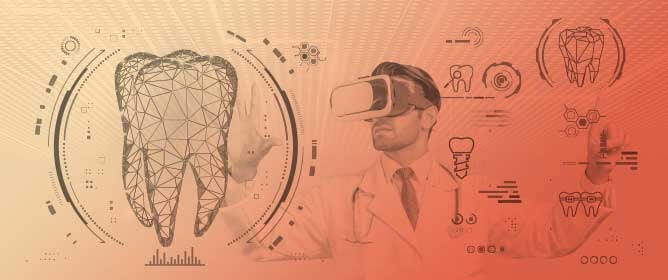
Craniosacral rhythm is a manual therapy technique that focuses on the subtle, rhythmic movement of cerebrospinal fluid as it flows through the central nervous system and musculoskeletal system. Through light and delicate manipulation of the cranial bones, membranes and soft tissues of the head and neck, it promotes rebalancing and release from tension in the body, improving the circulation of the cerebrospinal fluid and the individual’s health. In the field of prosthetic dentistry, in rare cases, such as a full arch or removable partial denture, the patient perceives a sensation of constriction even though all procedures have been used correctly. In this work, a new design fora removable partial denture is proposed that does not interfere with the primary respiratory mechanism, reducing the sensation of constriction in the patient. The materials used are the same as those used for a normal removable partial denture, and the technique used is the “lost wax” one: the novelty is that the prosthesis is made in two separate parts that are joined by a central connection; the result is a removable partial denture with a small central thickening in the main joint represented by the connection of the two pieces. Craniosacral breathing movements are favored with this prosthesis as the metal structure is hyperstatic towards intrusion movements, more or less virtual in the opposite direction, i.e., extrusion towards the vestibule.
Full article
(This article belongs to the Special Issue Innovative Prosthetic Devices Applied to the Human Body)
►
Show Figures

Figure 1


