The document provides notes on various neurological conditions:
- Cauda equine syndrome presents with acute motor and sensory loss in the lower extremities and requires urgent surgery after MRI to identify the site of injury, followed by bed rest and pain control.
- Stroke can be ischemic or hemorrhagic, with CT scan being the initial test to differentiate subtypes and rule out hemorrhage. Hypertension is a major risk factor.
- Multiple sclerosis treatments include corticosteroids for acute exacerbations and beta-interferon or glatiramer acetate to decrease relapse frequency.
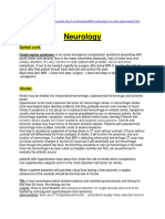
Neurology Spinal cord: Cauda equine syndrome: is an acute emergency compression syndrome presenting with acute motor and sensory loss in the lower extremities bilaterally, loss of rectal tone and urinary retention, its very common in older patient with prostatic cancer and bone metastasis. It has to be treated urgently by surgery after doing MRI to localize the site of injury after that patient should have bed rest and control the pain: Best initial test: MRI best next step: surgery best next step or discharge: bed rest +analgesics.
Stroke: Stroke may be divided into intracerebral hemorrhage, subarachnoid hemorrhage and ischemic stroke Hypertension is the most important risk factor for hemorrhagic stroke. In hemorrhagic stroke, focal neurological signs develop suddenly and they gradually worsen over minutes or hours Onset of symptoms is not abrupt as in subarachnoid hemorrhage or embolic stroke .Symptoms usually start during normal activity or may be precipitated by sex or strenuous activity. As the hemorrhage expands, headache, vomiting and altered mental status develop Patients with ischemic stroke usually dont have headache and impaired consciousness. Patients with ischemic strokes may have history of transient ischemic attacks. Patients with subarachnoid hemorrhage have sudden, dramatic onset of severe headache First step in all patients suspected of having stroke is CT scan without contrast .CTscan without contrast will differentiate the three subtypes of stroke. It will confirm the presence, size and location of hemorrhage. It is better than MRI in detecting intracranial hemorrhage CT scan may not show ischemic changes in the first 24 hours but it will essentially rule out hemorrhage Even though this patient has bruit, the first thing should be to rule out hemorrhage in the head Once the hemorrhage has been ruled out Doppler carotids and TEE can be performed to evaluate the embolic source
patients with hypertension have about four times the risk of stroke when compared to non-hypertensive subjects and it is the most important risk factor for stroke.
When a patient presents with painless visual loss lasting a few seconds, a duplex ultrasound of the carotids should be the first test of choice
TIA: a focal neurological deficit started suddenly and resolved spontaneously and belong for less than 24 hours .the etiology can be: embolic: there is atrial fibrlation and embolus production, its single episode and need to be treated by anticoagulant agents(heparin then warfarine) atheroembolic: there are HTN,hyperlipedimia,DM.and patient usually many episodes need to be treated with antiplatlet :aspirin or aspirin and dipyridamol if patient has TIA with aspirin ,clopidogrel if patient does not tolerate aspirin. CT or MRI are normal not like stoke In reversible ischemic neurologic deficit (RIND), the local symptomatology recovers in 24 hours to one-week period
Patient with cerebellar hemorrhage presents with ataxia, vomiting, occipital headache, gaze palsy, and facial weakness. There is no hemiparesis It is crucial to make early and correct diagnosis, as urgent surgical decompression may be life saving in such cases.
Vasospasm of the arteries at the base of the brain following subarachnoidal hemorrhage occurs in at about 30% of patients. Signs of ischemia appear at about 7 days after hemorrhage. Vasospasm is the major cause of morbidity and mortality in patients with subarachnoid hemorrhage
Always suspect lacunar stroke if patient presents with limited neurologic deficit .The principal cause of lacunar stroke is hypertension crisis that cause small vessel infarction or bleeding the 4 common lacunar syndromes: 1) Pure motor hemiparesis Manifested by a unilateral motor deficit involving the face, arm and, to a lesser extent, the leg .There may be a mild dysarthria But, there is no sensory, visual, or higher cortical dysfunction It usually results from lacunar infarction in the posterior limb of the internal capsule 2) Pure sensory stroke Patients complain of unilateral numbness, paresthesias, and a hemisensory deficit involving the face, arm, trunk, and leg. It results from a stroke in ventroposterolateral (VPL) nucleus of the thalamus.thats named thalamic stroke 3 )Ataxic-hemiparesis It is manifested by weakness that is more prominent in the lower extremity along with ipsilateral arm and leg incoordination .It also results from lacunar infarction in the posterior limb of the internal capsule 4) Dysarthria-clumsy hand syndrome: Lacunar stroke at the basis pontis usually causes this
The most common site of hypertensive hemorrhage is putamen (35%). Intemal capsule that lies adjacent to putamen is almost always involved leading to hemiparesis
Asymptomatic patients with carotid artery stenoses of 60 to 99 percent are considered to have a proven indication for CEA (carotid endarterectomy). Complete occlusion (100 percent stenosis) of the carotid artery is a contraindication to surgery
Parkinson: Always consider Shy. Dragger syndrome when a patient with Parkinsonism experiences orthostatic hypotension, impotence, incontinence, or other autonomic symptoms. Anti- Parkinsonism drugs are generally ineffective and treatment aims at intravascular volume expansion with fludrocortisone, salt supplementation, alpha adrenergic agonists, and application of constrictive garments to the lower body.
Anticholinergics are useful treatment for Parkinsonism patients whore < 70 (KAPLAN < 60)with disturbing tremor and minimal bradykinesia Parkinsonism tremor is a resting tremor
Dementia: Vascular dementia: sudden onset and step wise progression+ abnormal CT (lacunar infarcts)+ PMH of HTN ,hyperlipedemia ,DM and TIA. Pseudo dementia: patient is coming by himself complaining from forgetfulness since the same time of depressing event (living alone, dyeing of spouse) and there is no more than that. i.e. chief complaint is forgetfulness by the patient
Alzheimer: patient complains of many symptoms and the forgetfulness is one beside the failing in the dialy activity function,apraxia ,agnosia and aphasiathe important thing is that the patient is brought by a family members. At this time, the Alzheimers disease can be definitively diagnosed only by postmortem examination (brain autopsy).
Creutzfeldt-Jakob disease is characterized by a rapidly progressive dementia, myoclonus,
ataxia and characteristic findings of periodic high voltage complexes on EEG, the age of incidence is between 50-70 yrs old, bovine spongiform encephalopathy is a Variant Creutzfeldt- Jakob disease and represents bovine to human transmission. It occurs at much younger ages and the median age at onset is 29 yrs. the disease progression is slower. EEG is usually abnormal but there are no periodic high voltage complexes.
Recognize normal pressure hydrocephalus by the triad of gait disturbance, dementia and urinary incontinence. Other features are normal CSF pressures on lumbar puncture and enlarged ventricles on MRI. If repeated spinal taps leads to an improvement in symptoms, then ventriculo-peritoneal shunts can be considered as the definitive treatment for these patients
Two of the following core features are essential for a diagnosis of probable Dementia with Lew Body, and one is essential for possible DLB a) Fluctuating cognition with pronounced variations in attention and alertness b) Recurrent visual hallucinations that are typically well formed and detailed c) Spontaneous motor features of Parkinsonism
MG: Treatment: anticholine esterase (pyridostigmin,neostigmine) is the first initial treatment and it causes symptoms relief but not produce remission,after that patient consider for thymectomy or immunosuppressive. Thymectomy: it produce remission and effect for long term management in patient<60 yrs old. Immunosuppressive: (predinison or others) consider if patient between puberty and 60 and whose disease is not confined only to the extraocular musclesor after thymectomy done. Plasmapheresis or Intravenous immunoglobulins : Its effect is transient and cannt be used on long-term basis. It can also be used to stabilize the patient before thymectomy. It is also used in myasthenia crisis Treatment of myasthenia crisis consists of endotracheal intubation and withdrawal of anticholin esterases for many days, Plasmapheresis or IVIG may hasten the recovery in these patients but they are not the first step of management.also cholinergic crisis treated by withdrawal of anticholin esterase drugs.
Seizure: CT scan of brain without contrast is the initial diagnostic test of choice when a patient presents with impaired consciousness or new onset seizure to roll out SAH or tumor,that done in ER.in the neurology department the g.standard is EEG.
Infectious: Herpes mainly affects the temporal lobe of the brain and may present with acute onset (1 week duration) of focal neurological findings. Characteristic cerebralspinal fluid findings are lymphocyte pleocytosis, increased number of erythrocytes, and elevated protein HSV. CT or MRI are the initial test but polymerase chain reaction of the CSF is the gold standard.
MS: Internuclear ophthalmoplegia is a pathognomonic finding of multiple sclerosis and is due to demyelination of medial longitudinal fasciculus.
Acute exacerbations of MS are treated with corticosteroids. Beta-interferon or Glatiramer
acetate is used to decrease the frequency of exacerbations in patients with relapsing-remitting or secondary progressive form of MS
Headache: Pseudo tumor cerebri: benign intracranial hypertension/ young obese females/headache/ impaired absorption of CSF by arachnoid villi/ normal cerebrospinal fluid (CS F) analysis/ normal neuroimaging /papilledema/sometimes sixth (VI) nerve palsy/ no abnormality found on neuroimaging / history of exposure to glucocorticoids , vitamin or oral contraceptive pills/ treated by weight reduction in obese patients and with acetazolamide/ shunting, or optic nerve sheath fenestration is done to prevent blindness that is the most significant complication of this otherwise benign disorder. Once neuroimaging rules out space occupying lesion, lumbar puncture is the next step for diagnosis, repeated LP not used for Tx, empty sella seen on neuroimaging . Shrunken ventricles are seen on MRI
Verapamil is first line drug for prophylaxis of cluster headache. Prophylaxis should be started as soon as possible after the onset of acute attack. Other options for prophylaxis are prednisone, ergotamine, methysergide, cyproheptadine and indomethacin. Lithium is useful for prophylaxis of chronic form of cluster headache
Others: Brain mass lesions in HIV patient: toxoplasmosis is the most common cause and the lesion is multiple and ring like in the basal ganglia area..Never happened in patient taking TMP-SMX as prophylaxis. Lymphoma: suspected if CSF PCR is + for EBV and it is solitary mass in the per ventricular area.
GBS Vs Tickborne paralysis
Fever, Diarrhea before not present Sensory and reflexes loss sensory normal CSF:albumin-cytology dissociation normal Ascending paralysis progress during days to weeks hour to one day Palsmapheresis and immunoglobulines removing of the tick
Electromyography and conductions studies are the best diagnostic tests for polyneuropathy.
In an individual who has sudden onset of eye pain, photophobia and a mid dilated pupil, acute glaucoma must be ruled out. The best method to diagnose is tonometry Absence of forehead furrows indicates peripheral facial nerve palsy and excludes central causes,i.e upper half paralysis means peripheral palsy and couldnt be central palsy.
Primidone is an anticonvulsant agent, which can be used to treat benign essential tremor as a second line. However, it can also precipitate acute intermittent porphyria because Phenobarbital is one of its derivatives. Diagnosis of porphyria is made by assessing urine for porphobilinogen and presented with vague abdominal pain and hallucination.
Craniopharyngiomas: benign tumors, bimodal age distributionchildren and 55-65
years age group,The tumor is located above the sella turcica, and consists of multiple cysts, presents with hypopituitarism. In children, retarded growth is the most prominent feature (due to growth hormone and thyroid hormone deficiency), whereas sexual dysfunction is more prominent in adults. Women can present with amenorrhea, men with impotance. Since the tumor compresses the optic chiasm, bitemporal blindness is a classic sign of the disease. Headaches occur due to an increased intracranial pressure. Diagnosis is made with an MRI or CT scan, and treatment is with surgery and/or radiotherapy.
Caf-u-lai t spots, macrocephaly. feeding problems, short stature and learning disabilities are characteristic features of neurofibromatosis type 1(AD).
Hemi-neglect syndrome that is characterized by the ignoring of the left side of a space is due to the involvement of the right (non-dominant) parietal lobe.