Location via proxy:   [ UP ]  
[Report a bug]   [Manage cookies]                
Article

Microalgae Isolated from Singapore Mangrove Habitat as Promising Microorganisms for the Sustainable Production of Omega-3 Docosahexaenoic Acid

Wilmar Innovation Centre, Wilmar International Limited, 28 Biopolis Road, Singapore 138568, Singapore
*
Author to whom correspondence should be addressed.
These authors contributed equally to the work.
Biomass 2024, 4(3), 751-764; https://doi.org/10.3390/biomass4030042
Submission received: 30 April 2024 / Revised: 1 June 2024 / Accepted: 20 June 2024 / Published: 10 July 2024

Abstract

:
Docosahexaenoic acid (DHA, C22:6n-3) is an omega-3 fatty acid with beneficial effects for human health. In view of its increasing demand, DHA traditionally produced by marine fisheries will be insufficient, and an alternative sustainable source is urgently required. Here, we report the isolation and characterization of four novel microalgae strains, PLU-A, B, C and D, with a high DHA content of up to 45% from decayed mangrove samples collected from a coastal area in Singapore. Phylogenetic analysis revealed that these isolates were clustered with Schizochytrium sp. TK6 (OK244290.1) and were identified as Schizochytrium sp. strains. A medium optimization with Schizochytrium sp. PLU-D found a glucose-to-yeast extract ratio of 4:1 to be optimal for high biomass and lipid accumulation of up to 70% in shake flasks. In fed-batch fermentation scale-up with the Schizochytrium sp. PLU-D strain, this translates to 175 g/L dry biomass, 94 g/L lipid and 36.2 g/L DHA. Accordingly, the DHA titer obtained is superior to most of the scale-up production reported thus far, while the DHA content is comparable to two other commercially available DHA algae oils. These results suggest that Schizochytrium sp. PLU-D has high potential to be applied for the sustainable production of DHA.

1. Introduction

The green and sustainable production of nutraceuticals is emerging as a promising alternative to traditional chemical syntheses [1,2,3,4,5]. Notably, there is a growing demand for ω-3 long-chain fatty acids, particularly docosahexaenoic acid (DHA, C22:6n-3) and eicosapentaenoic acid (EPA, C20:5n-3), which are increasingly being consumed as dietary supplements due to their recognized health benefits. Primarily, DHA is the major ω-3 fatty acid present in the human brain and eyes and holds a significant role in the early development of the brain and retinas [6]. Studies have also reported the preventive effects of DHA on cardiovascular disease [7], a reduced risk of Alzheimer’s disease with DHA consumption [8] and its ability to reduce cancer cell viability and suppress cell proliferation [9,10]. Owing to its nutritional value, the global ω-3 fatty acid market was valued at USD 2.49 billion in 2019 and is expected to rise at an annual growth rate of 7% from the year 2020 to 2027 [11]. Traditionally, the primary origin of these ω-3 long-chain fatty acids has been marine fisheries which are rich in EPA and DHA [12]. However, there are concerns that the stable supply of fish oil may be disrupted by unsustainable fish farming, resulting in potential contamination from hazardous environmental pollutants with varied lipid composition and oil quality [13]. It is worth noting that fish do not have the ability to synthesize these fatty acids de novo but can obtain them through their food chain, such as by consuming marine microalgae [14].
To address this, efforts have been devoted to isolating and engineering oleaginous microbial strains as alternatives for the sustainable production of DHA-rich oils, such as Yarrowia lipolytica [15], Rhodotorula [16], Crypthecodinium conhii [17] and Schizochytrium sp. [18]. In particular, marine microalgae belonging to the Schizochytrium genus, a non-photosynthetic thraustochytrid, have drawn major interest due to their ability to produce high DHA content, amounting to 25–60% of the total fatty acids [19]. In general, there are two distinctive pathways for synthesizing DHA in marine microalgae strains, namely the aerobic elongase–desaturase pathway and the anaerobic polyketide-like synthase (PKS) pathway [20]. In the former case, following the synthesis of saturated C16:0 or C18:0 fatty acids by the canonical fatty acid synthetase (FAS) pathway, these fatty acids are then modified by the aerobic elongase–desaturase pathway via a series of alternate elongation and oxygen-dependent desaturation reactions to produce a range of long-chain polyunsaturated fatty acids (LC-PUFAs) [21]. In the latter, the anaerobic PKS pathway starts with acetyl-CoA as a precursor and undergoes repeated cycles of condensation, keto-reduction, dehydration and enoyl-reduction reactions to produce LC-PUFAs [22], with DHA and docosapentaenoic acid (DPA) as the major constituents [19,20]. For Schizochytrium sp. strains, DHA synthesis occurs anaerobically via the more efficient PKS pathway with relatively less expenditure of reducing power NADPH, thereby giving rise to an LC-PUFA profile mainly comprising of DHA and DPA. In this regard, microalgae belonging to the Schizochytrium genus are ideal candidates as high-level DHA producers.
To date, Schizochytrium sp. strains have been approved for DHA algae oil production in major countries and regions, including, but not limited to, the United States, the European Union, Australia and China. For example, the multi-national corporation giant DSM only recently commercialized their Schizochytrium sp.-derived DHA products, such as Life’s DHA® and Life’sTM OMEGA. Parallel to this, many new strains and processes associated with Schizochytrium sp.-derived DHA algae oil production were also developed. Schizochytrium sp. ABC101 was isolated from seawater samples near Jeju Island, South Korea, and its optimized fermentation process achieved 86 g/L DCW, with 37.2 g/L lipid and 16.7 g/L DHA [23]. In another study, Schizochytrium sp. HX-308 isolated from seawater samples in China produced 19 g/L lipid and 60% DHA through the addition of 4 g/L malic acid at the lipid accumulation stage [24]. The group continued to optimize the strain through adaptive laboratory evolution and achieved 38.1 g/L DHA after fermentation [25,26,27]. Likewise, Schizochytrium sp. PKU#Mn4 was isolated from the mangrove of coastal waters in China [28], and the strain was subjected to atmospheric and room-temperature plasma (ARTP) mutagenesis to improve its DHA content from 25.8% to 33.5% [29]. In this regard, the discovery and isolation of new proprietary Schizochytrium sp. strains with high biomass and DHA titers are highly desirable.
Singapore is an island country with several coastal mangrove habitats. However, there are limited reports on the isolation of novel marine microalgae strains from these locations. Here, we report the isolation and characterization of novel microalgae strains from mangrove samples collected from Pulau Ubin, Singapore. The preliminary analysis revealed four microalgae strains with potentially high lipid concentrations and DHA contents. Based on a phylogenetic analysis, these strains were identified as novel Schizochytrium sp. strains. The medium optimization of the glucose-to-yeast extract ratio in a shake flask culture was then performed with the Schizochytrium sp. PLU-D strain, leading to a significant improvement in biomass with high lipid accumulation. Subsequently, fed-batch fermentation was performed on a 2 L scale, thereby producing a high biomass and lipid concentration with DHA titers superior to most reported processes. This newly isolated PLU-D strain therefore serves as a potentially useful host for the sustainable production of DHA.

2. Materials and Methods

2.1. Sample Collection and Strain Isolation

Decayed mangrove samples, including fallen leaves, stems, flowers and fruits, were collected from Pulau Ubin (1.4126° N, 103.9577° E), an island located off Singapore’s northeastern coast (Figure S1a,b). The samples were first washed with sterilized seawater containing 0.5 g/L penicillin G (Sigma-Aldrich, Singapore) and 0.5 g/L streptomycin sulfate (Sigma-Aldrich, Singapore). The suspension was then filtered through a 40 μm filter to remove the debris before being streaked onto GPY (glucose–peptone–yeast extract) plates for isolation. The GPY plates contained 1 g/L peptone (Bio Basic, Singapore), 2 g/L yeast extract (Bio Basic, Singapore), 4 g/L glucose (Bio Basic, Singapore), 0.5 g/L penicillin G, 0.5 g/L streptomycin sulfate, 0.1 g/L fluconazole (Sigma-Aldrich, Singapore), 20 g/L sea salts (Sigma Aldrich, Singapore) and 10 g/L agar (Bio Basic, Singapore). The plates were then sealed and incubated at 28 °C in the dark for 3–5 d. Microalgae-like colonies were aseptically picked and streaked onto new GPY plates. Single colonies were isolated repeatedly until axenic cultures were obtained. The pure isolates were then preserved in 20% glycerol at −80 °C.

2.2. Strain Cultivation

The pure microalgae isolates were cultured in a 250 mL baffled flask with 50 mL of GY (glucose–yeast extract) medium containing 10 g/L yeast extract, 30 g/L glucose and 20 g/L sea salts at pH 6.0. The cultures were incubated in a rotary shaker (Infors HT, Switzerland) at 150 rpm and 28 °C for a period of 4 d, and the cell growth was monitored at regular intervals by measuring the dry cell weight (DCW). At the end of the cultivation, the cells were also harvested for lipid extraction to determine the total lipid concentration and fatty acid composition.

2.3. Strain Identification by 18S rRNA Sequences

Genomic DNAs (gDNA) were extracted using the Qiagen DNeasy Plant Mini Kit according to the manufacturer’s instructions (Qiagen, Singapore). The 18S rRNA sequences were amplified with the primers PF: 5′-CTGGTTGATCCTGCCAGTAGTC-3′ and PR: 5′-GTTAAGACTACGATGGTATCTAA-3′ from gDNA as the template. Each PCR reaction mixture contained 100 ng template gDNA, 1× PCR buffer, 1.0 mM MgSO4, 0.2 mM dNTPs, 0.3 μM of each primer and 1.0 U KOD DNA polymerase (TOYOBO, Singapore) in a 50 μL reaction volume. PCR amplification (Bio-Rad Thermal Cycler T100, Bio-Rad Laboratories, Inc., Hercules, CA, USA) was carried out using the following program: initial denaturation at 94 °C for 2 min, 30 cycles of [denaturation at 94 °C for 15 s, annealing at 30 s for 52 °C, extension at 68 °C for 1 min] and final extension at 68 °C for 5 min. The PCR products were then analyzed by agarose gel electrophoresis. The desired bands were excised from the gel and purified with the FavorPrep Gel/PCR Purification Mini Kit (Favorgen, Singapore) before being sequenced by 1st Base (Axil Scientific, Singapore).
The resulting 18S rRNA sequences were BLAST against available 18S rRNA sequences in the GenBank database via nucleotide BLAST [30]. Several 18S rRNA sequences were selected for the multiple-sequence alignment analysis, which was performed with the MUSCLE algorithm using MEGA 11 software [31]. An unrooted phylogenetic tree was then generated using the neighbor-joining algorithm.

2.4. Morphological Analysis

The cell morphological phenotypes were observed using an Olympus light microscope (Olympus, Tokyo, Japan) equipped with an Olympus DP74 color camera to verify the authenticity of microalgae-like morphology. The morphologies of the microalgae isolates were characterized with respect to vegetative cells, sporangium, ectoplasmic net, zoospores, amoeboid cells and binary divisions.

2.5. Measurement of Dry Cell Weight (DCW)

To determine the dry cell weight (DCW), 10 mL of culture broth was transferred to a pre-weighed centrifuge tube, and the cells were pelleted by centrifugation at 3500× g for 10 min at room temperature with an Eppendorf Centrifuge 5810 R (Eppendorf, Hamburg, Germany). The cell pellet was then washed twice with 10 mL distilled water and centrifuged as above before being dried at 60 °C until its weight remained constant.

2.6. Measurement of Total Lipids

The microalgae cells from 40 mL of the crude cell culture of each isolate were first pelleted by centrifugation at 3500× g for 10 min. The cell pellet was then resuspended in 20 mL of 0.1 M TrisHCl at pH 8.0, followed by the addition of 100 μL of Alcalase (Sigma-Aldrich, Singapore) and 100 μL of cellulase (Sigma-Aldrich, Singapore) to the cell suspension to initiate enzymatic hydrolysis. To promote complete enzymatic hydrolysis, the suspension was incubated in a 50 °C water bath (VWR, Singapore) with agitation at 150 rpm for 6 h. Following this, 12 mL of hexane was added to the suspension, vortexed and centrifuged at 3500× g for 10 min to promote lipid extraction. The extraction process was repeated two more times, and the hexane organic layer was combined into a pre-weighed centrifuge tube. The organic solvent was then removed by evaporation with reduced pressure using a Vacufuge Plus Concentrator (Eppendorf, Hamburg, Germany), and the extracted lipids were weighed and stored at −20 °C for further analysis.

2.7. Fatty Acid Composition Analysis

The extracted lipid was first derivatized into fatty acid methyl esters (FAMEs) prior to fatty acid composition analysis. To facilitate FAME derivatization, 10 mg of extracted lipid was first added to 1 mL of a 3 M hydrogen chloride solution in methanol (Sigma-Aldrich, Singapore) and incubated in a thermomixer (Eppendorf, Singapore) at 60 °C with agitation at 1000 rpm for 20 min before being cooled to room temperature. To extract the resulting FAMEs, 500 μL of hexane and 200 μL of distilled water were added to the reaction mixture and vortexed, followed by the centrifugation of the reaction mixture at 3500× g for 2 min. The hexane organic layer was then aliquoted to a fresh vial for FAME analysis.
To analyze the FAME composition, the hexane phase containing FAMEs was applied to a gas chromatography–mass spectrometer (Agilent Technologies, Santa Clara, CA, USA) coupled with a flame ionization detector (GC-MS/FID). The gas chromatography spectrometer (7890B, Agilent Technologies) was equipped with a 100 m × 0.25 mm ID × 0.25 μm film thickness HP-88 column and coupled to a mass spectrometer (5977, Agilent Technologies). Samples were injected in a split ratio of 1:10. Helium was used as the carrier gas, and the column flow rate was set at 1.3 mL/min. The initial oven temperature was set at 100 °C, and the inlet temperature was 250 °C. After 5 min, the oven temperature was increased by 4 °C/min to 200 °C and held for 10 min, followed by a further increase to 220 °C at a rate of 1 °C/min. The temperature of the FID was set at 250 °C with a makeup flow of 30 mL/min, a hydrogen flow of 40 mL/min and an air flow of 400 mL/min. The injection volume was set at 1 μL. Peaks were then identified based on the retention times and mass spectrometry of FAME standards (Sigma-Aldrich, Singapore).

2.8. Optimization of Carbon and Nitrogen Content

To optimize the concentration of carbon (glucose), Schizochytrium sp. PLU-D cells were cultured in a 250 mL baffled flask with 50 mL of medium containing 10 g/L yeast extract, 30–100 g/L glucose and 20 g/L sea salts at pH 6.0. The cultures were incubated in a rotary shaker at 150 rpm and 28 °C for a period of 4 d, and the total biomass, lipid concentration and fatty acid composition were determined at the end of the cultivation. To optimize the concentration of nitrogen content (yeast extract), the same culture conditions were used, with the glucose concentration kept at 90 g/L, while the yeast extract concentration was varied from 10 to 22.5 g/L. Similarly, the total biomass, lipid concentration and fatty acid composition were determined after the end of the cultivation.

2.9. Fed-Batch Fermentation

The microalgae isolate Schizochytrium sp. PLU-D was subjected to fed-batch fermentation in a 2 L Biostat B bioreactor (Sartorius, Germany) to determine its biomass, lipid concentration and DHA composition. A single colony of PLU-D was first inoculated in a 125 mL baffled flask containing 25 mL of GY medium, which consisted of 10 g/L yeast extract, 30 g/L glucose and 20 g/L sea salts at pH 6.0. The culture was incubated in a rotary shaker at 150 rpm and 28 °C for 2 d. Next, 10 mL of seed culture was inoculated into a 500 mL baffled flask containing 100 mL of preculture medium (10% v/v inoculum), which consisted of 22.5 g/L yeast extract, 90 g/L glucose and 20 g/L sea salts at pH 6.0. The preculture was incubated at 28 °C and 150 rpm for another 3 d. After 3 d, 70 mL of the preculture medium was inoculated into 700 mL of the fermentation medium, which contained 50 g/L yeast extract, 200 g/L glucose, 2.5 g/L NaCl, 4 g/L KH2PO4, 2.5 g/L MgSO4∙7H2O, 0.5 g/L KCl, 9.7 g/L Na2SO4, 0.1 g/L CaCl2, 10 mL/L trace element solution and 2 mL/L vitamin solution. The trace element solution contained 6 g/L Na2EDTA, 0.174 g/L FeCl3, 6.84 g/L H3BO3, 0.55 g/L MnCl2, 60 mg/L ZnCl2, 26 mg/L CoCl2∙6H2O, 52 mg/L NiSO4∙6H2O, 2 mg/L CuSO4∙5H2O and 4.26 mg/L Na2MoO4 and was filter-sterilized with a 0.22 μm membrane, while the vitamin solution contained 100 mg/L thiamine and 0.5 mg/L biotin and was filter-sterilized with a 0.22 μm membrane. Feed solution containing 100 g/L yeast extract and 400 g/L glucose was fed when a spike in the dissolved oxygen (DO) level was observed. The feed flow rate was first set at 0.14 mL/min and was increased every 8 h to ensure that the overall feed flow rate was maintained at 0.185 mL/L fermentation volume/min. A total of 500 mL of feed solution was fed into the fermenter after 94 h of fermentation. During fermentation, the DO was kept above 20% by manual adjustment of the air flowrate between 1 and 2 L/min and the O2 flowrate between 0 and 0.5 L/min throughout fermentation. For the entire fermentation process, the pH was maintained at 7.0 with 2 M NaOH and 0.5 M citric acid, and the temperature was kept at 28 °C, while the agitation was set at 700 rpm.

3. Results and Discussion

3.1. Isolation and Identification of Microalgae Strains

To isolate potential proprietary microalgae strains for the high production of DHA, decayed mangrove samples were first collected from Pulau Ubin, an island off Singapore’s northeastern coast (Figure S1a,b). A workflow was then developed to isolate single microalgae strains for the screening of high-level DHA producers (Figure S1c). Following several rounds of cultivation and purification, four axenic strains of microalgae-like microorganisms were isolated, namely PLU-A, PLU-B, PLU-C and PLU-D. These strains formed colonies with an opaquely white and flat creamy appearance on agar plates after incubation for 4 days at 28 °C.
In the identification of the isolated PLU strains, an NCBI blast was performed with the 18S rRNA sequences of the PLU isolates. Following a phylogenetic analysis of the 18S rRNA sequences of the most closely related species (Figure 1), three clusters were illustrated, with the four PLU-series strains being found in a tight cluster with Schizochytrium sp. TK6 (OK244290.1). These strains were also closely related to several Schizochytrium sp. strains, such as Schizochytrium sp. LY-2012 (JX847369.1), Schizochytrium sp. SH104 (KX379459.1) and Schizochytrium sp. ABC101 (MK265698). In this regard, the four strains were therefore identified as novel Schizochytrium sp., which were subsequently assigned as Schizochytrium sp. PLU-A (OP808218.1), Schizochytrium sp. PLU-B (OP808219.1), Schizochytrium sp. PLU-C (OP808220.1) and Schizochytrium sp. PLU-D (OP763747.1), respectively. The 18S rRNA sequences of these PLU isolates were also deposited into the NCBI database with their respective GenBank accession numbers (Figures S2–S5).

3.2. Characterization of Biomass, Lipid Concentration and DHA Content of Schizochytrium sp. Isolates

To evaluate the isolated strains for potential lipid accumulators with high DHA content, the four strains were cultured in a shake flask culture containing GY medium for 4 d, and their biomass, lipid concentration and DHA content were characterized. From the time-course growth of the PLU isolates (Figure S6), similar growth was observed for all four isolates when cultured in the GY medium. After a lag phase from 0 to 4 h, exponential cell growth was observed for all PLU isolates from 4 to 48 h, which was accompanied by an increase in biomass from 1.1–1.2 g/L to 13.9–14.4 g/L. From 48 to 96 h, all four PLU isolates entered a stationary phase, with their biomass remaining relatively constant. After 96 h, similar biomasses of 13.4–14.5 g/L were obtained for all the PLU isolates, while their lipid concentrations varied from 1.3 to 4.4 g/L (Figure 2a). The PLU-D isolate gave rise to the highest lipid concentration of 4.4 g/L, which corresponds to a lipid content of 32.3%, while PLU-B produced the lowest lipid concentration of 1.3 g/L, which amounts to 8.3% of its biomass.
To determine the DHA content and titers from the cultures of the respective PLU-isolates, a fatty acid composition analysis was also performed. Based on the fatty acid composition of the PLU isolates, the two most dominant fatty acids were DHA and palmitic acid (C16:0), which accounted for 41.6–45.8% and 35.9–38.6%, respectively (Figure 2b and Table S1), while the other LC-PUFA counterparts were found in minor quantities. The lack of diversity in the LC-PUFA composition in these PLU isolates further confirmed that the mechanism for DHA synthesis in Schizochytrium sp. occurs exclusively via the anaerobic PKS pathway, while the elongase–desaturase pathway appears to be non-functional [20]. Among the PLU isolates, PLU-C and PLU-D gave rise to relatively high DHA contents of 45.8% and 45.2%, respectively, while PLU-B showed the lowest DHA content of 41.6%. Correspondingly, PLU-D produced the highest DHA titer of 2.0 g/L, followed by PLU-C at 1.2 g/L, PLU-A at 0.74 g/L and, lastly, PLU-B at 0.54 g/L. Besides DHA, the PLU-series isolates also synthesized other valuable PUFAs, such as EPA and DPA, which accounted for 1.7–2.2% and 5.21–6.01%, respectively (Table S1). In addition, these strains also produced valuable odd-chain fatty acids (OCFAs) such as C15:0 and C17:0, which correspond to 4.1–4.8% and 0.6–0.8%, respectively (Table S1). In recent years, OCFAs have attracted much attention, and they have been found to be positively correlated with a lowered risk of cardiovascular disease and reduced mortality [32]. As such, the lipids from the PLU isolates may also provide additional health benefits as producers of high-DHA microalgae oil.
In Southeast Asia, home to one of the world’s largest and most diverse mangroves, significant efforts have also been undertaken to isolate high-DHA-producing microalgae strains. For example, more than 300 strains of thraustochytrids were isolated from 22 mangrove habitats across 12 different provinces in Thailand. When cultivated in shake flask conditions, these strains exhibited biomasses ranging from 0.3 to 8.9 g/L, with the total fatty acids accounting for 3.5–19.4% (w/w) of their biomass, along with a DHA content of up to 55.5% of the total fatty acids [33]. Likewise, 33 thraustochytrids were isolated from Malaysian mangrove habitats. These isolates had biomasses ranging from 1.1 to 5.8 g/L, with lipids constituting 3.3–15.8% (w/w) of their biomass, and a DHA content of up to 38.4% of total lipids [34]. In a separate study, a DHA-producing microalgae Aurantiochytrium sp. AW1 strain was also isolated from Malaysian coastal waters. This strain demonstrated the ability to produce 13.2 g/L biomass, from which 48.9% (w/w) of the total fatty acids was obtained, with 56.1% being DHA (3.6 g/L) [35]. Taking these into account, the biomass (13.7 g/L), lipid concentration (4.4 g/L; 32.3% of biomass) and DHA content (45.2%) of the PLU-D isolate were found to be higher or similar to these Southeast Asian tropical isolates under shake flask conditions. Schizochytrium sp. PLU-D was therefore chosen as the desired strain for the morphological analysis and subsequent culture optimization to increase its production of DHA. The Schizochytrium sp. PLU-D strain was also deposited under accession number DSM34665.

3.3. Morphology of Schizochytrium sp. PLU-D

Schizochytrium sp. PLU-D demonstrated the highest potential for DHA algae oil production among the PLU-series isolates, and its morphology was thus studied in detail. When examined under a microscope, the vegetative cells showed the typical spherical shape of Schizochytrium sp. strains, which were dispersed as single cells in the culture, with their sizes ranging from 9 to 20 μm in diameter (Figure 3a,b). The vegetative cells underwent a series of binary divisions to form diads, octads and decads, which are representative of small clusters of cells (Figure 3d,e). The zoosporangia developed from vegetative cells were larger in size, with a diameter within 10 to 25 μm, while rupture of the zoosporangium wall released small clusters of zoospores (Figure 3c). Amoeboid cells with an elongated shape with diameters ranging from 15 to 20 μm were also observed (Figure 3b,e). These amoeboid cells would become spherical and mature into zoosporangium, which would release zoospores [36].

3.4. Optimization of Carbon-to-Nitrogen Ratio

To increase the biomass and lipid concentration of the PLU-D culture in shake flask conditions, the concentrations of glucose as a carbon source and yeast extract as a nitrogen source were then optimized. For simplicity, throughout this paper, the C-N ratio refers to the ratio of glucose to yeast extract in the culture medium. It is well accepted that an increase in the C-N ratio, characterized by high glucose concentrations and nitrogen-depletion conditions, will lead to lipid accumulation in oleaginous cells [37,38,39]. This phenomenon arises due to a decrease in adenosine monophosphate (AMP)-dependent isocitrate dehydrogenase activity within the citric acid cycle, which promotes citrate export to the cytosol. Subsequent cleavage of citrate in the cytosol then generates acetyl-CoA, an essential precursor for lipogenesis [40,41]. As such, for carbon source optimization, the glucose concentration was first varied from 60 to 100 g/L, while the yeast extract concentration was kept at 10 g/L to evaluate the effects of increasing the C-N ratio from 6:1 to 10:1 (10YE-60G to 10YE-100G) (Figure 4a). When the C-N ratio was first raised from 3:1 (10YE-30G in GY medium) to 6:1 (10YE:60G), the biomass increased by 1.5-fold from 13.7 to 21.3 g/L, while the lipid concentration decreased from 4.4 to 2.4 g/L. A further increase in the C-N ratio from 6:1 (10YE-60G) to 10:1 (10YE-100G) resulted in a negligible improvement in biomass from 20.2 to 23.6 g/L. Similarly, the lipid concentration hovered between 2.4 and 3.8 g/L, which corresponds to a lipid content ranging from 11.4 to 15.9%. In the culture medium containing a C-N ratio ranging from 6:1 to 10:1, one likely reason for the marginal increase in biomass is that there is insufficient nitrogen available to support an increase in glucose concentrations. Observable cell lysis was also detected in the supernatant following centrifugation for all cultures containing 10YE-60G up to 10YE-100G, which explains the loss of lipids at C-N ratios ranging from 6:1 to 10:1. Since the highest biomass of 23.6 g/L was achieved with 10YE-90G, the glucose concentration was subsequently kept at 90 g/L to investigate the effects of increasing yeast extract concentration as the nitrogen source.
When the yeast extract concentration was increased from 11.25 to 18 g/L with the glucose concentration kept at 90 g/L (change in C-N ratio from 8:1 to 5:1), a steady increase in biomass was observed from 22.9 to 34.6 g/L (Figure 4b). However, negligible differences in the lipid concentration from 3.6 to 4.6 g/L were observed due to the lipid loss arising from noticeable cell lysis in the culture medium. With a further increase in the yeast extract concentration to 22.5 g/L (C-N ratio of 4:1), the nitrogen content sufficiently supported the biomass with no observable cell lysis. In this medium (22.5YE-90G), the biomass increased to 38.5 g/L, while the lipid concentration rose dramatically to 27.0 g/L, corresponding to a lipid content of 70.1%. From a fatty acid composition analysis of PLU-D in the 22.5YE-90G medium (Table S2), a fatty acid profile similar to that of the GY medium was obtained, in which 43.5% DHA was attained as compared to 45.2% in the GY medium. Correspondingly, this resulted in a titer of 11.7 g/L DHA, which is 5.9-fold greater than that obtained from PLU-D cultured in the GY medium (2.0 g/L).

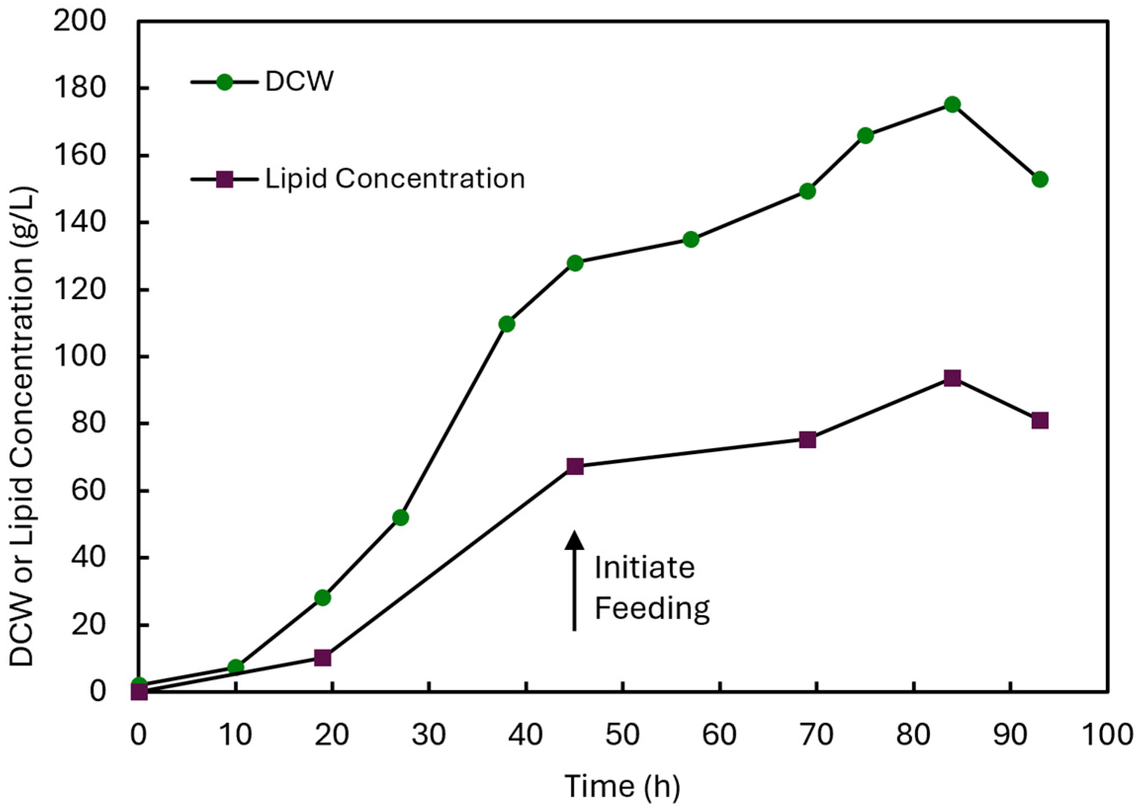
3.5. DHA Production by Schizochytrium sp. PLU-D in Fed-Batch Fermentation

To evaluate the scale-up production of DHA with the Schizochytrium sp. PLU-D strain, fed-batch fermentation was subsequently performed in a 2 L fermenter (Figure 5). The fermentation process began with a glucose batch phase in a medium with a C-N ratio of 4:1 (50YE-200G) for the accumulation of biomass. As shown in Figure 5, the biomass increased slightly from 2.0 to 7.3 g/L in the first 10 h, followed by a sharp increase from 7.3 to 128.0 g/L from 10 to 45 h. During this time, this was also accompanied by a slow and gradual increase in lipid concentration to 10.2 g/L at 19 h, followed by a sharp increase to 67.4 g/L at 45 h as the nitrogen source was depleted gradually. The initial glucose supplied at 200 g/L was almost fully consumed after 45 h, as indicated by a spike in the dissolved oxygen level (DO). Following this, a glucose fed-batch phase was initiated, in which feed solution containing the same C-N ratio of 4:1 (100YE-400G) was fed to the fermenter. During this stage, the presence of both carbon and nitrogen content in the feed was critical to promoting the continual accumulation of lipid and biomass without triggering cell lysis. From 45 to 84 h, the biomass continued to increase gradually from 67.4 to 175.3 g/L, while the lipid concentration also increased steadily from 67.4 to 93.6 g/L during the same time. At the point of harvest after 93 h, a slight decrease in the biomass and lipid concentration to 152.9 g/L and 81.2 g/L was observed, respectively.
A fatty acid composition analysis of the extracted lipids was also performed after 84 h to determine the maximum DHA titer obtained from the fermentation of PLU-D. Based on the fatty acid composition after 84 h, a similar fatty acid profile was obtained when compared to that of the 22.5YE-90G medium on the shake flask scale (Table S3). More pertinently, the lipid contained 38.7% of DHA, which corresponds to a titer of 36.2 g/L DHA obtained in the 2 L fermenter setup. To our knowledge, based on a comparison of the reported scale-up production of DHA in literature, this represents one of the highest DHA titers produced in a fermenter setup thus far (Table 1). From Table 1, the PLU-D strain produced superior DHA titers (36.2 g/L) as compared to other wild-type isolates, while higher DHA titers of 38.1 g/L and 41.4 g/L were obtained with the Schizochytrium sp. strains ALE-TF30 and M-6-23, respectively, following adaptive laboratory evolution with suitable inhibitors to increase DHA production [27,42]. In addition, drawing comparisons to the two fatty acid profiles of two commercial DHA algae oils available on the market (Table S3), both commercial algae oils have a DHA content of 38.4 and 39.3%, which is similar to that obtained with the PLU-D strain. For the other long-chain PUFAs, Nordic Naturals contained a higher EPA content of 19.8% as compared to 1.3% in GNC, while GNC produced a higher DPA content of 15.8% as compared to 1.8% in Nordic Naturals. In contrast, PLU-D contained lesser concentrations of EPA and DPA of 2.0% and 2.8%, respectively. Nonetheless, the Schizochytrium sp. PLU-D strain demonstrated its potential as a suitable strain for the sustainable production of DHA. Notwithstanding, an in-depth optimization of the fermentation process and the further development of the PLU-D strain may be necessary to attain the overarching goal of the green and commercially viable production of high-DHA microalgae oil.

4. Conclusions

Four microalgae strains were isolated from decayed mangrove samples collected from Pulau Ubin, Singapore, and were identified as Schizochytrium sp. through phylogenetic analysis. These PLU-series isolates achieved higher biomass, lipid and DHA content as compared to other wild-type microalgae strains isolated from Southeast Asia. The further optimization of the ratio of glucose to yeast extract in the medium with the PLU-D isolate on the shake flask scale resulted in an improvement in biomass with high lipid accumulation. Under the fed-batch fermentation process, the PLU-D strain was able to produce significantly high amounts of biomass and lipid, which makes it superior to most strains reported in literature thus far. This makes the Schizochytrium sp. PLU-D strain a potentially suitable microalgae strain for the commercial production of DHA. To achieve the eventual goal of commercially realizing the industrial-scale production of DHA, addressing the main bottleneck of reducing production costs may be necessary. This could be achieved by strain development via adaptive laboratory evolution or genetic engineering and further fermentation process optimization to raise DHA titers and productivity. Exploring DHA production using wastes as substitute substrates could also be an alternative approach to reduce production costs and promote the concept of a circular bioeconomy.

5. Patents

Wilmar International Limited has filed a patent application in Singapore (PCT/SG2023/050706) on 20th October 2023 relating to the use of the isolated Schizochytrium sp. strain PLU-D to produce fatty acids.

Supplementary Materials

The following supporting information can be downloaded at https://www.mdpi.com/article/10.3390/biomass4030042/s1. Figure S1: Location of mangrove samples and workflow for isolation of microalgae strains; Figure S2: 18S rRNA Sequence PLU-A (Genbank Accession Number: OP808218.1); Figure S3: 18S rRNA Sequence PLU-B (Genbank Accession Number: OP808219.1); Figure S4: 18S rRNA Sequence PLU-C (Genbank Accession Number: OP808220.1); Figure S5: 18S rRNA Sequence PLU-D (Genbank Accession Number: OP763747.1); Figure S6: Growth-curve of Schizochytrium sp. PLU-A, Schizochytrium sp. PLU-B, Schizochytrium sp. PLU-C, and Schizochytrium sp. PLU-D in shake-flask culture containing GY medium at 28 °C and 150 rpm over a period of 4 d. Table S1: Fatty acid compositions of Schizochytrium sp. PLU-A, Schizochytrium sp. PLU-B, Schizochytrium sp. PLU-C, and Schizochytrium sp. PLU-D cultured in GY medium; Table S2: Fatty acid compositions of Schizochytrium sp. PLU-D in shake-flask culture containing GY medium or 22.5YE-90G medium; Table S3: Comparison of fatty acid composition of Schizochytrium sp. PLU-D following fermentation with that of commercial oils.

Author Contributions

Conceptualization, G.K.B.K., S.N.K. and G.K.T.N.; methodology, software, validation, formal analysis, investigation, resources, and data curation, G.K.B.K., S.N.K., H.Z. and G.K.T.N.; writing—original draft preparation, G.K.B.K. and H.Z.; writing—review and editing, G.K.B.K., S.N.K. and G.K.T.N.; supervision, project administration, and funding acquisition, G.K.T.N. All authors have read and agreed to the published version of the manuscript.

Funding

This research is supported by A*STAR Singapore under its IAF-ICP (Award I2301E0021).

Institutional Review Board Statement

Not applicable.

Informed Consent Statement

Not applicable.

Data Availability Statement

The data presented in this study are available in this article.

Acknowledgments

The authors would like to thank the Wilmar Innovation Centre and A* STAR Singapore (Award I2301E0021) for their financial support. The authors would also like to thank Chua Nam-Hai for his suggestions and support of the work presented in this article. We would also like to thank Untzizu Elejalde, Lim Yeqin, Koh Dan Yu, Elvy Riani Wanjaya and Rebecca Lim Li Ting for their help in the analysis of our samples by GC-MS/FID. Finally, we would also like to thank the Singapore National Park Board (NParks) for the permit to collect mangrove samples and Yong Sze Yuen Kelvin for the facilitation of our sample collection in Pulau Ubin.

Conflicts of Interest

At the time of writing, G.K.B.K., S.N.K. and G.K.T.N. are employees of Wilmar International Limited, Singapore. Wilmar International Limited has filed a patent application in Singapore relating to the use of Schizochytrium sp. PLU-D to produce fatty acids. The authors declare no additional competing interests.

References

  1. Zhang, H.; Li, X.; Su, X.; Ang, E.L.; Zhang, Y.; Zhao, H. Production of Adipic Acid from Sugar Beet Residue by Combined Biological and Chemical Catalysis. ChemCatChem 2016, 8, 1500–1506. [Google Scholar] [CrossRef]
  2. Ng, A.M.J.; Yang, R.; Zhang, H.; Xue, B.; Yew, W.S.; Nguyen, G.K.T. A Novel Lipase from Lasiodiplodia theobromae Efficiently Hydrolyses C8-C10 Methyl Esters for the Preparation of Medium-Chain Triglycerides’ Precursors. Int. J. Mol. Sci. 2021, 22, 10339. [Google Scholar] [CrossRef]
  3. Chow, J.Y.; Nguyen, G.K.T. Rational Design of Lipase ROL to Increase Its Thermostability for Production of Structured Tags. Int. J. Mol. Sci. 2022, 23, 9515. [Google Scholar] [CrossRef] [PubMed]
  4. Kua, G.K.B.; Nguyen, G.K.T.; Li, Z. Enzyme Engineering for High-Yielding Amide Formation: Lipase-Catalyzed Synthesis of N-Acyl Glycines in Aqueous Media. Angew. Chemie Int. Ed. 2023, 62, e202217878. [Google Scholar] [CrossRef] [PubMed]
  5. Kua, G.K.B.; Nguyen, G.K.T.; Li, Z. Enzymatic Strategies for the Biosynthesis of N-Acyl Amino Acid Amides. ChemBioChem 2024, 25, e202300672. [Google Scholar] [CrossRef] [PubMed]
  6. Lauritzen, L.; Brambilla, P.; Mazzocchi, A.; Harsløf, L.B.S.; Ciappolino, V.; Agostoni, C. DHA Effects in Brain Development and Function. Nutrients 2016, 8, 6. [Google Scholar] [CrossRef] [PubMed]
  7. Khan, S.U.; Lone, A.N.; Khan, M.S.; Virani, S.S.; Blumenthal, R.S.; Nasir, K.; Miller, M.; Michos, E.D.; Ballantyne, C.M.; Boden, W.E.; et al. Effect of omega-3 fatty acids on cardiovascular outcomes: A systematic review and meta-analysis. eClinicalMedicine 2021, 38, 100997. [Google Scholar] [CrossRef] [PubMed]
  8. Chu, C.S.; Hung, C.F.; Ponnusamy, V.K.; Chen, K.C.; Chen, N.C. Higher Serum DHA and Slower Cognitive Decline in Patients with Alzheimer’s Disease: Two-Year Follow-Up. Nutrients 2022, 14, 1159. [Google Scholar] [CrossRef] [PubMed]
  9. Aslan, C.; Maralbashi, S.; Kahroba, H.; Asadi, M.; Soltani-Zangbar, M.S.; Javadian, M.; Shanehbandi, D.; Baradaran, B.; Darabi, M.; Kazemi, T. Docosahexaenoic acid (DHA) inhibits pro-angiogenic effects of breast cancer cells via down-regulating cellular and exosomal expression of angiogenic genes and microRNAs. Life Sci. 2020, 258, 118094. [Google Scholar] [CrossRef] [PubMed]
  10. Liu, Y.; Tian, Y.; Cai, W.; Guo, Y.; Xue, C.; Wang, J. DHA/EPA-Enriched Phosphatidylcholine Suppresses Tumor Growth and Metastasis via Activating Peroxisome Proliferator-Activated Receptor γ in Lewis Lung Cancer Mice. J. Agric. Food Chem. 2021, 69, 676–685. [Google Scholar] [CrossRef]
  11. Oliver, L.; Dietrich, T.; Marañón, I.; Villarán, M.C.; Barrio, R.J. Producing Omega-3 Polyunsaturated Fatty Acids: A Review of Sustainable Sources and Future Trends for the EPA and DHA Market. Resources 2020, 9, 148. [Google Scholar] [CrossRef]
  12. Wall, R.; Ross, R.P.; Fitzgerald, G.F.; Stanton, C. Fatty acids from fish: The anti-inflammatory potential of long-chain omega-3 fatty acids. Nutr. Rev. 2010, 68, 280–289. [Google Scholar] [CrossRef] [PubMed]
  13. Ganuza, E.; Anderson, A.J.; Ratledge, C. High-cell-density cultivation of Schizochytrium sp. in an ammonium/pH-auxostat fed-batch system. Biotechnol. Lett. 2008, 30, 1559–1564. [Google Scholar] [CrossRef] [PubMed]
  14. Burja, A.M.; Radianingtyas, H.; Windust, A.; Barrow, C.J. Isolation and characterization of polyunsaturated fatty acid producing Thraustochytrium species: Screening of strains and optimization of omega-3 production. Appl. Microbiol. Biotechnol. 2006, 72, 1161–1169. [Google Scholar] [CrossRef] [PubMed]
  15. Xue, Z.; Sharpe, P.L.; Hong, S.-P.; Yadav, N.S.; Xie, D.; Short, D.R.; Damude, H.G.; Rupert, R.A.; Seip, J.E.; Wang, J.; et al. Production of omega-3 eicosapentaenoic acid by metabolic engineering of Yarrowia lipolytica. Nat. Biotechnol. 2013, 31, 734–740. [Google Scholar] [CrossRef]
  16. Pino-Maureira, N.L.; González-Saldía, R.R.; Capdeville, A.; Srain, B. Rhodotorula Strains Isolated from Seawater That Can Biotransform Raw Glycerol into Docosahexaenoic Acid (DHA) and Carotenoids for Animal Nutrition. Appl. Sci. 2021, 11, 2824. [Google Scholar] [CrossRef]
  17. Mendes, A.; Reis, A.; Vasconcelos, R.; Guerra, P.; Lopes da Silva, T. Crypthecodinium cohnii with emphasis on DHA production: A review. J. Appl. Phycol. 2009, 21, 199–214. [Google Scholar] [CrossRef]
  18. Sahin, D.; Altindag, U.H.; Tas, E. Enhancement of docosahexaenoic acid (DHA) and beta-carotene production in Schizochytrium sp. using symbiotic relationship with Rhodotorula glutinis. Process Biochem. 2018, 75, 10–15. [Google Scholar] [CrossRef]
  19. Morabito, C.; Bournaud, C.; Maës, C.; Schuler, M.; Aiese Cigliano, R.; Dellero, Y.; Maréchal, E.; Amato, A.; Rébeillé, F. The lipid metabolism in thraustochytrids. Prog. Lipid Res. 2019, 76, 101007. [Google Scholar] [CrossRef]
  20. Ishibashi, Y.; Goda, H.; Hamaguchi, R.; Sakaguchi, K.; Sekiguchi, T.; Ishiwata, Y.; Okita, Y.; Mochinaga, S.; Ikeuchi, S.; Mizobuchi, T.; et al. PUFA synthase-independent DHA synthesis pathway in Parietichytrium sp. and its modification to produce EPA and n-3DPA. Commun. Biol. 2021, 4, 1–11. [Google Scholar] [CrossRef]
  21. Nagano, N.; Sakaguchi, K.; Taoka, Y.; Okita, Y.; Honda, D.; Ito, M.; Hayashi, M. Detection of Genes Involved in Fatty Acid Elongation and Desaturation in Thraustochytrid Marine Eukaryotes. J. Oleo Sci. 2011, 60, 475–481. [Google Scholar] [CrossRef] [PubMed]
  22. Meesapyodsuk, D.; Qiu, X. Biosynthetic mechanism of very long chain polyunsaturated fatty acids in Thraustochytrium sp. 26185. J. Lipid Res. 2016, 57, 1854–1864. [Google Scholar] [CrossRef]
  23. Ju, J.-H.; Ko, D.-J.; Heo, S.-Y.; Lee, J.-J.; Kim, Y.-M.; Lee, B.-S.; Kim, M.-S.; Kim, C.-H.; Seo, J.-W.; Oh, B.-R. Regulation of lipid accumulation using nitrogen for microalgae lipid production in Schizochytrium sp. ABC101. Renew. Energy 2020, 153, 580–587. [Google Scholar] [CrossRef]
  24. Ren, L.-J.; Huang, H.; Xiao, A.-H.; Lian, M.; Jin, L.-J.; Ji, X.-J. Enhanced docosahexaenoic acid production by reinforcing acetyl-CoA and NADPH supply in Schizochytrium sp. HX-308. Bioprocess Biosyst. Eng. 2009, 32, 837–843. [Google Scholar] [CrossRef] [PubMed]
  25. Ren, L.-J.; Ji, X.-J.; Huang, H.; Qu, L.; Feng, Y.; Tong, Q.-Q.; Ouyang, P.-K. Development of a stepwise aeration control strategy for efficient docosahexaenoic acid production by Schizochytrium sp. Appl. Microbiol. Biotechnol. 2010, 87, 1649–1656. [Google Scholar] [CrossRef]
  26. Ren, L.; Sun, X.; Zhang, L.; Huang, H.; Zhao, Q. Exergy analysis for docosahexaenoic acid production by fermentation and strain improvement by adaptive laboratory evolution for Schizochytrium sp. Bioresour. Technol. 2020, 298, 122562. [Google Scholar] [CrossRef] [PubMed]
  27. Sun, X.-M.; Ren, L.-J.; Bi, Z.-Q.; Ji, X.-J.; Zhao, Q.-Y.; Jiang, L.; Huang, H. Development of a cooperative two-factor adaptive-evolution method to enhance lipid production and prevent lipid peroxidation in Schizochytrium sp. Biotechnol. Biofuels 2018, 11, 65. [Google Scholar] [CrossRef] [PubMed]
  28. Liu, Y.; Singh, P.; Sun, Y.; Luan, S.; Wang, G. Culturable diversity and biochemical features of thraustochytrids from coastal waters of Southern China. Appl. Microbiol. Biotechnol. 2014, 98, 3241–3255. [Google Scholar] [CrossRef] [PubMed]
  29. Liu, L.; Bai, M.; Zhang, S.; Li, J.; Liu, X.; Sen, B.; Wang, G. ARTP Mutagenesis of Schizochytrium sp. PKU#Mn4 and Clethodim-Based Mutant Screening for Enhanced Docosahexaenoic Acid Accumulation. Mar. Drugs 2021, 19, 564. [Google Scholar] [CrossRef]
  30. Johnson, M.; Zaretskaya, I.; Raytselis, Y.; Merezhuk, Y.; McGinnis, S.; Madden, T.L. NCBI BLAST: A better web interface. Nucleic Acids Res. 2008, 36, W5–W9. [Google Scholar] [CrossRef]
  31. Tamura, K.; Stecher, G.; Kumar, S. MEGA11: Molecular Evolutionary Genetics Analysis Version 11. Mol. Biol. Evol. 2021, 38, 3022–3027. [Google Scholar] [CrossRef]
  32. Venn-Watson, S.; Lumpkin, R.; Dennis, E.A. Efficacy of dietary odd-chain saturated fatty acid pentadecanoic acid parallels broad associated health benefits in humans: Could it be essential? Sci. Rep. 2020, 10, 1–14. [Google Scholar]
  33. Unagul, P.; Suetrong, S.; Preedanon, S.; Klaysuban, A.; Gundool, W.; Suriyachadkun, C.; Sakayaroj, J. Isolation, fatty acid profiles and cryopreservation of marine thraustochytrids from mangrove habitats in Thailand. Bot. Mar. 2017, 60, 363–379. [Google Scholar] [CrossRef]
  34. Ou, M.-C.; Yeong, H.-Y.; Pang, K.-L.; Phang, S.-M. Fatty acid production of tropical thraustochytrids from Malaysian mangroves. Bot. Mar. 2016, 59, 321–338. [Google Scholar] [CrossRef]
  35. Manikan, V.; Nazir, M.Y.M.; Kalil, M.S.; Isa, M.H.M.; Kader, A.J.A.; Yusoff, W.M.W.; Hamid, A.A. A new strain of docosahexaenoic acid producing microalga from Malaysian coastal waters. Algal Res. 2015, 9, 40–47. [Google Scholar] [CrossRef]
  36. Kalidasan, K.; Vinithkumar, N.V.; Peter, D.M.; Dharani, G.; Dufossé, L. Thraustochytrids of mangrove habitats from andaman islands: Species diversity, PUFA profiles and biotechnological potential. Mar. Drugs 2021, 19, 571. [Google Scholar] [CrossRef]
  37. Ganuza, E.; Izquierdo, M.S. Lipid accumulation in Schizochytrium G13/2S produced in continuous culture. Appl. Microbiol. Biotechnol. 2007, 76, 985–990. [Google Scholar] [CrossRef]
  38. Ling, X.; Guo, J.; Liu, X.; Zhang, X.; Wang, N.; Lu, Y.; Ng, I.S. Impact of carbon and nitrogen feeding strategy on high production of biomass and docosahexaenoic acid (DHA) by Schizochytrium sp. LU310. Bioresour. Technol. 2015, 184, 139–147. [Google Scholar] [CrossRef]
  39. Patil, K.P.; Gogate, P.R. Improved synthesis of docosahexaenoic acid (DHA) using Schizochytrium limacinum SR21 and sustainable media. Chem. Eng. J. 2015, 268, 187–196. [Google Scholar] [CrossRef]
  40. Ratledge, C. Regulation of lipid accumulation in oleaginous micro-organisms. Biochem. Soc. Trans. 2002, 30, 1047–1050. [Google Scholar] [CrossRef]
  41. Ratledge, C. The role of malic enzyme as the provider of NADPH in oleaginous microorganisms: A reappraisal and unsolved problems. Biotechnol. Lett. 2014, 36, 1557–1568. [Google Scholar] [CrossRef] [PubMed]
  42. Zeng, L.; Bi, Y.; Guo, P.; Bi, Y.; Wang, T.; Dong, L.; Wang, F.; Chen, L.; Zhang, W. Metabolic Analysis of Schizochytrium Mutants with High DHA Content Achieved With ARTP Mutagenesis Combined with Iodoacetic Acid and Dehydroepiandrosterone Screening. Front. Bioeng. Biotechnol. 2021, 9, 738052. [Google Scholar] [CrossRef] [PubMed]
  43. Jakobsen, A.N.; Aasen, I.M.; Josefsen, K.D.; Strøm, A.R. Accumulation of docosahexaenoic acid-rich lipid in thraustochytrid Aurantiochytrium sp. strain T66: Effects of N and P starvation and O2 limitation. Appl. Microbiol. Biotechnol. 2008, 80, 297–306. [Google Scholar] [CrossRef] [PubMed]
  44. Huang, T.Y.; Lu, W.C.; Chu, I.M. A fermentation strategy for producing docosahexaenoic acid in Aurantiochytrium limacinum SR21 and increasing C22:6 proportions in total fatty acid. Bioresour. Technol. 2012, 123, 8–14. [Google Scholar] [CrossRef] [PubMed]
  45. Hoang, L.A.T.; Nguyen, H.C.; Le, T.T.; Hoang, T.H.Q.; Pham, V.N.; Hoang, M.H.T.; Ngo, H.T.T.; Hong, D.D. Different fermentation strategies by Schizochytrium mangrovei strain pq6 to produce feedstock for exploitation of squalene and omega-3 fatty acids. J. Phycol. 2018, 54, 550–556. [Google Scholar] [CrossRef] [PubMed]
  46. Yin, F.W.; Zhang, Y.T.; Jiang, J.Y.; Guo, D.S.; Gao, S.; Gao, Z. Efficient docosahexaenoic acid production by Schizochytrium sp. via a two-phase pH control strategy using ammonia and citric acid as pH regulators. Process Biochem. 2019, 77, 1–7. [Google Scholar] [CrossRef]
  47. Liu, L.; Zhu, X.; Ye, H.; Wen, Y.; Sen, B.; Wang, G. Low dissolved oxygen supply functions as a global regulator of the growth and metabolism of Aurantiochytrium sp. PKU#Mn16 in the early stages of docosahexaenoic acid fermentation. Microb. Cell Fact. 2023, 22, 1–13. [Google Scholar]
  48. Chang, G.; Gao, N.; Tian, G.; Wu, Q.; Chang, M.; Wang, X. Improvement of docosahexaenoic acid production on glycerol by Schizochytrium sp. S31 with constantly high oxygen transfer coefficient. Bioresour. Technol. 2013, 142, 400–406. [Google Scholar] [CrossRef]
Figure 1. Unrooted phylogenetic tree of PLU isolates and other publicly available microalgae strains based on 18S rRNA sequences. The tree was constructed by the neighbor-joining method in MEGA 11 software. A Bootstrap value of 500 was used for the analysis.
Biomass 04 00042 g001
Figure 2. Growth of Schizochytrium sp. PLU-A, Schizochytrium sp. PLU-B, Schizochytrium sp. PLU-C and Schizochytrium sp. PLU-D in shake flask culture containing GY medium. (a). Biomass and lipid concentration of PLU isolates after 4 d culture. (b). Fatty acid composition of PLU isolates after 4 d culture.
Biomass 04 00042 g002
Figure 3. Morphology of Schizochytrium sp. PLU-D grown in GY medium for 3 days. (a). Vegetative cells; (b). vegetative cells and an amoeboid cell (arrow); (c). release of zoospores from zoosporangium; (d). clusters of cells released from the binary cell division of an octad (white arrow) and a decad (black arrow); (e). an amoeboid cell (black arrow) and a diad from binary cell division (white arrow). The scale bar in (ac) is 20 μm, while the scale bar in (d,e) is 10 μm.
Biomass 04 00042 g003
Figure 4. Biomass and lipid concentration of Schizochytrium sp. PLU-D in shake flask culture containing varying glucose and yeast extract concentrations after 4 d culture. Shake flask conditions with an asterisk indicate observable cell lysis from the culture supernatant following centrifugation. (a). Effects of varying glucose concentrations with yeast extract kept at 10 g/L; (b). Effects of varying yeast extract concentrations with glucose kept at 90 g/L.
Biomass 04 00042 g004
Figure 5. Biomass and lipid concentration of Schizochytrium sp. PLU-D in a 2 L fermenter. The pH was controlled at pH 7.0 with 2 M NaOH and 0.5 M citric acid, and the temperature was maintained at 28 °C.
Biomass 04 00042 g005
Table 1. Comparison of biomass, lipid concentration and DHA titers from the fed-batch fermentation of thraustochytrid strains for DHA production.
EntryThraustochytrid StrainsScale Fermentation Time
(h)
Biomass
(g/L DCW)
Lipid
Concentration
(g/L)
Lipid
Content
(%)
DHA
Titer
(g/L)
DHA
Content
(%)
References
1Aurantiochytrium sp. ATCC PRA-276 (T66)5 L fermenter180100.058.058.017.430.0[43]
2Schizochytrium sp. G13/2S5 L fermenter4863.315.825.05.4734.6[13]
3Aurantiochytrium limacinum ATCCMYA-1381(SR21)5 L fermenter15061.840.365.220.350.4[44]
4Schizochytrium
mangrovei (PQ6)
30 L fermenter96105.345.743.45.912.9[45]
5Schizochytrium sp. CCTCC M209059 (HX-308)1500 L fermenter13271.035.850.417.548.9[25]
6Schizochytrium sp. CCTCC M209059 (HX-308)5 L fermenter120120.662.651.932.852.4[46]
7Schizochytrium sp. CCTCC M209059 (ALE-TF30)5 L fermenter120126.471.656.638.153.2[27]
8Schizochytrium sp. ABC1015 L fermenter8486.037.243.316.744.9[23]
9Aurantiochytrium sp. PKU#Mn165 L fermenter14456.719.033.56.031.6[47]
10Schizochytrium sp. LU3101 L baffled flask14488.652.058.724.747.5[38]
11Schizochytrium sp. ATCC 20,888 (S31)7.5 L fermenter96151.479.752.628.936.3[48]
12Schizochytrium sp. ATCC20888
(M-6-23)
5 L fermenter96154.888.257.041.446.9[42]
13Schizochytrium sp. PLU-D2 L fermenter84175.393.653.436.238.7This study
Disclaimer/Publisher’s Note: The statements, opinions and data contained in all publications are solely those of the individual author(s) and contributor(s) and not of MDPI and/or the editor(s). MDPI and/or the editor(s) disclaim responsibility for any injury to people or property resulting from any ideas, methods, instructions or products referred to in the content.

