GASTROENTEROLOGY 2002;122:406-414
Detection of Dysplastic Intestinal Adenomas Using EnzymeSensing Molecular Beacons in Mice
KATHARINA MARTEN,* CHRISTOPH BREMER,* KHASHAYARSHA KHAZAIE,~ MANSOUREH SAMENI,§
BONNIE SLOANE,§ CHING-HSUAN TUNG,* and RALPH WEISSLEDER*
*Center for Molecular Imaging Research, Massachusetts General Hospital and Harvard Medical School, Boston; ~Dana-Farber Cancer
Institute, Cancer Immunology & AIDS, Harvard Medical School, Boston, Massachusetts; §Department of Pharmacology, Wayne State
University School of Medicine, Detroit, Michigan
See editorial on page 571.
Background&Aims: Proteases play key roles in the
pathogenesis of tumor growth and invasion. This study
assesses the expression of cathepsin B in dysplastic
adenomatous polyps. Methods: Aged Apc M~n/+ mice
served as an experimental model for familial adenomatous polyposis. The 4 experimental groups consisted of
(a) animals injected with a novel activatable, cathepsin
B sensing near infrared fluorescence (NIRF) imaging
probe; (b) animals injected with a nonspeciflc NIRF; (c)
uninjected control animals; and (d) non-APCM~n/+ mice
injected with the cathepsin B probe. Lesions were analyzed by immunohistochemistry, Western blotting, reverse transcription-polymerase chain reaction, and optical imaging. Results: Cathepsin B was consistently
overexpressed in adenomatous polyps. When mice were
injected intravenously with the cathepsin reporter probe,
intestinal adenomas became highly fluorescent indicative of high cathepsin B enzyme activity. Even microscopic adenomas were readily detectable by fluorescence, but not light, imaging. The smallest lesion
detectable measured 50 ~m in diameter. Adenomas
in the indocyanine green and/or noninjected group
were only barely detectable above the background.
Conclusions: The current experimental study shows that
cathepsin B is up-regulated in a mouse model of adenomatous polyposis. Cathepsin B activity can be used as a
biomarker to readily identify such lesions, particularly
when contrasted against normal adjacent mucosa. This
detection technology can be adapted to endoscopy or
tomographic optical imaging methods for screening of
suspicious lesions and potentially for molecular profiling
in vivo.
dentification and removal of colonic adenomatous polyps during endoscopy has been shown to reduce the
incidence of colorectal cancer. ~ Adenomatous polyps represent up to half of all colonic polyps and are particularly
worrisome if they are large (>1 cm) or multiple, have
I
extensive villous components, 2,3 and/or are highly dysplastic. 1 The latter feature in particular has been associated with the successive development of oncogenes and
the loss of tumor-suppressor genes 4 and ultimately leads
to carcinoma formation. However, apart from the size
criterion, it is currently clinically difficult to ascertain
the extent of dysplastic features 5 during colonoscopy in
vivo. The availability of techniques that allow in vivo
identification of small highly dysplastic adenomas from
innocuous lesions would be helpful to guide selective
removal of polyps.
The exact molecular events occurring during colorectal tumorigenesis are slowly emerging. The genesis of
familial adenomatous polyposis (FAP), as well as sporadic
colorectal neoplasms, is closely linked with genetic defects that result in carboxy-terminal truncations of the
adenomatous polyposis coli (APC) protein. Colorectal
tumors with intact APC genes were found to contain
activating mutations of ~-catenin. 6,v APC and [3-catenin
are key components of the W n t signaling pathway (see
reviewsS-U)). It is generally agreed that most colonic
cancers develop from adenomatous polyps. Common to
most tumors, several generic features become altered
during multistage tumor progression, most importantly,
the control of proliferation, the balance between cell
survival, and programmed cell death (apoptosis), interactions with host cells and extracellular matrix, angiogenesis, and metastatic dissemination. H'12 Proteolytic
enzymes have been shown to play an essential role in
many of these processes, in particular high cell turnover,
invasion, and angiogenesis, t3 Some of these proteases
include matrix-metalloproteases (e.g., MMP), serine proAbbreviations used in this paper: APC, adenomatous polyposis coli;
ICG, indocyanine green; FAP, familial adenomatous polyposis; MMP,
matrix-metalloproteases; NIR, near-infrared; NIRF, near infrared fluorescence; SI, signal intensities; TBC, target to background contrast.
© 2002 by the American Gastroenterological Association
0016-5085/02/$35.00
doi:10.1053/gast.2002.30990
�February 2002
FLUORESCENCEIMAGING OF INTESTINALADENOMAS 407
teases (e.g., urokinase-type plasminogen activator), aspartic proteases (e.g., cathepsin D), and cysteine proteases (e.g., cathepsin 8). 13,14 Metalloproteinase has been
reported to be direct targets of the W n t signaling pathway. t5,[6 Cathepsin B in particular has been shown to be
up-regulated in areas of inflammation, necrosis, angiogenesis, t3 and focal invasion iv of colorectal carcinomas
and in dysplastic adenomas. 18,19
O u r laboratory has been interested in developing protease-specific sensor molecules that can be used for the
noninvasive in vivo m o n i t o r i n g of enzyme activities. 2° 22
W e have developed optically based, protease activatable
fluorescent sensors that operate in the near-infrared
( N I R ) region for m a x i m u m light tissue penetration. 2°,23
The goal of the current study was to determine whether
cathepsin B protease activity could be identified within
dysplastic adenomatous polyps and whether targeting of
this enzyme could be used for the detection of small
dysplastic adenomatous lesions. W e chose to investigate
adenomatous polyps in the A P C Min/+ mouse which is
heterozygous for a germ-line m u t a t i o n in the mouse
homologue of the h u m a n A P C gene. 24 These animals
develop multiple adenomas in the small and large bowel
which simulate dysplastic adenomatous polyps found in
h u m a n disease. 24 26
Materials
and Methods
Mouse Model
A P e i~[in/+ mice (n = 24, age 7 - 28 weeks) were
obtained from the Jackson Laboratories (Bar Harbor, ME).
Mice were randomly divided into 3 experimental groups: (1)
animals receiving an intravenous injection of the cathepsin
B-sensitive NIRF probe (n = 10; 142 adenomas; mean age,
15.8 weeks; 2 nmol Cy5.5); (2) animals receiving an intravenous injection with indocyanine green (ICG), a nonactivatable
fluorochrome (n = 4 mice, 44 adenomas; mean age, 16 weeks;
200 btg); and (3) noninjected control mice (n = 2 mice; 18
adenomas; mean age, 18 weeks). In addition, we also included
a cohort of strain matched non-APC mice (C57BL6, Jackson
Laboratories, Bar Harbor, ME, group d) and injected them
with the cathepsin B-sensitive NIRF probe (n = 3; no true
adenomas; mean age, 12 weeks; 2 nmol Cy5.5). The animal
protocol was approved by the Institutional Review Board.
Mice were anesthetized (90 mg/kg ketamine and 10 mg/kg
xylazine intraperitoneally) for intravenous injections and killed
24 hours later under halothane inhalation, and the entire bowel
was removed for light and NIR fluorescence imaging and for
correlative studies. Adenomas were identified after trypan blue
staining using a dissection microscope. With this technique it
is easy to distinguish a lymph node (Peyer's patch) from an
adenoma; the former has a smooth surface and occurs often in
pairs, whereas the latter has typical pedunculated/tubular or
sessile/villous morphology. A total of 142 polyps (group a) and
30 "polyp-like" lesions (group [a] and [d]) were imaged (Table
1), whereas lesions in these groups were identified by dissecting microscopy. An additional, 6 animals were used for in vitro
analyses and 2 animals (n = 30 polyps) for hematoxylin and
eosin staining of polyps.
Histology
Bowel tissue of a subset of animals (30 adenomas) was
fixed in phosphate-buffered formalin for 24 hours, paraffinembedded, sectioned into 6-Ftm thin slices and stained with
hematoxylin and eosin. Immunohistochemistry for cathepsin B
expression was performed on frozen sections using a primary
polyclonal anti-cathepsin B antibody (Santa Cruz Biotechnology, Santa Cruz, CA). Binding of the primary antibody was
revealed using an alkaline phosphatase-labeled secondary antibody. NBT/BCIP substrate (Boehringer-Mannheim, IN) was
used to visualize specific alkaline phosphatase activity. Sections
were counterstained with nuclear fast red. Control sections
were processed identically but using only the secondary antibody.
Fluorescence confocal microscopy was also performed on
normal and APC Min/+ mouse intestine. Sections of the paraffinembedded tissues were placed on glass slides, subjected to
deparaffination in xylene and ethanol, and microwaved twice
for 2-5 minutes each. 2v Sections were blocked with 10%
normal donkey serum for 1 hour at room temperature. Sections
were incubated with a 1:400 dilution of primary antibody
(polyclonal anti-cathepsin B immunoglobulin [Ig]G, developed, and characterized for specificity by us) overnight at room
temperature. After washing 5 × in phosphate-buffered saline
(PBS), they were reacted for 1 hour at room temperature with
a 1:100 dilution of secondary antibody (fluorescein-conjugated
donkey anti-rabbit IgG) plus 5% normal donkey serum and
washed 5 × with PBS. Immunofluorescence caused by binding
of the primary antibody to cathepsin B was observed on a Zeiss
LSM 310 confocal microscope (Zeiss, Thornwood, NY). Controis were run in the absence of primary antibody.
Reverse Transcription-Polymerase
Chain Reaction
Mice were killed, and the bowel was removed, flushed
with cold phosphate-buffered saline, opened longitudinally,
and examined under a stereomicroscope. Adenomas were ex-
Table 1. Sensitivity and Specificity
Visible light imaging
with magnification
NIR fluorescence
Lesions
Positive
Negative
Positive
Negative
142 polyps
30 polyp-like lesions
70
18
72
12
136
2
6
28
NOTE. Results are based on 142 polyps in APCM~n/+ mice and 30
polyp-like lesions (APCM'n/÷ mice and C57BL6 mice). The polyps were
identified by trypan blue staining of mucosa and visualizing bowel
specimen under a dissection microscope.
�408 MARTENET AL.
cised and snap frozen in liquid nitrogen. Normal intestinal
mucosa from the vicinity of the adenomas was collected in a
similar fashion. Reverse transcription-polymerase chain reaction (RT-PCR) was performed using an ABI Prism 7700
Sequence BioDetector (PE Biosystems, Foster City, CA) with
SYBR-green fluorescence detection according to the manufacturer's instructions (Perkin Elmer, Foster City, CA). Total
RNA was extracted using Trizol (Gibco BRL) and reverse
transcribed using Superscript IIRT (Gibco BRL) and oligo(dT)15 priming. Cathepsin B primers were intron-spanning
and included (5' to 3') AGG TTC GGT CAG AAA TGG
CTT, and (5' to 3') ATC CTT CTT TCT TGC CTG CTG.
Beta-Actin-1 primers were (5' to 3') TGG AAT CCT GTG
GCA TCC ATG AAA C, and (5' to 3') TAA AAC GCA GCT
CAG TAA CAG TCC G. Cycling conditions were as follows:
5 minutes at 95°C, 2 cycles (45 seconds) at 94°C, 45 seconds
at 60°C, 1 minute at 72°C, 2 cycles (45 seconds) at 94°C, 45
seconds at 58°C, 1 minute at 72°C, 2 cycles (45 seconds) at
94°C, 45 seconds at 56°C, 1 minute at 72°C, and 34 cycles (45
seconds) at 94°C, 45 seconds at 54°C, 1 minute at 72°C,
followed by 10 minutes at 72°C. Actin and cathepsin B
reactions were analyzed on the same plate. Relative starting
quantities of complementary DNA (cDNA) for each tissue
sample were determined using standard curves made from six
1:3 serial dilutions of wild-type Balb/c total spleen cDNA.
Standard curves were plotted as dilution factor versus threshold cycle. Values for both actin and cathepsin B expression in
adenoma and normal control tissue samples were determined
by putting the experimentally determined threshold cycle
values into the standard curve formula. Raw data were normalized for relative amount of total cDNA and tabulated.
GASTROENTEROLOGYVol. 122, No. 2
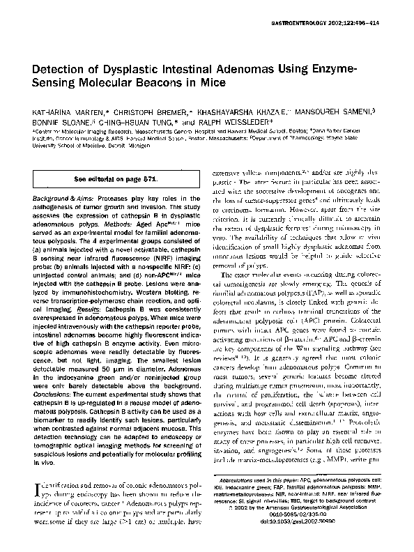
probe (Figure 1) and a nonspecific fluorochrome (indocyanine
green), in clinical use for retinal angiography28 The NIRF
probe contained Cy5.5 monofunctional dye (Amersham Pharmacia Biotech, UK) reporters adjacent to . . K - K . . . cleavage
sites on a macromolecular assembly, described in more detail
elsewhere, e° Activatability by cathepsin B and quality control
was performed for each batch synthesized. The assembly consisted of a synthetic graft copolymer containing partially pegylated (5 kilodaltons) poly-L-lysine (35 kilodaltons), similar to
what had been used clinically, e9 The injection dose (2 nmol
Cy5.5 per animal and time of imaging after injection) had
previously been optimized in non-APC M'"/+ mice bearing
other pathologies. 2°m
ICG (Akorn, IL, 2 mg/mL) was freshly prepared and used for
intravenous injections (200 b~g per animal). ICG was used as a
control to test the hypothesis that the fluorescence signal is
mainly caused by activation of the NIRF probe rather than
nonspecific accumulation.
MPEG
NH 2
MPEG
L!s--Lys--Lys--Lys--L!s
Western Blotting
Adenomas and healthy mucosa of APC M'n/+ mice were
homogenized in lysis buffer (50 mmol/L Tris-HC1 pH 7.4, 100
mmol/L NaC1, 10 mmol/L CaCI2 containing 0.25% Triton
X-100 and "complete" protease inhibitor cocktail; Boehringer
Mannheim, Germany). Total protein content was determined
using a bicinchoninic acid protein assay (Pierce, Rockford, IL).
Equal amounts of adenoma and mucosa extracts (4.5-30 I~g)
were loaded onto a 10% sodium dodecyl sulfate polyacrylamide gel. After separation proteins were transferred to a
polyvinylidine difluoride membrane (Bio-Rad Laboratories,
Hercules, CA). After blocking for 1 hour (3% bovine serum
albumin in phosphate-buffered saline [PBS]), membranes were
incubated with the primary anti-cathepsin B polyclonal antibody (Bio-Rad Laboratories, Hercules, CA) and a secondary
antibody conjugated with alkaline phosphatase (Sigma, St.
Louis, MO). Alkaline phosphatase activity was revealed with
NBT/BCIP substrate (Boehringer Mannheim, Germany).
Membranes were scanned and lanes were analyzed digitally
(Kodak Digital Science 1D software, Rochester, NY).
NIR Fluorochrome Probes
Two different near-infrared fluorescence (NIRF) probes
were used in this study: an activatable cathepsin B sensing
g
athepsin B
activation
MPEG
NH2
Lys~Lys--
LIs
y ~
MPEG
NH 2
I
HOOC ~ Ly s ~ Lys
I
NH
-O-
NH
h 'ox
-@-
Figure 1. Schematic diagram of the cathepsin B probe and its activation. The imaging probe consists of a cleavable indocyanine containing graft copolymer. The fluorochromes are essentially nonfluorescent in their native state caused by energy resonance transfer among
fluorochromes. On enzymatic cleavage the agent becomes fluorescent in the near-infrared ( h v e x = excitation wavelength, h v e m = emission wavelength).
�February 2002
FLUORESCENCEIMAGINGOF INTESTINALADENOMAS 409
Imaging and Image Analysis
Bowel samples were imaged immediately after excision
using a custom built NIRF reflectance imaging system. 23 The
system consisted of a light-tight chamber equipped with a
130-watt halogen white light source and an excitation filter
system (610-650 nm, Omega Optical, Brattleboro, VT).
Light diffusers were used to homogeneously distribute light
over the field of view. A 12-bit monochrome CCD camera
(Kodak, Rochester, NY) equipped with a f/1.2 12.5-75 mm
zoom lens and an emission bandpass filter (Omega Optical,
Brattleboro, VT) was used to detect fluorescence (Cy5.5 680720 nm and ICG 780-820 nm emission). Images were acquired over 3 minutes and analyzed using commercially available software (Kodak Digital Science 1D software, Rochester,
NY). Regions of interests were obtained from the entirety of
each adenoma and from adjacent size matched intestinal mucosa. Mean signal intensities (SI) were recorded.
The target (adenoma) to background (mucosa) contrast
(TBC) was calculatedas follows:
TBC(%) = ([SI.d
.....
-SI ....... ]/SI ...... ) × 100
(i.e., a value of 100 will represent a 100% higher fluorescence
signal of the adenoma compared with mucosa).
All results are presented as mean + standard error of the
mean (SEM). Statistical analysis of the 2 groups was conducted
using a 2-tailed Student t test for unpaired samples. A P value
<0.05 was considered to be significant.
Results
Adenomas (n = 204) ranged in size from 50 btm
to 6 m m in diameter (mean 2 mm). It has previously
been shown that the majority of cells found in tumors of
APC Min/+ mice do not display any of the differentiation
markers found in the various cell types of the intestinal
epithelium and therefore represent undifferentiated
cells. 26 Analysis of histologic sections showed the characteristic architecture of dysplastic crypts located at the
luminal surface of the mucosa, with underlying, wellspaced, nondysplastic crypts. In some lesions, high-grade
dysplasia, based on the identification of full-thickness
crypt epithelial nuclear stratification and loss of cytoplasmic mucinous differentiation, was present. Dysplasia in
general was characterized by tall, hyperchromatic disorderly cells with cigar-shaped nuclei and concomitant
crypt budding (Figure 2). Immunohistochemistry and
fluorescence confocal microscopy was positive for cathepsin B expression throughout the adenoma in epithelial
and stroma cells (Figure 2). W i t h progressive stages of
dysplasia, cathepsin B expression levels were successively
higher. By Western blotting, adenomas had a 36% -+
8.6% higher cathepsin B protein level when compared
with adjacent normal mucosa. RT-PCR showed cathep-
sin B m R N A to be about 35% -+ 8.3% higher compared
with adjacent mucosa on average.
In the noninjected animals, N I R imaging revealed a
similar signal intensity of adenomas compared with that
of adjacent mucosa (Figure 3). However, adenomas in
animals injected with the cathepsin B probe showed a
remarkably higher TBC (Figure 3). On average, contrast
was highest in large adenomas (220% + 97%), presumably caused by the higher amount of converting enzyme
per lesion. To prove that fluorescence signal intensity was
caused by enzyme activation in adenomas and not only
nonspecific accumulation, a cohort of animals was injected with fluorescent ICG. Adenomas (n = 44) in these
animals showed significantly lower TBC contrast as those
having received the cathepsin B sensing probe (TBC =
34% - 4% vs. 119% + 71%, P < 0.01, Figure 3).
Figure 4 summarizes the visible light and the N I R
appearance of jejunal bowel segments in the different
experimental groups. The N I R images are scaled equally
and clearly show the high conspicuity of adenomas using
the cathepsin B reporter probe. Figure 5 summarizes the
appearance of colonic adenomatous polyps. Interestingly,
even minute adenomas, not detectable by light imaging,
became easily detectable by NIRF imaging.
Table 1 summarizes quantitative data on lesion detectability using either visible light or N I R fluorescence.
Sensitivity for NIRF imaging was 96% with a specificity
of 93%. The sensitivity of light imaging was 49% and
the specificity 40%.
Discussion
The current results indicate that adenomatous
polyps show moderately elevated cathepsin B expression
and high-enzyme activity. The enzyme activity within
these lesions was ubiquitous and was highest in larger
colonic polyps with high degrees of dysplasia. These
lesions were highly conspicuous and even adenomas of
sizes as small as 50 lzm in diameter could be readily
identified. The results from this pilot study have 5
practical implications: (1) cathepsin B protease may play
a role in early alterations leading to tumor formation; (2)
such proteolytic enzymes and potentially other "biomarkers" can be used for in vivo molecular imaging of
suspicious lesions; (3) multiwavelength imaging with
different probes may facilitate "typing" of lesions; (4) the
strategy can be used to improve the detection of adenomas (particularly partly obscured sessile lesions); and (5)
the probe technology could be readily adapted to conventional endoscopy or even external tomographic N I R
imaging of bowel.
�410
MARTEN ET AL.
GASTROENTEROLOGYVol. 122, No. 2
Figure 2. Adenomas in the APCMin/+ model. (A-C) Hematoxylin-eosin stain of small (A) and a large (B, C) colonic adenoma (asterisk). The latter
shows the characteristic architecture of dysplastic epithelium with tortuous and branching crypts and hyperchromatic nuclei. (A, B) Objective
magnification: 2-fold, (C) 20-fold. (D-l) Fluorescence confocal microscopy. (D) Normal intestine; (E-I) Adenomas representing progressive stages
of dysplasia from (E) least to (/) most dysplastic. Note the increasing cathepsin B expression levels.
Role of Proteases in Premalignant Lesions
The involvement of proteases in cancers is well
established; however, recently there has been mounting
evidence that proteases are also involved in early alterations leading to tumor formation. 13 For example, increased immunostaining and altered localization of cathepsin B has been observed in late human adenomas 3°
and has been associated in particular with high-grade
dysplasia) 1 In the latter study, cathepsin B-positive
tumor cells were observed in 67% of adenomas but in
100% of adenomas with high-grade dysplasia or adenocarcinomas. Additional evidence for the importance of
protease involvement comes from animal studies. When
APC Min/+ mice, such as those used in this study, are
crossed with matrilysin (matrix metalloprotease) deficient mice, the development of spontaneous intestinal
polyps is decreased) 2 Our own data show that cathepsin
B was up-regulated in intestinal adenomas in the
�February 2002
FLUORESCENCE IMAGING OF INTESTINAL ADENOMAS
4110¸
3-6ram
T
"~ 300 ¸
<3ram
200
a.
T
,00:
,7-,
Control
ICG
[~
±
411
enomas were observed at the site of infection within 1
week after inoculation of the virus and loss of both APC
alleles. However, in conflict with a simple dominantnegative mode of action of the truncated APC, over-
_k
Cathepsin B probe
Figure 3. Conspicuity of adenomatous polyps. Data show the TBC.
Adenomas investigated with the cathepsin B imaging probe show a
TBC between 100% and 350%, at which adenomas appear as "light
bulbs" against dark mueosa (Figure 4).
A P C Min/+ m o u s e both at the m R N A and protein level.
The exact mechanism by which cathepsin B is up-regulated remains to be investigated. One working hypothesis is that it could be up-regulated through the W n t
signaling pathway similar to the up-regulation of matrix
metalloproteases. Alternative possibilities include an indirect effect of epithelial hyperproliferation or activation,
mesenchyme response to signals from the transformed
epithelial cells, or caused by the inflammatory response
and recruitment of activated host immune cells. In any
case, it is unlikely that cathepsin activation is caused by
a secondary genetic/oncogenic event other than loss of
APC, because cathepsin activation can be already detected in very early adenomas. Studies with transgenic
knock-out mice have confirmed that loss of the wild-type
APC allele is the critical secondary genetic event determining the transition from normal epithelium ro polyps. 33-36 These studies have been confirmed by conditional knock-out of both alleles of APC in adult m i c e ) v
In this case exon 14 of the mouse APC gene was flanked
with loxP sequences, and the Cre recombinase was provided to the adult mice through intrarectal infection
with recombinant adenoviral vectors encoding Cre. Ad-
Figure 4. Light and NiR fluorescence imaging of jejunal adenomas.
(A, B) Light images and (C-D) NIRF images of (A, C) a noninjected
6-month-old APCMrn/+ mouse and (B, D) a 6-month-old APCMin/+
mouse injected with the NIRF probe. (A) In the noninjected mouse,
multiple adenomas can be seen on the light image, (C) but are
essentially not fluorescent. (D) With injection of the NIRF probe, the
adenomas are clearly identified by NIRF imaging (Scale bar, 5 ram).
Figure 5. Colonic adenomas in a 7-month-old APCMin/+ mouse 24
hours after injection of the NIRF probe. (A) The light image shows 3
adenomas at a size range of 2-5 mm in diameter. Multiple small
adenomas are readily detectable by NIRF imaging (B, arrows), but can
barely be seen on the light image. (C) Correlative transillumination
microscopy revealing the small adenoma (shown in B, right arrow) to
be approximately 50 i~m in size (arrow).
�412
MARTEN ETAL.
GASTROENTEROLOGYVol. 122, No. 2
expression of truncated APC genes in transgenic mice
did not lead to polyposis. 35 Another area of investigations lies in determining the mechanism by which cathepsin B may increase cell proliferation for example by
activating growth factors (e.g., fibroblast growth factor,
epidermal growth factor, transforming growth factor-B,
vascular endothelial growth factor) or by liberating them
from the extracellular matrix. 38 Irrespective of these areas
of ongoing investigation, it is clear through the current
data and that of other investigators, 13 that proteases may
play important roles in premalignant lesions.
Reporter
Probe
The recent development of targeted NIRF, 39 activatable NIR fluorochromes, 2o red-shifted fluorescent
proteins, 4° and bioluminescent probes 4~ is slowly opening the road toward in vivo molecular imaging. The
cathepsin B probe used in the current study was a
quenched NIR probe previously used to detect malignant lesions. 2° One significant advantage of fluorescent
over other reporters (e.g., isotopes, iodinated agents for
radiograph) is that they can be "silenced" and "activated," enabling the design of molecular switches or
"beacons" (Figure 1). The probe used in this study and
probes in other studies 2°-22 are nonfluorescent in their
native (quenched) state and become highly fluorescent
after target interaction, resulting in signal amplification
of up to several hundred-fold, depending on the specific
design. The dosing and timing of imaging of these
probes has previously been optimized and was adapted in
the current study. Using activatable probes in particular
has several major advantages over single fluorochromes
attached to affinity molecules (such as antibodies). Most
important, quenching results in reduction of background
"noise" by several orders of magnitude and a single
enzyme can cleave multiple fluorochromes resulting in
efficient signal amplification. This advantage is best exemplified by our results (Figure 3) in which we show that
the TBC is an order of magnitude higher with the
cathepsin B probe than when nonspecific fluorochromes
are used. By choosing the appropriate substrate spacer, a
series of very specific enzyme probes can be developed,
and multiple probes can potentially be used for multispectrum imaging.
Detection
Technology
Inherently linked to the development of the previously described reporter probes2°-= and the molecular analysis of cancers and precancerous lesions, 13,17,3°is the need to
develop detection technology that can accurately quantitate
NIR fluorochrome concentrations and fluorescence activation in vivo. The current study used CCD technology to
examine the bowel specimen in reflectance model, 23 similar
to the technology used during fiberoptic endoscopy. The
current detection technology could thus be easily adapted t o
real-time endoscopy or even multiwavelength channel endoscopy. The adaptation would essentially require a separate
NIR light source, appropriate filters, and a sensitive CCD
camera for detection. NIR light has been shown to travel up
to 7-10 cm through tissue using Food and Drug Administration class I-3 laser sources. With advanced technology
for single photon counting or very low-noise detection
systems, it is also feasible to potentially record the molecular
signatures from outside the abdomen. Recently, optical
tomography with NIR light has been described42 and being
facilitated by rigorous mathematical modeling of light
propagation in tissue and technological advancements in
photon sources and detection techniques. It is clear that the
newly described probe armamentarium and novel NIR photon detection technology stand a good chance to significantly impact on our capability of imaging molecular targets in vivo.
Clinical Implications
The current study has several clinical implications. NIRF endoscopy using enzyme-sensing imaging
probes may become a complementary tool (molecular
endoscopy) to other types of endoscopy such as chromoe n d o s c o p y 43,44 or light-induced fluorescence spectroscopy.45-47 We believe that the required NIRF imaging
technology could be easily integrated into existing endoscopic systems, to provide high-resolution, real-time
imaging of larger intestinal surface areas. A second clinical application may be in externally usable tomographic
NIRF imaging, by which the underlying colon could be
imaged from outside the abdominal surface. Based on the
current probe, a number of different enzyme or targetsensing molecules could be designed for in vivo sensing
of broader ranges of biomarkers. Finally, such reporter
probes may become useful in monitoring of therapy with
protease inhibitors using other forms of endoscopy or
laparoscopy. 22 It is also conceivable that the technology
could be used to identify other pathologies, e.g., active
inflammation in ulcerative colitis. In conclusion, NIRF
imaging using protease-activatable imaging probes may
have a significant impact on diagnosis of a very early
stage of intestinal disease. With further advances in
technology and chemistry, we are likely to see significant
advances in optical imaging in vivo and sensing.
References
1. Winawer SJ, Fletcher RH, Miller L, Godlee F, Stolar MH, Mulrow
CD, Wootf SH, Glick SN, Ganiats TG, Bond JH, Rosen L, Zapka JG,
Olsen SJ, Giardiello FM, Sisk JE, Van Antwerp R, Brown-Davis C,
�February 2002
2.
3.
4.
5.
6.
7.
8.
9.
10.
11.
12.
13.
14.
15.
16.
17.
18.
19.
20.
21.
22.
Marciniak DA, Mayer RJ. Colorectal cancer screening: clinical
guidelines and rationale. Gastroenterology 1997;112:594-642.
O'Brien MJ, Winawer SJ, Zauber AG, Gottlieb LS, Sternberg SS,
Diaz B, Dickersin GR, Ewing S, Geller S, Kasimian D, et al. The
National Polyp Study. Patient and polyp characteristics associated with high-grade dysplasia in colorectal adenomas. Gastroenterology 1990;98:371-379.
Atkin WS, Morson BC, Cuzick J. Long-term risk of colorectal
cancer after excision of rectosigmoid adenomas. N Engl J Med
1992;326:658-662.
Christofori G, Hanahan D. Molecular dissection of multi-stage
tumorigenesis in transgenic mice. Semin Cancer Biol 1994;5:
3-12.
Pfau PR, Sivak MV. Endoscopic diagnostics. Gastroenterology
2001;120:763-781.
Morin PJ, Sparks AB, Korinek V, Barker N, Clevers H, Vogelstein
B, Kinzler KW. Activation of beta-catenin-Tcf signaling in colon
cancer by mutations in beta-catenin or APC. Science 1997;275:
1787-1790.
Sparks AB, Morin PJ, Vogelstein B, Kinzler KW. Mutational analysis of the APC/beta-catenin/Tcf pathway in colorectal cancer.
Cancer Res 1998;58:1130-1134.
van Es JH, Giles RH, Clevers HC. The many faces of the tumor
suppressor gene APC. Exp Cell Res 2001;264:126-134.
Fearon ER, Dang CV. Cancer genetics: tumor suppressor meets
oncogene. Curr Biol 1999;9:R62-R65.
Kinzler KW, Vogelstein B. Lessons from hereditary colorectal
cancer. Cell 1996;87:159-170.
Compagni A, Christofori G. Recent advances in research on
multistage tumorigenesis. Br J Cancer 2000;83:1-5.
Gryfe R, Swallow C, Bapat B, Redston M, Gailinger S, Couture J.
Molecular biology of colorectal cancer. Curr Probl Cancer 1997;
21:233-300.
Koblinski JE, Ahram M, Sloane BF. Unraveling the role of proteases in cancer. Clin Chim Acta 2000;291:113-135.
Kountouras J, Boura P, Lygidakis NJ. New concepts of molecular
biology for colon carcinogenesis. Hepatogastroenterology 2000;
47:1291-1297.
Brabletz T, Jung A, Dag S, Hlubek F, Kirchner T. Beta-catenin
regulates the expression of the matrix metalloproteinase-7 in
human colorectal cancer. Am J Pathol 1999;155:1033-1038.
Crawford HC, Fingleton BM, Rudolph-Owen LA, Goss KJ, Rubinfeld B, Polakis P, Matrisian LM. The metalloproteinase matrilysin
is a target of beta-catenin transactivation in intestinal tumors.
Oncogene 1999;18:2883-2891.
Emmert-Buck MR, Roth MJ, Zhuang Z, Campo E, Rozhin J, Sloane
BF, Liotta LA, Stetler-Stevenson WG. Increased getatinase A
(MMP-2) and cathepsin B activity in invasive tumor regions of
human colon cancer samples. Am J Pathol 1994;145:12851290.
Hazen LG, Bleeker FE, Lauritzen B, Bahns S, Song J, Jonker A,
Van Driel BE, Lyon H, Hansen U, Kohler A, Van Noorden CJ. Comparative localization of cathepsin B protein and activity in colorectal cancer. J Histochem Cytochem 2000;48:1421-1430.
Herszenyi L, Plebani M, Carraro P, De Paoli M, Roveroni G, Cardin
R, Tulassay Z, Naccarato R, Farinati F. The role of cysteine and
serine proteases in co~orectal carcinoma. Cancer 1999;86:
1135-1142.
Weissleder R, Tung CH, Mahmood U, Bogdanov A Jr. In vivo
imaging of tumors with protease-activated near-infrared fluorescent probes. Nat Biotechnol 1999;17:375-378.
Tung C, Mahmood U, Bredow S, Weissleder R. In vivo imaging of
proteolytic enzyme activity using a novel molecular reporter. Cancer Res 2000;2000:4953-4958.
Bremer C, Tung CH, Weissleder R. In vivo molecular target assessment of matrix metalloproteinase inhibition. Nat Med 2001;
7:743-748.
FLUORESCENCE IMAGING OF INTESTINAL ADENOMAS
413
23. Mahmood U, Tung C, Bogdanov A, Weissleder R. Near infrared
optical imaging system to detect tumor protease activity. Radiology 1999;213:866-870.
24. Moser AR, Pitot HC, Dove WF. A dominant mutation that predisposes to multiple intestinal neoplasia in the mouse. Science
1992;247:322-324.
25. Merritt AJ, Gould KA, Dove WF. Polyclonal structure of intestinal
adenomas in Apc M'n / + mice with concomitant loss of Apc+
from all tumor lineages. Proc Natl Acad Sci U S A 1997;94:
13927-13931.
26. Shoemaker AR, Gould KA, Luongo C, Moser AR, Dove WF. Studies of neoplasia in the min mouse. Biochim Biophys Acta 1997;
1332:F25-F48.
27. Moin K, Day NA, Sameni M, Hasnain S, Hirama T, Sloane B.
Human tumour cathepsin B. Comparison with normal liver cathepsin B. Biochem J 1992; 285(Pt 2):427-434.
28. Slakter JS, Yannuzzi LA, Guyer DR, Sorenson JA, Orlock DA.
Indocyanine-green angiography. Curt Opin Ophthalmol 1995;6:
25-32.
29. Callahan RJ, Bogdanov A Jr, Fischman AJ, Brady TJ, Weissleder R.
Preclinical evaluation and phase I clinical trial of a 99mTc-labeled
synthetic polymer used in blood pool imaging. AJR Am J Roentgenol 1998;171:137-143.
30. Campo E, Munoz J, Miquel R, Palacin A, Cardesa A, Sloane BF,
Emmert-Buck MR. Cathepsin B expression in colorectal carcinomas correlates with tumor progression and shortened patient
survival. Am J Pathol 1994;145:301-309.
31. Khan A, Krishna M, Baker SP, Banner BF. Cathepsin B and
tumor-associated laminin expression in the progression of colorectal adenoma to carcinoma. Mod Pathol 1998;11:704-708.
32. Su LK, Kinzler KW, Vogelstein B, Preisinger AC, Moser AR, Luongo C, Gould KA, Dove WF. Multiple intestinal neoplasia caused
by a mutation in the murine homolog of the APC gene. Science
1992;256:668-670.
33. Levy DB, Smith KJ, Beazer-Barclay Y, Hamilton SR, Vogelstein B,
Kinzler KW. Inactivation of both APC alleles in human and mouse
tumors. Cancer Res 1994;54:5953-5958.
34. Luongo C, Moser AR, Gledhill S, Dove WF. Loss of Apc+ in
intestinal adenomas from Min mice. Cancer Res 1994;54:59475952.
35. Oshima M, Dinchuk JE, Kargman SL, Oshima H, Hancock B,
Kwong E, Trzaskos JM, Evans JF, Taketo MM. Suppression of
intestinal poiyposis in Apc delta716 knockout mice by inhibition
of cyclooxygenase 2 (COX-2). Cell 1996;87:803-809.
36. Shoemaker AR, Moser AR, Dove WF. N-ethyI-N-nitrosourea treatment of muttiple intestinal neoplasia (Min) mice: age-related
effects on the formation of intestinal adenomas, cystic crypts,
and epidermoid cysts. Cancer Res 1995;55:4479-4485.
37. Shibata H, Toyama K, Shioya H, Ito M, Hirota M, Hasegawa S,
Matsumoto H, Takano H, Akiyama T, Toyoshima K, Kanamaru R,
Kanegae Y, Saito I, Nakamura Y, Shiba K, Noda T. Rapid colorectal adenoma formation initiated by conditional targeting of the
Apc gene. Science 1997;278:120-123.
38. Taipale J, Keski-Oja J. Growth factors in the extracellular matrix.
FASEB J 1997;11:51-59.
39. Becker A, Hessenius C, Licha K, Semmler W, Wiedenmann B,
Groetzinger C. Receptor targeted optical imaging of tumors with
near infrared fluorescent ligands. Nat Biotech 2001;19:327331.
40. Gross LA, Baird GS, Hoffman RC, Baldridge KK, Tsien RY. The
structure of the chromophore within DsRed, a red fluorescent
protein from coral. Proc Natl Acad Sci U S A 2000;97:1199011995.
41. Contag P, Olomu I, Stevenson D, Contag C. Bioluminescent
indicators in living mammals. Nat Med 1998;4:245-247
42. Ntziachristos V, Yodh AG, Schnall M, Chance B. Concurrent MRI
�414
43.
44.
45.
46.
47.
MARTEN ETAL.
and diffuse optical tomography of breast after indocyanine green
enhancement. Proc Natl Acad Sci U S A 2 0 0 0 ; 9 7 : 2 7 6 7 - 2 7 7 2 .
Acosta MM, Boyce HW. Chromoendoscopy--where is it useful?
J Clin Gastroenterol 1998;27:13-20.
Axelrad AM, Fleischer DE, Geller AJ, Nguyen CC, Lewis JH, AIKawas FH, Avigan MI, Montgomery EA, Benjamin SB. High-resolution chromoendoscopy for the diagnosis of diminutive colon
polyps: implications for colon cancer screening. Gastroenterology
1996;110:1253-1258.
Schomacker KT, Frisoli JK, Compton CC, Flotte TJ, Richter JM,
Deutsch TF, Nishioka NS. Ultraviolet laser-induced fluorescence
of colonic polyps. Gastroenterology 1 9 9 2 ; 1 0 2 : 1 1 5 5 - 1 1 6 0 .
Wang TD, Van Dam J, Crawford JM, Preisinger EA, Wang Y, Feld
MS. Fluorescence endoscopic imaging of human colonic adenomas. Gastroenterology 1 9 9 6 ; 1 1 1 : 1 1 8 2 - 1 1 9 1 .
Wang TD, Crawford JM, Feld MS, Wang Y, Itzkan I, Van Dam J. In
vivo identification of colonic dysplasia using fluorescence endoscopic imaging. Gastrointest Endosc 1 9 9 9 ; 4 9 : 4 4 7 - 4 5 5 .
GASTROENTEROLOGY Vol. 122, No. 2
Received June 27, 2001. Accepted October S, 2001.
Address requests for reprints to: Ralph Weissleder, M.D., Ph.D,
Center for Molecular Imaging Research, Massachusetts General Hospital,
Building 149, 13th Street, 5403 Charlestown, Massachusetts 02129.
e-mail: weissleder@helix.mgh.harvard.edu; fax: (617) 726-5708.
Supported by National Institute of Health grants P50 CA86355, R33
CA88365, and N01-C097065; the German Research Foundation (K.M.
and C.B.); and a DFCI grant "National Colorectal Cancer Research"
(K.K.).
Drs. Marten and Bremer contributed equally to this article.
We thank Anja Siermann for her excellent technical assistance in
performing the animal dissections and the RT-PCR, and Colin Martin
for valuable advice on performing the RT-PCR. We would like to
acknowledge the kind gift of some APC Min mice from Dr. Christoph
Peters, University of Freiburg, Germany, and Dr. Umar Mahmood for
providing the optical imaging system as well as advice in data acquisition and interpretation.
�