POSTERS
a c-met inhibition may decrease tumor recurrence and increase
survival in RFA treated HCC patients.
P0295
CD4+CD25+CD127LOW REGULATORY T CELLS PLAY
PREDOMINANT ANTI-TUMOR SUPPRESSIVE ROLE IN HEPATITIS B
VIRUS ASSOCIATED HEPATOCELLULAR CARCINOMA
S. Sharma1 , R. Khosla1 , P. David1 , A. Rastogi2 , A.K. Vyas1 ,
A. Bhardwaj3 , A. Sahney3 , R. Maiwall3 , G. Ramakrishna1 , S.K. Sarin3 ,
N. Trehanpati1 . 1 Research, 2 Pathology, 3 Hepatology, Institute of liver
and biliary sciences, New Delhi, India
E-mail: trehanpati@gmail.com
Background and Aims: Hepatocellular carcinoma (HCC) is the
second leading cause of cancer death worldwide and hepatitis B is
one of the common causes. T regulatory cells (Tregs) are strong
immunomodulators and are likely to play major role in HCC
development. We hypothesize that HBV infection expands Treg
mediated immunosuppression creating ideal microenvironment for
oncogenic transformations. Therefore, we investigated T regulatory
cells CD4+CD25+CD127low and their regulation by TGF-b, IL-10
and PD1 in patients with HBV related HCC (HBV-HCC) vs. nonHBV-HCC.
Methods: HBV-HCC (n = 17), non-HBV-HCC (n = 22; NASH =16,
alcohol related=6) and Chronic hepatitis B infection (CHBV;
n = 10) patients were recruited for study. Whole blood
immunophenotyping was done by multicolor flow cytometry.
Functionality of isolated CD4+CD25+CD127low Treg was assessed
by in vitro suppression assay. Expression of FoxP3, IL-10, PD-1,
TGF-b and Notch in Tregs and liver explants was analyzed by flow
cytometry, immuno-histochemistry (IHC) and qRT-PCR.
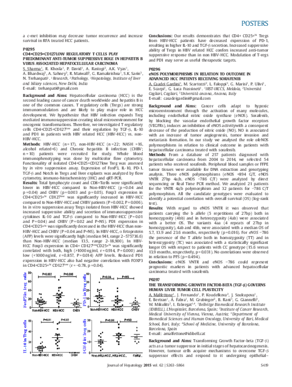
Results: Total lymphocyte count and CD8+Tcells were significantly
lower in HBV-HCC compared to Non-HBV-HCC (p = 0.04 and
p = 0.04) and CHBV (p = 0.003 and p = 0.05). Foxp3 expression in
CD4+CD25+hi CD127low was significantly increased in HBV-HCC
compared to Non-HBV-HCC and CHBV patients (P = 0.002, P = 0.006).
In in vitro suppression assay Tregs isolated from HBV-HCC showed
increased suppressive ability and secretion of immunosuppressive
cytokines IL-10 and TGF-b compared to Non-HBV-HCC (P = 0.01
and P = 0.04) and CHBV (P = 0.02 and P=NS). PD1 expression in
CD4+CD25+hi was significantly decreased in the HBV-HCC than nonHBV-HCC and CHBV (P = 0.04 and P=NS). In HBV-HCC, a fetoprotein
(AFP) levels were significantly high (median 941, range 2–575736.6)
than Non-HBV-HCC (median 13.5, range 2–18,900). In HBVHCC Foxp3 expression in CD4+ CD127low CD25+hi was significantly
correlated with both, high (>1000 ng/ml, r = 0.914, P = 0.000) and
low (<1000 ng/ml, r = 0.857, P = 0.014) AFP levels. Reduced PD1
expression in HBV-HCC also had negative correlation with FOXP3
in CD4+CD25+hi CD127low (r = −0.78, p = 0.04).
Conclusions: Our results demonstrates that CD4+ CD25+hi Tregs
from HBV-HCC patients have decreased expression of PD-1,
resulting in higher IL-10 and TGF-b secretion. Increased suppressive
ability of Tregs in HBV related HCC confers increased anti-tumor
suppressive response than in non HBV-HCC. Modulation of T-regs
and PD1 may serve as useful therapeutic targets.
P0296
eNOS POLYMORPHISMS IN RELATION TO OUTCOME IN
ADVANCED HCC PATIENTS RECEIVING SORAFENIB
A. Caadei Gardini1 , M. Scartozzi2 , L. Faloppi3 , G. Marisi1 , P. Ulivi1 ,
E. Scarpi1 , G. Luca Frassineti1 . 1 IRST-IRCCS, Meldola, 2 Università
Cagliari, Cagliari, 3 Università ancona, Ancona, Italy
E-mail: casadeigardini@gmail.com
Background and Aims: Cancer cells adapt to hypoxic
microenvironment through the activation of many molecules,
including endothelial nitric oxide synthase (eNOS). Sorafenib,
by blocking the vascular endothelial growth factor receptors
(VEGFRs), induces an inhibition of eNOS activitywith a consequent
decrease of the production of nitric oxide (NO). NO is associated
with an increase of tumor angiogenesis, tumor invasion and
metastasis formation. In our study we analysed the role ofeNOS
polymorphisms in relation to clinical outcome in patients with
hepatocellular carcinoma treated with sorafenib.
Methods: From a database of 257 patients diagnosed with
hepatocellular carcinoma from 2004 to 2014, we selected 54
patients who received sorafenib. Peripheral blood samples or FFPE
tumor tissues were available for DNA extraction and genotyping
analysis. Three eNOS polymorphisms (eNOS +894 G/T, eNOS
VNTR 27bp 4a/b, eNOS −786 C/T) were analyzed by direct
sequencing or Real Time PCR method. We analyzed 21 patients
for the VNTR 4a/b polymorphism and 32 patients for −786 C/T
polymorphism. All the candidate genotypes were evaluated to
identify a potential correlation with overall survival (OS) (log-rank
test).
Results: With regard to eNOS VNTR it was observed that
patients carrying the b allele (5 repetitions of 27bp) both in
homozygosity (4bb) and in heterozygosity (4ab) were associated
with a better OS. The variants 4aa (4 repeats of 27bp in
homozygosity), 4ab and 4bb, were associated with a median OS of
5.7, 13.9 and 23.6 months, respectively (p = 0.016). For eNOS −786
the presence of the T allele both in homozygosity (TT) and in
heterozygosity (TC) was associated with a statistically significant
longer OS with respect to patients with CC genotype (15.6 versus
13.9 months, respectively, p = 0.031). No correlations were observed
in relation to PFS (p = 0.494).
Conclusions: eNOS VNTR and eNOS −786 could represent
prognostic markers in patients with advanced hepatocellular
carcinoma treated with sorafenib.
P0297
THE TRANSFORMING GROWTH FACTOR-BETA (TGF-b) GOVERNS
HUMAN LIVER TUMOR CELL PLASTICITY
A. Malfettone1 , J. Fernando1 , P. Koudelkova2 , J. Soukupova1 ,
E. Bertran1 , À. Fabra1 , M. Grubinger2 , B. Rani3 , G. Giannelli3 ,
W. Mikulits2 , I. Fabregat1,4 . 1 Bellvitge Biomedical Research Institute
(IDIBELL), L’Hospitalet, Barcelona, Spain; 2 Institute of Cancer Research,
Medical University of Vienna, Vienna, Austria; 3 Department of
Biomedical Sciences and Human Oncology, University of Bari, Medical
School, Bari, Italy; 4 School of Medicine, University of Barcelona,
Barcelona, Spain
E-mail: amalfettone@idibell.cat
Background and Aims: Transforming Growth Factor-beta (TGF-b)
acts as a tumor suppressor in initial stages of hepatocarcinogenesis.
However, tumour cells acquire mechanisms to overcome TGF-b
suppressor effects and respond to it undergoing epithelial–
Journal of Hepatology 2015 vol. 62 | S263–S864
S419
�POSTERS
mesenchymal transition (EMT). In some tumour cells a link exists
between EMT and the expression of stem cell markers. Aim of this
study was to analyze if autocrine activation of the TGF-b pathway in
hepatocellular carcinoma (HCC) cells may control the expression of
Cancer Stem Cell (CSC) markers concomitant with the acquisition
of mesenchymal properties.
Methods: Different HCC cell lines with different autocrine
expression of TGF-b and epithelial–mesenchymal phenotypes were
analysed. TGF-b Receptor I (TbRI) expression was stably silenced
by shRNA. EMT and SC marker expression was analyzed by
Flow Cytometry, Immunofluorescence and Real-Time PCR. Sphereforming and colony-formation assays were performed to explore
the biological properties of liver CSCs.
Results: Epithelial liver tumour cells expressed EpCAM and CD133,
whereas mesenchymal-like cells expressed CD44. Cancellation
of the TbRI in Hep3B (with a mixed epithelial–mesenchymal
phenotype) prevented the TGF-b-induced EMT, but also induced
a mesenchymal–epithelial transition (MET). Relevantly, TbRI knockdown decreased the basal expression of EpCAM and CD133 and
reduced the formation of liver spheroids and number of clones.
The TbRI knock-down in HLE and HLF (mesenchymal-like cells)
decreased significantly the expression of the Snail family genes,
but unexpectedly did not produce a full MET. Nevertheless, TbRI
knock-down decreased expression of CD44, which correlated with
a lower ability to form liver spheroids and clones. Interestingly,
chronic treatment of Hep3B cells with TGF-b induced progressive
down-regulation of EpCAM and CD133 and up-regulation of CD44,
concomitant with the appearance of a mesenchymal phenotype.
Furthermore, these cells formed liver spheres and colonies more
efficiently.
Conclusions: TGF-b not only modulates the EMT phenotype of HCC
cells, but also the expression of CSC genes, although it is not clear
yet the cross-talk among both processes. A mesenchymal phenotype
and CD44 expression are associated with poor prognosis in HCC.
Results of this study further support that activation of the TGF-b
pathway may be considered a therapeutic target in HCC.
Acknowledgements: People Programme (Marie Curie Actions) of
the FP7–2013, under REA grant agreement #PITN-GA-2012-316549
(IT-LIVER).
P0298
GLUCOSE TRANSPORTER ISOFORM 1 (GLUT1) EXPRESSION
DETERMINES HEPATIC METASTASIS OF MELANOMA CELLS
A. Koch1 , S.A. Lang1 , P. Wild2 , A. Bosserhoff3 , C. Hellerbrand1 .
1
University Hospital Regensburg, Regensburg, Germany; 2 University
Hospital Zurich, Zurich, Switzerland; 3 University of Erlangen, Erlangen,
Germany
E-mail: claus.hellerbrand@ukr.de
Background and Aims: The facilitative glucose transporter isoform
1 (GLUT1) is the key rate-limiting factor in glucose transport into
cancer cells, and we have previously shown that GLUT1 is a tumorpromotor in hepatocellular carcinoma, while its expression is at the
detection limit in normal hepatocytes.
The aim of this study was to analyze whether GLUT1 expression
and a high capacity for glucose uptake, respectively, are general
pro-cancerogenic factors in the liver.
Methods: We used malignant melanoma as a model-tumor, which
is known to preferentially metastasize to the liver.
Results: Similar as observed in HCC, GLUT1 expression was
enhanced in melanoma cell lines compared to primary
melanocytes, as well as in melanoma compared to naevi.
Immunohistochemical analysis of a tissue micro array consisting
of 140 human melanoma tissues showed that GLUT1 expression
was significantly enhanced in metastasis compared to primary
tumors. GLUT1 expression in primary tumors correlated with tumor
staging, and most importantly, with progression- and overallS420
survival. To determine the role of GLUT1 in melanoma metastasis,
GLUT1 expression was suppressed in the murine melanoma
cell line B16 (i) by stable transfection with shRNA and (ii) by
using the selective GLUT1-Inhibitor WZB117. GLUT1 suppression
caused decreased anaerobic glycolysis and lactate secretion, and
inhibited proliferation and migration of B16 cells. Moreover, GLUT1
suppression lowered apoptosis resistance of melanoma cells. Next,
B16 cell clones with and without GLUT1 suppression were subjected
to an established model of hepatic metastasis, in which tumor
cells were injected into the spleen of syngeneic mice, from where
they metastasize into the liver via the portal circulation. GLUT1
suppressed cells formed significantly less metastases and hepatic
metastases derived from GLUT1 suppressed B16 cells revealed less
immune-cell infiltration and more apoptosis as assessed by CD3immunohistochemistry and TUNEL staining.
Conclusions: Our data promote the hypothesis that high glucose
levels in the portal circulation and the liver, and the capacity
to utilize those, respectively, promote hepatic metastasis. Our
data indicate enhanced apoptosis resistance of tumor cells
and known immunomodulatory effects of lactate as potential
underlying mechanisms of this phenomenon. GLUT1, which is
almost selectively expressed in malignant cells but not in healthy
liver or other non-malignant tissues, appears as an attractive
therapeutic target for hepatic metastasis.
P0299
SORAFENIB EFFECT ON MITOCHONDRIAL FUNCTION PROVIDES
A TARGET FOR INCREASING HCC THERAPY EFFICACY
A. Tutusaus1 , M. Stefanovic1 , J.C. Fernandez-Checa1 , M. Marı́1 ,
A. Morales1 . 1 IIBB-CSIC/Hospital Clinic-IDIBAPS-CIBEREHD, Barcelona,
Spain
E-mail: amorales@clinic.ub.es
Background and Aims: Multikinase inhibitor sorafenib has limited
efficacy in the treatment of advanced hepatocellular carcinoma
(HCC). Novel therapies, in combination with sorafenib or in
monotherapy, are demanded to increase drug efficacy in HCC
treatment. The lack of positive results from other drugs, underscores
the importance of identifying weaknesses in HCC biology that
current approaches have not recognized. A mitochondrial effect of
sorafenib has been previously reported, although its participation
in sorafenib toxicity and HCC therapy has drawn little attention.
Methods: Hepatoma cell lines (HepG2 and Hep3B) were treated
with sorafenib and Bcl2-inhibitors. Western blots in total, cytosolic
and mitochondrial extracts and qPCRs were performed after
sorafenib exposure. ROS production, mitochondrial membrane
permeabilization, caspase activity and ATP measurements were
analyzed in sorafenib-treated hepatoma cells. Tumor growth was
determined after subcutaneous injection of HepG2 cells on the
flanks of nude mice.
Results: Sorafenib induces a rapid decline in mitochondrial
membrane potential, with production of reactive oxygen species
(ROS) and reduction in the levels of the antiapoptotic Bcl-2-family
protein MCL-1. However, cytochrome c release from mitochondria
and/or ATP depletion after sorafenib exposure were not observed
during hours, suggesting modest mitochondrial contribution in cell
death. Bcl-2 levels, that play an important role in mitochondrial
dependent cell death, were not affected by sorafenib, so we decided
to evaluate the effect of several Bcl-2 inhibitors (HA-14, ABT-263,
ABT-767 and AT-101) on sorafenib toxicity in hepatoma cells. Among
them, ABT-263 (Navitoclax) a potent inhibitor of Bcl-xL and Bcl-2
now in Phase II clinical trials for leukemia and solid tumors, was
the drug that exhibited higher capacity to potentiate sorafenib
effects on Hep3B and HepG2 cells. ABT-263 co-administration with
sorafenib induced quick release of cytochrome c and enhanced
caspase-3 activity. Moreover, in vivo administration of ABT-263
combined with sorafenib greatly potentiated sorafenib effects,
Journal of Hepatology 2015 vol. 62 | S263–S864
�
 Isabel Fabregat