Academia.edu no longer supports Internet Explorer.
To browse Academia.edu and the wider internet faster and more securely, please take a few seconds to upgrade your browser.
A case of Myasthenia Gravis masquerading as GBS
A case of Myasthenia Gravis masquerading as GBS
 Uzzwal Mallick
2018, Bangladesh Critical Care Journal
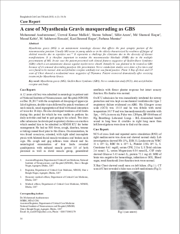
Myasthenia gravis (MG) is an autoimmune neurologic disease that affects the post synaptic portion of the neuromuscular junction. Usually MG occur in young adults or in the elderly characterized by weakness of fatigue of skeletal muscles due to repetitive use.1-2 It represents a challenge for clinicians due to the diversity of disease manifestations. It is therefore important to monitor the neuromuscular blockade (NMB) due to the multiple presentations of MG. In our case the patient presented with clinical features suggestive of Guillen Barre Syndrome (GBS) which is an autoimmune disease against myelin nerve sheath. Initially he was planned to be treated as GBS because of it’s unusual descending paralysis like presentation. Nerve conduction studies were done a few times and were found to be normal. Anti acetylcholine receptor antibody test was found positive. Plane X Ray of chest and CT scan of Chest showed a mediastinal mass suggestive of Thymoma. Patient recovered dramatically afte...
Related Papers
The Egyptian Journal of Neurology, Psychiatry and Neurosurgery
Occurrence of Guillain-Barré syndrome and Myasthenia Gravis in an elderly maleIntroduction The occurrence of both Guillain-Barré syndrome(GBS) and myasthenia gravis (MG) in the same individual is rare. The underlying pathophysiology was assumed to be autoimmune humoral mechanisms and molecular mimicry with a cross-reaction between autoantibodies and myelin sheath of peripheral nerves and acetylcholine receptors of the neuromuscular junction (NMJ). Case description A 68-year-old male known diabetic and hypertensive with good drug compliance presented with acute onset quadriparesis with bulbar involvement for 1 day. On examination, he had mild neck flexion weakness and bulbar weakness. He had flaccid quadriparesis with absent deep tendon reflexes and negative Babinski. The rest of the neurological examination was normal. Discussion and evaluation Blood and electrophysiological studies showed evidence of demyelinating polyradiculoneuropathy with temporal dispersion suggestive of Guillain-Barré syndrome. He was treated with intravenous immunoglobin and complete r...
Generalized myasthenia gravis is a rare case of autoimmune wherein the antibodies destroy the post-sinaptic acetylcholine receptors at skeletal muscle’s neuromuscular junctions. The clinical presentation is specific distributin of motoric deficit without sensoric deficit which diminished with rest and worsens with excessive use. We report a case of a woman 52 yo with symptoms of ptosis, diplopia and dificulty of swallowing. Repetitive nerve stimulation showed >10% decrement and prostigmin test was positive. The patient was treated and showed clinical improvement.
Journal of Neuromuscular Diseases
Myasthenia Gravis: Unusual Presentations and Diagnostic Pitfalls2016 •
Myasthenia gravis (MG) is the archetypic disorder of both the neuromuscular junction and autoantibody-mediated disease. In most patients, IgG1-dominant anti-bodies to acetylcholine receptors cause fatigable weakness of skeletal muscles. In the rest, a variable proportion possesses antibodies to muscle-specific tyrosine kinase while the remainder of seronegative MG is being explained through cell-based assays using a receptor-clustering technique and, to a lesser extent, proposed new antigenic targets. The incidence and prevalence of MG are increasing, particularly in the elderly. New treatments are being developed, and results from the randomised controlled trial of thymec-tomy in non-thymomatous MG, due for release in early 2016, will be of particular clinical value. To help navigate an evidence base of varying quality, practising clinicians may consult new MG guidelines in the fields of pregnancy, ocular and generalised MG (GMG). This review focuses on updates in epidemiology, immunology, therapeutic and clinical aspects of GMG in adults.
Journal of the Peripheral Nervous System
Unusual neurophysiological and immunological findings in myasthenia gravis: a case report2004 •
Archives of the Balkan Medical Union
Characterization of myasthenia gravis using clinical classification and repetitive nerve stimulation2021 •
2011 •
A 56 year old male presented with features of easy f atiguability, episodic hoarseness and nasal quality of voice accompanied by nasal regurgitation of liquids for three years. Neurological examination revealed lingual atrophy with fasciculations, pyramidal tract signs and features of neuromuscular junction dysfunction. Moreover, CT scanning of thorax revealed thymic enlargement. The co-occurrence of myasthenia gravis with motor neurone disease is being reported for its rarity.
RELATED PAPERS
International Journal of Clinical Practice
Experience in implementation of cardiovascular absolute risk assessment and management in Australian general practice2010 •
Revue d’élevage et de médecine vétérinaire des pays tropicaux
Antibiorésistance des souches de Salmonella gallinarum isolées en aviculture moderne en zones périurbaines au Mali2019 •
2018 •
2016 •
FEMS Microbiology Ecology
Dynamics of the microbial community during growth of the house dust mite Dermatophagoides farinae in culture2019 •
Nephro-Urology Monthly
Evaluation of Factors Affecting Wrist Radio-Cephalic Arteriovenous Fistula Maturation: A Single Institutional Observational Study2021 •
Vii Connepi Congresso Norte Nordeste De Pesquisa E Inovacao
Adsorção do corante têxtil turquesa remazol utilizando a eichornia azurea (raiz, caule e folha) como bioadsorvente2012 •