B I OD I V E R S I TA S
Volume 22, Number 5, May 2021
Pages: 2636-2645
ISSN: 1412-033X
E-ISSN: 2085-4722
DOI: 10.13057/biodiv/d220522
First report of bullet wood (Mimusops elengi) sudden decline disease
caused by Ceratocystis manginecans in Indonesia
R. PRATAMA1, A. MUSLIM2,♥, S. SUWANDI2, N. DAMIRI2, S. SOLEHA1
1Agriculture
Sciences Graduate Program, Faculty of Agriculture, Universitas Sriwijaya. Jl. Padang Selasa No. 524, Bukit Besar, Palembang 30139, South
Sumatra, Indonesia
2 Laboratory of Phytopathology, Department of Plant Protection, Faculty of Agriculture, Universitas Sriwijaya. Jl. Raya Palembang-Prabumulih Km 32,
Indralaya, Ogan Ilir 30662, South Sumatra, Indonesia. Tel.: +62-711-580663, Fax.: +62-711-580276, ♥email: a_muslim@unsri.ac.id,
rahmatpratama@pps.unsri.ac.id
Manuscript received: 14 January 2021. Revision accepted: 18 April 2021.
Abstract. Pratama R, Muslim A, Suwandi S, Damiri N, Soleha S. 2021. First report of bullet wood (Mimusops elengi) sudden decline
disease caused by Ceratocystis manginecans in Indonesia. Biodiversitas 22: 2636-2645. Ceratocystis manginecans cause wilt and death
of plants in several important crops and native vegetation in Indonesia. Ceratocystis wilt was recently found to be causing substantial
mortality in bullet wood (Mimusops elengi) in South Sumatra. The aim of this study was to describe the symptomatology of the new
disease and characterize isolates of C. manginecans obtained from diseased bullet wood plants. Diseased plants showed substantial
discoloration of the woody xylem and wilt-type symptoms of the foliage, with the eventual death of the whole plant. Isolations from
infected plants yielded fungi that were similar morphologically to C. manginecans, with typical hat-shaped ascospores and light-colored
perithecial bases. Sequencing of the internal transcribed spacer (ITS) and β-tubulin of the isolates confirmed their identification,
grouping them with C. manginecans and separating them from all other Ceratocystis species. This is the first report of C. manginecans
in Indonesia causing wilt and death on bullet wood. C. manginecans is an important pathogen, and strategies to reduce losses need to be
established in Indonesia because the aggressiveness of C. manginecans to bullet wood has been shown in inoculation experiments
Keywords: Ceratocystidaceae, molecular phylogeny, pathogenicity, Sapotaceae
INTRODUCTION
Bullet wood (Mimusops elengi) belongs to the family
Sapotaceae, common English names are Asian Bulletwood,
Bullet Wood Tree, Indian Medlar, Red Coondoo Spanish
Cherry and it is known in Indonesia as Tanjung. The
species is native to India, Sri Lanka, the Andaman Islands,
Myanmar, Indo-China, Peninsular Malaysia and Vanuatu;
it has been introduced and cultivated elsewhere. M. elengi
can grow in tropical and subtropical climates. This plant
thrives in areas with high humidity and seasonal rainfall
and seasonal dry periods (Lim 2012). The bullet wood trees
range from small to large, and are found in all parts of
Indonesia where bullet wood is cultivated in gardens as an
ornamental tree, for medicines and planted along avenues
because of its fragrant flowers (Seth 2003).
M. elengi is widely used for medicine, and various parts
of M. elengi Linn. (Sapotaceae) have been used widely in
traditional Indian medicine for the treatment of pain,
inflammation and wounds. M. elengi stem bark would be a
possible therapeutic candidate having cytotoxic and antitumor potential (Kumar et al. 2016); it also has
antibacterial and antifungal uses (Ali et al. 2008). At
present, M. elengi is used as the synthesis of calcium
phosphate nanoparticles that is easy, eco-friendly and
scalable (Pokale et al. 2014).
Several types of pathogenic fungi have been identified
to cause disease in M. elengi plants. Curvularia lunata
caused die-back in India (Khatun et al. 2011);
Pestalotiopsis clavispora caused leaf blight (Lokesh et al.
2017). Ceratocystis was first isolated from a single tree of
bullet wood showing sudden decline in Thailand.
Symptoms displayed by the diseased trees include gum
exudation from the trunks and wilting and loss of the dark
green foliage with a corresponding browning of leaves on
single branches. In this study they did not confirm the
pathogen with a Koch postulates test detail (Pornsuriya and
Sunpapao 2015). Recently we have observed many bullet
wood trees showing similar symptoms with C.
manginecans decline in many locations in South Sumatra,
Indonesia.
Ceratocystis manginecans include many economically
important plant pathogens. This pathogen has caused a
sudden decline and has led to the death of thousands of
Mangifera indica trees in Oman with Hypocryphalus
mangifera vector (Al Adawi et al. 2013). In Indonesia, C.
manginecans caused die-back on Acacia mangium and A.
crassicarpa plantations in Riau (Tarigan et al. 2010),
whereas in Vietnam recently, C. manginecans caused wilt
disease in Dalbergia tonkinensis and Chukrasia tabularis
(Chi et al. 2019a; Chi et al. 2020); in Pakistan, this
pathogen also causes wilt disease in Albizia lebbeck
(Razzaq et al. 2020). Commonly C. manginecans cause
yellowing of leaves and rapid wilting of leaves was
observed on individual branches in affected trees that
ultimately spread to the canopy followed by the death of
the whole tree. Dark brown to black tissue discoloration
was observed in the woody xylem tissues of infected trees.
�PRATAMA et al. – Mimusops elengi sudden decline disease caused by Ceratocystis manginecans
This study aimed to identify the cause of a new
outbreak of wilt disease causing a sudden decline to the
trees, wilted canopies, and tree death in M. elengi in South
Sumatera, Indonesia. This study was also conducted to
describe the characteristics of the pathogen and confirm
Koch’s postulates test.
MATERIALS AND METHODS
Disease symptoms and specimen collection
The distribution and impact of the C. manginecans
disease on M. elengi were determined from roadside trees
planting in Jakabaring (Palembang) and Kayuagung (Ogan
Komering Ilir) and the agricultural field of Sriwijaya
University in Indralaya (Ogan Ilir), South Sumatra,
Indonesia. Symptoms of wilt diseases were evaluated as
follows: the extent of lesion development from
discoloration of bark and wood, the extent of foliar wilting
or loss and tree death.
Samples of diseased trunks were collected from two to
six-year-old trees from September 2019 to April 2020.
Wood samples were taken from lesions of wilted trees
using a knife sterilized in 70% ethanol. The wood samples
collected from M. elengi showed brown to black streaking
in the woody xylem. Each sample was wrapped in tissue
paper and placed in a coolbox. The same day, the wood
samples (1–20 mm length, 1–2 mm thick) were sandwiched
between two slices of fresh carrot and placed on sterile dry
paper in plastic boxes at 25°C following the method of Li
et al. (2014). After 5-10 days, hat-shaped spores of putative
Ceratocystis pathogens were placed on 2% (w/v) malt
extract agar (MEA) (Merck, Germany), and incubated at 25
°C in a laboratory. When cultures had grown to several cm
in diameter, hyphal tips were sub-cultured onto new MEA
and potato dextrose agar (PDA) (Merck, Germany) plates
and incubated at 25-28 °C. Morphological traits of fruiting
bodies and spores were observed under an optical Olympus
CX33 microscope (Olympus Corporation, Japan).
Genomic DNA extraction, PCR amplification, and
sequencing
DNA isolation used YeaStar Genomic DNA Kit (Zymo
Research Corporation, California, USA). To extract
genomic DNA, cultures were incubated for five days to
allow sufficient mycelial growth in potato dextrose broth
(PDB) (Merck, Germany). Mycelium was purified with
sterile filter paper (Whatman) and transferred to 1.5 mL
Eppendorf tubes. The quantity and quality of DNA
extracted were evaluated with a spectrophotometer
(NanoDrop ND-1000; Thermo Fisher, Waltham, MA,
USA) to calibrate the concentration and purity of DNA as
PCR templates.
The PCR amplification reactions were conducted on a
T-100 thermal cycler (Bio-Rad, Hercules, CA, USA).
Amplifications were carried out in 50 μl reactions
containing 20 μl DreamTaq Green PCR Master Mix
(Eppendorf, Germany) (DreamTaq DNA Polymerase, 2X
DreamTaq Green buffer, dNTPs, and 4 mM MgCl2), 1,5 μl
of each forward and reverse primer, 4 μl of DNA template
2637
and 23 μl sterilized water. The PCRs were performed with
a C1000 Touch™ thermal cycler (Bio-Rad, USA). The
PCR cycling parameters were as follows: initial
denaturation for 5 min at 95°C, followed by 35 cycles at
95°C for 30 s, 56°C for 45s and 72°C for 1 min.
Amplification was completed at 72°C for 10 min and the
PCR product was stored at 10°C (Chi et al. 2020).
PCR amplifications were made for two gene regions,
including part of the b-tubulin (BT) using primers βt1a
(TTCCCCCGTCTCCACTTCTTCATG)
and
βt1b
(GACGAGATCGTTCATGTTGAACTC) (Oliveira et al.
2015a), and the ITS using ITS1 and ITS4. The resulting
PCR products were submitted to 1st BASE (Malaysia) for
forward and reverse sequencing reactions. Raw sequence
data were assembled, examined, and manually edited using
Genestudio 2.1.1.5 (Genestudio, Suwanee, Georgia) and
BioEdit software (van der Nest et al. 2019). The DNA
sequences were compared to the GenBank database via the
nucleotide-nucleotide BLAST search interface located at
the National Center for Biotechnology Information,
Bethesda, USA. Relevant sequences were transferred with
NoteTab Light v7.2.
Phylogenetic analyses
The sequences of Ceratocystis spp. closely related to
the one from Mimusops elengi were retrieved from
GenBank. Phylogenetic sequences from different gene
regions were aligned using Mesquite v3.5 (Maddison and
Maddison 2018) (http://mesquiteproject.org) and corrected
manually. Phylogenetic trees based on a concatenated data
set of the ITS and βt were computed and analyzed as a
single dataset. Maximum Parsimony (MP) analyses were
performed in MEGA v. 10 (Kumar et al. 2016; Paul et al.
2018) with 1000 bootstrap replications.
Pathogenicity tests
Pathogenicity studies were conducted on two
agroforestry plants, A. mangium and M. elengi. Plants had
stem diameters of 2–3 cm and heights <1 m. The
pathogenic potential of isolates was evaluated by the under
bark inoculation method described by Deidda et al. (2016).
Bark was wounded to expose the cambium using a 4 mm
cork borer, and discs of agar bearing mycelium taken from
the margins of actively growing, 2-week-old cultures on
2% MEA (Tarigan et al. 2010; Tarigan et al. 2011; Chi et
al. 2019a, Chi et al. 2020), Ceratocystis isolates were
placed with the mycelium facing the cambium. Ten plants
of each tree species were inoculated with sterile MEA
plugs to serve as controls. All inoculation points and the
ends of the logs were covered with masking tape and
polyethylene films, respectively, to prevent desiccation of
the inoculum and cambium, and to reduce contamination.
The nursery trial evaluated seven Ceratocystis strains
isolated from M. elengi (CAME30813, CAME30814,
CAME30815, CAME30816, CAME30817, CAME30818
and CAME30819) and one strain (CAW30814) from A.
mangium. There were ten replicate plants per treatment in
each row plot in each of the blocks. After 45 days, lesion
(L) length and foliar symptoms severity were recorded.
Representative wood samples were taken from within
�2638
B I OD I V E R S I TA S 22 (5): 2636-2645, May 2021
lesions outside the inoculation area, and the pathogen
reisolated and sequenced for Koch’s postulates test.
Foliar symptoms severity (FS) was assessed using a
scale of 0 to 4: 0 = healthy; 1 = lower leaves yellow; 2 =
slight wilting; 3 = severe wilting; 4 = dead (Muslim et al.
2003; Chi et al. 2019a; Muslim et al. 2019).
The pathogenicity test data were analyzed using SAS
university edition software package. Analysis of variance
(ANOVA) and Tukey's honestly significant difference
(Tukey's HSD) test were used to determine whether there
were significant differences in comparisons of means of
different treatments.
RESULT AND DISCUSSION
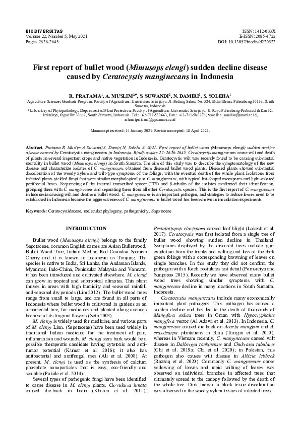
Symptoms of Mimusops elengi wilt disease
We observed disease symptoms from September 2019
to April 2020, in various places that are widely planted in
the Indralaya (Ogan Ilir), Jakabaring (Palembang) and
Kayuagung (Ogan Komering Ilir) areas, South Sumatra,
Indonesia. The disease was found scattered throughout the
planted area, with symptoms of partially dead trees, foliar
wilting or loss and tree death (Figure 1a). Initially, the
leaves of infected plants lost turgor and brightness, with
yellowing symptoms in the older leaves, followed by
wilting and death of the plant. Symptomatic plant stems
showed xylem discoloration (Figure 1b), infections
generally started in the roots and then moved upwards in
the stem ultimately reaching the upper branches of the
entire plant, and the plants ultimately died (Figure 1c).
Death of adjacent plants indicates transmission of root
infection because these pathogens are also known as soilborne pathogens. The severity of the infection is also
caused by pruning the branches using tools previously used
to cut the infected plants.
Observation of diseased plant xylem tissue in crosssections of the stem showed dark brown lesion formation in
the cambium (inner bark region) towards vascular tissue
(Figure 1d). In the initial stage of plant infection, the foliar
symptoms, wilt and the fruits appeared normal, but as the
infection progressed, the fruits of affected plants were
smaller, shriveled, wrinkled and dry. Many of the bark
beetle vectors of C. manginecans, Hypocryphalus
mangiferae were found around bullet wood diseases
(Figure 1e). Testing by the Li et al. (2014) method showed
that Ceratocystis had grown on the carrots, and ascomata of
C. manginecans with necks supporting sticky masses of
ascospores on the carrot slices (Figure 1.F).
Sampling and isolation
Seven isolates of C. manginecans were collected from
diseased bullet wood (M. elengi) (Figure 2). There were
three
isolates
(CAME30815,
CAME30816 and
CAME30817) from Ogan Ilir (Indralaya); two isolates
(CAME30818 and CAME30814) from Jakabaring
(Palembang); and two isolates (CAME30819 and
CAME30813) from Kayuagung (Ogan Komering Ilir). We
also isolated one isolate (CAW30814) from diseased
acacia, A. mangium in the agricultural field of Sriwijaya
University, Indralaya.
B
A
D
E
C
F
Figure 1. Symptoms of Ceratocystis manginecans wilt disease in bullet wood: a. tree death of M. elengi: b. sap stain mold on bullet
wood, c. wilted leaves of bullet wood, d. sap stain mold on bullet wood, e. The bark beetle vector of C. manginecans, Hypocryphalus
mangiferae, f. isolation of the fungus from discolored xylem showing dark mycelium and sporulation on the carrot slices
�PRATAMA et al. – Mimusops elengi sudden decline disease caused by Ceratocystis manginecans
2639
A
B
C
D
E
F
G
H
Figure 2. Isolates of Ceratocystis manginecans and related species grew on malt extract agar (MEA) for 7 d at 25 oC. A, B, C:
Ceratocystis CAME30815, CAME30816 and CAME30817, from Mimusops elengi in Sriwijaya University, Indralaya. D, E, F:
Ceratocystis CAME30819, CAME30813 and CAME30814 from Mimusops elengi in Jakabaring, Palembang. G: Ceratocystis
CAME30818, from Mimusops elengi in Kayuagung, Ogan Komering Ilir. H: Ceratocystis CAW30814, from Acacia mangium in
Indralaya
Fungal morphology
Seven isolates were morphologically indistinguishable
(Table 2). At 7–14 days of incubation at 25 oC on MEA,
cultures were pale brown to dark brown and produced a
banana-like odor. Mycelium on MEA was grey, and the
reverse side of the colony olivaceous grey; submerged
mycelium darkened as the ascomata developed, forming
fine, radiating fibrils. Ascomata developing within seven
days and mature within ten days, superficially or partly
embedded in the agar, dark brown to black (Figure 3a).
Ascomatal bases were submerged or on the agar surface,
dark bases dark brown to black, base subglobose to globes,
(134.58-) 169.12 - 276.29 (-310.83) μm long and (122.91-)
161.89-244.14 (-283.13) μm wide in diameter (Figure 3a).
Ascomata necks were erect, occasionally curved, black at
the base becoming subhyaline towards the apex, smooth to
crenulate, (346.51-) 454.94-720.16 (-828.59) μm long
including ostiolar hyphae (Figure3b). Ascospores were hatshaped, (3.61-) 5.64-6.23 (-6.93) μm length and (2.06-)
2.279-3.67 (-3.85) μm width (Figure 3f). Barrel conidia
(8.62-) 8.85-12.79 (-13.25) μm length and (5.89-)
4.12x6.87 (-8.67) μm width. Bacilliform conidia (9.05-)
10.82-22.32 (-35.97) μm length and (2.01-) 2.83-5.71 (8.87) μm width (Figure 3c). Chlamydospores oval, thickwalled, smooth, (8.21-) 9.15-16.21 (-18.50) μm length and
(4.92-) 6.46-15.81 (14.65) μm width (Figure 3e).
Sequence analysis
To confirm the identity of the wilt pathogen, the ITS
and β-tubulin 1 gene sequences of two isolates from bullet
wood (M. elengi) were compared to reference sequences
downloaded from the gene bank database (NCBI GenBank)
(Table 1) and indicated that isolates from Indonesia were
grouped within the C. fimbriata. s.l species complex and
were most closely related to C. manginecans. PCR
amplification resulted in fragments of ~550 base pairs (bp)
in size which had 100% homology with C. manginecans
(Figure 4). Bootstrap values were equal to or greater than
50%, derived from 1000 iterations.
Pathogenicity
The results of the pathogenicity tests of seven isolates
(CAME30813, CAME30814, CAME30815, CAME30816,
CAME30817, CAME30818, and CAME30819) on M.
elengi and one isolate (CAW30814) from A. mangium are
shown in Table 3. All isolates tested showed a varying
reaction to lesion and foliar symptoms (Figure 5). For the
pathogenicity test on M. elengi, seven isolates from M.
elengi as well as one isolate from A. mangium strongly
infected the wood and produced significant lesion lengths
ranging from 3.99 to 10.47 cm and showed significant
differences from the control. Three isolates from M. elengi
(CAME30815; CAME30819; CAME30818) as well as one
isolate from A. mangium (CAW30814) showed high
pathogenicity on the foliar symptom (with foliar severity
index of 2.3-3.6 and lesion length 6.52-10.47 cm), while
the other isolates (CAME30817; CAME30816;
CAME30814,
CAME30813)
showed
moderate
pathogenicity to M. elengi (foliar severity index of 1.4-2.0
and lesion length 3.99-6.02 cm). When the isolates were
tested for their pathogenicity on A. mangium as the primary
host of the pathogen, two isolates (CAME30815;
CAME30819) showed strong pathogenicity on lesion
length (13.76-11.89 cm) and also provided high
pathogenicity on foliar severity index (4), where all plants
tested were dead. Three isolates (CAW30814;
�2640
B I OD I V E R S I TA S 22 (5): 2636-2645, May 2021
CAME30818;
CAME30817)
showed
moderate
pathogenicity on lesion length 8.71-10.14 cm and foliar
severity index 2.8-3.2, while the other isolates
(CAME30816, CAME30814, CAME30813) showed low
pathogenicity on lesion length 7.19-8.63 cm and foliar
severity index 1.9-2.4.
Table 1. Ceratocystis isolates considered in the phylogenetic analyses
C.manginecans
C.manginecans
C.manginecans
C.manginecans
C.manginecans
C.manginecans
C.manginecans
C.manginecans
C.manginecans
C.manginecans
C.manginecans
C.manginecans
C.manginecans
C.manginecans
C.albifundus
C.caryae
C.smalleyi
Mimusops elengi
Mimusops elengi
Mangifera indica
Mangifera indica
Mangifera indica
Mangifera indica
A. crassicarpa
A. crassicarpa
Acacia mangium
A. crassicarpa
Acacia mangium
Hypocryphalus mangifera
Mangifera indica
Hypocryphalus mangifera
A. mearnsii
C. cordiformis
C. cordiformis
Indonesia
Indonesia
Oman
Pakistan
Pakistan
Pakistan
Indonesia
Indonesia
Indonesia
Indonesia
Indonesia
Pakistan
Oman
Oman
RSA
U.S.A
U.S.A
R.Pratama
R.Pratama
M. Deadman
A.Al-Adawi
A.Al-Adawi
A. Al-Adawi
M. Tarigan
M. Tarigan
M. Tarigan
M. Tarigan
M. Tarigan
A. Al-Adawi
M. Deadman
M. Deadman
J. Roux
J. Johnson
G. Smalley
Gene region/GeneBank
accession no
ITS
BT
MT373423
Submitted
MT373424
Submitted
AY953383
EF433308
EF433304
EF433313
EF433305
EF433314
EF433302
EF433311
EU588663
EU588642
EU588662
EU588641
EU588659
EU604671
EU588665
EU588644
EU588658
EU588638
EF433303
EF433312
AY953385
EF433310
AY953384
EF433309
DQ520638
EF070429
EF070424
EF070439
EF070420
EF070436
C. populicola
C.atrox
Populus sp.
E. grandis
Poland
Australia
J. Gremmen
M.J. Wingfield
EF070418
EF070415
EF070434
EF070431
C. polycroma
C. obpyriformis
C.pirilliformis
Syzygium aromaticum
A. mearnsii
E. nitens
Indonesia
South Africa
Australia
M.J. Wingfield
R.N. Heath
M.J. Wingfield
AY528970
EU245003
AF427105
AY528966
EU244975
DQ371653
Isolate no
CAME30819
CAME30818
CMW13851
CMW23643
CMW23641
CMW23634
CMW21125
CMW21123
CMW22581
CMW21132
CMW22579
CMW23628
CMW13854
CMW13852
CMW4068
CMW14793
CMW14800
CBS114724
CMW14789
CMW19385
CBS115778
CMW11424
CMW23808
CMW6579
Identify
Host
Geographic
origin
Collector
Table 2. Morphological comparisons of Ceratocystis manginecans and other phylogenetically closely related species
Character
Ascomata base
Ascomata base average
Ascomata neck
Ascomata neck average
Ascospores
Ascospores average
Bacilliform conidia
Bacilliform conidia average
Barrel-shaped conidia
Barrel-shaped conidia
average
Clamydospore
Clamydospore average
Ceratocystis manginecans
(from M. elengi)
(134.58-) 169.12 - 276.29 (-310.83) x
(122.91-) 161.89-244.14 (-283.13)a
220.01x211.63b
(346.51-) 454.94-720.16 (-828.59)
568.41
(3.61-) 5.64-6.23 (-6.93) x (2.06-) 2.279-3.67
(-3.85)
5.62 x 3.93
(9.05-) 10.82-22.32 (-35.97) x (2.01-) 2.835.71 (-8.87)
16.56x4.27
(8.62-) 8.85-12.79 (-13.25) x (5.89-)
4.12x6.87 (-8.67)
11.497 x 15.82
Ceratocystis acaciivora
Ceratocystis manginecans
(from A. mangium)
(from A. mangium)
(105-) 131-175 (-206) x (107- (132.1-) 175.3 (−233.2)
) 125-167 (-188)
(8.21-) 9.15-16.21 (-18.50) x (4.92-) 6.4615.81 (14.65)
11.13 x 14.18
(10-) 12-14 (-15) x (7-) 8-12 (10.1-) 13.1 (−15.5) x
(-14)
(6.1-) 9.2 (−11.1)
(301-) 348-448 (-522)
(327.3-) 452.7 (−556)
5-7 x 3-4
(3.1-) 4.2 (−4.7) x (4.1-)
5.5 (−6.8)
(11-) 14-22 (-29) x 3-5
(14.6-) 18.6 (−30.7)x (3.0) 4.6 (−5.4)
(8-) 9-11 (-13) x 4-6
(6.8) 8.1 (−10.6)
Reference
This study
M. Tarigan et al. (2010)
Chi et al. (2019)
Note: All measurements are in µm. a Measurements are presented in the format [(minimum-) (average-standard deviation) - (averagestandard deviation) (-maximum)]. b Measurements are presented in the format minimum x maximum
�PRATAMA et al. – Mimusops elengi sudden decline disease caused by Ceratocystis manginecans
2641
Table 3. Pathogenicity of Ceratocystis isolates on Mimusops elengi and Acacia mangium under nursery condition
M. elengi
A. mangium
Lesion length (cm)
Foliar symptoms
Lesion Length (cm)
Foliar symptoms
CAME30815
10
10.47e
3.6
13.77d
4
CAME30819
10
8.29de
3.1
11.89cd
4
CAW30814
10
7.35cd
2.8
10.14bcd
3.2
CAME30818
10
6.52bcd
2.3
9.92bcd
3.1
CAME30817
10
6.02bcd
2
8.72bc
2.8
CAME30816
10
5.27bc
1.8
8.47bc
2.4
CAME30814
10
4.93bc
1.5
8.64bc
2
CAME30813
10
3.99b
1.4
7.19b
1.9
Control (MEA)
10
0.1a
0
0.1a
0
Fpr
<0.001
<0.001
Note: Values followed by the same letters in a column are not different among isolates at P=0.05 according to Tukey’s HSD multiple
range test.
Isolates
Host test
D
B
E
A
C
F
Figure 3. Morphological characteristics of Ceratocystis isolated from M. elengi stem lesion: A. Globose ascomata with long neck, B.
Divergent ostiolar hyphae, C. Barrel-shaped conidia, D. Conidiophore/phialide, E. Chlamydospores, F. Hat-shaped ascospores. Scale
bars: A = 100 µm; B, C, D, E = 10 µm;F = 5 µm
�2642
B I OD I V E R S I TA S 22 (5): 2636-2645, May 2021
Figure 4. Phylogenetic tree constructed by MEGA with Maximum Parsimony (MP). The tree was built using combined sequence data
of the ITS region and b-tubulin gene and their related species from GenBank. Consistency (CI), retention (RI), and composite indexes
(CoI) are 0.714286, 0.837209, and 0.664843 for all sites and parsimony-informative sites. The percentage of replicate trees in which the
associated taxa clustered together in the bootstrap test (1000 replicates) is shown next to the branches. Bootstrap values >50% are
indicated above the branches. The analysis involved 28 nucleotide sequences. All positions containing gaps and missing data were
eliminated. There were 493 positions in the final dataset
A
B
C
D
Figure 5. Response after 45 days of Mimusops elengi seedlings to under-bark inoculation with mycelium of Ceratocystis manginecans.
A. Wilting of seedlings; B. Lesions on the stem; C. No lesions on the stem; D. Normal foliage on the control seedling which received
MEA only and appeared healthy. Yellow arrow indicates the point of inoculation, red arrows show the lesion boundary
�PRATAMA et al. – Mimusops elengi sudden decline disease caused by Ceratocystis manginecans
A
2643
B
Figure 6. Correlation between foliar symptoms severity (FS) and lesion length (cm): A. Mimusops elengi, B. Acacia mangium
There was a strong linear correlation between foliar
symptom severity and lesion length for M. elengi and A.
mangium (Figure 6). The Koch’s postulates test was
conducted by reisolating the pathogen, and its identity
confirmed to be the same as C. manginecans.
Discussion
The results of this study show clearly that C.
manginecans was responsible for the wilt outbreak disease
that has recently appeared on the M. elengi agricultural
field in Indralaya (Ogan Ilir) and roadside trees in
Jakabaring (Palembang) and Kayuagung (Ogan Komering
Ilir), South Sumatra. This is the first report of a
Ceratocystis wilt disease on M. elengi in Indonesia.
Formerly the disease was reported in Thailand (Pornsuriya
and Sunpapao 2015), however, they just observed a single
tree and they did not conduct pathogenicity tests and
observe morphological characteristics. In Indonesia
Tarigan et al. (2010; 2011) reported C. manginecans
caused wilt and die-back disease in A. mangium plantations
in Riau and Suwandi et al. (2021) have reported infection
of this disease on Lansium domesticum tree.
Mimusops elengi infected with Ceratocystis has wilted
foliage symptoms, sudden wilt, decline and branch drying,
and progressive loss of the canopy resulting in tree death.
M. elengi trees showed typical symptoms of infection by
the Ceratocystis fungus; the same was true of a serious wilt
pathogen of A. mangium and A. crassicarpa in Indonesia
and Vietnam (Tarigan et al. 2011; Thu et al. 2012; Thu et
al. 2016). Vascular wilt, wood stain and stem cankers are
the most characteristic symptoms of infection by C.
manginecans in woody trees (Harrington 2013). Rot in
roots or stems, vascular wilt, sapwood discoloration and
cankers are symptomatic of plants infected by C.
manginecans species (Oliveira et al. 2015b).
The comparison of morphological characteristics and
gene sequences (β-tubulin and ITS) of the isolates
examined in this study were similar to those in descriptions
given for C. manginecans isolated from diseased Acacia
trees which form part of the C. fimbriata s. l. complex,
which is typified by C. fimbriata sensu stricto (Engelbrecht
and Harrington 2005; Tarigan et al. 2011). The two isolates
from Indralaya (Ogan Ilir), Jakabaring (Palembang) and
Kayuagung (Ogan Komering Ilir) have 100% identical
sequences of ITS and Beta Tubulin representing a single
clone of the pathogen. This supports the view that M.
elengi wilt disease in Indralaya (Ogan Ilir), Jakabaring
(Palembang) and Kayuagung (Ogan Komering Ilir)
emerged from a single introduction of a single haplotype
and the population has expanded clonally to infect M.
elengi trees in many parts of all three regions.
Ceratocystis manginecans was reported in Indonesia
associated with wilt and die-back disease on Acacia spp.
(Tarigan et al. 2011). It was found that many acacia plants
were attacked by Ceratocystis disease and Hypocryphalus
mangifera insects in the field raised the suspicion that M.
elengi were infected by Ceratocystis in acacia plants. H.
mangifera is a vector insect for the spread of Ceratocystis
in the world (Van Wyk et al. 2007; Masood et al. 2008; AlAdawi et al. 2013; Fourie et al. 2016). Pruning M. elengi
branches using equipment that has previously been infected
with Ceratocystis also increases the severity of the disease;
this can be seen by the number of plants that are attacked
after pruning with tools used to prune Acacia plants that are
attacked by Ceratocystis. Cankers of Ceratocystis were
associated with branch wounds from pruning, pruning time
and pruning technique on plants (Chi et al. 2019b). In this
study, infected bullet wood was observed surrounding
infected acacia by C. manginecans with abundant H.
mangifera. This might be caused by spore contamination
on cutting tools after pruning diseased acacia and spore
spreading by insect vector H. mangifera.
The pathogenicity of C. manginecans to these hosts was
demonstrated in inoculation trials and it is clear that the
fungus is responsible for the widespread death of these
trees. It was also possible to show that isolates of the
pathogen from these trees are able to infect and kill other
plants. The eight C. manginecans isolates all formed
lesions on the stems of M. elengi seedlings when inoculated
under the bark in nursery cloches. The most pathogenic
was CAME30815 and CAME30819 isolate from M. elengi
and the CAW30814 isolate from A. mangium resulted in
�2644
B I OD I V E R S I TA S 22 (5): 2636-2645, May 2021
foliar symptoms with severity index 3.6, 3.1, and 2.8 after
45 days from inoculation with pathogen. When the isolates
were tested on seedling A. mangium, the seedlings were
infected and mostly dead as they were the main host plant
and susceptible to C. manginecans (Tarigan et al. 2011).
Previous researchers of C. manginecans on M. elengi in
Thailand have reported molecular identification C.
manginecans by primers combination the ITS, β-tubulin
(βt) and transcribed elongation factor 1-α (TEF1-α) gene
regions with one isolate. In our research, we tested two
isolates for sequencing, seven isolates for pathogenicity,
and Koch’s postulates assay using three isolates
(CAME30815, CAME30816 and CAME30817) from Ogan
Ilir (Indralaya); two isolates (CAME30818 and
CAME30814) from Jakabaring (Palembang); and two
isolates (CAME30819 and CAME30813) from Kayuagung
(Ogan Komering Ilir) and one isolate (CAW30814) from
diseased Acacia, A. mangium. All isolates showed the
ability to infect both bullet wood and Acacia, and all
isolates can be reisolated confirming Koch’s postulates.
The wilt disease of M. elengi appears to be serious and
it is clearly a new host tree or pathogen association that has
apparently occurred due to a host shift. This category of
diseases is increasing in importance in plants, and they can
devastate native trees that have not previously encountered
them (Roy 2001; Anderson et al. 2004; Slippers et al. 2005;
Woolhouse et al. 2005; Desprez-Loustau et al. 2007;
Wingfield et al. 2010). In this regard, the wilt disease of M.
elengi can impact seriously on the natural biodiversity of
Indonesia, and studies should be instituted to understand
them better. The findings of this study will give knowledge
of the characteristics of the symptoms of the disease, their
causes and help to minimize the spread of wilt disease in
M. elengi in plantations or roadside trees and to consider its
possible pathogenicity.
This study presents the first report of Ceratocystis wilt
or sudden decline disease of bullet wood in Indonesia and
the discovery of a fungus that has been identified as C.
manginecans. The disease of bullet wood that gave rise to
this study is serious and management options to reduce its
incidence are required C. manginecans is an aggressive
pathogen and a deeper understanding of its role in tree
death will be important in the future.
ACKNOWLEDGEMENTS
This research was funded by a PMDSU scholarship in
the fiscal year of 2019-2021 according to the Director of
Research and Community Service, Directorate of Research
and Community Service (DRPM), Directorate General for
Research and Development, Ministry of Research,
Technology, and Higher Education Indonesia, Number:
068/SP2H/AMD/LT/DRPM/2020 chaired by Ahmad
Muslim.
REFERENCES
Al Adawi AO, Barnes I, Khan IA, Al Subhi AM, Al Jahwari AA,
Deadman ML, Wingfield BD, Wingfield MJ. 2013. Ceratocystis
manginecans associated with a serious wilt disease of two native
legume trees in Oman and Pakistan. Australas Plant Pathol 42:179193. DOI: 10.1007/s13313-012-0196-5.
Ali MA, Mozid MA, Yeasmin MS, Khan AM, Sayeed MA. 2008. An
evaluation of antimicrobial activities of Mimusops elengi Linn. Res J
Agric Biol Sci 4: 871-874.
Anderson PK, Cunningham AA, Patel NG, Morales FJ, Epstein PR,
Daszak P. 2004. Emerging infectious diseases of plants: Pathogen
pollution, climate change and agrotechnology drivers. Trends Ecol
Evol 19:535-544. DOI: 10.1016/j.tree.2004.07.021.
Chi NM, Nhung NP, Trang TT, Thu PQ, Hinh TX, Nam NV, Quang DN,
Dell B. 2019a. First report of wilt disease in Dalbergia tonkinensis
caused by Ceratocystis manginecans. Australas Plant Pathol 48: 439445. DOI: 10.1007/s13313-019-00643-1.
Chi NM, Thu PQ, Hinh TX, Dell B. 2019b. Management of Ceratocystis
manginecans in plantations of Acacia through optimal pruning and
site selection. Australas Plant Pathol 48: 343-350. DOI:
10.1007/s13313-019-00635-1.
Chi NM, Trang TT, Nhung NP, Quang DN, Son VM, Tuan TA, Mai LT,
Hung TX, Nam NV, Thu PQ, Dell B. 2020. Ceratocystis wilt in
Chukrasia tabularis in Vietnam: Identification, pathogenicity and
host tolerance. Australas Plant Pathol 50: 17-27. DOI:
10.1007/s13313-020-00754-0.
Deidda A, Buffa F, Linaldeddu BT, Pinna C, Scanu B, Deiana V, Satta A,
Franceschini A, Floris I. 2016. Emerging pests and diseases threaten
Eucalyptus camaldulensis plantations in Sardinia, Italy. Iforest 9:
883-891. DOI: 10.3832/ifor1805-009.
Desprez-Loustau ML, Robin C, Buee M, Courtecuisse R, Garbaye J,
Suffert F, Sache I, Rizzo DM. 2007. The fungal dimension of
biological invasions. Trends Ecol Evol 22: 472-480. DOI:
10.1016/j.tree.2007.04.005.
Engelbrecht CJB, Harrington TC. 2005. Intersterility, morphology and
taxonomy of Ceratocystis fimbriata on sweet potato, cacao and
sycamore. Mycologia 97:57-69. DOI: 10.3852/mycologia.97.1.57.
Fourie A, Wingfield MJ, Wingfield BD, Thu PQ, Barnes I. 2016. A
possible centre of diversity in South East Asia for the tree pathogen,
Ceratocystis manginecans. Infect Genet Evol 41: 73-83. DOI:
10.1016/j.meegid.2016.03.011.
Harrington TC. 2013. Ceratocystis diseases. In: Gonthier P (eds).
Infectious Forest Diseases. CABI, Wallingford.
Khatun S, Cakilcioglu U, Chakrabarti M, Ojha S, Chatterjee NC. 2011.
Biochemical defense against die-back disease of a traditional
medicinal plant Mimusops elengi Linn. European J Med Plants 1: 4049. DOI: 10.9734/EJMP/2011/247.
Kumar H, Savaliya M, Biswas S, Nayak PG, Maliyakkal N, Setty MM,
Gourishetti K, Pai KSR. 2016. Assessment of the in vitro cytotoxicity
and in vivo anti-tumor activity of the alcoholic stem bark
extract/fractions of Mimusops elengi Linn. Cytotechnology 68: 861877. DOI: 10.1007/s10616-014-9839-4.
Li J, Gao J, Han YH, Sun YX, Huang Q. 2014. First report of Ceratocystis
fimbriata-caused wilt of Eriobotrya Japonica in China. Plant Dis 98:
1270. DOI: 10.1094/PDIS-12-13-1290-PDN.
Lim TK. 2012. Mimusops elengi. Edible Medicinal and Non-Medicinal
Plants. Springer Science+Business Media, New York. DOI:
10.1007/978-94-007-1764-0.
Lokesh S, Raghavendra VB, Sugnanachar N, Melappa G. 2017. First
Report of leaf blight of bakul (Mimusops elengi Linn) caused by
Pestalotiopsis clavispora (G.F. Atk.) Steyaert in India. J Plant Physiol
Pathol 5: 1-3.
Maddison WP, Maddison DR. 2018. Mesquite: A Modular System for
Evolutionary Analysis. Available via: http://mesquiteproject.org
Masood A, Saeed S, Sajjad A. 2008. Characterization and damage patterns
of different bark beetle species associated with mango sudden death
syndrome in Punjab, Pakistan. Pak Entomol 30: 163-168.
Muslim A, Horinouchi H, Hyakumachi M. 2003. Biological control of
Fusarium wilt of tomato with Hypovirulent Binucleate Rhizoctonia in
greenhouse
conditions.
Mycoscience
44:77-84.
DOI:
10.1007/S10267-002-0084-x.
Muslim A, Hyakumachi M, Kageyama K, Suwandi S, Pratama R. 2019. A
rapid bioassay to evaluate efficacy of Hypovirulent Binucleate
Rhizoctonia in reducing Fusarium Crown and root rot of tomato. The
Open Agric J 13: 27-33. DOI: 10.2174/1874331501913010027.
Oliveira SS, Harrington TC, Ferreira MA, Damacena MB, Al-Sadi AM,
Al-mahmooli HIS, Alfenas AC. 2015a. Species or genotypes?
Reassessment of four recently described species of the Ceratocystis
�PRATAMA et al. – Mimusops elengi sudden decline disease caused by Ceratocystis manginecans
wilt pathogen, Ceratocystis fimbriata, on Mangifera indica.
Mycology 105: 1229-1244. DOI: 10.1094/PHYTO-03-15-0065-R.
Oliveira LSS, Harrington TC, Freitas RG, McNew D, Alfenas AC. 2015b.
Ceratocystis tiliae sp. nov., a wound pathogen on Tilia Americana.
Mycologia 107: 986-995. DOI: 10.3852/14-273.
Paul CN, Nam SS, Kachroo A, Kim HY, Yang JW. 2018.
Characterization and pathogenicity of sweet potato (Ipomoea batatas)
black rot caused by Ceratocystis fimbriata in Korea. Eur J Plant
Pathol: 7-8. DOI: 10.1007/s10658-018-1522-8.
Pokale P, Shende S, Gade A, Rai M. 2014. Biofabrication of calcium
phosphate nanoparticles using the plant Mimusops elengi. Environ
Chem Lett 12: 393-399. DOI: 10.1007/s10311-014-0460-8.
Pornsuriya C, Sunpapao A. 2015. A new sudden decline disease of bullet
wood in Thailand is associated with Ceratocystis manginecans. Aust
Plant Dis Notes 10:26-31. DOI: 10.1007/s13314-015-0176-z.
Razzaq K, Anjum R, Hanif S, Sultan A. 2020. First report of Ceratocystis
manginecans causing Siris (Albizia lebbeck) wilt in Pakistan. Plant
Dis 104: 1-3. DOI: 10.1094/PDIS-10-19-2057-PDN.
Roy BA. 2001. Patterns of association between crucifers and their flowermimic pathogens: Host jumps are more common than co-evolution or
co-speciation.
Evolution
55:41-53.
DOI:
10.1111/j.00143820.2001.tb01271.x.
Seth MK. 2003. Trees and their economic importance. Bot Rev 69: 321376. DOI: 10.1663/0006-8101(2004)069[0321:TATEI]2.0.CO;2.
Slippers B, Stenlid, J, Wingfield, MJ. 2005. Emerging pathogens: Fungal
host jumps following anthropogenic introduction. Trends Ecol Evol
20: 420-421. DOI: 10.1016/j.tree.2005.05.002.
Suwandi S, Irsan C, Hamidson H, Umayah A, Asriyani KD. 2021.
Identification and Characterization of Ceratocystis fimbriata Causing
Lethal Wilt on the Lansium Tree in Indonesia. Plant Pathol J 37:124136. DOI: 10.5423/PPJ.OA.08.2020.0147
2645
Tarigan M, Roux J, Wingfield MJ, VanWyk M, Tjahjono B. 2010. Three
new Ceratocystis spp. in the Ceratocystis moniliformis complex from
wounds on Acacia mangium and A. crassicarpa. Mycoscience 51: 5367. DOI: 10.1007/S10267-009-0003-5.
Tarigan M, Roux J, Van Wyk M, Tjahjono B, Wingfield MJ. 2011. A new
wilt and die-back disease of Acacia mangium associated with
Ceratocystis manginecans and C. acaciivora sp. nov. in Indonesia. S
Afr J Bot 77: 292-304. DOI: 10.1016/j.sajb.2010.08.006.
Thu PQ, Quynh DN, Dell B. 2012. Ceratocytis sp. causes crown wilt of
Acacia spp. planted in some ecological zones of Vietnam. J Plant Prot
5: 24-29.
Thu PQ, Chi NM, Tam TTT. 2016. Ceratocystis wilt disease of Acacia
auriculiformis, Acacia mangium and Acacia hybrid in Vietnam. Sci
Tech J Agric Rural Dev 8:134-140.
Van der Nest MA, Steenkamp ET, Roodt D, Soal NC, Palmer M, Chan
WY, Wilken PM, Duong TA, Naidoo K, Santana QC, Trollip C, Vos
LD, van Wyk S, McTaggart AR, Wingfield MJ, Wingfield BD. 2019.
Genomic analysis of the aggressive tree pathogen Ceratocystis
albifundus.
Fungal
Biol
123:
351-363.
DOI:
10.1016/j.funbio.2019.02.002.
Van Wyk M, Al Adawi AO, Khan IA, Deadman ML, Al Jahwari AA,
Wingfield BD, Ploetz R, Wingfield MJ. 2007. Ceratocystis
manginecans sp. nov., causal agent of a destructive mango wilt
disease in Oman and Pakistan. Fungal Divers 27: 213-230.
Wingfield MJ, Slippers B, Wingfield BD. 2010. Novel association
between pathogens, insects and tree species threaten world forests. N
Z J For Sci 40: 95-110.
Woolhouse MEJ, Haydon DT, Antia R. 2005. Emerging pathogens: the
epidemiology and evolution of species jumps. Trends Ecol Evol 20:
238-244. DOI: 10.1016/j.tree.2005.02.009.
�
 nurhayati damiri