CASE REPORT – OPEN ACCESS
International Journal of Surgery Case Reports 77 (2020) 634–637
Contents lists available at ScienceDirect
International Journal of Surgery Case Reports
journal homepage: www.casereports.com
An unusual presentation of pleomorphic adenoma in a patient with
thalassemia: A case report
Abdelrahman R. Alsaleh a , Adham A. Aljariri a , Baraa A. Wazwaz b , Hasan A. Haider a ,
Waheed Rahman a , Abdulqadir J. Nashwan c,d,∗
a
Otolaryngology Department, Ambulatory Care Center (ACC), Hamad Medical Corporation (HMC), Doha, Qatar
Pathology Department, Hamad General Hospital (HGH), Hamad Medical Corporation (HMC), Doha, Qatar
c
Hazm Mebaireek General Hospital (HMGH), Hamad Medical Corporation (HMC), Doha, Qatar
d
University of Calgary in Qatar (UCQ), Doha, Qatar
b
a r t i c l e
i n f o
Article history:
Received 14 September 2020
Received in revised form
18 November 2020
Accepted 18 November 2020
Available online 21 November 2020
Keywords:
Infratemporal fossa
Pleomorphic adenoma
Spontaneous infarction
Surgery
Parapharyngeal abscess
Thalassemia
a b s t r a c t
We report the first case of spontaneous infarction occurring in pleomorphic adenoma located in infratemporal fossa mimicking parapharyngeal abscess, including clinical presentation, workups, and surgical
approach. The final diagnosis was confirmed on a histological basis. The combination of CT scan and MRI
might be helpful in distinguishing neoplasia from abscess collection in this area. Surgical intervention is
the treatment of choice.
© 2020 The Authors. Published by Elsevier Ltd on behalf of IJS Publishing Group Ltd. This is an open
access article under the CC BY license (http://creativecommons.org/licenses/by/4.0/).
1. Introduction
Pleomorphic adenoma (PA) or benign mixed tumor is a benign
heterogeneous tumor composed of variable epithelial and myeoepithelial components. PA is the most common salivary gland
neoplasia that also can occur in the respiratory tract or nasal cavity
[1].
Although infarction of PA after fine-needle aspiration has been
documented thoroughly, unprovoked infarction of PA has remained
as an unusual entity reported in the literature [2]. To our knowledge, our case report is the first case of spontaneous infarction
occurring in pleomorphic adenoma located in the infratemporal
fossa mimicking parapharyngeal abscess.
The infratemporal fossa (ITF) is located posterolaterally to the
maxilla and maxillary antrum. It is bounded anteriorly by the maxilla and posteriorly by the glenoid fossa as well as the mandible.
Medially, the ITF is bounded by the lateral pterygoid plates. The roof
Abbreviations: PA, Pleomorphic adenoma; ITF, Infratemporal fossa; WBC, White
blood cells; AP, Anteroposterior; TR, Transverse; CC, Craniocaudal; H&E, Hematoxylin and eosin; MRI, Magnetic resonance imaging.
∗ Corresponding author at: Doha, 3050, Qatar.
E-mail address: ANashwan@hamad.qa (A.J. Nashwan).
contains the foramen ovale and foramen spinosum and contains the
pterygoid muscles.
Neoplasia involving the ITF region may arise from tissues in the
region. However, more often, they result from the extension of the
surrounding structures. Rarely, metastatic lesions can be seen in
this region. Due to its concealed localization, tumors may remain
unnoticed for quite some time; thus, signs and symptoms often
appear late, insidious and may be falsely attributed to other disease
processes [3].
2. Case presentation
A 20-year-old Mediterranean housewife female patient presented walk into the emergency department with a 5-day history
of severe sore throat associated with difficulty of swallowing and
trismus, which was not relieved with supportive treatments and
oral antibiotics. Apart from minor thalassemia, her medical history
was insignificant, and insignificant past surgical history; patient
denied having fever, weight loss, night sweat, and loss of appetite,
patient has no family history of malignancy, anyhow she reports
multiple similar episodes of recurrent sore throat that responded
to co-amoxiclav oral antibiotic.
Clinical examination revealed a left-sided peritonsillar swelling
with a mild deviation of the uvula to the right side; she also had
painful and tender submandibular lymph nodes on the left side
https://doi.org/10.1016/j.ijscr.2020.11.101
2210-2612/© 2020 The Authors. Published by Elsevier Ltd on behalf of IJS Publishing Group Ltd. This is an open access article under the CC BY license (http://creativecommons.
org/licenses/by/4.0/).
CASE REPORT – OPEN ACCESS
A.R. Alsaleh et al.
International Journal of Surgery Case Reports 77 (2020) 634–637
Fig. 2. Representative sections of the tumor showing infarcted white parts, surgical
margin inked green.
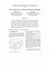
Fig. 1. Coronal Cut of Neck CT with contrast.
as well. The fiberoptic exam showed mild medialization of the
left parapharyngeal wall. Major salivary glands examination was
unremarkable. Her routine blood tests were within normal limits
except for the picture of microcytic hypovolemic anemia consistent with her thalassemia condition and elevated C-reactive protein
(CRP) that was 28.9 (WBC count6.0 with no leukocytosis). Suspected to have a peritonsillar collection; aspiration was attempted
in the emergency room that, however, yielded no pus. A computerized tomography (CT) neck with contrast (Fig. 1) was done
to the patient and showed evidence of a large left parapharyngeal collection with enhancing peripheral wall and non-enhancing
contents. The swelling measured 3.4 × 2.7 × 3.5 cm in the maximum anteroposterior (AP), transverse (TR) and craniocaudal (CC)
dimensions, respectively. Multiple enlarged elongated benign looking submandibular and upper jugular groups of lymph nodes with
preserved fatty hilum, largest measuring 9 mm in the short axis in
the left submandibular region. Hence, the patient was presumed
to have a left parapharyngeal collection - likely an abscess with
enlarged lymph nodes.
The patient shifted to the operating theater for incision and
drainage of left parapharyngeal abscess; trans-cervical approach
was made initially by the ORL-HNS consultant to drain the suspected abscess. However, no pus yielded, repeated aspiration also
did not yield anything. Therefore, a trans-oral approach followed
where incision anterior to the anterior tonsillar pillar was done.
With further dissection until reaching the left infratemporal fossa,
the upper pole of a mass was noticed and was hard, surrounded
by necrotic tissue. This full mass was delivered completely through
transoral incision.
Post-operative, the patient tolerated the surgery very well and
started diet short after the operation without any limitations; she
was discharged on postoperative day 3 with analgesia.
The patient followed for 1 year and she was doing fine without
any signs of recurrence, neurological deficit or palatal dysfunctional
issues; no further imaging workup was done for the patient as the
progression of the disease along with the final pathological report
indicate a benign pathology.
Histopathological; grossly, the specimen consisted of a single
irregular firm tan brown soft tissue nodule measuring 4 × 3 ×
2.5 cm. Cut surfaces showed brown heterogeneous soft tissue with
multiple scattered white firm parts largest measured 1.5 × 1.5 ×
0.5 cm abutting surgical margin (Fig. 2).
Fig. 2: Representative sections of the tumor showing infarcted
white parts. Surgical margin inked green.
Fig. 3. A: Tumor with a background of hemorrhage, inflammation, and a focus of
normal salivary glands (asterisk) (H&E magnification x 40). B: infarcted part of the
tumor (asterisk) with abundant squamous metaplasia and keratinization (arrow
heads) (H&E magnification x 100).
Microscopically, the extensively sampled specimen showed
myoepithelial cells scattered within myxoid stroma intermingled
with sheets of degenerate ductal epithelial cells together with
prominent squamous metaplasia and abundant keratinization.
Focal hemorrhage, inflammation, and necrosis were also noted.
A rim of normal salivary gland tissue was identified. The overall appearances were consistent with an infarcted pleomorphic
adenoma with florid squamous metaplasia (Fig. 3). No malignant
features were identified.
Fig. 3 - Tumor with a background of hemorrhage, inflammation,
and a focus of normal salivary glands (asterisk) (H&E magnification
x 40).
635
CASE REPORT – OPEN ACCESS
A.R. Alsaleh et al.
3. Discussion
Tumors of the ITF present a diagnostic and a surgical challenge due to the often-occult nature of such lesions as well as
the complexity of the local anatomy. Neoplasia of ITF can be
classified as primary, secondary or metastatic [4]. Among the
malignant tumors in the ITF region, adenoid cystic carcinoma,
adenocarcinoma and squamous cell carcinoma are the most common malignant tumor encountered in this location [4]. In contrast,
nasopharyngeal fibroma is frequently found in ITF benign lesions.
To our knowledge, only three cases of pleomorphic adenoma in
the ITF region were reported and we believe that we report the
first case of an infarcted pleomorphic adenoma in the ITF region
[5–7]. Several assumptions have been discussed regarding the origin of all these salivary gland tissue abnormal locations. Ferlito
assumes the existence of scattered heterotopic salivary gland tissue in the head and neck region, and in the pituitary gland region
specifically, external auditory meatus, nasal fossae, sternoclavicular joint, mandibula, and cervical soft tissues. Heterotopic salivary
gland tissues in these areas, all have the potential to develop into a
pleomorphic adenoma.
CT scan remains a substantial image modality in diagnosing
tumors of the ITF as it helps to determine the extent of the disease, its local spread, and radiologic characteristics of the tumor
may help to some extent in determining the type of the tumor.
Furthermore, Magnetic resonance imaging (MRI) should be considered as a modality whenever the suspicion of the vascular lesion
is present or in cases of atypical presentation as in our case, the
palate is the most common site of minor salivary gland PA followed by the lip, buccal mucosa and floor of the mouth, tongue,
tonsil pharynx, retromandibular area and nasal cavity, parapharyngeal space PA can originate de novo or from the deep lobe of the
parotid gland and then extend by the stylomandibular tunnel into
the parapharyngeal space.
Parapharyngeal tumors are rare, constituting less than 0.5% of
head and neck neoplasms [8]. Of these, PA is the most common
benign tumor, representing around 40% [9].
Due to its concealed location, lesions often appear late; in
addition, surgical planning is confounded by proximity to vital
structures, namely intracranial structures, the orbit, sinuses, and
the nasopharynx. Indicating the difficulty of access, numerous surgical approaches have been described.
Although the prognosis of PA is generally good, yet the conventional watchful waiting and clinical follow up is not sufficient,
while periodic radiological evaluation might be needed to detect
any recurrence post-surgical excision.
While many surgeons acclaim complete surgical excision is
the only valid treatment option for pleomorphic adenoma, other
reports illustrated good results with adjuvant radiotherapy against
incompletely resected tumors due to its location that renders
them inoperable, However, this remains a matter of controversy
[2].
Although seems to be relatively rare, spontaneous infarction
resembling abscess formation should be taken into consideration
in the differential diagnosis of such cases. In atypical case presentations, one might consider MRI to differentiate between abscess
and neoplasia in the ITF region. This case has been reported in line
with the SCARE Guideline 2018 [10].
International Journal of Surgery Case Reports 77 (2020) 634–637
transcervical approach allows excellent control of the tumor and
neurovascular elements but the transoral approach might be also
needed according to the case nature.
Declaration of Competing Interest
No conflict of interest.
Funding
Non sponsored and no fund received.
Ethical approval
The paper describes a case report therefore no special ethical
consideration required.
Consent
Written informed consent was obtained from the patient for
publication of this case report and accompanying images. A copy
of the written consent is available for review by the Editor-in-Chief
of this journal on request.
Author contribution
ARA: Data Collection, Literature Search, Manuscript Preparation,
AAA: Manuscript Preparation & revision,
HAH, WR: Manuscript Preparation,
BAW: pathology slides preparation,
AJN: Manuscript Revision and submission.
All authors read the final manuscript and agreed on it.
Registration of research studies
N/A.
Guarantor
Dr Abdelrahman R. Alsaleh
AAlsaleh@hamad.qa
00974-55176528
Provenance and peer review
Not commissioned, externally peer-reviewed.
Acknowledgment
We thank the patient for letting us share her case.
References
[1] K. Behzatoglu, et al., Spontaneous infarction of a pleomorphic adenoma in
parotid gland: diagnostic problems and review, Diagn. Cytopathol. 32 (6)
(2005) 367–369.
[2] M. Clark, et al., CT of intranasal pleomorphic adenoma, Neuroradiology 41 (8)
(1999) 591–593.
[3] R. Tiwari, et al., Tumors of the infratemporal fossa, Skull Base Surg. 10 (1)
(2000) 1–9.
[4] J.J. Conley, Tumors of the infratemporal fossa, Arch. Otolaryngol. 79 (1964)
498–504.
[5] T. El-Hadi, et al., Plemorphic adenoma of the infratemporal space: a new case
report, Int. J. Otolaryngol. 2009 (2009) 529350.
[6] L.E. Gurey, C.D. Brook, S.M. Parnes, Pleomorphic adenoma of the
infratemporal fossa: case report and literature review, Laryngoscope 120
(Suppl 4) (2010) S151.
[7] K. Jeyanthi, et al., Pleomorphic adenoma in the infra-temporal space: the first
case report, Head Neck Pathol. 1 (2) (2007) 173–177.
4. Conclusion
ITF Infarcted neoplasms may present clinically as an abscess,
in these situations, the combination of CT scan and MRI might be
a helpful diagnostic tool in distinguishing neoplasia from abscess
collection in this area. Treatment of ITF tumors is surgical. The
636
CASE REPORT – OPEN ACCESS
A.R. Alsaleh et al.
[8] J.G. Batsakis, N. Sneige, Parapharyngeal and retropharyngeal space diseases,
Ann. Otol. Rhinol. Laryngol. 98 (4 Pt 1) (1989) 320–321.
[9] K.V. Hughes 3rd., K.D. Olsen, T.V. McCaffrey, Parapharyngeal space neoplasms,
Head Neck 17 (2) (1995) 124–130.
International Journal of Surgery Case Reports 77 (2020) 634–637
[10] R.A. Agha, M.R. Borrelli, R. Farwana, K. Koshy, A. Fowler, D.P. Orgill, For the
SCARE Group, The SCARE 2018 Statement: updating consensus surgical CAse
REport (SCARE) guidelines, Int. J. Surg. 60 (2018) 132–136.
Open Access
This article is published Open Access at sciencedirect.com. It is distributed under the IJSCR Supplemental terms and conditions, which
permits unrestricted non commercial use, distribution, and reproduction in any medium, provided the original authors and source are
credited.
637
Academia.edu no longer supports Internet Explorer.
To browse Academia.edu and the wider internet faster and more securely, please take a few seconds to upgrade your browser.
An unusual presentation of pleomorphic adenoma in a patient with thalassemia: A case report
International Journal of Surgery Case Reports, 2020
...Read more
Related Papers
Diagnostic Cytopathology, 2009
Download


PLEOMORPHIC ADENOMA: CLINICAL CASE REPORT (Atena Editora), 2022
Download


Head and Neck Pathology, 2007
Download

Diagnostic and interventional radiology (Ankara, Turkey), 2008
Download


Diagnostic Cytopathology, 2009
Download

Acta Cytologica, 2001
Download

مجلة العمارة والفنون والعلوم الإنسانیة, 2020
Download

Analele Universităţii "Dunărea de Jos" din Galaţi Fascicula XI Construcţii navale/ Annals of "Dunărea de Jos" of Galati Fascicle XI Shipbuilding, 2021
Download


Download

