The Journal of Neuroscience, January 2, 2013 • 33(1):133–149 • 133
Systems/Circuits
Short-Term Plasticity Explains Irregular Persistent Activity
in Working Memory Tasks
David Hansel1 and German Mato2
Laboratory of Neurophysics and Physiology and Institute of Neuroscience and Cognition, University Paris Descartes, 75270 Paris Cedex 06, France, and
Comisión Nacional de Energía Atómica and Consejo Nacional de Investigaciones Cientificas y Técnicas, Centro Atómico Bariloche and Instituto Balseiro,
8400 San Carlos de Bariloche, Argentina
1
2
Persistent activity in cortex is the neural correlate of working memory (WM). In persistent activity, spike trains are highly irregular, even
more than in baseline. This seemingly innocuous feature challenges our current understanding of the synaptic mechanisms underlying
WM. Here we argue that in WM the prefrontal cortex (PFC) operates in a regime of balanced excitation and inhibition and that the
observed temporal irregularity reflects this regime. We show that this requires that nonlinearities underlying the persistent activity are
primarily in the neuronal interactions between PFC neurons. We also show that short-term synaptic facilitation can be the physiological
substrate of these nonlinearities and that the resulting mechanism of balanced persistent activity is robust, in particular with respect to
changes in the connectivity. As an example, we put forward a computational model of the PFC circuit involved in oculomotor delayed
response task. The novelty of this model is that recurrent excitatory synapses are facilitating. We demonstrate that this model displays
direction-selective persistent activity. We find that, even though the memory eventually degrades because of the heterogeneities, it can be
stored for several seconds for plausible network size and connectivity. This model accounts for a large number of experimental findings,
such as the findings that have shown that firing is more irregular during the persistent state than during baseline, that the neuronal
responses are very diverse, and that the preferred directions during cue and delay periods are strongly correlated but tuning widths
are not.
Introduction
Working memory (WM), the ability to temporarily hold, integrate, and process information to produce goal-directed behavior, is crucial to higher cognitive functions, such as planning,
reasoning, decision-making, and language comprehension (Baddeley, 1986; Fuster, 2008). The persistent activity recorded in
neocortex during WM tasks is thought to be the main neuronal
correlate of WM (Fuster and Alexander, 1971; Miyashita and
Chang, 1988; Goldman-Rakic, 1995). For example, in an
oculomotor-delayed response (ODR) task in which a monkey has
to remember the location of a stimulus for several seconds to
make a saccade in its direction, a significant fraction of the neurons in the prefrontal cortex (PFC) modify their activity persistently and selectively to the cue direction during the delay period
(Funahashi et al., 1989, 1990, 1991; Constantinidis et al., 2001;
Takeda and Funahashi, 2007). The classical view is that this reflects a multistability in the dynamics of the PFC circuit because
Received July 19, 2012; revised Oct. 24, 2012; accepted Oct. 26, 2012.
Author contributions: D.H. and G.M. designed research; D.H. and G.M. performed research; D.H. and G.M. analyzed data; D.H. and G.M. wrote the paper.
This research was conducted within the scope of the France-Israel Laboratory of Neuroscience (FILN-LEA) and
supported by UniNet EU excellence network, ANR-09-SYSC-002-01 and Project SECyT-ECOS A04B04. We thank S.
Funahashi, R. Guetig, Y. Loewenstein, C. Levenes, G. Mongillo, and C. van Vreeswijk for fruitful discussions. We are
also thankful to Y. Loewenstein, G. Mongillo, and C. van Vreeswijk for critical reading of our manuscript. We also
thank Ecole de Neuroscience Paris Ile-de-France.
Correspondence should be addressed to David Hansel, University Paris Descartes, Laboratory of Neurophysics and
Physiology, 45 Rue des Saints-Pères, 75270 Paris, France. E-mail: david.hansel@univ-paris5.fr.
DOI:10.1523/JNEUROSCI.3455-12.2013
Copyright © 2013 the authors 0270-6474/13/330133-17$15.00/0
sensory inputs are the same in the precue and in the delay periods
but neuronal activity is different (Hebb, 1949; Hopfield, 1984;
Amit, 1995; Amit and Brunel, 1997; Wang, 2001).
The persistent activity of PFC neurons is highly irregular temporally (Shinomoto et al., 1999; Compte et al., 2003; Shafi et al.,
2007). For instance, it has been reported that in ODR tasks, the
coefficient of variation of the interspike interval distribution is
close to 1, and even ⬎1 for many of the neurons (Compte et al.,
2003). Previous modeling works have attempted to account for
this irregularity (Brunel, 2000; van Vreeswijk and Sompolinsky,
2004; Renart et al., 2007; Roudi and Latham, 2007; Barbieri and
Brunel, 2008). However, in all these models, unless parameters
are tuned, the neurons fire much too regularly in memory states.
Hence, accounting in a robust way for highly irregular firing and
persistent activity remains challenging.
Irregular firing of cortical neurons is naturally explained if one
assumes that cortical networks operate in a regime in which excitation is strong and balanced by strong inhibition (Tsodyks and
Sejnowski, 1995; van Vreeswijk and Sompolinsky, 1996, 1998;
Vogels et al., 2005; Lerchner et al., 2006; Vogels and Abbott,
2009). It is therefore tempting to assume that in WM tasks the
PFC network expresses a multistability between balanced states.
We argue here that this requires that the nonlinearities underlying the persistence of activity in PFC are primarily in the neuronal
interactions and not in the neurons as assumed previously. We
also argue that short-term plasticity is a possible substrate for
these nonlinearities. Specifically, we propose that the short-term
facilitating excitatory synapses recently reported in PFC (Hempel
�134 • J. Neurosci., January 2, 2013 • 33(1):133–149
Hansel and Mato • Short-Term Plasticity and Working Memory
et al., 2000; Wang et al., 2006) play an essential role in WM. To
illustrate these claims, we develop a new network model for spatial WM based on this assumption. We show that the model
displays highly irregular spontaneous activity as well as persistent, selective, and highly irregular delay activity. Importantly, it
also displays a great deal of the diversity observed in the delay
activity of PFC neurons (Funahashi et al., 1989, 1990; Asaad et al.,
1998; Romo et al., 1999; Takeda and Funahashi, 2002; Brody et
al., 2003; Shafi et al., 2007).
A brief account of this study has been presented in abstract
form (Hansel and Mato, 2008).
Materials and Methods
The network models. In this paper we consider two network models made
of spiking neurons. One model (Model I) has an unstructured connectivity. The second model (Model II) represents a network in PFC involved in an ODR task and has a ring architecture (Ben-Yishai et al., 1995;
Hansel and Sompolinsky, 1996).
Single neuron dynamics. Both models are made up of an excitatory and
an inhibitory population of integrate-and-fire neurons. The membrane
potential of a neuron follows the dynamics:
dV
⫽ ⫺ V ⫹ I rec共t兲 ⫹ Iext共t兲,
dt
(2)
(3)
2
i is
N␣
the direction of the cue for which the sensory input into neuron (i, ␣) is
maximum; and erase is the direction of the saccade that is actually performed. An estimate of erase is given by the direction of the population
vector computed from the activity of the neurons at the end of the delay
period (see below). We assume that the angles, i,␣, are uniformly distributed between 0 and 360°.
For simplicity we take A␣␥共t兲 ⫽ Â␣␥H␥共t兲 (␥ ⫽ cue, erase), where Â␥ is
constant, H cue(t) ⫽ 1 during the cue period, and H cue(t) ⫽ 0 otherwise.
Similarly, H erase(t) ⫽ 1 when the memory erasing input is present and
H erase(t) ⫽ 0 otherwise.
Connectivity of the networks. We define the connectivity matrix of the
network by Ji␣,j ⫽ 1 if neuron (j, ) is connected presynaptically to
neuron (i, ␣) and Ji␣,j ⫽ 0 otherwise. In Model I the connectivity is
where cue is the direction in which the cue is presented; i, ␣ ⫽
冉 冊
关兴2
,
2
2␣
(4)
where [] ⫽ min(兩兩, 2 ⫺ 兩兩) and C␣ is a normalization that ensures
that the total number of inputs a neuron in population ␣ receives from
population  is on average K␣. The range of the interactions is characterized by the parameters ␣. The resulting network architecture is a
probabilistic version of the architecture of the ring model, in which neurons are all connected with probability 1, whereas the strength of their
interactions depends on their distance on the ring (Ben-Yishai et al.,
1995; Hansel and Sompolinsky, 1996; Compte et al., 2000).
Synaptic interactions. We model the recurrent synaptic input current
into neuron (i, ␣) as:
I i,rec␣ 共t兲 ⫽
The first term on the right hand side represents the background input,
which is constant in time and depends solely on the target population.
The second term represents the transient sensory inputs related to the cue
to be memorized. The third term represents a transient input that erases
the memory at the end of the delay period. In Model I, Ii,␥ ␣ 共t兲 (␥ ⫽ cue,
erase) is homogeneous (i.e., Ii,␥ ␣ 共t兲 ⬅ A␣␥ 共t兲 does not depend on i). In
model II:
I i,␥ ␣ 共 t 兲 ⫽ A ␣␥ 共 t 兲关 1 ⫹ ⑀ ␣␥ cos共i, ␣ ⫺ ␥ 兲兴,
P ␣ 共 兲 ⫽ C ␣ exp
(1)
where is the membrane time constant, I rec(t) is the total recurrent
synaptic current the neuron receives from all other cells in the network
connected to it and I ext(t) represents feedforward inputs from outside
the network. Whenever the membrane potential of the neuron reaches
the threshold, VT, it fires an action potential and its voltage is reset to VR.
We take VT ⫽ 20 mV, VR ⫽ ⫺3.33 mV. The time constants of the
neurons are ⫽ 20 and 10 ms for excitatory and inhibitory neurons
respectively, in accordance with standard values (Somers et al., 1995).
The external inputs. Simulation of a memory task (Model I) or of the
ODR task (Model II) requires a stimulus that depends on time. First,
there is a precue period that allows the system to settle in a baseline state.
In the second stage, a transient input is applied to simulate the cue, after
which the input returns to the previous value. Finally another transient
input is applied to erase the memory that has been stored. Thus, the
external feedforward input into neuron i ⫽ 1, . . ., N␣ in population ␣ ⫽
E, I (hereafter neuron i, ␣) is:
I i,ext␣ 共t兲 ⫽ I␣b ⫹ Ii,cue␣ 共t兲 ⫹ Ii,erase
␣ 共t兲.
unstructured. Therefore the probability, Pi␣,j, that Ji␣,j ⫽ 1 depends
only on ␣ and : Pi␣,j ⫽ K␣/N, where N is the number of neurons in
population  ⫽ E, I and K␣ is the average number of connections a
neuron in population ␣ receives from population .
The architecture of Model II is consistent with the columnar functional anatomy of the monkey PFC (Goldman-Rakic, 1987, 1988, 1995;
Rao et al., 1999). The probability of connection between two neurons is
given by Pi␣,j ⫽ P␣(兩i␣ ⫺ j兩) with:
冘
Ji ␣ , j  G␣ uj  ,n xj  ,n f␣ 共t ⫺ tj  ,n 兲,
(5)
jn
where G␣ is a constant that measures the maximal synaptic current
neuron (i, ␣) receives from neuron (j, ), tj,n is the time of the n-th spike
fired by neuron (j, ), xj,n is the amount of synaptic resources available
at its synaptic terminals before this spike, and uj,n is the fraction of these
resources used by this spike. The dynamics of these two variables are
responsible for the short-term plasticity (STP) and we model them as
(Markram et al., 1998):
冉
u j  ,n⫹1 ⫽ u j  ,n exp ⫺
⌬tj  ,n
f
冊 冉
冉
⫹ U 1 ⫺ uj  ,n exp ⫺
⌬tj  ,n
f
冊冊
(6)
冉
xj,n⫹1 ⫽ xj,n共1 ⫺ uj,n⫹1兲exp ⫺
⌬tj  ,n
r
冊
冉
⫹ 1 ⫺ exp ⫺
冊
⌬tj  ,n
,
r
(7)
where ⌬tj,n is the n-th interspike interval of neuron (j, ); r and f are
the recovery and the facilitation time constants of the synapse, and U the
maximal utilization parameter.
Finally, the function f␣(t) in Equation 5 describes the dynamics of
individual postsynaptic currents (PSCs). It is given by f␣(t) ⫽ (1/␣)
exp(⫺t/␣)H(t) where ␣ is the synaptic decay time, and H(t) ⫽ 0
[respectively (resp.) 1] for t ⬍ 0 (resp. t ⬎ 0) (Dayan and Abbott, 2001).
Network size, connectivity, and scaling of network parameters with connectivity. The total number of neurons in the network is N ⫽ NE ⫹ NI
with NE ⫽ 0.8 N and NI ⫽ 0.2 N. The average total number of synaptic
inputs a neuron receives, K, is assumed to be the same in the two populations. We take: KEE ⫽ KIE ⬅ KE ⫽ 0.8 K and KII ⫽ KEI ⬅ KI ⫽ 0.2 K.
Unless specified otherwise we take N ⫽ 80,000 and K ⫽ 2000.
The balanced regime is mathematically well defined only in the limit
N 3 ⬁, K 3 ⬁ while K ⬍⬍ N, and the strength of the recurrent interactions and the external inputs are scaled as (van Vreeswijk and Sompolinsky, 1998):
b
␣,
â␣␥
g ␣
冑K 
(8)
I ␣b ⫽ i ␣b 冑K E
(9)
 ␣␥ ⫽ â ␣␥ 冑K E ,
(10)
G ␣ ⫽
where g␣, i and do not depend on K. This scaling ensures that, in a
wide range of parameters, the temporal fluctuations in the synaptic in-
�Hansel and Mato • Short-Term Plasticity and Working Memory
J. Neurosci., January 2, 2013 • 33(1):133–149 • 135
Table 1. Parameters of the synaptic interactions for the unstructured and spatial
working memory networks
gEE (AMPA)
gEE (NMDA)
gIE (AMPA)
gIE (NMDA)
gEI
gII
U
r
f
EE
IE
EI
II
Unstructured
Spatial WM
560 mV ms
560 mV ms
67.2 mV ms
7.4 mV ms
⫺138.6 mV ms
⫺90.6 mV ms
0.03
200 ms
450 ms
—
—
—
—
533.3 mV ms
533.3 mV ms
67.2 mV ms
7.4 mV ms
⫺138.6 mV ms
⫺90.6 mV ms
0.03
200 ms
450 ms
60°
70°
60°
60°
puts remain finite and do not depend on the connectivity when the
connectivity is large, whereas the time-average excitatory and inhibitory
inputs increase and balance each other (van Vreeswijk and Sompolinsky,
1996, 1998). In that limit, even though the excitatory and inhibitory
inputs become infinitely large, the temporal mean and SD of the fluctuations of the total inputs remain finite and on the same order of the
neuronal threshold.
For finite connectivity, the balance of excitation and inhibition is only
approximate. Therefore, to qualify the dynamic regime of the network as
balanced, it is important to check the robustness to increasing K. As
explained in Results, it is essential to verify that the domain of the parameters in which the multistability between balanced states occurs does not
vanish in the limit of large N and large K. To test for this robustness, we
performed numerical simulations with a network of size up to N ⫽
320,000 neurons and connectivities as large as K ⫽ 32,000.
Synaptic parameters. Unless specified otherwise, the parameters of the
models used in our simulations are those given in Table 1.
In both models, pyramidal cells form a mixture of fast (AMPA) and
slow (NMDA) synapses on other pyramidal cells and interneurons. Both
components share the same connectivity matrices Ji␣,jE but differ in their
synaptic strength (g AMPA and g NMDA, respectively) and in the decay time
constant of their PSCs. Equation 5 must be interpreted as including the
sum over both components. The decay time constant of the excitatory
postsynaptic currents are 3 and 50 ms for AMPA and NMDA synapses,
respectively. We tested to confirm that taking longer time constants for
the NMDA synapses (for instance 80 ms, as reported by Wang et al.,
2008) has no impact on the results (data not shown).
The voltage dependence of the NMDA currents was not included in
the simulations depicted in this paper. However, we have verified that
including voltage dependence while keeping the STP of the recurrent
excitation (EE) synapse interactions does not qualitatively change the
conclusions of our work. For that purpose, we multiplied the synaptic
, by the same voltage-dependent factor as in Compte et
strength, G␣NMDA
E
NMDA
al. (2000). Since this factor is always ⬍1, we had to increase GEE
and
NMDA
GIE
by some constant factor equal to 5.
NMDA
We define R␣ as follows: R␣ ⫽ g␣AMPA
⫹ g␣AMPA
). In most of
冫共 g␣ E
E
E
the simulations we took RE ⫽ 0.5 and RI ⫽ 0.9. This accounts for the fact
that NMDA synapses are more abundant between pyramidal cells than
between pyramidal cells and interneurons (Thomson, 1997). All the inhibitory interactions have a decay time constant of 4 ms (Bartos et al.,
2001, 2002). We verified that the properties of the model were robust
with respect to the values of the excitation and inhibition decay-time
constants, as well as with respect to the ratios RE and RI.
The recurrent excitation between pyramidal cells displays short-term
plasticity. For simplicity, in the simulations described in this paper, we
assumed that this is the case for AMPA as well as for NMDA interactions.
However, we verified that the properties of the network are essentially the
same if only AMPA synapses display STP (provided that the synaptic
AMPA
is increased appropriately to compensate for the absence
strength GEE
of facilitation in NMDA synapses). The parameters of the STP were
chosen such that the network displays multistability in a broad range of
background inputs. The chosen values for the recovery and the facilitation time constants were compatible with the in vitro data of Wang et al.
(2006). The utilization parameter, U, was in the range of the lower values
reported in that study. We assume that excitatory synapses to inhibitory
neurons as well as all inhibitory synapses do not display STP. Therefore
for these synapses xj,n ⫽ uj,n ⫽ 1.
With the parameters in Table 1, the maximal postsynaptic potentials
(PSPs) of the various connections are as follows: 4.3 10 ⫺2 mV and 0.14
mV for the NMDA and AMPA components of the EE synapses, respectively; ⫺2.3 mV for the EI synapses, ⫺2.5 mV for the II synapses; and 2.5
10 ⫺2 and 1 mV for the NMDA and AMPA components of the excitatory
to the inhibitory neurons, respectively. Note that the PSPs generated by
individual excitatory connections are substantially weaker than those
generated by inhibitory ones. This is partially compensated for by excitatory connectivity that is larger by a factor of 4 than the inhibitory connectivity, and by greater activity for inhibitory than for excitatory
neurons. Moreover, the neurons receive an excitatory tonic input. Altogether, excitation and inhibition inputs balance approximately as we
show in the results.
Numerical simulations. Simulations were performed using a secondorder Runge–Kutta scheme with a fixed time step, ␦t ⫽ 0.1 ms, supplemented by an interpolation scheme for the determination of the firing
times of the neurons (Hansel et al., 1998).
Characterization of the irregularity in action potential firing. We quantify the irregularity of the discharge of a neuron by the coefficient of
variation (CV ) of its interspike interval (ISI) distribution defined by:
CV ⫽
冓 共 ␦ n ⫺ 冓 ␦ n 冔 兲 2 冔 1/ 2
,
冓 ␦ n冔
(11)
where ␦n is n-th ISI of the neuron and 冓 . . . 冔 denotes an average overall
number of spikes it has fired.
We also evaluate the coefficient of variation CV2. CV2 is computed by
comparing each ISI (␦n) to the following ISI (␦n⫹1) to evaluate the degree
of variability of ISIs in a local manner (Holt et al., 1996). It is defined by:
CV 2 ⫽ 2
冓兩 ␦ n ⫺ ␦ n⫹1 兩冔
.
冓 ␦ n ⫹ ␦ n⫹1 冔
(12)
For a Poisson spike train, CV ⫽ CV2 ⫽ 1.
Evaluation of the phase diagrams. To evaluate which regions in the
space of parameters display persistent activity, we use the following procedure: In simulating the network, we slowly increase the external input
IE (while keeping the rest of the parameters constant) and monitor the
mean and spatial modulation of the network activity. When IE has
reached a predefined value of sufficient size, we continue the simulations
while decreasing IE back to its initial value. This generates a hysteresis
curve, which enables us to identify the bistability region for that point in
the parameter space. The procedure is repeated for different values of the
parameters (e.g., GEE) to obtain a phase diagram.
Simulation of the delayed response task in Model II. At the beginning of
a trial, the network is initialized from random initial conditions. After 3 s
(representing the fixation period in the experiment), the cue is presented
and the related feedforward input occurs for ⌬tcue ⫽ 0.5 s. The delay
period goes from 3.5 to 6.5 s. The transient input, which erases the
memory, begins at t ⫽ 6.5 s and has a duration of ⌬terase ⫽ 1 s.
Quantification of the single neuron directional selectivity in Model II.
Tuning curves were estimated by simulating 20 trials for each of the eight
cues from 0 to 315° in intervals of 45°. Using a bootstrap method, we
determined whether the task-related activity of a neuron was directionally tuned (Constantinidis et al., 2001). For each neuron we evaluated the
quantity Oi, ␣ ⫽ 关 冘 r2兴1/ 2 where r is the firing rate of the neuron during
the delay period averaged over the 20 trials with the cue presented in
direction . We compared the obtained value of Oi,␣ with the one obtained after randomly permuting the angles of each trial before averaging. If
the second quantity is smaller than the first one for 99% of the permutations,
we consider that the activity is significantly directionally tuned.
We quantified the degree of directional selectivity with the circular
variance (CircVar) (Mardia, 1972) defined by CircVar ⬅ 1 ⫺ c1/c0 where
�136 • J. Neurosci., January 2, 2013 • 33(1):133–149
Hansel and Mato • Short-Term Plasticity and Working Memory
ck ⫽ 兩⌺ r exp(ik)兩. A broad tuning curve (badly selective response)
corresponds to CircVar close to 1 whereas for very sharp tuning CircVar
is close to 0.
The tuning curves of a large fraction of the neurons can be well fitted to
a von Mises function defined as:
r 共 兲 ⫽ A ⫹ B exp
冉
冊
cos共 ⫺ 兲 ⫺ 1
.
D
(13)
We estimated the parameters A, B, , D for each neuron by minimizing
the quadratic error of the fit: E ⫽ ⌺ 共r共 兲 ⫺ r 兲2 冫2 , where the sum
is over the eight directions of the cue and is the trial-to-trial SD of the
response. The estimate of the preferred direction (PD) of the neuron is
given by PD ⫽ 180°/. The sharpness of the tuning curve [tuning width
(TW)] above baseline can be computed from the formula:
冢
180⬚
cos⫺1 1 ⫹ D log
TW ⫽
冉 冊冣
1 ⫹ exp ⫺
2
2
D
.
(14)
The quality of the fit is estimated by evaluating the 2 distribution for 4
degrees of freedom (8 points minus 4 parameters) (Press et al., 1992).
This probability characterizes the goodness-of-fit. Bad fits correspond to
extremely low values of the probability, q. We consider that the fit is good
if q ⬎ 0.001. To determine the spatial modulation of the network activity
and population vector in Model II, let us denote by fj␣(t) the firing rate of
neuron (j, ␣) averaged over a time window of 50 ms around time t. To
characterize the spatial modulation of the activity of population ␣ at time
t we computed:
Z ␣共 t 兲 ⫽
冘
f j ␣ 共 t 兲 exp共ij, ␣ 兲冫
j
⫽ M ␣ 共 t 兲 exp共i⌿␣ 共t兲兲,
冘
fj ␣ 共t兲
(15)
j
(16)
where M␣ is the modulus of the complex number Z␣ and ␣ is its argument. Note that the real and imaginary parts of Z␣(t) are the components
of the population vector at time t for population ␣. If the network activity
is homogeneous, M␣(t) is ⬃0, whereas for a very sharply modulated
activity profile, M␣(t) is ⬃1. In our simulations, we always found that
E(t) is approximately equal to I(t). The preferred direction, erase, of
the feedforward input that erases the memory trace was taken to be the
value of E ⫽ I at the end of the delay period.
Results
Irregular firing in cortex in vivo and balance of excitation
and inhibition
Cortical neurons fire irregularly (Burns and Webb, 1976; Softky
and Koch, 1993; Bair et al., 1994). The neurons that fire less
irregularly are those in primary motor cortices, in supplementary motor cortices, or in association with or in motor areas,
such as parietal regions (Maimon and Asaad, 2009; Shinomoto
et al., 2009), where the CV of the ISI distributions of the
neurons are in the range CV ⫽ 0.5– 0.8. The neurons that fire
more irregularly are in sensory areas and in prefrontal cortex
where the CVs are ⬃1.
Remarkably, recent experimental studies in monkeys performing WM tasks have reported that the level of temporal irregularity with which PFC neurons fire during the delay period is
comparable to, if not higher than, what is observed in spontaneous activity or during the fixation period (Shinomoto et al., 1999;
Compte et al., 2003; Shafi et al., 2007).
The highly irregular activity of cortical neurons in vivo has
long appeared paradoxical in view of the large number of their
synaptic inputs (Softky and Koch, 1992, 1993; Holt et al., 1996).
This is because the temporal fluctuations of the postsynaptic current produced by K ⬎⬎ 1 presynaptic afferents firing asynchronously are much smaller, by a factor 1/冑K, than its average.
Accordingly, since in vitro neurons fire regularly in response to
weakly noisy input (Connors et al., 1982), one would expect that
firing in vivo would be only weakly irregular. A generic solution to
this problem posits nearly balanced strong excitatory and inhibitory synaptic inputs such that their temporal fluctuations, although much smaller than their means taken separately, are
comparable to the average total input and to the neuronal threshold (van Vreeswijk and Sompolinsky, 1996, 1998). Modeling
studies have shown that balanced states emerge in a robust manner without fine tuning of parameters from the collective dynamics of recurrent neuronal networks (van Vreeswijk and
Sompolinsky, 1996, 1998, 2004; Amit and Brunel, 1997; Lerchner
et al., 2006; Hertz, 2010). The balance mechanism has been applied to account for the high variability of spontaneous activity as
well as sensory-evoked neuronal activity in cortex (van Vreeswijk
and Sompolinsky, 2004; Lerchner et al., 2006). Can it also provide
a natural framework to account for the spiking irregularity observed during WM tasks?
In balanced states, the level of activity of macroscopic ensembles of neuronal populations are largely independent of singlecell intrinsic properties (van Vreeswijk and Sompolinsky, 1998).
This can be understood heuristically as follows. Let us consider a
network comprising one excitatory population ( E) and one inhibitory population ( I) (Fig. 1 A). The state of each population is
characterized by its activity, f␣, ␣ ⫽ E, I. Assuming a stationary
state of the network, this activity is related to the total input into
the population ␣, h␣, via f␣ ⫽ S␣(h␣) where S␣ is the sigmoidal
input– output transfer function of population ␣ and hE and hI are
given by (see for instance Dayan and Abbott, 2001):
h E ⫽ G EE f E ⫺ G EI f I ⫹ I E
(17)
h I ⫽ G IE f E ⫺ G II f I ⫹ I I ,
(18)
where the constants G␣ measure the efficacy of the interactions
between population  and ␣ and IE and II are external inputs to
the network.
If the excitation is too strong, the inputs hE and hI and therefore the activities of the populations reach the saturation levels of
SE and SI. Conversely, for overly strong inhibition, hE and hI are
below the (soft) threshold of SE and SI and network activity is very
low. An appropriate balance of excitation and inhibition is necessary to prevent the network from being in one of these extreme
regimes. This occurs if the activities of the two populations obey
the conditions:
G EE f E ⫺ G EI f I ⫹ I E ⬇ 0
(19)
G IE f E ⫺ G II f I ⫹ I I ⬇ 0,
(20)
which express the very fact that inhibition balances excitation.
These balance conditions do not depend on the input– output
transfer functions of the populations. They fully determine the
population activities as a function of the external inputs. Since
these equations are linear, there is generically a unique solution
for given values of IE and II. Hence the network cannot exhibit
more than one balanced state. This effective washout at the macroscopic level of the neuronal intrinsic properties is a remarkable
feature of the balance regime. As a matter of fact, it can be derived
in large networks of randomly connected binary neurons (van
Vreeswijk and Sompolinsky, 1998), of randomly connected spiking integrate-and-fire neurons (Renart et al., 2010), or of
integrate-and-fire networks (Lerchner et al., 2004; van Vreeswijk
and Sompolinsky, 2004).
�Hansel and Mato • Short-Term Plasticity and Working Memory
J. Neurosci., January 2, 2013 • 33(1):133–149 • 137
number of neurons induces a transition to the other state. We will
term the state in which the activity is lower as baseline and the
state in which the activity is elevated as persistent.
Bistability between balanced states in a simplified rate model
with nonlinear synaptic interactions
It is clear that the linearity of Equations 19 and 20 stems from the
assumption that the interactions between the neurons are linear;
namely, that the synaptic inputs are proportional to the presynaptic firing rates and linearly sum up.
We first relax this assumption by considering that the recurrent excitatory interactions depend nonlinearly on the activity of
the excitatory neurons. This means that we replace GEE in Equations 19 and 20 by a term GEEFEE( fE), which depends nonlinearly
on the activity of the excitatory population. Equations 19 and 20
are therefore replaced by:
G EE S EE f E ) ⫺ G EI f I ⫹ I E ⫽ 0
(21)
G IE f E ⫺ G II f I ⫹ I I ⫽ 0,
(22)
with SEE( fE) ⫽ fEFEE( fE). Expressing fI as a function of fE using
Equation 22 and inserting in Equation 21 we get:
fE ⫺ Q
⫽ S EE 共 f E 兲 ,
G
(23)
G ⫽ G II G EE / 共 G IE G EI 兲 ⬎ 0,
(24)
where
Figure 1. Bistability between balanced states sustained by nonlinear recurrent excitation in
a two-population rate model. A, Architecture of the model. B, Graphic solution of the balance
equations for the sigmoidal synaptic transduction function, SEE( fE): Black curve: The function
SEE( fE). Straight lines: y ⫽ ( fE ⫺ Q)/G, Q ⫽ 0.5; blue: G ⫽ 20; green: G ⫽ 3.3; red: G ⫽ 9.3.
The intersections between the straight lines and the black curve correspond to the possible
states of the network. For G ⫽ 20 and G ⫽ 3.3, the network has only one stable state. For G ⫽
9.3, it is bistable and also displays one unstable state (indicated by the symbol u). C, Nonlinear
input– output transfer function FEE( fE) ⫽ (a ⫹ bfE)/(1 ⫹ cfE ⫹ df2E ) with a ⫽ 0.03, b ⫽
0.0135 s, c ⫽ 0.0195 s, d ⫽ 2.7 10 ⫺3 s 2 (top). D, Phase diagram of the network. Background
inputs to the E and I populations are equal (IE ⫽ II). Parameters: GEI ⫽ 2.5, GIE ⫽ 9, GII ⫽ 3.
According to the classical theory of WM, the selective persistent activity observed during the delay period reflects the coexistence of many collective stable states of the network dynamics in
PFC. The argument above implies that for these states to be balanced, other nonlinearities than those present in the input– output transfer functions of the neurons are required. This
prompted us to inquire which nonlinearities other than those of
single neurons are sufficient to achieve multistability between
balanced states.
Bistability between balanced states can be robustly sustained
by nonlinear recurrent interactions
The simplest form of persistent activity is exhibited by neural
networks that possess two stable states that differ by the level of
activity of the neurons. If the network is in one of these states, it
remains there until an appropriate perturbation of a macroscopic
and Q ⫽ 共GII IE /GEI ⫺ II 兲/GIE . Equation 23 determines fE as a
function of the model parameters. Note that this equation is formally equivalent to the one that determines the firing rate of a
population of excitatory neurons with an input– output transfer function, SEE, coupled recurrently with linear interactions
of strength G, receiving an external input GQ. It can be solved
graphically: its solutions are given by the intersections of the
straight line y ⫽ ( fE ⫺ Q)/G with the curve y ⫽ SEE( fE).
Of particular interest is the case in which SEE has a sigmoidal
shape (Fig. 1B). Then, for G small (Fig. 1B, blue line) or G large (red
line) only one solution exists (blue and red points, respectively). For
intermediate G (green line), three solutions coexist (green points).
One corresponds to a low activity state and another to a high activity
state. In the third solution (point u) the activity is at an intermediate
level. Stability analysis reveals that the low and high activity states are
stable whereas the intermediate state is unstable. Therefore, a network with such nonlinear recurrent excitatory interactions can display bistability between two balanced states.
As an example, we consider the function FEE plotted in Figure
1C. We numerically solved Equation 23 for different values of the
external input and the strength of the recurrent excitation. The
resulting phase diagram is plotted in Figure 1 D. It shows that
there is a large domain in the parameter space where the network
displays bistability. In this domain, the balanced conditions,
Equations 21 and 22, are fulfilled. Hence, the bistability is between balanced states.
Similar analyses can be performed when nonlinearities are
present in II, EI, or IE interactions. This shows that bistability
between balanced states can also occur if the II interactions are
nonlinear with a sigmoidal transfer function. However, nonlinearities only in EI or IE interactions are not sufficient to sustain
bistability of balanced states (results not shown).
�138 • J. Neurosci., January 2, 2013 • 33(1):133–149
Nonlinearities induced by facilitating recurrent excitatory
synapses can sustain bistability between balanced states in a
spiking network
The analysis above provides insights into the possibility of achieving bistability between balanced states in large neuronal networks
in which excitatory recurrent synaptic currents are sigmoidal
functions of the firing rates of the presynaptic neurons. Synapses
exhibiting STP with facilitation at a low presynaptic firing rate
display input– output transfer functions that exhibit this feature.
This suggests that STP may underlie bistability of balanced states.
Let us note that it has been previously found that synapses with
STP can give rise to bistability in a fully connected network of
excitatory integrate-and-fire neurons, although in this case no
irregular firing is observed (Hempel et al., 2000).
We investigated this hypothesis in a network consisting of two
large populations of integrate-and-fire neurons with random and
unstructured connectivity (see Materials and Methods for details). The recurrent excitatory synapses are endowed with STP
described according to the model of Markram et al. (1998). The
efficacy of an EE connection, GEEux, is the product of the maximal efficacy GEE, the amount of available synaptic resources x,
and the utilization fraction of resources u. At each presynaptic
spike, the synapse depresses due to depletion of neurotransmitter
and it also facilitates due to calcium influx. As a result, the variable x is reduced by a quantity ux (depression) and the fraction u
increases (facilitation). Between spikes, u relaxes to its baseline
level, U, and x recovers to 1, with time constants f and r, respectively. The parameters we use for the STP are given in Table 1.
Figure 2 A depicts the steady-state input– output transfer function for these parameters when the presynaptic spike train has
Poisson statistics or when it is periodic. In both cases, the
shape is similar to the one in Figure 1C. Note that the shape of
the input– output transfer function depends only weakly on
the spike statistics.
We performed extensive numerical simulations to study the
dependence of the network steady states on the model parameters. Figure 2 B shows the phase diagram of the model as a function of the strength of the recurrent excitation and the
background inputs. All other parameters of the model are given
in Tables 1 and 2. It is qualitatively similar to the phase diagram of
our nonlinear rate model (Fig. 1 D). It displays a wide region of
bistability between a low (baseline) and an elevated (persistent)
activity state. In this region, the network prepared in the baseline
state remains in that state. However, a transient input of appropriate intensity and duration induces a switch of the network to
the activity-elevated state. The network persists in that state until
another appropriate transient input switches it back to baseline
(Fig. 2C).
The balance regime is characterized by excitatory and inhibitory inputs into neurons that are much larger than the neuronal
threshold, whereas the temporal mean and temporal fluctuations
of the total (net) inputs are comparable to the threshold. Figure
3A shows the excitatory (red), inhibitory (blue), and total synaptic currents (black) to an excitatory neuron in the network in the
baseline and in the persistent states. In both situations, the time
average of the excitatory current into this neuron is much larger
than the threshold. However, it is compensated for to a large
extent by a strong inhibition. This results in a total input whose
temporal mean is below threshold at a distance comparable to the
amplitude of the input temporal fluctuations. As a result, in baseline as well as during the delay period, the action potentials this
neuron fires are driven by the temporal fluctuations. The resulting spike trains are highly irregular in both epochs.
Hansel and Mato • Short-Term Plasticity and Working Memory
Figure 2. Bistability between balanced states induced by STP in recurrent excitation in a
two-population network of integrate-and-fire neurons. Parameters as in Table 1. We keep the
b
b
relationship IE ⫽ I I . A, The transduction function of the recurrent excitatory synapses
facilitates (resp. depresses) at a low (resp. high) presynaptic firing rate. This function was
computed by simulating the model synapse (Eqs. 6, 7 in Materials and Methods) and the stationary value (after 5 s of simulation) of the product ux averaged over 100 trials. Dashed line,
Periodic input. Dots, Input with Poisson statistics. B, Phase diagram of the network (GEI ⫽ 2.6).
The star indicates the parameters used in C. C, Top, The population average activity of the
excitatory (solid line, low activity state: fE ⫽ 1.25 Hz; high activity state: fE ⫽ 3.9 Hz) and
inhibitory (dashed line, low activity state: fI ⫽ 4.1 Hz; high activity state: fI ⫽ 9.32 Hz) populations. Bottom, External inputs (background plus transient inputs).
The histograms plotted in Figure 3B show that these features
are not specific to this particular neuron. For all the neurons, the
mean excitatory and inhibitory currents are much larger than the
threshold in baseline as well as in the persistent state. However, in
both states the mean net input is comparable, in absolute value, to
the threshold and the input fluctuations. It is in general below
threshold, but the fluctuations are large enough to bring the
membrane potential of the neurons above threshold. This is clear
from the histogram of the mean inputs plus 1.5 SDs plotted in
green in Figure 3B. The distribution of the membrane potentials
in the two populations can be seen in Figure 3C. We can see that
even if the firing rate is higher in the delay state than in baseline,
the membrane potentials tend to be smaller in the second case
�Hansel and Mato • Short-Term Plasticity and Working Memory
J. Neurosci., January 2, 2013 • 33(1):133–149 • 139
Table 2. Parameters of the external current for the unstructured and spatial
working memory networks
b
E
cue
E
cue
⑀E
erase
E
⑀Eerase
b
I
cue
I
⑀Icue
erase
I
⑀Ierase
i
â
â
i
â
â
Unstructured
Spatial WM
1.66 mV
4.66 mV
0
2.66 mV
0
0.83 mV
2.33 mV
0
1.83 mV
0
1.66 mV
2.4 mV
0.17
5.2 mV
0.23
0.83 mV
1 mV
0
3.7 mV
0.28
because the mean total input decreases. The activity of the neurons is highly irregular in both states. This is depicted in Figure
3D, where the spike rasters of a subset of neurons are plotted. This
is also confirmed by Figure 3E, which plots the CV of the ISI of
2000 neurons as a function of their averaged firing rates.
Interestingly, in the persistent state, the mean net inputs into
the neurons are more negative than during baseline. However,
the resulting mean hyperpolarization of the neurons is compensated for by an increase in their input temporal fluctuations in
such a way that the neuronal activity is larger in the persistent
states than in baseline. The firing is more irregular in the persistent state: the histogram of the CV of the ISI distributions is
shifted toward values larger than during baseline (Fig. 3E). This is
a consequence of the increase in the input temporal fluctuations.
Note that our model does not incorporate the voltage dependence of NMDA synapses (Jahr and Stevens, 1990). This is another nonlinearity that will not be washed out in the balanced
state. We have checked that these nonlinearities do not affect the
qualitative behavior of the model when STP is present, but they
are incapable by themselves of generating persistent activity in a
balanced state. This is because as the firing rate increases the
mean total input decreases and the mean membrane potential
decreases also (Fig. 3C). The increase in the firing rate is allowed
only by the increase in the fluctuations, but the NMDA synapse
filters those fluctuations and is dominated by the mean voltage.
Therefore the voltage-dependent NMDA synapse will not become potentiated as firing rate increases, and cannot provide a
suitable substrate for WM.
Robustness of the bistability regime with respect to
connectivity changes
We investigated the robustness of the bistability regime in our
model with respect to changes in connectivity K by simulating the
network with different values of K while the strength of the interactions and the external inputs are scaled according to Equations
8, 9, and 10 (van Vreeswijk and Sompolinsky, 1996, 1998, 2004).
This scaling guarantees that the temporal fluctuations in the total
inputs into the neurons remain similar while increasing K.
Changing the connectivity from K ⫽ 2000 to K ⫽ 4000 has
some effect in the phase diagram of the network as indicated by
the comparison of the solid and dashed lines in Figure 4 A (left).
The lower boundary of the bistable region moves slightly upward
but at the same time the upper boundary also moves in the same
direction. The latter move is larger than the former. Hence, in
fact, the bistable region is slightly larger for K ⫽ 4000. This suggests that the phase diagram remains essentially the same when K
increases. Figure 4A (right) plots the critical value of the background
current on the boundary of the bistable region for GEE ⫽ 1.6 V ms for
Figure 3. Inhibition balances excitation in the baseline as well as in the persistent state. A,
Input currents into an excitatory neuron. Red, Excitatory input (recurrent plus background).
Blue, Inhibitory input. Black, Total input (excitatory plus inhibitory). The firing rate and the CV of
the ISI histogram of this neuron are as follows: baseline: f ⫽ 1.4 Hz, CV ⫽ 1.2; delay: f ⫽ 16.6
Hz, CV ⫽ 1.9. B, Population histograms of the inputs into the neurons normalized to the firing
threshold. Blue, Time-averaged inhibitory input. Red, Time-averaged excitatory input. Black,
Time-averaged total input. Green, Time-averaged total input plus 1.5 SDs of the total input
fluctuations. All neurons are included in the histograms. Vertical line corresponds to threshold.
C, Population histograms of the membrane potentials. Red, Excitatory population. Blue, Inhibitory population. The membrane potentials of all the neurons are included in the histograms.
The potentials are sampled with a rate of 0.1 Hz. Vertical line corresponds to threshold. D, Spike
trains of 200 excitatory neurons during baseline and delay periods. E, CV versus firing rates. One
thousand neurons in each population are included. The histograms of the CVs and the firing
rates are plotted in Figure 4. The results plotted in C and D were obtained in simulations 100 s
long.
�140 • J. Neurosci., January 2, 2013 • 33(1):133–149
Hansel and Mato • Short-Term Plasticity and Working Memory
tory currents (blue) as well as the mean (black) and the fluctuations (green) of the net inputs into one excitatory neuron. The
mean excitation and the mean inhibition increase proportionally
to 冑K. This contrasts with the mean and the fluctuations of the
net inputs, which remain almost constant and on the order of the
neuronal threshold. These features indicate that the baseline as
well as the elevated activity states are balanced independently
of K. Finally, Figure 4 D,E plots the histograms of the single
neuron firing rates and CV for different values of K. It is clear
that increasing the connectivity has almost no effect on these
distributions.
We therefore conclude that the bistability, the excitation and
inhibition balance, the irregularity, and the heterogeneity of the
neuronal activity are robust in our network with respect to
change in connectivity.
Figure 4. The bistable regime is robust with respect to changes in the average connectivity,
K. A, Left, Phase diagram for K ⫽ 2000 (solid line) and K ⫽ 4000 (dashed line). The dots show
the critical value of the background current below which the network displays bistability for
gEE ⫽ 1.6 V ms and K ⫽ 2000, 4000, 8000, 16,000, and 32,000 (left to right). Right, The critical
value of the background current below which the network displays bistability as a function of K.
For the simulations with K ⫽ 2000, the network size was N ⫽ 80,000. For the other values of K,
N ⫽ 160,000. B, The average activity of the excitatory population versus time for different
values of K. Red, K ⫽ 2000; green, K ⫽ 4000; black, K ⫽ 8000. The network size is N ⫽ 80,000
and the synaptic strength is gEE ⫽ 1.23 V ms. C, The population averages of the mean excitatory
input (red), total inhibitory input (blue), mean net input (black), and of the input fluctuations
(green). Left, Baseline. Right, Delay period. D, Histograms of the firing rates for K ⫽ 2000 (red),
K ⫽ 4000 (green), and K ⫽ 8000 (black); N ⫽ 80,000. All the neurons in the two populations
are included. Left, Baseline. Right, Delay period. The histograms are very similar for all the three
values of the connectivity. E, Histograms of the CV for K ⫽ 2000 (red), K ⫽ 4000 (green), and
K ⫽ 8000 (black); N ⫽ 80,000. CVs were estimated from spike trains 100 s long. All the neurons
with a firing rate larger than 0.5 Hz in the two populations are included. Left, Baseline. Right,
Delay period. The histograms are almost identical for all three values of the connectivity.
K ⫽ 2000 up to K ⫽ 32,000. The overall variation of the critical
current suggests that it saturates as K becomes very large.
The properties of the dynamical states of the network for the
reference set of parameters (see Tables 1, 2) are also compared in
Figure 4 for K ⫽ 2000, 4000, and 8000. Figure 4 B confirms the
robustness of the bistability with respect to K. Indeed, the network is bistable, the population average activities in the two coexisting stable states are the same, and the overall dynamics of
activity during the switch on/off are very similar for all the values
of K tested. Figure 4C plots, as a function of K, the population
average of the mean excitatory currents (red) and mean inhibi-
Network mechanism underlying visuospatial WM
The classical framework to investigate visuospatial WM in primates is the ODR task schematically represented in Figure 5A. In
this task, a monkey needs to remember the location of a stimulus
for a delay period of several seconds and make a saccade in that
direction at the end of the period (Funahashi et al., 1989). Electrophysiological recordings performed in dorsolateral prefrontal
cortex of primates has revealed that neurons in this region modify
their activity persistently and selectively to the cue direction during the delay period (Funahashi et al., 1989, 1990, 1991; Constantinidis et al., 2001; Takeda and Funahashi, 2007). It is believed
that this selective persistent activity is the neural correlate of information on the location of the cue that has to be memorized to
perform the saccade at the end of the delay period.
A theoretical framework to account for this selective persistent delay activity is a recurrent network made of identical neurons with the geometry of a ring and a connectivity pattern such
that the interaction between two neurons depends solely on their
distance on the ring (Camperi and Wang, 1998; Compte et al.,
2000). With sufficiently strong and spatially modulated recurrent
excitation and appropriate inhibition, the network operates in a
regime of multistability between a state in which the activity is
homogeneous and a set of states characterized by a bumpy activity
profile. The “bump” can be localized at an arbitrary location if the
network connectivity is tuned so that it is rotationally invariant. During the cue period, a transient stimulus tuned to a specific location
on the ring, corresponding to the direction to be memorized, selects
the state in which the bump peaks at that location. After the stimulus
is withdrawn, the network remains in this state. Therefore, the network is able to encode the memory of the cue location.
A crucial ingredient in this hypothesis is that neuronal populations respond in a nonlinear fashion to external inputs. This is
essential not only to generate the persistence of neuronal activity
during the delay but also its bumpy localized profile (i.e., selectivity) (Hansel and Sompolinsky, 1998). In all the models studied
so far to account for selective persistent activity in ODR tasks
(Compte et al., 2000; Barbieri and Brunel, 2008), the population
nonlinearities are induced by nonlinear input– output transfer
functions of single neurons. As argued above, the network in
these models cannot display baseline as well as memory balanced
states because of the washout at the population level of the latter
nonlinearities. In the following we show that this becomes possible if the nonlinearities are generated by short-term facilitation in
the recurrent excitatory synapses.
�Hansel and Mato • Short-Term Plasticity and Working Memory
J. Neurosci., January 2, 2013 • 33(1):133–149 • 141
their distance according to Equation 4.
The neurons receive a homogeneous
background input that is constant in time.
During the cue period, the neurons receive an additional input. This stimulus
depends on the direction of the cue according to Equation 3. It is maximum for
cue ⫽ . Another transient input erases
the memory at the end of the delay period.
A crucial feature of the model is that excitatory recurrent synapses display STP as
in Figure 2 A. See Materials and Methods
for the details of the model.
We studied this network using numerical simulations that mimic the experimental protocol of an ODR task.
Figure 5B shows a typical trial. At the
start, during the precue period, the network activity is low and homogeneous:
the network is in its baseline state. The
visual cue is presented for 500 ms in direction cue ⫽ 180°. Subsequently the
neurons near ⫽ 180° elevate their activity. Upon removal of the cue, the network relaxes to a state with a “bumpy”
pattern of activity localized near cue.
The network activity remains elevated
and localized close to ⫽ cue (Fig. 5C),
maintaining memory of the cue direction during the 3 s delay period. Eventually, the transient input at the end of
the delay period erases this memory.
For the duration of the turn-on input
we have chosen 0.5 s, as this is the typical
duration of the cue period in the ODR
task experiments (Funahashi et al.,
1989). We have also assumed that the
cue-related input is tuned to account for
the direction selectivity of the cue period activity of PFC neurons. With the
parameters we have chosen, the cuerelated input, averaged over all directions, is not very different from the
background input. The modulation
with the direction, , is 0.17. So the
maximum/minimum of this input are
Figure 5. The PFC network model. A, Left, The visuomotor WM task. A trial begins while the monkey fixates the screen center. only 17% larger/smaller than the backA visual cue appears for 0.5 s. The monkey must memorize the cue direction during a 3 s delay period while it maintains fixation. At
ground. In fact, our simulations indithe end of the delay it must make a saccade in the cue direction and restore fixation. Right, Model architecture. Red dots, Excitatory
cate that the mean cue-related input can
(pyramidal) neurons. Blue dots, Inhibitory interneurons. Neurons are arranged according to their preferred directions . The
probability of connection decreases as a function of the difference between the preferred directions. The interaction strengths are be taken as the same as for the backGEE, GIE, GEI, and GII. The background currents IbE , IIb are applied uniformly to all the neurons. The transient currents IEcue, IIcue, IEerase, ground input (results not shown). The
and Ierase
are applied as explained in Materials and Methods. B, One trial of the task simulated in the network. Cue direction, 180°. most critical parameter is , which
E
Fixation, cue, and delay periods are denoted by F, C, and D, respectively. Top, Left, Spike rasters. Ten percent of the excitatory needs to be at least 0.1. Therefore, the
neurons are included. Top, Right, Spatial profile of the time-averaged activity of the neurons during the delay period. Twenty-five percent transient input necessary to activate the
of the neurons are represented. Middle, Population-averaged activity of excitatory neurons versus time. Bottom, External input to excit- persistent state does not need to be large
atoryneuronsversustime.C,Thepositionofthebumpversustimeduringcue(C)anddelay(D)periodsforeightdirectionsofthecue(every compared with background.
45°, dashed lines). The blue vertical line denotes the beginning of the delay period (see Materials and Methods for details).
As far as we know, nothing is known experimentally about the strength and the duBalanced visuospatial WM can be sustained by STP
ration of the external inputs responsible for turning the switch off.
We consider a large recurrent network of integrate-and-fire neuSimulations show that erasing of the memory can be achieved in
rons with a ring architecture (Fig. 5A; Materials and Methods).
various ways in our model. In fact, a broad range of stimulus paramEach neuron is parametrized by its location, , on the ring. The
eters and stimulus durations can achieve switch-off. The most effiprobability of connection between two neurons decreases with
cient way is a sufficiently strong and long-lasting feedfoward
�142 • J. Neurosci., January 2, 2013 • 33(1):133–149
Hansel and Mato • Short-Term Plasticity and Working Memory
inhibitory input into the excitatory population, or a strong excitatory input on the inhibitory neurons. However, such stimuli suppresses almost completely the activity in all the neurons. This is not
observed in experiments. In the parameters of our reference set, both
populations of neurons are excited by the transient stimulus that
erases the memory. The excitation of the interneurons induces an
inhibition on the pyramidal cells. This inhibition is to some extent
balanced by the feedforward excitatory input these neurons receive
via the erasing stimulus. As a result, this input reduces their activity
and only some of the pyramidal neurons display a transient comFigure 6. Spatial profiles of the time-averaged inputs into excitatory neurons demonstratplete suppression of activity. Note that according to Table 2 with the
ing the balance of excitation and inhibition in the spatial WM model. A, Baseline. B, Delay
parameters of the simulations described in the paper, the mean (over
period. Black curves, Top to bottom, Excitatory input, total input, and inhibitory input. Red,
directions) of the switch-off-related input is approximately 2 (for
Excitatory input averaged with a square filter with a width of 50 neurons to smooth fast spatial
excitatory neurons) and 3 (for inhibitory neurons) times larger than
fluctuations. Blue, Inhibitory input averaged with the same filter. Green, Total input plus 1.5 SDs
the background input. Hence the switch-off input is larger than the
of the total synaptic input fluctuations. All the inputs are normalized to the threshold (dashed
background, but only moderately.
line).
To show that there is a balance of excitation and inhibition during the precue as
well as during the delay period, we computed the time average of each neuron’s
excitatory, inhibitory, and total synaptic
inputs. The spatial profiles of these signals
are depicted in Figure 6 for the excitatory
neurons. During the precue period, these
inputs display heterogeneities on a short
spatial scale, due to the randomness of the
connectivity. After averaging over these
fluctuations, the profile of activity is essentially homogeneous. This is in contrast
to the delay period, during which these
inputs are modulated, in line with the fact
that the mnemonic activity encodes the
direction of the cue. Another difference
between the two periods is that excitatory
and inhibitory inputs into the neurons are
larger during the delay than during the
precue period. However, during both periods, inhibition approximately balances
excitation and action potentials are driven
by temporal fluctuations in the inputs
(compare green line with threshold). In- Figure 7. The activity of the neurons during the delay period is selective to the direction of the cue and the spike trains
terestingly, the temporal mean of the total are highly irregular for preferred as well as for nonpreferred cue directions. Rasters and PSTH (bins, 50 ms) for one
excitatory cell. Eight cue directions and 20 trials/cue direction. Vertical lines indicate the cue period (blue) and the end of
inputs tends to be lower during the delay the delay period (red). Center, tuning curves of the neuron for the delay (black; PD, 170°; TW, 38°) and cue (green; PD, 199°;
than during the precue period. This is TW, 68°) periods. Note that for this neuron the TWs are different but the PDs are similar. Error bars: SD estimated over the
compatible with the elevation of the activ- 20 trials. Dashed line, Baseline firing.
ity of a large fraction of the neurons during the delay period because the
fluctuations also tend to increase. This is similar to what we found
the trial-averaged firing rate was higher (preferred directions)
in our simulations of the unstructured network studied above
or lower (nonpreferred directions) than the activity of that
(e.g., Fig. 3).
neuron averaged both over trials and over the eight directions
The activity of most of the neurons in our network is
(Compte et al., 2003). These results show that all the neurons
direction-selective during the cue period as well as during the
in both periods fire in a very irregular manner and that, during
delay. This is depicted in Figure 7 for one excitatory neuron.
the delay, the firing is more irregular when the cue was preNote that its tuning curves during the two periods have
sented at preferred directions. We also investigated whether
slightly different preferred directions (PDs) and different tunslow firing rate nonstationarities can account for part of the
ing widths (TWs). The discharge pattern of the neuron at
irregularity. To do that we computed CV2, which takes into
account difference between adjacent interspike intervals (see
baseline is highly irregular (CV ⫽ 0.9). It is also strongly irMaterials and Methods). We found (Fig. 8, dashed lines) that
regular during the delay whether the cue is presented at PD
the behavior is now much more similar to a Poisson process.
(CV ⫽ 1.6) or away from it (CV ⫽ 1.3). In Figure 8 we show
This is also compatible with the results found in Compte et al.
the distribution of CV over the whole population in the precue
(2003). In Figure 8 we also show CV in baseline period versus
and the delay periods. For the delay period the CVs were comCV in delay period for all the neurons in the network. We can
puted separately for preferred (Fig. 8, left) and nonpreferred
see that there is no correlation between the two values.
directions (Fig. 8, right), defined as those directions for which
�Hansel and Mato • Short-Term Plasticity and Working Memory
J. Neurosci., January 2, 2013 • 33(1):133–149 • 143
The tuning curves of the responses
during the cue period are also diverse (Fig.
10 A, green lines). For instance, for some
neurons the optimal response is larger for
the cue than for the delay whereas for others the reverse is observed. The simulations also revealed strong correlations
between the preferred direction of the delay and the cue responses. In contrast, the
tuning widths in the two epochs are only
weakly correlated (Fig. 10 B). Note that in
Fig. 10 B (left) one can see eight faint horizontal stripes associated with the eight
equally spaced directions used to evaluate
the tuning curves. This is because for very
narrow tuning curves, the sampling of the
directions is too coarse to obtain a precise
estimate of the preferred directions. This
effect involves ⬍0.1% of the neurons.
Figure 11 plots the poststimulus time
histograms (PSTHs) of the activity for
several neurons. It shows that the firing
rate dynamics of the neurons during the
task are also diverse. In particular, alFigure 8. Top, Histograms of the CV (solid line) and CV2 (dashed line) for activities during fixation (baseline), and delay periods. though for many neurons activity remains
Delay-P, preferred directions. Delay-NP, nonpreferred directions. (see Materials and Methods). For each neuron, CV and CV2 were
essentially constant during the delay pecomputed from 20 trials per cue direction. Only directions with average firing rate ⬎2 Hz are included. Bottom, CV in baseline
riod (Figs. 1, 3, 11C), for others it ramps
versus CV in delay for all the neurons.
up (Figs. 2, 4, 11C). Some neurons display
a phasic response to the cue (Figs. 1, 2,
11C), whereas others do not (Figs. 3, 4,
11C). In fact, visual inspection of the
PSTH for a sample of 200 neurons (data
not shown) indicates a phasic cue response at the preferred direction for approximately half of them.
This diversity is also an outcome of the
balanced regime in which the network is
operating. Because of the randomness in
connectivity, the excitatory and the inhibitory inputs fluctuate spatially. Although
the spatial fluctuations are much smaller
Figure 9. Diversity in the tuning curves. A, Histogram of the tuning width for neurons with tuning curves well fitted with a von
than the spatial average for each of these
Mises function. Solid, Excitatory neurons (TW, 48 ⫾ 14°). Dashed, Inhibitory neurons (TW, 61 ⫾ 16°). B, Histograms of the
inputs, they are comparable in size in the
difference ⌬PD ⫽ PD ⫺ between the PD of a neuron and its location in the network for these neurons. C, Histograms of the
total inputs. As a result, the spatial fluccircular variance for all the neurons (solid, excitatory neurons; dashed, inhibitory neurons).
tuations in connectivity substantially affect the discharge of the neurons (van
The selectivity properties and the dynamics of the delay
Vreeswijk and Sompolinsky, 1996, 1998, 2004) inducing diactivity are diverse
versity in single neuron activity properties.
The neurons in our model display a diversity of selectivity propThe network dynamics are multistable in a broad range of the
erties. Although most neurons (98%) have selective delay activity
background
(selectivity evaluated by the bootstrap method), only half have
inputs
tuning curves well fitted to a von Mises function (Eq. 13). MoreWe assessed the robustness of multistability with respect to
over the width of the tuning curves of these neurons are broadly
changes in the background inputs. Figure 12 depicts the bifurcadistributed (Fig. 9A) and the preferred directions are diverse even
tion diagram for the network dynamic states when the backfor nearby neurons (Fig. 9B). Neurons with tuning curves badly
ground input is varied. This was done by running the dynamics of
fitted to a von Mises function also display a broad dispersion in
the network (with the parameters of Table 1) while changing IbE
the degree of selectivity as quantified by the circular variance
(keeping IIb ⫽ IbE 冫2) very slowly. The network was initialized in
(Mardia, 1972) (Fig. 9C). Other aspects of the diversity in tuning
the low activity state with a small value of IbE just above the threshcurves are depicted in Figure 10 A. Note in particular that, deold current of the excitatory neurons, IbE ⬇ 20 mV . Figure 12
pending on the neuron, for a cue that is opposite to the preferred
plots the hysteresis behavior of the spatial average ( A) and the
direction, the delay activity can be suppressed (also Fig. 7), simspatial modulation ( B) of the network activity, as defined in the
ilar to, or enhanced compared with baseline (Fig. 10 A, black
lines).
Materials and Methods section, as a function of I bE . By increas-
�144 • J. Neurosci., January 2, 2013 • 33(1):133–149
Hansel and Mato • Short-Term Plasticity and Working Memory
ing I bE slowly, the network remains in a
state of low activity up to a value of I bE
⬇ 80 mV, beyond which this state no
longer exists. The network then settles in a
state of elevated activity where it remains
while IbE keeps increasing. At IbE ⫽ 100 mV
the direction of the changes of I bE is reversed and it starts to decrease. The network now tracks the state of elevated
activity until I bE ⬇ 24 mV, when it
ceases to exist. This shows that in a
broad range of background inputs the
network displays multistability between
states that differ by their level of activity
and by their spatial profile.
Selectivity depends on the range of
the inhibition
Importantly, the multistability of the network and the selectivity of the neurons are
robust to changes in the connectivity K.
This is shown in Figure 13A, which plots
the distribution of the circular variance
for three different values of the connectivity (K ⫽ 2000, 4000, and 8000). It shows
that for larger connectivity the neurons
tend to be more selective, but that this effect saturates for large values of K.
Figure 14 plots the bifurcation diagram for different values of the spatial
range of the inhibitory interactions. It
shows that, even though the range of the
inhibition is shorter than the range of the Figure 10. A, Diversity in the shapes of the tuning curves. Black, Delay. Green, Cue. Dashed line, Baseline average firing rate. All
excitation, the network displays multista- the neurons are excitatory except for the last neurons in the second line, which are inhibitory. B, Comparison of 2the tuning
properties of the neurons during the cue and delay periods. Left, The preferred directions are strongly correlated (R ⫽ 0.94).
bility. It also shows that the domain of
Right, The tuning widths are weakly correlated (R 2 ⫽ 0.02). Only neurons with tuning curves well fitted with von Mises functions
multistability is larger when the inhibition for the two periods are included.
is broader. It can also be seen that in the
multistable regime, the spatial average fir2000; Renart et al., 2003). Hence, the memory trace encoded in
ing rates in the baseline as well as in persistent states depend only
the location of the bump of network activity fades during the
weakly on the range of the inhibition (Fig. 14, left). The dependelay period at a rate that depends on the velocity of this drift.
dency of the spatial modulation of the activity profile in the perIf the drift is too fast, this trace cannot be conserved for the
sistent state is more pronounced: for broader inhibition the
duration of the delay and the selectivity of the neurons to the
modulation is larger (Fig. 14, right). This corresponds to an
cue direction will be impaired. However, if the drift is suffiincrease in the degree of direction selectivity of the neurons
ciently slow, the network can still function properly to encode
during the delay period when inhibition is broader. This is
the position of the cue, provided the delay duration is not
shown in Figure 13B and in Table 3. The fraction of neurons
overly lengthy.
with good fit to a circular variance, also given in Table 3, is also
In our network model, the connectivity is random and hence
sensitive to the range of the inhibitory interactions. The
is
heterogeneous.
As a result, the dynamics of the network posbroader the inhibition, the smaller this fraction becomes.
sesses
only
a
small
number of attractors, as shown in Figure 15A
These results indicate that these parameters (range of inhibi(left).
The
eight
trajectories
of the bump plotted in that figure
tion and connectivity) can be varied in a very broad range and the
correspond to different directions of presentation of the stimulus
network still displays selective persistent activity.
during the cue period. After a relatively long time all the trajectories converge toward one of two possible locations. In other
Encoding of the cue direction in the position of the
words, there are only two persistent attractors. However, if the
activity bump
delay period is not too long, the position of the cue is still strongly
In theory, the existence of a continuous set of persistent attractors
correlated at the end of this period (Fig. 15A, right).
is necessary to maintain memory of the cue direction in the netIn fact, the accuracy with which the location of the cue can be
work. This continuity is destroyed by very small spatial heterogememorized
decreases with the duration of the delay period. To
neities in the connectivity or in the intrinsic properties of the
estimate the rapidity of the memory degradation, we simulated
neurons. In the presence of such heterogeneities, the network
Nr ⫽ 100 realizations of the network for delay periods of 15 s. For
state during the delay period drifts toward a discrete attractor of
each realization we computed, as a function of time, the location
the dynamics that is only very weakly correlated with the cue
of the bump of activity, k(t) (k ⫽ 1, . . ., Nr), as explained in
position (Tsodyks and Sejnowski, 1995; Zhang, 1996; Seung et al.,
�Hansel and Mato • Short-Term Plasticity and Working Memory
J. Neurosci., January 2, 2013 • 33(1):133–149 • 145
state. Therefore, it is a diffusion process
and ⌺(t) ⬀ 冑t. For very long times, the
dynamics converge toward one of the attractors therefore ⌺(t) saturates. For intermediate values of t, the drift is
dominated by the effect of the heterogeneity in the connectivity. It takes the form of
a directed random walk and ⌺(t) increases
linearly with time: ⌺(t) ⬇ Vt. The rate of
this increase, V, is an estimate of the drift
velocity and thus of the rapidity of memory deterioration during the delay period.
For the parameters of Table 1, we find a
drift velocity of approximately 1.8°/s (Fig.
15B). Hence, the typical error in encoding
the direction of the cue in the memory
trace in this network is not ⬎6° if the delay
period is 3 s in duration.
The drift velocity depends on the network size, N, and on its connectivity, K.
This is depicted in Figure 16, which plots
log V as a function of log N for two values
of K. The best linear fit of the data points
reveals that the slope is very close to 1⁄2 for
Figure 11. Diversity in the firing rate dynamics during cue, delay, and response periods. Poststimulus histograms (100 trials these two cases. This means that the velocincluded) for five neurons. A–D, Different excitatory neurons for a cue direction at their PDs. E, F, One excitatory neuron with PD ⫽
ity of the drift scales is V ⬀ 1/ 冑N with a
161° and a cue at 135° (E) and at 225° (F ).
prefactor that decreases with the connectivity. This prefactor also depends on
other parameters of the network. For instance, we found that it
increases with the background input and therefore with the average activity of the network in the persistent state.
The scaling of the drift velocity can be understood in terms of
the fluctuations in the neuronal input. For a bump that involves a
finite fraction of the network [i.e., a number of neurons which is
O( N)] and if the fluctuations are uncorrelated, the drift velocity
should scale as the fluctuation of the input on the individual
neuron divided by 冑N. In Zhang (1996) these fluctuations come
from perturbations in connections on the order of 1/ 冑N. This
means that the fluctuation in the total input for a given neuron is
Figure 12. Bifurcation diagram as IbE is varied. A, Spatial average activities of excitatory
on the order of 1/ 冑N and the drift velocity is O(1/ 冑N). In Renart
neurons (solid) and inhibitory (dashed). B, Spatial modulation of the activities of the two popet al. (2003) the fluctuations come from intrinsic heterogeneity
ulations. All parameters are given in Table 1. The arrow indicates the value of the current used in
and are order 1, so the drift velocity will be O(1/ 冑N). In our case
simulations.
we have O( K) inputs into each neuron, each one of them scaling
as 1/冑K[. Therefore the total fluctuations per neuron are on the
order of 1 and the drift velocity scales as O(1 冑N). The prefactor
Materials and Methods. Defining ␦k(t) ⫽ k(t) ⫺ k(0) (t ⫽ 0 at
should
remain finite in the limit of very large K because the
the beginning of the delay period), we evaluated:
quenched fluctuations do not vanish in the balanced regime.
N
Proving rigorously this conjecture is an interesting problem that
1 r
␦ ⌿ k共 t 兲 ,
(25)
⌬共t兲 ⫽
deserves further research.
N
k⫽1
Itskov et al. (2011) have recently studied a ring model of visuospatial WM with rate-based neuronal dynamics in which the
and:
selective delay activity was generated by the nonlinearity of the
neurons. In the presence of heterogeneities the dynamics had
Nr
1
only a small number of attractors. However, they showed that
共t兲 ⫽
␦ ⌿ k共 t 兲 2.
(26)
N
short-term facilitation in the recurrent interactions can slow
k⫽1
down the drift of the bump dramatically. This is because it
Clearly ⌬(t) is a small quantity on the order of 1/ 冑Nr. This is
selectively amplifies synapses from neurons that have been
activated by the cue, which tends to pin down the bump at its
because, after averaging over the realizations of the connectivity,
initial position. A similar effect explains the slowness of the
the network displays rotational symmetry. This is in contrast with
drift in our PFC model. Therefore, facilitation in the recurrent
⌺(t), which does not vanish even though Nr is large, as depicted in
excitatory interactions plays two roles in our mechanism: (1)
Figure 15B. At short time, the drift is dominated by the effect of
the fast noise generated by the network dynamics in the balanced
it induces nonlinearities that sustain the persistence of the
冘
冘
冑冘
�146 • J. Neurosci., January 2, 2013 • 33(1):133–149
Hansel and Mato • Short-Term Plasticity and Working Memory
Figure 13. A, Dependence of the width of the tuning curves on the network connectivity. Left,
Distribution of the circular variance for the excitatory neurons. Right, Distribution of the circular variance for the inhibitory neurons. K ⫽ 2000 (red), K ⫽ 4000 (blue), K ⫽ 8000 (green). Other parameters as in Tables 1 and 2. B, Dependence of the width of the tuning curves on the range of
inhibition. Left, Distribution of circular variance for the excitatory neurons. Right, Distribution of the circular variance for the inhibitory neurons. II ⫽ EI ⫽ 60° (red), II ⫽ EI ⫽
80° (black), and II ⫽ EI ⫽ 40° (green). All the other parameters (but II, EI) are given in
Table 1.
Figure 15. A, The direction of the population vector estimated from the activity of the
excitatory neurons versus time. All parameters are as in the reference set. At that time the cue is
presented at t ⫽ 3 s for a duration of 500 ms in one of the eight directions. From bottom to top
(in degrees): 0, 45, 90, 135, 180, 225, 270, 315. The total simulation time is 150 s (left) and 10 s
(right). B, ⌺(t) (in degrees) versus time (see definition in Materials and Methods). The size of
the network is NE ⫽ 64,000, NI ⫽ 16,000. The averaging was performed over Nr ⫽ 100
realizations of the network. All parameters are as in the reference set.
Figure 14. Dependence of the bifurcation diagram on the range of inhibition. A, Spatial average
activity of the excitatory neurons for the following: II ⫽ EI ⫽ 60° (red), II ⫽ EI ⫽ 80° (black),
and II ⫽ EI ⫽ 40° (green). B, Spatial modulation of the activity of the excitatory neurons. All
parameters (but II, EI) are given in Tables 1 and 2.
Table 3. Tuning width and percentage of neurons with good tuning, fq>0.001, as a
function of the width of the inhibitory interactions, ⴝ II ⴝ EI
TWE
TWI
fq⬎0.001
40°
60°
80°
50 ⫾ 13°
48 ⫾ 14°
38 ⫾ 13°
60 ⫾ 14°
61 ⫾ 16°
49 ⫾ 16°
75%
44%
23%
activity and (2) it slows down the degradation of the memory
trace.
In our PFC model, the spatial fluctuations in the connectivity
give rise to strong heterogeneities. As a result, the bump of activity that should encode the direction of the cue drifts during the
Figure 16. The drift velocity of the bump versus the network size. Circles, Average connectivity as in the reference set. Triangles, Average connectivity smaller by a factor of 2. All other
parameters are as in the reference set. Lines correspond to the best linear fit: y ⫽ a0 ⫹ a1x.
Solid: a0 ⫽ 6.193, a1 ⫽ ⫺0.499; correlation coefficient, 0.991. Dashed: a0 ⫽ 6.372, a1 ⫽
⫺0.5; correlation coefficient, 0.996.
delay period toward a location that is weakly correlated with the
stimulus. This leads to an eventual loss of memory. However, we
found that the drift is slow. It is on the order of 2°/s in a network
with 80,000 neurons and connectivity K ⫽ 2000 and even slower
for a larger network. For plausible size and connectivity one can
easily obtain a drift of 0.5–1°/s, which is in line with experimental
data (White et al., 1994, Ploner et al., 1998).
�Hansel and Mato • Short-Term Plasticity and Working Memory
Discussion
Experimental data support the hypothesis that the cortex operates in states in which excitation is balanced by inhibition
(Destexhe et al., 2003; Shu et al., 2003; Haider et al., 2006). Modeling studies (van Vreeswijk et al., 1996, 1998; Lerchner et al.,
2004, Vogels et al., 2005; Vogels and Abbott, 2009; Hansel and
van Vreeswijk, 2012) have argued that this can explain in a natural way why neurons in vivo fire so irregularly in spontaneous as
well as in sensory-evoked activity. In the present work, we argued
that balance of excitation and inhibition can also explain the high
irregularity of neuronal firing in persistent activity but that this
requires synaptic nonlinearities. This is because, if the synaptic
interactions are linear, neuronal nonlinearities wash out at the
population level in balanced states, thus precluding more than
one balanced state.
Our first result is that in an unstructured network, nonlinearities induced by short-term facilitation in recurrent excitation are
appropriate to generate bistability between balanced states in a
very robust way. We then demonstrated that in a network model
of PFC, these nonlinearities can also sustain the selectivity of the
delay activity as recorded in PFC during ODR tasks. Interestingly,
we found in our simulations that the mean input into a neuron is
more hyperpolarizing during the delay period than in baseline.
Concomitantly, the temporal fluctuations in the input also increase to guarantee that the activity is larger during the delay. This
explains why in our model the spike trains are typically more
irregular during the delay period than during baseline, in qualitative agreement with the experimental results of Compte et al.
(2003). Similar features were found in the mean-field theory of
the integrate-and-fire network with stochasticity and short-term
plasticity in neuronal interactions studied by Mongillo et al.
(2012). However, this effect may be model and parameter dependent and more modeling as well as experimental works are required to further probe its significance.
Another important feature of our PFC model is the diversity
across neurons in the neuronal tuning curves and in the dynamics. This occurs although all the neurons (in a given population)
are identical. In fact this is another hallmark of the balanced
regime in which the network operates. Such diversity is also observed in experimental data (Funahashi et al., 1989, 1990; Takeda
and Funahashi, 2007). Remarkably, in our model, the preferred
directions of the neurons during the cue and during the delay
periods are highly correlated whereas the tuning widths are very
weakly correlated in agreement with experimental reports (Funahashi et al., 1990).
An essential property of the mechanism we have proposed is
its robustness. Even if the average connectivity, K, is very large,
the unstructured network as well as our PFC model display multistability of balanced states in which the activity is driven by
temporal fluctuations in synaptic inputs. The temporal irregularities and the heterogeneities in the neuronal firing are very robust
features that depend only weakly on K for very large K. This
agrees with the work of Mongillo et al. (2012).
This contrasts with what happens in networks with linear synaptic interactions. In this case a coexistence of several states in
which the neuronal firing is driven by the temporal fluctuations
rather than by the mean inputs can in theory be achieved. However, this requires an increasingly precise tuning of the parameters as the connectivity increases (Renart et al., 2007). This is
because the response of populations to external inputs becomes
more linear as the connectivity increases. As a result, accounting
for both spatiotemporal irregularity and mnemonic activity in
J. Neurosci., January 2, 2013 • 33(1):133–149 • 147
models with linear synaptic interactions requires fine-tuning of
the network size and connectivity (Renart et al., 2007), the coding
level (van Vreeswijk and Sompolinsky, 2004; Lundqvist et al.,
2010), synaptic efficacies (Roudi and Latham, 2007), and on the
level of fast noise (Barbieri and Brunel, 2008). Durstewitz and
Gabriel (2007) argued that a network with voltage-dependent
NMDA synapses can significantly display enhanced variability
when it operated in a chaotic close-to-bifurcation regime. This
presumably also needs fine-tuning of the parameters in the large
size limit to insure the right membrane potential distribution.
Facilitation has been found in vitro in synapses in a population
of pyramidal neurons in PFC (Hempel et al., 2000; Wang et al.,
2006). Wang et al. (2006) fitted the dynamics of these synapses to
the same model of STP that we used here (Tsodyks and Markram,
1997) and found a broad diversity for parameters f, r, and U.
The conditions under which STP gives rise to multistablity in
balanced networks have been already investigated by Mongillo et
al. (2012) in the infinite connectivity limit for a network of
integrate-and-fire neurons. The conditions we found in our
models are qualitatively similar. In the case of the rate model
(Eqs. 21, 22), they can be summarized as follows. On the one
hand, the effective synaptic strength [FEE( fE)] must be nonmonotonic (i.e., synaptic transmission must be facilitating at
low firing rates). This can be obtained if U is sufficiently small
and r ⬍ f. If U increases, the region displaying facilitation
becomes smaller, but that can be compensated by decreasing
r. Another necessary condition is that the maximum value of
FEE must be larger than 1/G (Eq. 24). This quantity depends on
the couplings but it can be made small enough by taking a large
value of GEE. Our simulations of the integrate-and-fire model
agree with those conditions. They show that the range of the
background input in which our PFC model displays persistent
selective activity decreases when U increases when all other
parameters are kept constant. However, if one also changes
GEE, keeping UGEE constant (or, alternatively, decreasing r),
there is a selective delay activity for U as large as 0.12. The STP
parameters f, r can be changed by ⫾20% without losing
multistability even if all the other parameters are kept constant. These numbers are similar to those reported in the phase
diagrams computed by Mongillo et al. (2012). However, if the
STP parameters are changed too much, the network model
loses its selective delay activity. This is compatible with recent
experimental studies that have identified altered STP in PFC
circuits as a neural substrate underlying impairment in WM
(Fénelon et al., 2011; Arguello and Gogos, 2012).
Mongillo et al. (2008) proposed a mechanism for WM based
on STP that differs from the one we have investigated here. It does
not require reverberating activity. It relies on the fact that synapses displaying STP keep a transient mark of changes in network
activity over time scales of several hundred milliseconds. Mongillo et al. (2008) argued that the mark left by the cue period
activity can underlie WM for a duration of ⬍1 s. Similarly, in our
mechanism, there should be a mark of persistent activity outlasting the end of the delay period. This prediction can be examined
experimentally (e.g., by comparing the activity of the neurons in
the postsaccadic and precue periods in an ODR task). STP dynamics can be also expressed in the gradual building up of the
activity at the beginning of the delay period. This should be observed preferentially in neurons for which the phasic response
during the cue period is not too strong. This effect can be seen
in the examples of Figure 11C,F. Another interesting prediction of our model is that the strong heterogeneity in the neuronal properties manifests itself in an uncorrelated way
�148 • J. Neurosci., January 2, 2013 • 33(1):133–149
between the different periods. For instance, the CV values
between the two periods do not correlate (Fig. 8). Similarly, no
correlation can be found for tuning widths between cue and
delay periods (Fig. 11).
To conclude, the highly irregular activity observed both during fixation and delay periods of WM tasks challenges network
mechanisms of WM. We argued in favor of a new mechanism in
which nonlinearities in synaptic interactions play a central role in
sustaining multistability in neocortical networks. We claimed
that the short-term facilitation observed in populations of pyramidal neurons in PFC is a plausible physiological substrate for
these nonlinearities. We illustrated this in a model of visuospatial
WM that accounts for the first time in a comprehensive and
robust way for the observed persistence selectivity, temporal irregularity, and diversity of delay activity of neurons in PFC during ODR tasks. This framework is general and can also be applied
to other WM tasks.
References
Amit DJ, Brunel N (1997) Model of global spontaneous activity and local
structured activity during delay periods in the cerebral cortex. Cereb Cortex 7:237–252. CrossRef Medline
Amit DJ (1995) Modeling brain functions: the world of attractor neural
networks. Cambridge, UK: Cambridge UP.
Arguello PA, Gogos JA (2012) Genetic and cognitive windows into circuit
mechanisms of psychiatric disease. Trends Neurosci 35:3–13. CrossRef
Medline
Asaad WF, Rainer G, Miller EK (1998) Neural activity in the primate prefrontal cortex during associative learning. Neuron 21:1399 –1407.
CrossRef Medline
Baddeley AD (1986) Working memory. Oxford, UK: Oxford UP.
Bair W, Koch C, Newsome W, Britten K (1994) Power spectrum analysis of
bursting cells in area MT in the behaving monkey. J Neurosci 14:2870 –
2892. Medline
Barbieri F, Brunel N (2008) Can attractor network models account for the
statistics of firing during persistent activity in prefrontal cortex? Front
Neurosci 2:114 –122. CrossRef Medline
Bartos M, Vida I, Frotscher M, Geiger JR, Jonas P (2001) Rapid signaling at
inhibitory synapses in a dentate gyrus interneuron network. J Neurosci
21:2687–2698. Medline
Bartos M, Vida I, Frotscher M, Meyer A, Monyer H, Geiger JR, Jonas P
(2002) Fast synaptic inhibition promotes synchronized gamma oscillations in hippocampal interneuron networks. Proc Natl Acad Sci U S A
99:13222–13227. CrossRef Medline
Ben-Yishai R, Lev Bar-Or RL, Sompolinsky H (1995) Theory of orientation
tuning in visual cortex. Proc Natl Acad Sci U S A 92:3844 –3848. CrossRef
Medline
Brody CD, Hernández A, Zainos A, Romo R (2003) Timing and neural
encoding of somatosensory parametric working memory in macaque prefrontal cortex. Cereb Cortex 13:1196 –1207. CrossRef Medline
Brunel N (2000) Persistent activity and the single-cell frequency-current
curve in a cortical network model. Network 11:261–280. CrossRef
Medline
Burns BD, Webb AC (1976) The spontaneous activity of neurones in the
cat’s cerebral cortex. Proc R Soc Lond B Biol Sci 194:211–223. CrossRef
Medline
Camperi M, Wang XJ (1998) A model of visuospatial working memory in
prefrontal cortex: recurrent network and cellular bistability. J Comput
Neurosci 5:383– 405. CrossRef Medline
Compte A, Brunel N, Goldman-Rakic PS, Wang XJ (2000) Synaptic mechanisms and network dynamics underlying spatial working memory in a
cortical network model. Cereb Cortex 10:910 –923. CrossRef Medline
Compte A, Constantinidis C, Tegner J, Raghavachari S, Chafee MV,
Goldman-Rakic PS, Wang, XJ (2003) Temporally irregular mnemonic
persistent activity in prefrontal neurons of monkeys during a delayed
response task. J Neurophysiol 90:3441–3454. CrossRef Medline
Connors BW, Gutnick MJ, Prince DA (1982) Electrophysiological properties of neocortical neurons in vitro. J Neurophysiol 48:1302–1320.
Medline
Constantinidis C, Franowicz MN, Goldman-Rakic PS (2001) The sensory
Hansel and Mato • Short-Term Plasticity and Working Memory
nature of mnemonic representation in the primate prefrontal cortex. Nat
Neurosci 4:311–316. CrossRef Medline
Dayan P, Abbott LF (2001) Theoretical neuroscience: computational and
mathematical modeling of neural systems. Cambridge, MA: MIT.
Destexhe A, Rudolph M, Paré D (2003) The high-conductance state of neocortical neurons in vivo. Nat Rev Neurosci 4:739 –751. CrossRef Medline
Durstewitz D, Gabriel T (2007) Dynamical basis of irregular spiking in
NMDA-driven prefrontal cortex neurons. Cereb Cortex 17:894 –908.
Medline
Fénelon K, Mukai J, Xu B, Hsu PK, Drew LJ, Karayiorgou M, Fischbach GD,
Macdermott AB, Gogos JA (2011) Deciency of Dgcr8, a gene disrupted
by the 22q11.2 microdeletion, results in altered short-term plasticity in
the prefrontal cortex. Proc Natl Acad Sci U S A 108:4447– 4452. CrossRef
Medline
Funahashi S, Bruce CJ, Goldman-Rakic PS (1989) Mnemonic coding of visual space in the monkey’s dorsolateral prefrontal cortex. J Neurophysiol
61:331–349. Medline
Funahashi S, Bruce CJ, Goldman-Rakic PS (1990) Visuospatial coding in
primate prefrontal neurons revealed by oculomotor paradigms. J Neurophysiol 63:814 – 831. Medline
Funahashi S, Bruce CJ, Goldman-Rakic PS (1991) Neuronal activity related
to saccadic eye movements in the monkey’s dorsolateral prefrontal cortex.
J Neurophysiol 65:1464 –1483. Medline
Fuster JM (2008) The prefrontal cortex. Fourth edition. London, UK:
Elsevier.
Fuster JM, Alexander GE (1971) Neuron activity related to short-term
memory. Science 173:652– 654. CrossRef Medline
Goldman-Rakic PS (1987) Circuitry of primate prefrontal cortex and regulation of behavior by representational memory. In: Handbook of physiology. The nervous system. Higher functions of the brain, pp 373– 417.
Bethesda, MD: American Physiological Society.
Goldman-Rakic PS (1988) Topography of cognition. Parallel distributed
networks in primate association cortex. Annu Rev Neurosci 11:137–156.
CrossRef Medline
Goldman-Rakic PS (1995) Cellular basis of working memory. Neuron 14:
477– 485. CrossRef Medline
Haider B, Duque A, Hasenstaub AR, McCormick DA (2006) Neocortical
network activity in vivo is generated through a dynamic balance of excitation and inhibition. J Neurosci 26:4535– 4545. CrossRef Medline
Hansel D, Mato G (2008) Balanced excitation and inhibition and shortterm plasticity: a new paradigm for working memory. Abstract Soc Neurosci. Washington, 2008.
Hansel D, Sompolinsky H (1996) Chaos and synchrony in a model of a
hypercolumn in visual cortex. J Comput Neurosci 3:7–34. CrossRef
Medline
Hansel D, Sompolinsky H (1998) Modeling feature selectivity in local cortical circuits. In: Methods in neuronal modeling. From synapses to networks. Second edition (Koch C, Segev I, eds). Cambridge, MA: MIT.
Hansel D, van Vreeswijk C (2012) The mechanism of orientation selectivity
in primary visual cortex without a functional map. J Neurosci 32:4049 –
4064. CrossRef Medline
Hansel D, Mato G, Meunier C, Neltner L (1998) On numerical simulations
of integrate-and-fire neural networks. Neural Comput 10:467– 483.
CrossRef Medline
Hebb DO (1949) The organization of behavior. New York: Wiley.
Hempel CM, Hartman KH, Wang XJ, Turrigiano GG, Nelson SB (2000)
Multiple forms of short-term plasticity at excitatory synapses in rat medial prefrontal cortex. J Neurophysiol 83:3031–3041. Medline
Hertz J (2010) Cross-correlations in high-conductance states of a model
cortical network. Neural Comput 22:427– 447. Medline
Holt GR, Softky WR, Koch C, Douglas RJ (1996) Comparison of discharge
variability in vitro and in vivo in cat visual cortex neurons. J Neurophysiol
75:1806 –1814. Medline
Hopfield JJ (1984) Neural networks and physical systems with emergent
collective computational abilities. Proc Natl Acad Sci U S A 81:3088 –
3092. Medline
Itskov V, Hansel D, Tsodyks M (2011) Short-term facilitation may stabilize
parametric working memory trace. Front Comput Neurosci 5:1–19.
Medline
Jahr CE, Stevens CF (1990) Voltage dependence of NMDA-activated macroscopic conductances predicted by single-channel kinetics. J Neurosci
10: 3178:3182.
�Hansel and Mato • Short-Term Plasticity and Working Memory
Lerchner A, Ahmadi M, Hertz J (2004) High-conductance states in a meanfield cortical network model. Neurocomputing 58:935–940. CrossRef
Lerchner A, Ursta C, Hertz J, Ahmadi M, Ruffiot P, Enemark S (2006) Response variability in balanced cortical networks. Neural Comput 18:634 –
659. CrossRef Medline
Lundqvist M, Compte A, Lansner A (2010) Bistable, irregular firing and
population oscillations in a modular attractor memory network. PLos
Comput Biol 6:e1000803. CrossRef Medline
Maimon G, Assad JA (2009) Beyond Poisson: increased spike-time regularity across primate parietal cortex. Neuron 62:426 – 440. CrossRef Medline
Mardia KV (1972) Statistics of directional data. London, UK: Academic.
Markram H, Wang Y, Tsodyks M (1998) Differential signaling via the same
axon of neorcortical pyramidal neurons. Proc Natl Acad Sci U S A 95:
5323–5328. CrossRef Medline
Miyashita Y, Chang HS (1988) Neuronal correlate of pictorial short-term
memory in the primate temporal cortex. Nature 331:68 –70. CrossRef
Medline
Mongillo G, Barak O, Tsodyks M (2008) Synaptic theory of working memory. Science 319:1543–1546. CrossRef Medline
Mongillo G, Hansel D, van Vreeswijk C (2012) Bistability and spatiotemporal irregularity in neuronal networks with nonlinear synaptic transmission. Phys Rev Lett 108:158101. CrossRef Medline
Ploner CJ, Gaymard B, Rivaud S, Agid Y, Pierrot-Deseilligny C (1998) Temporal limits of spatial working memory in humans. Eur J Neurosci 10:
794 –797. CrossRef Medline
Press W, Vetterling W, Teukolsky S, Flannery B (1992) Numerical recipes in
FORTRAN 77: the art of scientific computing. Cambridge, UK: Cambridge UP.
Rao SG, Williams GV, Goldman-Rakic PS (1999) Isodirectional tuning of
adjacent interneurons and pyramidal cells during working memory: evidence for microcolumnar organization. J Neurophysiol 81:1903–1916.
Medline
Renart A, Song P, Wang XJ (2003) Robust spatial working memory through
homeostatic synaptic scaling in heterogeneous cortical networks. Neuron
38:473– 485. CrossRef Medline
Renart A, Moreno-Bote R, Wang XJ, Parga N (2007) Mean-driven and
fluctuation-driven persistent activity in recurrent networks. Neural Comput 19:1– 46. CrossRef Medline
Renart A, de la Rocha J, Bartho P, Hollender L, Parga N, Reyes A, Harris KD
(2010) The asynchronous state in cortical circuits. Science 327:587–590.
CrossRef Medline
Romo R, Brody CD, Hernández A, Lemus L (1999) Neuronal correlates
of parametric working memory in the prefrontal cortex. Nature 399:
470 – 473. CrossRef Medline
Roudi Y, Latham PE (2007) A balanced memory network. PLoS Comput
Biol 3:e141. CrossRef Medline
Seung HS, Lee DD, Reis BY, Tank DW (2000) Stability of the memory of eye
position in a recurrent network of conductance-based model neurons.
Neuron 26:259 –271. CrossRef Medline
Shafi M, Zhou Y, Quintana J, Chow C, Fuster J, Bodner M (2007) Variability
in neuronal activity in primate cortex during working memory tasks.
Neuroscience 146:1082–1108. CrossRef Medline
Shinomoto S, Kim H, Shimokawa T, Matsuno N, Funahashi S, Shima K,
Fujita I, Tamura H, Doi T, Kawano K, Inaba N, Fukushima K, Kurkin S,
Kurata K, Taira M, Tsutsui K, Komatsu H, Ogawa T, Koida K, Tanji J, et
al. (2009) Relating neuronal firing patterns to functional differentiation
of cerebral cortex. PLoS Comput Biol 5:e1000433. CrossRef Medline
Shinomoto S, Sakai Y, Funahashi S (1999) The Ornstein-Uhlenbeck process
J. Neurosci., January 2, 2013 • 33(1):133–149 • 149
does not reproduce spiking statistics of neurons in prefrontal cortex.
Neural Comput 11:935–951. CrossRef Medline
Shu Y, Hasenstaub A, McCormick DA (2003) Turning on and off recurrent
balanced cortical activity. Nature 423:288 –293. CrossRef Medline
Softky WR, Koch C (1992) Cortical cells should fire regularly, but do not.
Neural Comput 4:643– 646. CrossRef
Softky WR, Koch C (1993) The highly irregular firing of cortical cells is
inconsistent with temporal integration of random EPSPs. J Neurosci 13:
334 –350. Medline
Somers DC, Nelson SB, Sur M (1995) An emergent model of orientation
selectivity in cat visual cortical simple cells. J Neurosci 15:5448 –5465.
Medline
Takeda K, Funahashi S (2002) Prefrontal task-related activity representing
visual cue location or saccade direction in spatial working memory tasks.
J Neurophysiol 87:567–588. Medline
Takeda K, Funahashi S (2007) Relationship between prefrontal task-related
activity and information flow during spatial working memory performance. Cortex 43:38 –52. CrossRef Medline
Thomson AM (1997) Activity-dependent properties of synaptic transmission at two classes of connections made by rat neocortical pyramidal
axons in vitro. J Physiology 502:131–147. CrossRef Medline
Tsodyks MV, Markram H (1997) The neural code between neocortical pyramidal neurons depends on neurotransmitter release probability. Proc
Natl Acad Sci U S A 94:719 –723. CrossRef Medline
Tsodyks M, Sejnowski T (1995) Rapid switching in balanced cortical network models. Network 6:1–14. CrossRef
Tsodyks M, Sejnowski T (1997) Associative memory and hippocampal
place cells. Adv Neural Inf Process Syst 6:81– 86.
van Vreeswijk C, Sompolinsky H (1996) Chaos in neuronal networks
with balanced excitatory and inhibitory activity. Science 274:1724 –
1726. CrossRef Medline
van Vreeswijk C, Sompolinsky H (1998) Chaotic balanced state in a model
of cortical circuits. Neural Comput 10:1321–1371. CrossRef Medline
van Vreeswijk C, Sompolinsky H (2004) Irregular activity in large networks
of neurons. In: Methods and models in neurophysics, lecture notes of the
XXX Les Houches Summer School (Chow C, Gutkin B, Hansel D,
Meunier C, Dalibard J, eds). London, UK: Elsevier Science and
Technology.
Vogels TP, Abbott LF (2009) Gating multiple signals through detailed balance of excitation and inhibition in spiking networks. Nat Neurosci 12:
483– 491. CrossRef Medline
Vogels TP, Rajan K, Abbott LF (2005) Neural network dynamics. Annual
Rev Neurosci 28:357–376. CrossRef
Wang H, Stradtam GG 3rd, Wang XJ, Gao WJ (2008) A specialized NMDA
receptor function in layer 5 recurrent microcircuitry of the adult rat prefrontal cortex. Proc Natl Acad Sci U S A 105:16791–16796. CrossRef
Medline
Wang XJ (2001) Synaptic reverberations underlying mnemonic persistent
activity. Trends Neurosci 24:455– 463. Medline
Wang Y, Markram H, Goodman PH, Berger TK, Ma J, Goldman-Rakic PS
(2006) Heterogeneity in the pyramidal network of the medial prefrontal
cortex. Nat Neurosci 9:534 –542. CrossRef Medline
White JM, Sparks DL, Stanford TR (1994) Saccades to remembered target
locations: an analysis of systematic and variable errors. Vision Res 34:79 –
92. CrossRef Medline
Zhang K (1996) Representation of spatial orientation by the intrinsic dynamics of the head-direction cell ensemble: a theory. J Neurosci 16:2112–
2126. Medline
�
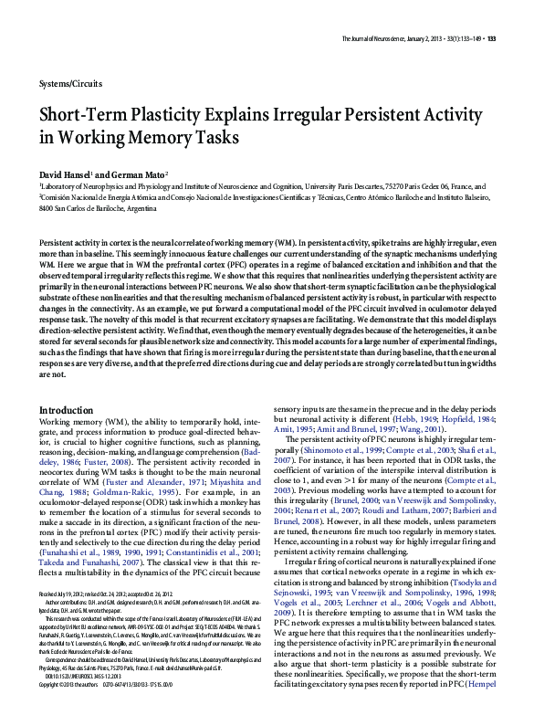
 David Hansel