Academia.edu no longer supports Internet Explorer.
To browse Academia.edu and the wider internet faster and more securely, please take a few seconds to upgrade your browser.
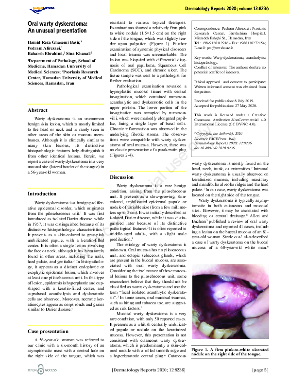
Oral warty dyskeratoma: An unusual presentation
 Pedram Alirezaei
Related Papers
JAAD Case Reports
Oral warty dyskeratoma of the retromolar trigone: An unusual presentation of a rare lesionAnil, S., V. T. Beena, et al. (1994). "Oral squamous cell carcinoma in a case of dyskeratosis congenita." Annals of dentistry 53(1): 15-18. Oral manifestations of dyskeratosis congenita (DCG) have received little attention in dental literature. This report is a case of dyskeratosis congenita in a 17-year-old female which was associated with oral lesions such as leukoplakia, superimposed candidal infection, desquamative gingivitis, and severe periodontitis. Histopathologic examination of the granular lesion on the right lateral border of the dorsum of the tongue showed well-differentiated squamous cell carcinoma. Etiopathogenesis, clinical features, and laboratory findings of this disease are discussed.
2008 •
Dermatology Reports
A case of post-inflammatory warty dyskeratoma of the chest: Other dermoscopic featuresWarty Dyskeratoma (WD) is a rare condition consisting in single or multiple papular or nodular lesions of the skin or of the oral mucosamucosa. Histologically, a cupshaped epidermal invagination centred by a plug of epidermal hyperparakeratosis with suprabasal acantholysis and dyskeratosis is typically observed. A case of post-inflammatory WD, which was also observed by dermoscopy, is described. Dermoscopy showed an eight-shape whitish collarette surrounded by light brown pigmentation. A central white structureless area with an adjacent rosette were observed. Some small rust-coloured blood crusts were also observed in the centre of the lesion; no prominent vascular pattern was detected. The etiopathogenesis of this benign neoplasm could be multifactorial. Dermoscopy of WD is not specific but may help to ruling out other skin tumors.
Oral Medicine & Pathology
Synchronous occurrence of oral squamous cell carcinoma and Warthin tumor2009 •
Annals of Applied Bio-Sciences
Tumour-like lesions of oral cavity: A clinicopathological study of 95 cases2017 •
Malaysian Family Physician
Overview of common oral lesionsThis article summarises common oral lesions that clinicians may face in everyday practice by categorising them by clinical presentation: ulcerated lesions, white or mixed white–red lesions, lumps and bumps, and pigmented lesions. The pathologies covered include recurrent aphthous stomatitis, herpes simplex virus, oral squamous cell carcinoma, geographic tongue, oral candidosis, oral lichen planus, pre-malignant disorders, pyogenic granuloma, mucocele and squamous cell papilloma, oral melanoma, hairy tongue and amalgam tattoo. The objective of this review is to improve clinician knowledge and confidence in assessing and managing common oral lesions presenting in the primary care setting.
RELATED PAPERS
Archaeological and Anthropological Sciences
Between cities and villages: the livestock economy in historical Palestine. Namdar, Gadot and Sapir-Hen 2024. Archaeological and Anthropological Sciences.2024 •
Healthy lifestyle of Ukrainians during war: Primary analysis of survey data
Здоровий спосіб життя українців під час війни: первинний аналіз даних опитування (стаття 2024)Spirale Arts Lettres Sciences Humaines
Le tour de l’Île. Séminaire La bête et le souverain, Volume II (2002-2003), de Jacques Derrida, Galilée, 407p.Séminaire La bête et le souverain, Volume II (2002-2003), de Jacques Derrida, Galilée, 407p2012 •
Diari de Tarragona
Les falles de la nit de Reis d'alguns pobles del Camp de Tarragona2024 •
IOP Conf. Series: Materials Science and Engineering
The Smart Parking System Using Ultrasonic Control Sensors2021 •
Catholic Religious Encyclopedia
THE RELIGIOUS LEGACY OF EDUCATING INDIA: (THE CONTRIBUTION OF CATHOLIC RELIGIOUS CONGREGATIONS IN THE FIELD OF EDUCATION IN INDIA: PAST, PRESENT AND THE FUTURE2020 •
2022 •
The Journal of neuroscience : the official journal of the Society for Neuroscience
Sphingosine-1-Phosphate (S1P) Impacts Presynaptic Functions by Regulating Synapsin I Localization in the Presynaptic Compartment2016 •
SHS Web of Conferences
L’approche phraséologique du roman médiéval : une voie de caractérisation générique ?2020 •
Biological Psychiatry
Distinct Altered Neurochemistry in the Hippocampus of Individuals With Bipolar Disorder and a Comorbid Anxiety Disorder: Evidence From 1H MRS2020 •
Research in arts and education
Forest Disputes: Socially Engaged Art and Forest Science for Understanding Sustainability Challenges2024 •
Journal of obstetrics and gynaecology of India
Laparoscopic specimen retrieval bags2014 •
Neuropsychopharmacology
Neural Correlates of Reward-Based Spatial Learning in Persons with Cocaine Dependence2013 •
2023 •
2019 •
RELATED TOPICS
- Find new research papers in:
- Physics
- Chemistry
- Biology
- Health Sciences
- Ecology
- Earth Sciences
- Cognitive Science
- Mathematics
- Computer Science