International Immunopharmacology 11 (2011) 331–341
Contents lists available at ScienceDirect
International Immunopharmacology
j o u r n a l h o m e p a g e : w w w. e l s ev i e r. c o m / l o c a t e / i n t i m p
Review
Immunomodulatory and therapeutic activity of curcumin
Raghvendra M. Srivastava a, Sarvjeet Singh b, Shiv K. Dubey c, Krishna Misra d, Ashok Khar e,⁎
a
Department of Otolaryngology, Hillman Cancer Centre, University of Pittsburgh Cancer Institute, Pittsburgh, PA 15213, USA
Department of Internal Medicine, Division of Cardiology, University of Texas Southwestern Medical Center, Dallas, Texas 75390, USA
c
Department of Internal Medicine, Division of Hematology-Oncology, University of Michigan Medical School, Ann Arbor, Michigan 48109, USA
d
Division of Bioinformatics, Indian Institute of Information Technology, Allahabad, India
e
CMBRC, Apollo Hospitals Educational and Research Foundation, Apollo Health City, Jubilee Hills, Hyderabad 500033, India
b
a r t i c l e
i n f o
Article history:
Received 1 July 2010
Accepted 22 August 2010
Available online 8 September 2010
Keywords:
Curcumin
Anti-inflammatory
Anti-cancerous
Immune and metabolic diseases
a b s t r a c t
Inflammation is a disease of vigorous uncontrolled activated immune responses. Overwhelming reports have
suggested that the modulation of immune responses by curcumin plays a dominant role in the treatment of
inflammation and metabolic diseases. Observations from both in-vitro and in-vivo studies have provided
strong evidence towards the therapeutic potential of curcumin. These studies have also identified a plethora
of biological targets and intricate mechanisms of action that characterize curcumin as a potent ‘drug’ for
numerous ailments. During inflammation the functional influence of lymphocytes and the related cross-talk
can be modulated by curcumin to achieve the desired immune status against diseases. This review describes
the regulation of immune responses by curcumin and effectiveness of curcumin in treatment of diseases of
diverse nature.
© 2010 Elsevier B.V. All rights reserved.
Contents
1.
2.
3.
4.
5.
6.
7.
8.
Introduction . . . . . . . . . . . . . . . . . . . . . . . . . . . . . . . . . . . . . . . . . . . . .
Immunomodulatory action of curcumin on T lymphocytes . . . . . . . . . . . . . . . . . . . . . . .
Immunoinhibitory action of curcumin on dendritic cells (DCs) . . . . . . . . . . . . . . . . . . . . .
Immunomodulatory effect of curcumin on natural killer (NK) cells . . . . . . . . . . . . . . . . . . .
Immunomodulatory effect of curcumin on monocytes and macrophages (Mϕ) . . . . . . . . . . . . . .
Immunomodulatory effect of Curcumin on B cells . . . . . . . . . . . . . . . . . . . . . . . . . . .
Immunomodulatory effect of curcumin on neutrophils and eosinophils and mast cells and its anti-oxidant
Curcumin in health and disease . . . . . . . . . . . . . . . . . . . . . . . . . . . . . . . . . . . .
8.1.
Role of curcumin in the neoplastic diseases . . . . . . . . . . . . . . . . . . . . . . . . . . .
8.2.
Curcumin in cardiovascular disease . . . . . . . . . . . . . . . . . . . . . . . . . . . . . . .
8.3.
Curcumin in neurodegenerative disease. . . . . . . . . . . . . . . . . . . . . . . . . . . . .
8.4.
Immunomodulatory action of curcumin in the prevention of inflammatory diseases . . . . . . . .
9.
Concluding remarks and future perspectives . . . . . . . . . . . . . . . . . . . . . . . . . . . . . .
References . . . . . . . . . . . . . . . . . . . . . . . . . . . . . . . . . . . . . . . . . . . . . . . .
. . . . .
. . . . .
. . . . .
. . . . .
. . . . .
. . . . .
properties
. . . . .
. . . . .
. . . . .
. . . . .
. . . . .
. . . . .
. . . . .
.
.
.
.
.
.
.
.
.
.
.
.
.
.
.
.
.
.
.
.
.
.
.
.
.
.
.
.
.
.
.
.
.
.
.
.
.
.
.
.
.
.
.
.
.
.
.
.
.
.
.
.
.
.
.
.
.
.
.
.
.
.
.
.
.
.
.
.
.
.
.
.
.
.
.
.
.
.
.
.
.
.
.
.
.
.
.
.
.
.
.
.
.
.
.
.
.
.
.
.
.
.
.
.
.
.
.
.
.
.
.
.
.
.
.
.
.
.
.
.
.
.
.
.
.
.
.
.
.
.
.
.
.
.
.
.
.
.
.
.
.
.
.
.
.
.
.
.
.
.
.
.
.
.
.
.
.
.
.
.
.
.
.
.
.
.
.
.
331
332
333
334
334
335
336
336
336
337
338
338
339
339
1. Introduction
Abbreviations: Ag, antigen; Ab, antibody; NO, nitric oxide; LPS, lipopolysaccharide;
ConA, concanavalin A; AP-1, activator protein 1; NF-κB, nuclear factor-kappaB; NF-AT,
nuclear factor of activated T cells; PMA, phorbol 12-myristate 13-acetate; PHA,
phytohaemagglutinin; ROS, reactive oxygen species; ROIs, reactive oxygen intermediates; COX-2, cyclooxygenase-2; APC, antigen presenting cells; DCs, dendritic cells; IDO,
indoleamine 2,3-dioxygenase.
⁎ Corresponding author.
E-mail address: ashok.khar@aherf.net (A. Khar).
1567-5769/$ – see front matter © 2010 Elsevier B.V. All rights reserved.
doi:10.1016/j.intimp.2010.08.014
Turmeric is a mixture of compounds related to curcumin known as
curcuminoids consisting of curcumin [i.e.diferuloylmethane or 1,7-bis
(4-hydroxy-3-methoxy-phenyl) hepta-1, 6-diene-3, 5-dione)] as the
major component, demethoxycurcumin, bisdemethoxycurcumin and
cyclocurcumin [1] (Fig. 1). Curcumin has been in use for its medicinal
benefits since centuries but the first documented case of its use as a
drug emerged only in 1937 when it was utilized to treat biliary
disease. Since then its therapeutic potential has been explored in
332
R.M. Srivastava et al. / International Immunopharmacology 11 (2011) 331–341
even minor fluctuations in the cellular redox milieu [10,11]. These
transcription factors in turn control cell cycle, differentiation, stress
response and other physiological processes [12–15]. The intricate
mechanism of action of curcumin involves various biological targets viz
transcription factors: NF-AT, AP-1, signal transducers and activator of
transcription (STAT), p53 and kinases: mitogen-activated protein
kinases, cytokines release, and the receptors found on different immune
cell type. These actions of curcumin greatly affect the innate and
adaptive arms of immunity, especially in the pathological conditions.
Curcumin effectively modulates the function of T cells, B cells, dendritic
cells (DCs), monocytes, macrophages (mφ) and neutrophils. Overwhelming reports have supported the anti-inflammatory action of
curcumin and its potential role in the therapy of numerous immune cell
related diseases. Although curcumin does not have a drug profile yet, the
safety and non-toxic effect of oral curcumin (12 g/day) which is much
higher than its regular in-take as food supplement have been
established by the drug governing agency [16]. Recently, the pre-clinical
and clinical studies that were conducted at different places have been
reviewed [17]. However, there are certain limitations concerning the
use of curcumin as a drug. Due to its insolublility in water, curcumin has
very poor bioavailability, its cellular uptake is slow and it gets
metabolized very fast once inside the cell. Therefore it requires
repetitive oral doses in order to achieve significant concentration inside
the cells for any physiological effects. To address these limitations a large
number of curcumin analogues have been prepared that have shown
improved uptake, metabolism and activity.
In this review we discuss the effect and applications of curcumin
across a spectrum of pathological conditions involving immune cells,
metabolic targets and diseases.
Fig. 1. Curcuminoids present in turmeric.
2. Immunomodulatory action of curcumin on T lymphocytes
inflammatory diseases, neoplastic disease, cardiovascular and neurodegenerative disease, diabetes, cystic fibrosis and other disorders. Due
to a vast number of biological targets and virtually no side effects,
curcumin has achieved the potential therapeutic interest to cure
immune related, metabolic diseases and cancer [2–7] (Table 1).
Majority of the studies suggested that the biological effects of
curcumin are mainly derived from its ability to either bind directly
to various proteins such as cyclooxygenase-2 (COX-2), lipoxygenase,
GSK3b and several other regulatory enzymes or by its ability to
modulate intracellular redox state [1,8,9]. Modulation of cellular
redox homeostasis exerts an indirect but more global effect on a
number of cellular processes, since several critical transcription
factors such as activator protein 1 (AP1), nuclear factor-kappaB (NFκB), nuclear factor of activated T cells (NF-AT), p53 etc. are sensitive to
Sikora et al. demonstrated that the mitogen concanavalin A (ConA)
stimulated and the spontaneous proliferation of rat thymocytes could
be inhibited by curcumin (50 μM) and similar anti-proliferative
effects of curcumin on ConA-stimulated Jurkat T cell line were also
reported. In contrast, the similar dose of curcumin could protect rat
thymocytes and Jurkat T cells from dexamethasone and ultra-violet
irradiation induced apoptosis, respectively. These bimodal effects of
curcumin were correlated with the suppressive effects of curcumin on
AP-1 transcription factor activation; however no effect of curcumin
was seen on AP-1 under normal conditions [18]. In contrast to the
study of Sikora et al., an independent study showed that curcumin
(50 μM) could induce cell death in the normal quiescent and
proliferating human lymphocytes through caspase-3 activation but
Table 1
The potential of curcumin was shown in the various diseases involving multiple mechanisms in the respective cell types.
Diseases
Cell types
Mechanisms of action
References
Alzheimer disease
Monocytic THP-1 cell line,
peripheral blood monocytes
Th-17 producing T cells, TLR4
and TLR 9 expressing T cells.
Anti-inflammtory: by blocking the amyloid peptide induced expression of TNF-α, IL-1β,
MCP-1, IL-8, MIP-1β and CCR5
Anti-inflammatory: blocking of EAE incidences by blocking IL-6; IL-21 signaling, and the
differentiation of Th-17 producing T cells, modulation of the function of TLR-4 and TLR-9 on T
cells, blocking of IL-12 signaling
Anti-inflammatory: decreased the frequency of eosinophils and the inflammatory cells by the
regulation iNOS, inhibition of the IgE and Ag-induced degranulation of mast cells.
Anti-inflammatory: suppressed ROIs generation, Blocks crystal induced neutrophil activation,
suppressed arthritis Ag-induced T cell proliferation
Suppressed pp38, suppressed pro-inflammatory IL-1β and enhanced IL-10 level
[59,61]
Multiple sclerosis
Allergy
Arthritis
Inflammatory bowel disease
Eosinophils, bronchoalveolar
inflammatory cells, mast cells
Neutrophils, T cells
Intestinal mucosal biopsies of
patients.
Psoriasis
Keratinocytes
Inflammatory cardiovascular Myocardial tissue, endothelial
disorders
cell line
Wound healing
Infilitrating Mϕs, keratinocytes,
fibroblasts
Inflammatory type II
Infiltrating Mϕs, adipose tissue,
diabetes
hepatic tissue
[31–33]
[75–77]
[34,70,71]
[151,152]
Blocks TNF-α mediated activation of cells
[153]
Blocks neutrophils activation. Attenuate the plasma level of IL-10, IL-8 and TNF-α. Blocks TNF-α [78,153]
induced pro-inflammatory responses in cell line
Anti-androgen receptor signaling activity, decreased local TNF-α level
[58]
Reduced Mϕs frequency in the adipose tissue, reduced expression of TNF-α, MCP-1 and reduced [7]
NF-κB activity in the hepatic tissue
R.M. Srivastava et al. / International Immunopharmacology 11 (2011) 331–341
without DNA degradation. This study also highlighted that curcumin
affects the viability of proliferating T cells much severely than
quiescent T cells [19,20]. Furthermore Deters et al. demonstrated
that curcumin (2.8–10 μM) can significantly abrogate the proliferation of peripheral blood mononuclear cells (PBMC) induced by OKT3
mAb (a human TCR/CD3 complex Ab) [21]. The concentration range of
curcumin used by Deters et al. was similar to the concentrations that
was shown to significantly affect human T cell proliferation induced
by various distinct stimuli viz. phorbol 12-myristate 13-acetate
(PMA), CD28, phytohaemagglutinin (PHA) [22]. Although direct
suppressive effects of curcumin on superantigen induced proliferation
of T cells were demonstrated in several studies as described before,
few studies also demonstrated that T cells fail to get appropriate
amount of co-stimulatory signals from curcumin-treated Ag presenting cells (APCs), as curcumin (20–30 μM) aborted the upregulation of
CD86 and CD83 in response to the APC maturation stimuli. This
inhibitory effect of curcumin on T cells was independent of the HLADR levels on the respective APCs as the HLA-DR level was not
downregulated by the curcumin. Curcumin, however, could also
reduce the DCs' dependent allogenic CD4+ T cell proliferation in a
mixed lymphocyte reaction assay at 1:16 ratio of DCs to T cells. In this
study a probable affect of curcumin on the cytoskeleal elements of DCs
was argued in this context, which may be attributed to its inhibitory
anti-proliferative effects [23]. Another study demonstrated a significant increase in ConA-stimulated proliferation of splenic cells at
6.25 μM curcumin and a significant decrease in proliferation at
12.5 μM and a complete blockage of proliferation with 25 μM
curcumin, which confirmed the distinct function of curcumin at
variable concentrations. Also, curcumin irreversibly inhibited the
induction of lymphoproliferation by other mitogens and alloantigens.
As the in-vivo effects of curcumin are highly dependent on the
bioavailable concentration of curcumin, it is indeed a daunting agenda
to correlate the in-vitro activities of curcumin and in-vivo responses in
the pathological conditions, especially in the localized pathological
conditions like non-metastatic tumors of different origins [24]. As
highlighted in the review so far, a variety of results indicated the invitro T cell immunosuppressive properties of curcumin in terms of T
cell death, as well as blocking the proliferation capacity of T cells.
Nevertheless curcumin has been in use since centuries and its
consumption has not been associated with any immunocompromised
disorders; seriously arguing the significance of its immunosuppressive properties on T cells that have been reported under in-vitro
conditions. We had demonstrated that T cells that were harvested
from the curcumin-injected (40 mg/kg/day; i.p) animals showed
enhanced lymphoproliferation and a similar proliferative effect of
curcumin was also observed when T cells were stimulated with ConA
and PHA in conjunction with curcumin. Our study also provided
evidence of specific lymphoproliferative effect of curcumin in-vivo, by
using cyclosporine A, a potent immunosuppressant drug. Interestingly, the enhanced Antigen (Ag)-specific T cell proliferation was also
observed in curcumin-injected rats that had received a highly
immunogenic AK-5 histiocytoma cells as a source of tumor Ag [25].
The evidence for the enhanced frequency of CD4+T cells was
furthermore reported in another spontaneously generated tumor
model of adenoma in C57BL/6J-Min/+ (Min/+) mouse that were fed
with 0.1% dietary curcumin. In a statistically controlled lymphocyte
infiltration setup, this study reported an enhanced number of CD8+,
CD4+ and CD3+ T cells in the curcumin fed animals. In this model the
spontaneous polyp formation in the mucosa was significantly reduced
by the curcumin administration and the anti-tumor mechanism was
correlated with the enhanced cytokine level due to the increased
number of activated CD4+ T cells, although no direct evidence for
such a conclusion was described in this study. Also, this study showed
enhanced level of B cells in the intestinal mucosa, but no role or
increase in the number of monocytes was found in this spontaneous
tumor model system [26]. In another elegant tumor model, in which
333
tumor growth could disintegrate the thymus morphology, curcumin
(50 mg/kg body weight) restored the thymic integrity including CD3+
T cell frequency and served as immunoprotective compound during
carcinogenesis. This effect of curcumin was attributed mechanistically
to the anti-oxidant properties of curcumin because this tumor
induced oxidative stress in thymic T cells [27]. As an extension of
this work the same group demonstrated that curcumin could prevent
the tumor induced apoptosis of thymocytes as well as restoration of
the frequency of CD4+ T/CD8+ T cells in the same tumor model
system with the same dose of curcumin. Mechanistically it was also
shown that curcumin modulated Jak-3/Stat-5 activity to restore the
immune cell frequency and activity [28,29]. Curcumin decreased IL12-induced STAT4 phosphorylation but enhanced the (interferon)
IFN-β-induced STAT4 phosphorylation; curcumin decreased IL-12
induced IFN-γ production and IL-12 Rβ1 and β2 expression, whereas
it enhanced IL-10 production and IFN receptor (IFNAR) subunits 1 and
2 expression. Curcumin also increased IFN-α-induced IL-10 and
IFNAR1 expression. Pretreatment with curcumin decreased IFN-αinduced IFNAR2 expression and failed to modify the level of IFN-αinduced phospho-STAT4 activation. These findings favour the distinct
mode of action of curcumin when T cells get activated with different
stimuli and also confirmed the multifarious targets of curcumin in the
activated T cells [30]. It was recently acknowledged that IL-17
producing Th1 cells play an instrumental role in the established
model of experimental autoimmune encephalomyelitis (EAE), which
mimics multiple sclerosis. Oral curcumin (100 or 200 mg/kg body
weight) administration in the rats suppressed the frequency of
inflammatory cells in the spinal cord along with lowering the
frequency of paralytic incidences, which was the disease marker in
this model. The decreased level of IL-17, transforming growth factor
beta (TGF-β), IL-6, IL-21, STAT3 expression and STAT3-phosphorylation was reported in curcumin-treated groups. Also, it was shown that
curcumin blocks the differentiation of Th-17 cells by blocking STAT-3
transcription in T cells. Moreover curcumin inhibited neural AgMBP68–86 peptide specific lymphocytes responses and IL-17 mRNA
expression. These recent evidences furthermore proved the significance of curcumin in the IL-17 mediated disorders [31]. Bright and
colleague reported that T cells expressing Toll-like receptors-4 and 9
(TLR4 and TLR9) play an instrumental role in the pathogenesis of EAE
model. Curcumin treatment led to the decrease in the expression of
PLPp139–151 and MOGp35–55 Ag-induced TLR4 and TLR9 on the
CD4+ T cells and CD8+ T cells, which also ameliorated this disease. It
was found that TLR 4 and TLR9 acted as co-stimulatory receptors to
enhance the proliferation and cytokine production in response to the
specific agonists [32]. Previously Bright's group had also reported
that curcumin can inhibit IL-12 production in spleen cells, Mϕ and
microglia and curcumin can inhibit EAE by blocking IL-12 signaling in
T lymphocytes [33]. Curcumin also inhibited the proliferation of
mouse splenic T cells that were stimulated with ConA and in a model
of type II collagen (CII)-induced arthritis (CIA) in which T cell
proliferation was induced by bovine type II collagen Ag. Moreover,
curcumin reduced anti-CII IgG2a Ab in the serum of CIA mouse [34].
3. Immunoinhibitory action of curcumin on dendritic cells (DCs)
Being at the centre of various immunological responses, DCs
control various pathogenic conditions and recently several groups
have investigated the action of curcumin on DCs' function. In a
detailed study Kim et al. reported for the first time that curcumin, at a
dose of up to 25 μM, inhibits DC maturation and the related
immunostimulatory function. They also showed that more than
50 μM concentration was toxic for DCs. Surprisingly however various
studies have used 50 μM concentration in different immune cells as
described elsewhere in this review and the discrepancy between the
uses of different concentration of curcumin reflects the variable dose
sensitivities of different immune cells and cell lines to curcumin.
334
R.M. Srivastava et al. / International Immunopharmacology 11 (2011) 331–341
However, it remains to be investigated if curcumin dose sensitivity of
immature and mature DCs results in distinct biological outcomes. Kim
et al. also showed that curcumin could suppress the lipopolysaccaride
(LPS) mediated surface overexpression of CD86, CD80 and MHCII
expression in murine DCs during maturation but at the same time
curcumin treatment increased the FITC-dextran particle uptake
significantly. These observations provided the evidence to modulate
DC mediated specific immune response in the autoimmune disorders
for regulating the function of T cells [35]. Park and colleagues reported
that the pretreatment with curcumin (1–25 μM) could also suppress
the LPS (200 ng/ml) induced indoleamine 2,3-dioxygenase (IDO)
production in bone marrow derived-DCs (BMDCs). However, curcumin enhanced the COX-2 expression by three fold and prostaglandin
E2 production by two fold in the LPS treated DCs. The enhanced
prostaglandin E2 level by curcumin was attributed to the suppression
of LPS-induced IDO production in this study. The curcumin or
prostaglandin E2 treated DCs showed reduced proliferation of OVAspecific CD8+ T cells that was induced by LPS [36]. Although
intravenous LPS (3 μg) injection reduces the splenic blood flow by
31% and reduces the access of Ag to the mouse spleen [37], a high dose
of LPS (1.5 mg/kg body weight, i.p.) induced heavy IDO in splenic DCs
and pre-injection of curcumin (50 mg/kg body weight, i.p.) inhibited
the LPS-induced IDO production in the splenic DCs [36]. In contrast to
the effect of curcumin on BMDCs showing enhanced COX-2
expression, another study showed the dose dependent (2–16 μM)
inhibiton of the production of LPS (0.2 ng/ml) induced COX-2 in the
BV2 microglial cells. This contrasting results on the COX-2 level in the
BV2 microglial cells and BMDCs may be due to a very high difference
in the LPS concentration (0.2 vs 200 ng/ml) or LPS serotype that were
used in these studies. Moreover, a distinct cellular response to LPS
could not be ruled out in different cell types that may greatly differ in
the density of LPS receptor or co-receptors [38]. IFN-γ regulates
multiple elements of DCs response and since it is being used for
monocyte derived-DCs (MoDCs) conditioning in cancer therapy, it can
drive DCs for potent Th1 polarizing activities. Although IFN-γ (5 to
500 IU/ml) treatment was shown to upregulate CD86, CD38, CCR7 on
MoDCs in 48 h [39], no significant increase in CD86 and CD80 level
was found at 24 h after 200 IU/ml of IFN-γ treatment in murine
BMDCs. However, IFN-γ (100 IU/ml) upregulated IDO production in
BMDCs and curcumin (1–25 μM) inhibited the functions as well as
level of IDO in IFN-γ stimulated murine BMDCs. Thus curcumin
reversed the IDO-mediated reduced T cell proliferation function. This
study also showed that curcumin can modulate IFN-γ induced IDO
expression by affecting Janus kinase 1 (JAK) and protein kinase C δ
(PKC) signaling [40]. In a similar direction, additional data showed
that curcumin-treated DCs led to the development of anergic CD4+T
cells and curcumin-treated DCs could also induce regulatory T cells
(Tregs) development. More interestingly, curcumin-treated DCs also
promoted the production of IL-10 and αAlDHAa1 (α retinal
dehydrogensae). These retinoids function as the regulators of mucosal
immune responses. Curcumin induced Treg cells inhibited Ag-specific
T cell activation in-vitro and could inhibit colitis caused by Ag-specific
pathogenic T cells in-vivo. These findings supported the important
role of curcumin in the modulation of DCs' function to achieve
tolerogenic responses [41]. Curcumin (1 μM) itself could suppress the
LPS-induced IL-12/23p40 production in-vitro, whereas IL-10 (2.5 ng/
ml) and curcumin (0.1 μM) at their suboptimal concentrations acted
synergistically to suppress the LPS-induced IL-12/23p40 production
from DCs. However, no appreciable therapeutic effect of the dietery
curcumin was noticed in the IL10-deficient mice having Th1 mediated
colitis. Also, no significant improvement in the colitis was observed as
curcumin failed to modify the pathogenic T cells in IL-10 deficient
mouse. These results comprehensively confirmed the dependence of
curcumin on IL-10 for its immunoinhibitory actions. Nevertheless it
provided the evidence that a very little bioavailable concentration of
curcumin might effectively modify the overall immune response in
combination with IL-10. Curcumin and IL-10 also acted synergistically
to inhibit the NF-κB activity in intestinal epithelial cells and thus could
provide the additional benefits without influencing the function of
immune cells. These results also provoke the possibility of a
combinatorial approach to investigate the immunohibitory action of
curcumin with other immunohibitory molecules viz. TGF-β, prostaglandins etc. [42]. Such investigations will be valuable to specify the
action of curcumin in various pathogenic conditions in future.
4. Immunomodulatory effect of curcumin on natural killer
(NK) cells
NK cells directly participate in the killing of tumor cells after the
recognition of stress inducible ligands and killing involves the
induction of cell death by perforin and granzyme B. Various
investigators have directly measured the NK cell activity against
tumor cells both, in-vitro and in-vivo. In the initial studies, curcumin
feeding (1, 20 or 40 mg/kg) up to five weeks showed no effect on the
NK cell activity in rats but enhanced the antibody (Ab) responses in
rats [43]. In another study, Yadav et al. showed that curcumin
treatment can augment NK cell cytotoxicity in-vitro that can further
be enhanced by IFN-γ treatment [44]. The generation of IL-2 induced
non-specific cytotoxic LAK cells (similar to cytotoxic NK cells) in the
presence of curcumin (at 10–20 μM/l) was evaluated by Gau et al. and
the cytotoxic activity of LAK cells was determined against NK sensitive
YAC-1 lymphoma cells. The results showed little effect on the
generation of LAK cell-mediated cytotoxicity, whereas higher dose
(30 μM/l) of curcumin inhibited the cytotoxic LAK cell generation [24].
Few serious concerns for the use of curcumin in the melanoma
treatment were however raised, which were based on the facts that
NK cells from healthy donors treated with curcumin (10 or 20 μM/l)
and IL-12 (10 or 50 ng/ml) secreted less amount of IFN-γ. Moreover,
curcumin-treated NK cells also showed reduced granzyme B to kill
K562 and A375 melanoma cell lines and curcumin slightly reduced
production of IFN-γ by NK cells in the presence of A375 melanoma
and K562 target cell lines. Although this study found the direct effect
of curcumin on tumor cells it could not provide appreciable reasons
for the use of curcumin as the ‘modifiers of NK mediated immune
responses’ in favour of its use in anti-tumor therapies [45]. Similarly,
our previous study had shown that curcumin injections for prolonged
duration had no effect on the NK cell activity in-vivo, during the
progression of ascites tumor [25]. However, we observed larger solid
tumor with curcumin in the transplanted subcutaneous AK-5 tumor
that interestingly underwent rapid spontaneous regression. Also, an
enhanced activation of NK cells was observed after curcumin
treatment that correlated with the response of curcumin on the
tumor in-vivo [46]. Such an effect clearly describes the effect of
curcumin as ‘NK cells modifier’, however its in-vivo effects may be
highly dependent on the specific pathology of the diseases. Recently,
tumor derived exosomes attracted much attention as they can
effectively modulate the anti-tumor immune responses. Zhang et al.
showed that curcumin enhanced the proteasomal degradation of
tumor derived exosomal proteins that inhibit IL2-induced NK cell
activity against breast carcinoma, partially restoring the NK cell
activity against tumor. Such an action of curcumin displayed that
curcumin can also target the immune escape strategies that are
critical for the immune responses [47].
5. Immunomodulatory effect of curcumin on monocytes and
macrophages (Mϕ)
Monocyte recruitment at the inflammatory site plays a vital role in
the inflammatory response. Curcumin inhibited the tumor necrosis
factor α (TNF-α) induced adhesion of monocytes on human
endothelial cells. The TNF-α induced upregulation of Inter-Cellular
Adhesion Molecule 1 (ICAM-1), vascular cell adhesion molecule-1
R.M. Srivastava et al. / International Immunopharmacology 11 (2011) 331–341
(VCAM-1) and endothelial cell leukocyte adhesion molecule-1
(ELAM-1) on monocytes was completely inhibited by curcumin. The
curcumin mediated blocking of these adhesion molecules was
attributed to the inhibitory effect on NF-κB activation. These results
showed the promising activities of curcumin in the local inflammatory
responses like arthritis as well as in metastasis [48]. PMA or LPSinduced production of inflammatory cytokines viz. TNF-α, IL-8,
macrophage inflammatory protein 1 alpha (MIP-1α), monocyte
chemoattractant protein (MCP-1) and IL-1β in monocytes and
alveolar Mϕs was significantly inhibited by curcumin in a dose
dependent manner [49]. Lim et al. also showed that curcumin blocked
the enhanced expression and secretion of PMA induced inflammatory
cytokine MCP-1 in U937 monocytic cell line [50]. Although curcumin
completely blocked LPS mediated NO production in RAW264.7 cell
line, it enhanced the phagocytosis of fluorescent beads and the surface
expression of CD14. The enhancement of pahgocytic activity and the
surface expression of CD14 followed a similar pattern, however no
direct role of curcumin mediated enhanced CD14 surface expression
was described for the phagocytic capcity [51]. Previously, an
independent study had also shown a significant increase in Mϕ
phagocytic activity in curcumin-treated animals. These actions of
curcumin on Mϕs also described the enhanced scavenging capacity
under non-inflammatory conditions [52]. Prolonged alcohol treatment led to oxidative stress and the mononuclear cells obtained from
alcoholic animals showed lesser capacity for collagen surface
attachment; however alcohol in conjunction with curcumin showed
normal adhesion potential of mononuclear cells. This study showed
the reduction in the toxic effect of alcohol by curcumin in the
prolonged duration [53]. Pretreatment with curcumin inhibited the
LPS-induced TLR-2 mRNA and NF-κB level in RAW264.7 cells [54].
Treatment with bisdemethoxycurcumin inhibited the LPS-induced
NO production in RAW264.7 cells, which was abrogated by blocking
the activity or the expression level of heme oxygenase-1. It was also
shown that anti-inflammatory effects mediated by bisdemethoxycurcumin signaling to heme oxygenase-1 involve **Ca2+/calmodulinCaMKII-ERK1/2-Nrf2 cascade in RAW264.7 Mϕ cells [55]. Sumanont et
al. have furthermore reported that curcumin manganese complex
(CpCpx) and diacetylcurcumin manganese complex (AcylCpCpx)
have greater NO radical scavenging activity than their parent
compounds, curcumin and acetylcurcumin, respectively [56].
In diabetic condition, a massive increase in the inflammatory
cytokines has been reported. To evaluate the anti-inflammatory effect
of curcumin under high glucose mediated inflammatory responses,
Jain et al. studied the effect of curcumin and placebo supplementation
on plasma level of TNF-α, IL-6, MCP-1, glucose and oxidative stress in
streptozotocin-treated diabetic rats [57]. Curcumin treatment significantly reduced the high glucose mediated upregulation of inflammatory cytokines along with increasing the lipid peroxidation.
However curcumin had no effect on the reduced insulin level under
diabetic conditions. In this study, the anti-inflammatory action of
curcumin on the high glucose induced IL-8, TNF-α, IL-6, MCP-1 was
also shown by using human promonocytic U937 cell line.
Androgen receptor is a nuclear receptor that translocates to the
nucleus following ligand binding and modulates the function of
various genes. In the healing skin androgen receptor were detected in
infilitrating Mϕs, keratinocytes and in dermal fibroblasts that
indicated its possible function in the healing process. Androgen
receptor activity in the presence of 5α-dihydrotestosterone induced
TNF-α promoter activity in Mϕs. The curcumin derivative ASC-J9
disrupts the androgen receptor and its co-regulator interaction
resulting in the increased androgen receptor degradation and the
decreased androgen receptor transactivation. The topical application
of ASC-J9 cream in mouse resulted in quick wound healing and also
decreased local TNF-α expression. This study concluded that the
curcumin derivative ASC-J9, which acts by inhibiting androgen
receptor activity, could be utilized in wound healing as an anti-
335
inflammatory agent [58]. Alzheimer's disease is a complex disorder
mainly characterized by deposition of large amount of amyloid-β (Aβ)
peptide and subsequent massive inflammatory response. Heavy
infilitration of monocytes and Mϕ has been observed in the affected
tissue with Aβ deposition. Giri et al. demonstrated that both Aβ1–40
and fibrilar Aβ1–42 peptide are abundantly present in the plasma of
patients along with the increase in the level of cytokines TNF-α and
IL-1β and chemokines MCP-1, IL-8 and MIP-1β. Activation of
transcription factors AP-1 and EGR-1 regulates the level of cytokines
and chemokines in THP-1 monocytic cells and in peripheral blood
monocytes [59]. Based on the ability of curcumin to block inflammation as well as to modulate the activities of β-secretase and
acetylcholinesterase, in-vitro and in-vivo studies with curcumin led
to suppressed Aβ deposition and aggregation in experimental animals
[60]. In the evaluation study on the role of curcumin in Alzheimer
disorder Giri et al. furthermore showed that curcumin could block the
Aβ1–40-induced expression of TNF-α, IL-1β, MCP-1, IL-8, MIP-1β and
CCR5. Also, it was reported that curcumin could inhibit Aβ-induced
Egr-1 DNA-binding activity. These results provided the mechanism of
the anti-inflammatory action of curcumin in this disease [61].
6. Immunomodulatory effect of Curcumin on B cells
Decoté-Ricardo et al. evaluated the effects of curcumin on murine
spelnic B cells. LPS-induced IgM secretion as well as CpG and TLR4induced proliferation of B cells was inhibited following curcumin
treatment. However curcumin failed to exert anti-proliferative effect
when the B cell prolifearion was induced by the T-independent type 2
stimuli anti-delta-dextran or by the anti-IgM Ab. Moreover curcumin
(10 μM) had no effect on the calcium mobilization induced by antiIgM (10 μg/ml) Ab. Interestingly, however, curcumin inhibited the
TLR ligand and anti-IgM induced phosphorylation of ERK, Iκ-B and
p38 kinase along with inhibiting NF-κB activation. These observations
indicated the anti-inflammatory effects of curcumin in the B cell
response [62]. Another study described that the mitogen-LPS-induced
proliferation of B cells can be dose dependently inhibited by curcumin
(1–20 μM); additionally the LPS-induced secretion of IgG1 and IgG2a
was inhibited by curcumin. However, the curcumin mediated
inhibition of IgG1 secretion was more pronounced than the inhibition
of IgG2a secretion [63]. An independent study also described that
curcumin (10 μM) can also inhibits the production of IgE from rat
splenocytes [64]. Epstein barr virus (EBV) can immortalize human B
lymphocytes in-vitro and immortalization is promoted by the
oxidative stress induced by potent immunosuppressive drug cyclosporine A and with hydrogen peroxide. Curcumin (20 μM) aborted the
EBV induced B cell immortalization process. This effect of curcumin
may be exploited to prevent post-transplant lymphoproliferative
disorders in patient receiving cyclosporine A, which otherwise may
promote EBV induced B cell immortalization [65]. Later on, it was
found that the curcumin modulates this immortalization process by
enhanced apoptosis in the virus infected B cells [66]. In animals with
spontaneous polyps in the intestinal mucosa, curcumin treatment
resulted in 40% increase in B cell numbers in the intestinal mucosa,
suggesting the therapeutic responses to curcumin [26].
B cell receptor (BCR) signaling regulates the induction of apoptosis
in chronic lymphocytic lymphoma. The central mediator of BCRsignaling is the spleen tyrosine kinases, that govern the function and
survival of B cells, and a high level of phosphorylated spleen tyrosine
kinase was found in lymphoma cells in comparison to healthy B cells
[67]. Curcumin differentially modulated the cytotoxicity of primary
chronic lymphocytic lymphoma in comparison to healthy B cells [67].
Rats that received 1, 20 or 40 mg/kg curcumin for 5 weeks showed
significantly enhanced IgG only at 40 mg/kg levels whereas animals
receiving lower dietary concentrations (1 or 20 mg/kg) of curcumin
had same IgG level as that of control with no dietary curcumin. These
observations suggest that a threshold level of bioavailable curcumin is
336
R.M. Srivastava et al. / International Immunopharmacology 11 (2011) 331–341
also needed to modulate the IgG mediated responses [43]. A recent
study showed that curcumin (6 to 50 μM) could suppress the
expression of division dependent upregulation of activation-induced
cytosine deaminase, which plays pivotal role in the Ig class switch
recombination and somatic hyper-mutation and participates in
tumorigenesis. Also the decrease in the recovery of IgG + classswitched B cells within the divided population was observed. These
observations suggest the potential of curcumin in the treatment of B
cell autoimmune disease [68].
7. Immunomodulatory effect of curcumin on neutrophils and
eosinophils and mast cells and its anti-oxidant properties
Several independent studies have provided the evidence that
curcumin can act on various aspects of neutrophil function, in a
stimulus specific manner and may thus dampen the neutrophil
mediated inflammatory response [69]. Chemotactic peptide Nformyl-methionyl-leucyl-phenylalanine (FMLP) and zymosan activated plasma induced aggregation of the monkey neutrophils could
be inhibited by the curcumin (1 mM). FMLP peptide, zymosan and
arachidonic acid induced production of oxygen radical was attenuated
by the curcumin treatment. Calcium ionophore A23187 could nullify
the curcumin effect by interfering with the effect of curcumin in
neutrophils [69]. Neutrophils play significant role in the damage of
joint tissue in the rheumatoid arthritis. A recent study demonstrated
the reduced level of oxygen radical generation by neutrohphils upon
treatment with curcumin both in-vitro and in-vivo. Adjuvant induced
arthritis enhanced the neutrophil frequency in the blood that remain
unaltered by curcumin. The stimulation of neutrophils by PMA led to
increased level of PKC isozymes, α and β II, which was abrogated by
curcumin treatment without interfering with neutrophils vital
functions [70]. Similarly, the crystal induced neutrophil activation
that served as a model of induced arthritis or rheumatoid arthritis
condition was inhibited by curcumin [71]. Oral administration of
curcumin (40–60 mg/kg body weight) increased survival of mice by
70% in response to heavy dose of LPS (40 mg/kg body weight).
Moreover curcumin suppressed the LPS mediated neutrophil infiltration in liver that was the primary cause of liver damage. However, the
reduction of infiltration was limited to the liver only, because whereas
hepatic venules had same frequency of neutrophils as that of without
curcumin. The reduction of LPS-induced infiltration of neutrophils
was also correlated with the reduced levels of ICAM-1 and VCAM-1 in
the liver tissue that influence neutrophil adhesion [72]. Without
affecting the viability, curcumin (100 μM) significantly reduced the IL8 induced chemotactic activity of neutrophils in dose dependent
manner and curcumin modulated this chemotaxis by dampening the
IL-8 induced Ca++ ion mobilization. Surface CXCR1 and CXCR2 were
internalized upon IL-8 treatment and curcumin treatment enhanced
the intracellular level of CXCR1 and CXCR2 in conjunction with IL-8,
which indicated that the effect of curcumin on the reduced migration
of neutrophils might be attributed to the reduced IL-8 receptors.
However, curcumin itself downregulated surface IL-8 receptor CXCR1
and CXCR2 and also blocked the recycling of these receptors on
neutrophils. The Rab GTPase family (Ras superfamily of monomeric G
proteins) plays pivotal role in the cellular transport mechanism.
Interestingly, both CXCR1 and CXCR2 showed enhanced binding with
Rab11 upon curcumin treatment, which could potentially block the
recovery of IL-8 to cell surface. This study revealed the intricate
mechanism that curcumin triggers to achieve anti-inflammatory
responses meadiated by neutrophils [73]. LPS induced lung damage
and reduction in lung and bronchoalveolar lavage fluid protein
content, which was accompanied by enhanced numbers of neutrophils and elevated myeloperoxidase activity in cell-free lavage.
Elevation in the cytokine-induced neutrophil chemoattractant-I
protein level was seen in response to LPS in the lung tissue, which
was significantly reduced by the pretreatment with curcumin. This
shows an important protective response of curcumin by dampening
neutrophil function in lung injury [74].
In a murine model of asthama, which was induced by OVA-Ag and
which had airway hyper-responsiveness to allergens, curcumin (i.p,
10 or 20 mg/kg body weight) decreased the frequency of eosinophils
and the inflammatory cells, inhibited iNOS (inducible nitric oxide
synthase) expression in lungs and also suppressed the level of IL-4
and IL-5 in bronchoalveolar lavage fluid [75]. An interesting study
involving the action of curcumin on mast cells indicated that
curcumin reversibly inhibits the degranulation of mast cells along
with inhibiting secretion of IL-4 and TNF-α. The evaluation of the antiallergic affect of curcumin was performed by utilizing passive
cutaneous anaphylaxis in the mouse ear model. Oral administration
of curcumin (50 mg/kg) suppressed the mast cell dependent IgE and
Ag-induced local passive cuataneous anaphylaxis [76,77]. Effect of
curcumin during myocardial ischemia/reperfusion injury with cardioplegia was also investigated [78]. The postoperative increase in the
IL-8, IL-10, TNF-α levels in the plasma was decreased by curcumin.
Also curcumin inhibited the activation of neutrophils in myocardium
that was estimated by the myloperoxidase activity assay [78].
8. Curcumin in health and disease
Due to the fact that curcumin has been shown to be associated
with a number of physiological processes and that it has a wide
variety of cellular targets, its therapeutic role has been studied in
several inflammatory and non-inflammatory disorders. In this section,
we discuss most recent findings related to its direct application in
health and disease.
8.1. Role of curcumin in the neoplastic diseases
Curcumin has received maximum attention owing to its antitumor properties. Several hundred reports in the last two decades
have shown its ability to selectively kill transformed cells across
almost all types of tumors. Curcumin can exert its anti-tumor effects
at two levels, (i) at the level of tumorigenesis or (ii) in selectively
inducing apoptosis in tumor cells. Huang et al. have discussed the
anti-carcinogenic effects of curcumin in duodenal and colon cancer in
mice. In this study, dietary curcumin could significantly reduce tumor
load during both pre initiation and post initiation of chemical induced
carcinogenesis [79]. Similarly, curcumin application inhibited the
induction of epidermal DNA synthesis and the tumor promotion in
skin following 12-0-tetradecanoyl phorbol-13-acetate (TPA) treatment [80] as well as benzopyrene induced DNA adducts and skin
tumors and DMBA induced skin tumors [79]. Rao et al. have shown
that curcumin (200 ppm) in the diet could significantly suppress
azoxymethane-induced colonic aberrant crypt foci formation, which
are early preneoplastic lesions, and colon tumor incidence and tumor
multiplicity [81]. These effects of curcumin in inhibiting tumorigenesis involve inhibition of arachidonic acid metabolism; decrease in
TPA induced ornithine decarboxylase activity and inhibition of DNA
synthesis. It was thought that metabolites of arachidonic acid such as
HPETEs, HETEs, leukotrienes and prostaglandins play an important
role in TPA induced inflammation and tumor promotion [82,83].
Similarly ornithine decarboxylase (ODC) is a rate limiting enzyme in
polyamine synthesis [81] and its overexpression has been linked with
cell transformation and carcinogenesis in skin, breast and colon. Thus,
it is logical to speculate that inhibiting arachidonic acid metabolism
and/or ODC activity shall result in an inhibition of tumorigenesis. In
addition, curcumin has also been shown to cleave β-catenin, which
impairs Wnt signaling and cell–cell adhesion pathways, which are
critical in the development and promotion of many types of tumors
including colorectal cancer [84]. Curcumin also induced downregulation of cyclin D1 expression and CDK-4 activity in breast and
squamous cell carcinoma cell lines [85]. The suppression of cyclin
R.M. Srivastava et al. / International Immunopharmacology 11 (2011) 331–341
D1 by curcumin led to inhibition of CDK-4 mediated phosphorylation
of retinoblastoma, which is a crucial step for the cell to pass through
the G1 phase of cell cycle and become transformed [86].
In addition to its inhibitory effects on neoplastic transformation,
curcumin has been shown to induce apoptosis in tumor cells by
various mechanisms, which include impairment of the ubiquitin
proteasome pathway, upregulation of proto-oncoprotein Bax, activation of caspases and induction of Fas receptor aggregation in a Fas
ligand dependent manner and the generation of free radicals [87–90].
One of the several possible mechanisms of apoptosis induction by
curcumin involves the inhibition of proteasome complex. In mouse
neuro 2a cells, exposure to curcumin revealed a dose dependent
decrease in the proteasome activity and an increase in the
ubiquitinated proteins. Curcumin also decreased the turnover of the
destabilized enhanced green fluorescent protein suggesting an
inhibition of the cellular proteasome machinery [90]. Another mode
of apoptosis induction by curcumin involves the upregulation of p53
in tumor cells. In human basal cell carcinoma, apoptosis induction by
curcumin resulted in induction of p53 and its downstream targets,
p21 waf1/cip1 and GADD45, which are known to regulate apoptosis
under stress conditions [91]. Work in our laboratory has shown that
curcumin induced apoptosis involves the production of reactive
oxygen intermediates (ROIs) and involves activation of caspase-3
[87]. A large number of reports confirm that curcumin induced typical
apoptotic mode of cell death in a wide variety of tumors complete
with mitochondrial depolarization and caspase-3 activation. However, some studies suggest that apoptosis induced by curcumin is
independent of caspase-3 [92,93]. Curcumin has also been reported to
induce an apoptosis like pathway, which is independent of not only
caspases but mitochondria as well [93]. These effects of curcumin in
Jurkat T cells were accompanied by DNA fragmentation into high but
not low molecular weight fragments and the frequency of opening of
the mitochondrial permeability transition pores in curcumin-treated
cells was decreased compared to the control untreated cells. However,
one of the most commonly shown effects of curcumin on tumors is its
ability to induce the opening of mitochondrial permeability transition
pore, which in turn induces the collapse of the mitochondrial
membrane potential, respiration impairment ultimately leading to
cell death [94,95]. This observed difference in curcumin's action with
respect to the opening of permeability transition pore could be
attributed to the large difference in its concentrations that were used
during these studies.
8.2. Curcumin in cardiovascular disease
The therapeutic effects of curcumin in the development and
progression of cardiovascular disease have been studied to some
depth in the last decade. Owing to its ability to regulate oxidant stress,
curcumin has been shown to be effective against cardiac hypertrophy,
cardiomyocyte apoptosis following myocardial infarction and ischemia/reperfusion injury [96–98]. Cardiac hypertrophy is the remodeling of the left ventricle following pressure or volume overload that
results in ventricular wall thickening and an increase in overall
cardiac dimensions. It begins as a compensatory process that becomes
maladaptive over time and leads to heart failure [99,100]. Development of hypertrophy involves activation of the calcium and redox
sensitive transcription factor NF-AT that brings about the metabolic
and biochemical changes within the cardiomyocyte [101]. Transcriptional activation associated with hypertrophy has been recently
shown to be regulated by acetylation and deacetylation events at
histone lysine tails [102]. Acetylation and deacetylation of histones is
carried out by enzymes called histone acetyl transferases (HATs) and
histone deacetylases (HDACs), respectively. Various HDACs have been
implicated in the pathogenesis of cardiac hypertrophy. For example,
loss of class 2 HDAC results in development of hypertrophy while loss
of class-1 HDAC confers resistance to hypertrophic growth [102,103].
337
Morimoto et al. studied the effects of curcumin on HAT and
progression of hypertrophy and subsequent decompensated heart
failure. They have shown that exposure of isolated neonatal rat
cardiomyocytes (NRCMs) to 5 or 10 μM curcumin completely
suppressed the induction of hypertrophic response following phenylephrine treatment, a known inducer of cardiac hypertrophy [97]. In
an in-vivo setting also, administration of curcumin prevented the
development of hypertension induced heart failure in salt sensitive
Dahl rat model of hypertension [97]. These data strongly suggest that
curcumin possesses anti-hypertrophic properties both in-vitro and invivo. It has been proposed that curcumin may inhibit hypertrophic
remodeling by two mechanisms (i) by inhibition of histone
acetylation through inhibition of HATs and (ii) by disrupting p300/
GATA4 transcriptional complex through a completely independent
mechanism. Curcumin has also been shown to inhibit p300 mediated
acetylation of p53, both in-vitro as well as in-vivo [104]. Similarly,
inhibition of NF-κB by curcumin could also be involved in the antihypertrophic effects of curcumin since NF-κB signaling is involved in
cardiomyocyte hypertrophy [105].
Oxidative stress is a major outcome determinant in myocardial
infarction and ischemia/reperfusion and curcumin's anti-oxidant
property has been shown to prevent isoproterenol induced myocardial necrosis in rats [106]. In models of experimentally induced
myocardial infarctions such as isoproterenol treatment, decrease in
lysosomal stability leading to increase in lysosomal autolytic enzymes
has been reported [107,108]. Curcumin has been shown to stabilize
membranes and thereby suppress the infarct induced increase in
myocardial lysosomal enzymes [109,110]. Besides, the generation of
free radicals following ischemia/reperfusion can also be controlled by
curcumin due to its strong anti-oxidant properties.
The cardiotoxicity associated with doxorubicin, a potent drug for
treatment of a broad array of cancers, is a major concern for cancer
patients [111]. Animal studies have shown that doxorubicin treatment
induces free radical generation and p53 activation, decreases glutathione and increases serum peroxidase and catalase [96,112]. Curcumin
treatment significantly attenuated the cardiotoxic effects of doxorubicin
[113]. The beneficial effect of curcumin in blockade of doxorubicin
cardiotoxicity can be linked to modulation of intracellular redox status
by curcumin. In a study by Feng et al., curcumin completely abrogated
the induction of glucose induced hypertrophy in cardiomyocytes [114].
Glucose induced cardiomyocyte hypertrophy is mediated by p300
upregulation and subsequent activation of p300 dependent transcription factors. Since, curcumin can inhibit p300, exposure to curcumin
prevented the induction of p300 mediated hypertrophic response in
cardiomyocytes [114]. In human patients curcumin could markedly
reduce the generation of glucose induced reactive oxygen species (ROS)
in red blood cells. Myocardial tissue from diabetic rats exhibited higher
levels of eNOS and iNOS mRNA and curcumin treatment considerably
inhibited the upregulation in both eNOS and iNOS transcript levels
[115]. Collectively, these studies have established the usefulness of
curcumin in the treatment of various cardiovascular ailments. However,
caution need to be exercised while reproducing curcumin's anti-tumor
effects in the cardiovascular system due to the extremely different
metabolic and biochemical nature of the two cell types. The molecular
pathways targeted by curcumin in tumor cells may not be targeted at all
in the cardiomyocytes or may be modulated in a markedly different
manner so as to drastically change the physiological outcome. One of
most important difference between the two cell types is the metabolic
signature; tumor cells have a higher dependence on glycolysis while
cardiomyocytes mainly depend on lipid oxidation for their metabolic
needs. Similarly, the proteome and the transcriptome within cardiomyocytes are regulated quite differently than in a transformed cell type.
This calls for a careful examination and analysis of curcumin dosage
along with mechanistic details of its physiological effects within
different cell types before establishing curcumin in any independent
or combinatorial drug regimen.
338
R.M. Srivastava et al. / International Immunopharmacology 11 (2011) 331–341
8.3. Curcumin in neurodegenerative disease
Brain is perhaps the most susceptible organ to oxidative damage
due to the highly oxidative intracellular environment of the neurons
and glial cells [116]. Oxidative stress has been shown to increase both
with normal brain ageing as well as with brain injury [117,118]. ROS
generation in the brain can enhance the production of nitric oxide by
activating neuronal nNOS and iNOS. Nitric oxide is a known mediator
of glutamatergic transmission and has been shown to be involved in
ageing and age related neurodegenerative disorders [117]. Accumulation of redox active metals such as iron, copper and zinc, due to high
levels of ascorbic acid in the brain that facilitates redox metal
reactions, aggravates the oxidative load in the brain [119]. An
elevation in the free radicals and oxidative stress in turn induces
the activation of NF-κB and other inflammatory molecules such as IL1β and TNF-α [120,121]. The neuroprotective effects of curcumin
have been described in a variety of stress models. In an oxidative
damage induced neurodegeneration model, Guangwei et al. have
shown the ability of curcumin to attenuate acrylonitrile induced
oxidative damage in the brain [122]. In this study, curcumin dose of
100 mg/kg of body weight prevented lipid peroxidation and glutathione depletion in response to acrylonitrile exposure. In another
study, curcumin could increase the cholinergic activity of neurons and
free radical scavenging in streptozotocin induced dementia in rats
[123]. The ability of curcumin to increase cholinergic activity in the
brain is mediated by an increase in the acetylcholinesterase enzyme. It
has been previously reported that curcumin attenuated diabetic
encephalopathy by a similar free radical scavenging effect and
increase in acetylcholinesterase activity [124].
Acute traumatic brain injury results in a widespread ‘secondary
brain damage’ following the primary mechanical damage. One of the
most critical mediators of the rather chronic secondary brain injury is
the oxygen derived free radical species. Kontos and Povlishock have
shown upregulation of the superoxide radical (O2−) in the brain
microvasculature immediately following acute injury [125,126]. The
superoxide radical can be generated from various enzymatic reactions
such as arachidonic acid cascade, oxidation of amine neurotransmitters, mitochondrial leakage and xanthine oxidase activity [127]. The
ability of curcumin to sacavenge oxygen derived free radicals has been
implicated in its potential as a neuroprotective agent. Dietary
curcumin supplementation has been shown to maintain energy
homeostasis after brain trauma [128]. Cerebral edema, a cause of
increased intracranial pressure and poor clinical outcome after acute
brain injury, was significantly controlled by pretreatment (75–
150 mg/kg body weight) as well as post treatment (300 mg/kg body
weight) with curcumin [129]. The protective effects of curcumin were
associated with inhibition of IL-1β expression and inhibition of
aquaporin-4 induction. Wakade et al. have shown that curcumin can
attenuate vascular inflammation following subarachnoid hemorrhage
while another study by Zhao et al. has described neuroprotection
conferred by curcumin after cerebral ischemia [130,131]. These
findings support the notion that intervention with curcumin
treatment at any point during the brain injury can change the clinical
outcome.
Effects of curcumin on the pathophysiology of Alzheimer's disease
have been studied somewhat extensively and several groups have
shown its ability to inhibit Aβ-plaque formation [132,133]. In a mouse
model of Alzheimer's disease, low doses of curcumin (160 ppm)
decreased the plaque burden and reduced the soluble as well as
insoluble forms of Aβ by about 50% [134]. Yang et al. have described
the ability of curcumin to inhibit the formation of Aβ-oligomers. They
have also shown that curcumin can bind to the amyloid plaques and
significantly reduce in-vivo plaque formation [135]. They have earlier
shown the efficacy of curcumin in reducing CNS lipid peroxidation
and iNOS [136], which in turn can lower the oxidative stress. One of
the possible mechanisms suggested in curcumin's ability to inhibit
plaque formation is the high affinity with which it binds redox
reactive metals such as copper and iron and therefore may act as a
potent anti-oxidant by chelating redox reactive metals [137]. Amyloid
plaque burden has been associated with depolarizing of the neuronal
membrane and enhanced glutamate-mediated excitotoxicity
[138,139] that results in impaired electrical firing of the neurons.
Curcumin administration has been shown to prevent misfiring of
neurons following Aβ burden in embryonic hippocampal neurons
[140]. In nitrosourea induced neurotoxicity, curcumin administration
prevented increase in the activity of glucose metabolic pathway
enzymes including hexokinase, LDH and SDH [141]. Similarly, in
mercury induced neurotoxicity, pretreatment with curcumin abrogated the increase in metallothinine mRNA and suppressed the toxic
and oxidative stress load following mercury exposure in rats [142].
Cumulatively, these studies suggest that curcumin can help in
maintaining the oxidative intraneuronal environment and thereby
protect brain from a variety of oxidative, toxic and mechanical
injuries.
8.4. Immunomodulatory action of curcumin in the prevention of
inflammatory diseases
Curcumin administration has been shown to be associated with a
positive outcome in a large number of chronic inflammatory diseases
due to its ability to inhibit NF-κB activation and subsequent inflammatory pathways. Starting with its use in biliary disease in 1937, curcumin
has now been shown to ameliorate almost all kinds of liver toxicity and
disease. Curcumin can inhibit the increase in serum ALT and AST
enzymes following iron induced liver toxicity [143]. The biochemical
and histopathological changes induced by ethanol toxicity were
abrogated by curcumin administration [144]. Curcumin could also
protect against thiodoacetamide induced hepatitis and cirrhosis in rats.
It also protected against carbon tetrachloride induced livertoxicity and
reversed carbon tetrachloride induced cirrhosis [145]. The underlying
mechanism for the effects of curcumin on liver involves its ability to act
as an oxidant and inhibit NFκB activation thereby inhibiting the
inflammatory signaling cascade. Curcumin could protect against
dinitrobenzene sulfonic acid induced model of murine colitis by
suppressing p38 kinase and IL-1β activation [146]. In a similar murine
model of inflammatory bowel disease, intragastric administration of
curcumin inhibted the increase in intestinal neutrophil infiltration and
serine protease activity, suggesting its promising therapeutic potential
in the treatment of inflammatory bowel disease [147]. Rheumatoid
arthritis is another chronic pro-inflammatory disease that has been
shown to be targeted by curcumin. Several studies have reported the
physiologically beneficial effects of curcumin in the management of
rheumatoid arthritis [148,149]. These studies have shown the ability of
curcumin to inhibit the increase in serum acidic glycoproteins and
matrix metalloproteinase expression that is generally associated with
the progression of disease in rheumatoid arthritis patients. Decreased
apoptosis of synovial fibroblasts is one of causes for joint inflammation
and stiffness in rheumatoid arthritis patients and Park et al. have shown
that curcumin could induce apoptosis in synovial fibroblast by
upregulation of proapoptotic genes including bax and a simultaneous
downregulation of anti-apoptotic genes including bcl-2 and XIAP [150].
Chronic inflammatory bowel disease is a life threatening disease
that affects children and adults. Elevated level of pp38 kinase was
seen in the intestinal mucosa of ulcerative colitis and Crohn's disease
biopsies, which was inhibited by curcumin (5–20 μM) ex-vivo.
Curcumin suppressed the production of pro-inflammatory cytokine
IL-1β and enhanced the production of IL-10 in the ex-vivo cultured
mucosal biopsies. However, it had modest yet consistent effect on the
reduction of IL-1β level [151]. Due to its strong inhibitory effects on
cyclooxygenases1 and cyclooxygeanse-2, lipoxygenase, TNF-α, IFN-γ,
iNOS and NF-κB, curcumin (360 mg/dose; 3 or 4 times/day for three
months) showed promising response in patients and could reduce
R.M. Srivastava et al. / International Immunopharmacology 11 (2011) 331–341
clinical relapse in patients with quiescent inflammatory bowel disease
[152]. TNF-α plays instrumental role in the pathogenesis of inflammatory skin disorder psoriasis and since curcumin is a strong antiinflammatory agent, its action in the HaCa T keratinocytes was
investiagted. Curcumin (20 μM) aborted the TNF-α induced expression
of IL-1β, IL-6, IL-8 and TNF-α in keratinocytes. Curcumin also blocked
the activation of NF-κBp65, pJNK, pp38 kinase activation and downregulated Cyclin E level. Without modulating the TNF receptor I and II
expression, TNF-α induced activation of NF-κB in human umbilical vein
endothelial cells was blocked by curcumin. Also, curcumin inhibited the
pJNK level, pP38 kinase level and STAT-3 activation along with lowering
the intracellular ROS level. The expression of ICAM-1, MCP-1, and IL8 was attenuated by curcumin at both mRNA and protein level. These
studies indicate the protective effect of curcumin in the treatment of
various pro-inflammatory diseases [153].
9. Concluding remarks and future perspectives
Immunomodulatory properties of curcumin are mostly immunosuppressive, but in some cases immunostimulative effects have been
reported. Although studies with inflammatory disease might direct
the investigators towards the exploration of only immunosuppressive
properties of curcumin, caution shall be exercised regarding the
immunostimulative effect of curcumin. Due to the potent neoplastic,
anti-inflammatory and immunoactivating properties, studying the
mechanism of the action of curcumin is an intriguing challenge.
Defining the basis of the appropriate concentration in the host for the
effective therapeutic response, synthesis of curcumin analogues with
improved properties and the effect of curcumin on the cross-talk
among activated lymphocytes are some of the direct questions that
remain to be answered.
References
[1] Singh S, Khar A. Biological effects of curcumin and its role in cancer
chemoprevention and therapy. Anticancer Agents Med Chem 2006;6(3):259–70.
[2] Egan ME, Pearson M, Weiner SA, Rajendran V, Rubin D, Glockner-Pagel J, et al.
Curcumin, a major constituent of turmeric, corrects cystic fibrosis defects. Science
2004;304(5670):600–2.
[3] Mall M, Kunzelmann K. Correction of the CF defect by curcumin: hypes and
disappointments. Bioessays 2005;27(1):9–13.
[4] Fiala M, Liu PT, Espinosa-Jeffrey A, Rosenthal MJ, Bernard G, Ringman JM, et al.
Innate immunity and transcription of MGAT-III and Toll-like receptors in
Alzheimer's disease patients are improved by bisdemethoxycurcumin. Proc
Natl Acad Sci USA 2007;104(31):12849–54.
[5] Hsu CH, Cheng AL. Clinical studies with curcumin. Adv Exp Med Biol 2007;595:
471–80.
[6] Miriyala S, Panchatcharam M, Rengarajulu P. Cardioprotective effects of
curcumin. Adv Exp Med Biol 2007;595:359–77.
[7] Weisberg SP, Leibel R, Tortoriello DV. Dietary curcumin significantly improves
obesity-associated inflammation and diabetes in mouse models of diabesity.
Endocrinology 2008;149(7):3549–58.
[8] Gafner S, Lee SK, Cuendet M, Barthelemy S, Vergnes L, Labidalle S, et al. Biologic
evaluation of curcumin and structural derivatives in cancer chemoprevention
model systems. Phytochemistry 2004;65(21):2849–59.
[9] Hong J, Bose M, Ju J, Ryu JH, Chen X, Sang S, et al. Modulation of arachidonic acid
metabolism by curcumin and related beta-diketone derivatives: effects on
cytosolic phospholipase A(2), cyclooxygenases and 5-lipoxygenase. Carcinogenesis 2004;25(9):1671–9.
[10] Coronella-Wood J, Terrand J, Sun H, Chen QM. c-Fos phosphorylation induced by
H2O2 prevents proteasomal degradation of c-Fos in cardiomyocytes. J Biol Chem
2004;279(32):33567–74.
[11] Bykov VJ, Lambert JM, Hainaut P, Wiman KG. Mutant p53 rescue and modulation
of p53 redox state. Cell Cycle 2009;8(16):2509–17.
[12] Sabapathy K, Klemm M, Jaenisch R, Wagner EF. Regulation of ES cell
differentiation by functional and conformational modulation of p53. EMBO J
1997;16(20):6217–29.
[13] Shaulian E, Karin M. AP-1 in cell proliferation and survival. Oncogene 2001;20(19):
2390–400.
[14] Shaulian E, Karin M. AP-1 as a regulator of cell life and death. Nat Cell Biol 2002;4(5):
E131–6.
[15] Burhans WC, Heintz NH. The cell cycle is a redox cycle: linking phase-specific
targets to cell fate. Free Radic Biol Med 2009;47(9):1282–93.
[16] Lao CD, Ruffin MTt, Normolle D, Heath DD, Murray SI, Bailey JM. Dose escalation
of a curcuminoid formulation. BMC Complement Altern Med 2006;6:10.
339
[17] Jurenka JS. Anti-inflammatory properties of curcumin, a major constituent of
Curcuma longa: a review of preclinical and clinical research. Altern Med Rev
2009;14(2):141–53.
[18] Sikora E, Bielak-Zmijewska A, Piwocka K, Skierski J, Radziszewska E. Inhibition of
proliferation and apoptosis of human and rat T lymphocytes by curcumin, a curry
pigment. Biochem Pharmacol 1997;54(8):899–907.
[19] Magalska A, Brzezinska A, Bielak-Zmijewska A, Piwocka K, Mosieniak G, Sikora
E. Curcumin induces cell death without oligonucleosomal DNA fragmentation
in quiescent and proliferating human CD8+ cells. Acta Biochim Pol 2006;53(3):
531–8.
[20] Sikora E, Bielak-Zmijewska A, Magalska A, Piwocka K, Mosieniak G, Kalinowska
M, et al. Curcumin induces caspase-3-dependent apoptotic pathway but inhibits
DNA fragmentation factor 40/caspase-activated DNase endonuclease in human
Jurkat cells. Mol Cancer Ther 2006;5(4):927–34.
[21] Deters M, Knochenwefel H, Lindhorst D, Koal T, Meyer HH, Hansel W, et al.
Different curcuminoids inhibit T-lymphocyte proliferation independently of
their radical scavenging activities. Pharm Res 2008;25(8):1822–7.
[22] Ranjan D, Johnston TD, Wu G, Elliott L, Bondada S, Nagabhushan M. Curcumin
blocks cyclosporine A-resistant CD28 costimulatory pathway of human T-cell
proliferation. J Surg Res 1998;77(2):174–8.
[23] Shirley SA, Montpetit AJ, Lockey RF, Mohapatra SS. Curcumin prevents human
dendritic cell response to immune stimulants. Biochem Biophys Res Commun
2008;374(3):431–6.
[24] Gao X, Kuo J, Jiang H, Deeb D, Liu Y, Divine G, et al. Immunomodulatory activity of
curcumin: suppression of lymphocyte proliferation, development of cellmediated cytotoxicity, and cytokine production in vitro. Biochem Pharmacol
2004;68(1):51–61.
[25] Varalakshmi C, Ali AM, Pardhasaradhi BV, Srivastava RM, Singh S, Khar A.
Immunomodulatory effects of curcumin: in-vivo. Int Immunopharmacol 2008;8
(5):688–700.
[26] Churchill M, Chadburn A, Bilinski RT, Bertagnolli MM. Inhibition of intestinal
tumors by curcumin is associated with changes in the intestinal immune cell
profile. J Surg Res 2000;89(2):169–75.
[27] Bhattacharyya S, Mandal D, Sen GS, Pal S, Banerjee S, Lahiry L, et al. Tumorinduced oxidative stress perturbs nuclear factor-kappaB activity-augmenting
tumor necrosis factor-alpha-mediated T-cell death: protection by curcumin.
Cancer Res 2007;67(1):362–70.
[28] Bhattacharyya S, Mandal D, Saha B, Sen GS, Das T, Sa G. Curcumin prevents
tumor-induced T cell apoptosis through Stat-5a-mediated Bcl-2 induction. J Biol
Chem 2007;282(22):15954–64.
[29] Bhattacharyya S, Md Sakib Hossain D, Mohanty S, Sankar Sen G, Chattopadhyay S,
Banerjee S, et al. Curcumin reverses T cell-mediated adaptive immune
dysfunctions in tumor-bearing hosts. Cell Mol Immunol 2010;7(4):306–15.
[30] Fahey AJ, Adrian Robins R, Constantinescu CS. Curcumin modulation of IFN-beta
and IL-12 signalling and cytokine induction in human T cells. J Cell Mol Med
2007;11(5):1129–37.
[31] Xie L, Li XK, Funeshima-Fuji N, Kimura H, Matsumoto Y, Isaka Y, et al.
Amelioration of experimental autoimmune encephalomyelitis by curcumin
treatment through inhibition of IL-17 production. Int Immunopharmacol
2009;9(5):575–81.
[32] Chearwae W, Bright JJ. 15-deoxy-Delta(12, 14)-prostaglandin J(2) and curcumin
modulate the expression of toll-like receptors 4 and 9 in autoimmune T
lymphocyte. J Clin Immunol 2008;28(5):558–70.
[33] Natarajan C, Bright JJ. Curcumin inhibits experimental allergic encephalomyelitis
by blocking IL-12 signaling through Janus kinase-STAT pathway in T lymphocytes. J Immunol 2002;168(12):6506–13.
[34] Moon DO, Kim MO, Choi YH, Park YM, Kim GY. Curcumin attenuates
inflammatory response in IL-1beta-induced human synovial fibroblasts and
collagen-induced arthritis in mouse model. Int Immunopharmacol 2010;10(5):
605–10.
[35] Kim GY, Kim KH, Lee SH, Yoon MS, Lee HJ, Moon DO, et al. Curcumin inhibits
immunostimulatory function of dendritic cells: MAPKs and translocation of NFkappa B as potential targets. J Immunol 2005;174(12):8116–24.
[36] Jung ID, Jeong YI, Lee CM, Noh KT, Jeong SK, Chun SH, et al. COX-2 and PGE2
signaling is essential for the regulation of IDO expression by curcumin in
murine bone marrow-derived dendritic cells. Int Immunopharmacol 2010;10
(7):760–8.
[37] Platt CD, Ma JK, Chalouni C, Ebersold M, Bou-Reslan H, Carano RA, et al. Mature
dendritic cells use endocytic receptors to capture and present antigens. Proc Natl
Acad Sci USA 2010;107(9):4287–92.
[38] Kang G, Kong PJ, Yuh YJ, Lim SY, Yim SV, Chun W, et al. Curcumin suppresses
lipopolysaccharide-induced cyclooxygenase-2 expression by inhibiting activator
protein 1 and nuclear factor kappab bindings in BV2 microglial cells. J Pharmacol
Sci 2004;94(3):325–8.
[39] Frasca L, Nasso M, Spensieri F, Fedele G, Palazzo R, Malavasi F, et al. IFN-gamma
arms human dendritic cells to perform multiple effector functions. J Immunol
2008;180(3):1471–81.
[40] Jeong YI, Kim SW, Jung ID, Lee JS, Chang JH, Lee CM, et al. Curcumin suppresses
the induction of indoleamine 2, 3-dioxygenase by blocking the Janus-activated
kinase-protein kinase Cdelta-STAT1 signaling pathway in interferon-gammastimulated murine dendritic cells. J Biol Chem 2009;284(6):3700–8.
[41] Cong Y, Wang L, Konrad A, Schoeb T, Elson CO. Curcumin induces the tolerogenic
dendritic cell that promotes differentiation of intestine-protective regulatory T
cells. Eur J Immunol 2009;39(11):3134–46.
[42] Larmonier CB, Uno JK, Lee KM, Karrasch T, Laubitz D, Thurston R, et al. Limited
effects of dietary curcumin on Th-1 driven colitis in IL-10 deficient mice suggest
340
[43]
[44]
[45]
[46]
[47]
[48]
[49]
[50]
[51]
[52]
[53]
[54]
[55]
[56]
[57]
[58]
[59]
[60]
[61]
[62]
[63]
[64]
[65]
[66]
[67]
[68]
[69]
[70]
[71]
R.M. Srivastava et al. / International Immunopharmacology 11 (2011) 331–341
an IL-10-dependent mechanism of protection. Am J Physiol Gastrointest Liver
Physiol 2008;295(5):G1079–91.
South EH, Exon JH, Hendrix K. Dietary curcumin enhances antibody response in
rats. Immunopharmacol Immunotoxicol 1997;19(1):105–19.
Yadav VS, Mishra KP, Singh DP, Mehrotra S, Singh VK. Immunomodulatory effects
of curcumin. Immunopharmacol Immunotoxicol 2005;27(3):485–97.
Bill MA, Bakan C, Benson Jr DM, Fuchs J, Young G, Lesinski GB. Curcumin induces
proapoptotic effects against human melanoma cells and modulates the cellular
response to immunotherapeutic cytokines. Mol Cancer Ther 2009;8(9):2726–35.
Bhaumik S, Jyothi MD, Khar A. Differential modulation of nitric oxide production
by curcumin in host macrophages and NK cells. FEBS Lett 2000;483(1):78–82.
Zhang HG, Kim H, Liu C, Yu S, Wang J, Grizzle WE, et al. Curcumin reverses breast
tumor exosomes mediated immune suppression of NK cell tumor cytotoxicity.
Biochim Biophys Acta 2007;1773(7):1116–23.
Kumar A, Dhawan S, Hardegen NJ, Aggarwal BB. Curcumin (Diferuloylmethane)
inhibition of tumor necrosis factor (TNF)-mediated adhesion of monocytes to
endothelial cells by suppression of cell surface expression of adhesion molecules
and of nuclear factor-kappaB activation. Biochem Pharmacol 1998;55(6):775–83.
Abe Y, Hashimoto S, Horie T. Curcumin inhibition of inflammatory cytokine
production by human peripheral blood monocytes and alveolar macrophages.
Pharmacol Res 1999;39(1):41–7.
Lim JH, Kwon TK. Curcumin inhibits phorbol myristate acetate (PMA)-induced
MCP-1 expression by inhibiting ERK and NF-kappaB transcriptional activity. Food
Chem Toxicol 2010;48(1):47–52.
Bisht K, Choi WH, Park SY, Chung MK, Koh WS. Curcumin enhances noninflammatory phagocytic activity of RAW264.7 cells. Biochem Biophys Res
Commun 2009;379(2):632–6.
Antony S, Kuttan R, Kuttan G. Immunomodulatory activity of curcumin. Immunol
Investig 1999;28(5–6):291–303.
Rajakrishnan V, Shiney SJ, Sudhakaran PR, Menon VP. Effect of curcumin on
ethanol-induced stress on mononuclear cells. Phytother Res 2002;16(2):171–3.
Matsuguchi T, Musikacharoen T, Ogawa T, Yoshikai Y. Gene expressions of Tolllike receptor 2, but not Toll-like receptor 4, is induced by LPS and inflammatory
cytokines in mouse macrophages. J Immunol 2000;165(10):5767–72.
Kim AN, Jeon WK, Kim BC. Up-regulation of heme oxygenase-1 expression
through CaMKII-ERK1/2-Nrf2 signaling mediates the anti-inflammatory effect of
bisdemethoxycurcumin in LPS-stimulated macrophages. Free Radic Biol Med
2010;49(3):323–31.
Sumanont Y, Murakami Y, Tohda M, Vajragupta O, Matsumoto K, Watanabe H.
Evaluation of the nitric oxide radical scavenging activity of manganese
complexes of curcumin and its derivative. Biol Pharm Bull 2004;27(2):170–3.
Jain SK, Rains J, Croad J, Larson B, Jones K. Curcumin supplementation lowers
TNF-alpha, IL-6, IL-8, and MCP-1 secretion in high glucose-treated cultured
monocytes and blood levels of TNF-alpha, IL-6, MCP-1, glucose, and glycosylated
hemoglobin in diabetic rats. Antioxid Redox Signal 2009;11(2):241–9.
Lai JJ, Lai KP, Chuang KH, Chang P, Yu IC, Lin WJ, et al. Monocyte/macrophage
androgen receptor suppresses cutaneous wound healing in mice by enhancing
local TNF-alpha expression. J Clin Invest 2009;119(12):3739–51.
Giri RK, Selvaraj SK, Kalra VK. Amyloid peptide-induced cytokine and chemokine
expression in THP-1 monocytes is blocked by small inhibitory RNA duplexes for
early growth response-1 messenger RNA. J Immunol 2003;170(10):5281–94.
Hamaguchi T, Ono K, Yamada M. Curcumin and Alzheimer's Disease. CNS
Neurosci Ther 2010;16(5):285–97.
Giri RK, Rajagopal V, Kalra VK. Curcumin, the active constituent of turmeric,
inhibits amyloid peptide-induced cytochemokine gene expression and CCR5mediated chemotaxis of THP-1 monocytes by modulating early growth
response-1 transcription factor. J Neurochem 2004;91(5):1199–210.
Decote-Ricardo D, Chagas KK, Rocha JD, Redner P, Lopes UG, Cambier JC, et al.
Modulation of in vitro murine B-lymphocyte response by curcumin. Phytomedicine 2009;16(10):982–8.
Sharma S, Chopra K, Kulkarni SK, Agrewala JN. Resveratrol and curcumin
suppress immune response through CD28/CTLA-4 and CD80 co-stimulatory
pathway. Clin Exp Immunol 2007;147(1):155–63.
Kuramoto Y, Yamada K, Tsuruta O, Sugano M. Effect of natural food colorings on
immunoglobulin production in vitro by rat spleen lymphocytes. Biosci Biotechnol
Biochem 1996;60(10):1712–3.
Ranjan D, Siquijor A, Johnston TD, Wu G, Nagabhuskahn M. The effect of
curcumin on human B-cell immortalization by Epstein–Barr virus. Am Surg
1998;64(1):47–51 discussion-2.
Ranjan D, Johnston TD, Reddy KS, Wu G, Bondada S, Chen C. Enhanced apoptosis
mediates inhibition of EBV-transformed lymphoblastoid cell line proliferation by
curcumin. J Surg Res 1999;87(1):1–5.
Buchner M, Fuchs S, Prinz G, Pfeifer D, Bartholome K, Burger M, et al. Spleen
tyrosine kinase is overexpressed and represents a potential therapeutic target in
chronic lymphocytic leukemia. Cancer Res 2009;69(13):5424–32.
Haque S, Lee H, Waqas B, Chiorazzi N, Mongini P. Anti-inflammatory curcumin
inhibits AID expression within cycling human B cells. J Immunol 2010;184(Apr):
96.21.
Srivastava R. Inhibition of neutrophil response by curcumin. Agents Actions
1989;28(3–4):298–303.
Jancinova V, Perecko T, Nosal R, Kostalova D, Bauerova K, Drabikova K. Decreased
activity of neutrophils in the presence of diferuloylmethane (curcumin) involves
protein kinase C inhibition. Eur J Pharmacol 2009;612(1–3):161–6.
Jackson JK, Higo T, Hunter WL, Burt HM. The antioxidants curcumin and
quercetin inhibit inflammatory processes associated with arthritis. Inflamm Res
2006;55(4):168–75.
[72] Madan B, Ghosh B. Diferuloylmethane inhibits neutrophil infiltration and
improves survival of mice in high-dose endotoxin shock. Shock 2003;19(1):
91–6.
[73] Takahashi M, Ishiko T, Kamohara H, Hidaka H, Ikeda O, Ogawa M, et al. Curcumin
(1, 7-bis(4-hydroxy-3-methoxyphenyl)-1, 6-heptadiene-3, 5-dione) blocks the
chemotaxis of neutrophils by inhibiting signal transduction through IL8 receptors. Mediat Inflamm 2007;2007:10767.
[74] Lian Q, Li X, Shang Y, Yao S, Ma L, Jin S. Protective effect of curcumin on
endotoxin-induced acute lung injury in rats. J Huazhong Univ Sci Technolog Med
Sci 2006;26(6):678–81.
[75] Moon DO, Kim MO, Lee HJ, Choi YH, Park YM, Heo MS, et al. Curcumin attenuates
ovalbumin-induced airway inflammation by regulating nitric oxide. Biochem
Biophys Res Commun 2008;375(2):275–9.
[76] Lee JH, Kim JW, Ko NY, Mun SH, Her E, Kim BK, et al. Curcumin, a constituent of
curry, suppresses IgE-mediated allergic response and mast cell activation at the
level of Syk. J Allergy Clin Immunol 2008;121(5):1225–31.
[77] Kurup VP, Barrios CS. Immunomodulatory effects of curcumin in allergy. Mol
Nutr Food Res 2008;52(9):1031–9.
[78] Yeh CH, Chen TP, Wu YC, Lin YM, Jing Lin P. Inhibition of NFkappaB activation
with curcumin attenuates plasma inflammatory cytokines surge and cardiomyocytic apoptosis following cardiac ischemia/reperfusion. J Surg Res 2005;125
(1):109–16.
[79] Huang MT, Wang ZY, Georgiadis CA, Laskin JD, Conney AH. Inhibitory effects
of curcumin on tumor initiation by benzo[a]pyrene and 7,12-dimethylbenz[a]
anthracene. Carcinogenesis 1992;13(11):2183–6.
[80] Berger AL, Randak CO, Ostedgaard LS, Karp PH, Vermeer DW, Welsh MJ.
Curcumin stimulates cystic fibrosis transmembrane conductance regulator Cl−
channel activity. J Biol Chem 2005;280(7):5221–6.
[81] Rao CV, Simi B, Reddy BS. Inhibition by dietary curcumin of azoxymethaneinduced ornithine decarboxylase, tyrosine protein kinase, arachidonic acid
metabolism and aberrant crypt foci formation in the rat colon. Carcinogenesis
1993;14(11):2219–25.
[82] Hammarstrom S, Hamberg M, Samuelsson B, Duell EA, Stawiski M, Voorhees JJ.
Increased concentrations of nonesterified arachidonic acid, 12 L-hydroxy-5, 8,
10, 14-eicosatetraenoic acid, prostaglandin E2, and prostaglandin F2alpha in
epidermis of psoriasis. Proc Natl Acad Sci USA 1975;72(12):5130–4.
[83] Higgs GA, Salmon JA, Spayne JA. The inflammatory effects of hydroperoxy and
hydroxy acid products of arachidonate lipoxygenase in rabbit skin. Br J
Pharmacol 1981;74(2):429–33.
[84] Jaiswal AS, Marlow BP, Gupta N, Narayan S. Beta-catenin-mediated transactivation and cell–cell adhesion pathways are important in curcumin (diferuylmethane)-induced growth arrest and apoptosis in colon cancer cells. Oncogene
2002;21(55):8414–27.
[85] Mukhopadhyay A, Banerjee S, Stafford LJ, Xia C, Liu M, Aggarwal BB. Curcumininduced suppression of cell proliferation correlates with down-regulation of
cyclin D1 expression and CDK4-mediated retinoblastoma protein phosphorylation. Oncogene 2002;21(57):8852–61.
[86] Sherr CJ. D-type cyclins. Trends Biochem Sci 1995;20(5):187–90.
[87] Khar A, Ali AM, Pardhasaradhi BV, Begum Z, Anjum R. Antitumor activity of
curcumin is mediated through the induction of apoptosis in AK-5 tumor cells.
FEBS Lett 1999;445(1):165–8.
[88] Bush JA, Cheung Jr KJ, Li G. Curcumin induces apoptosis in human melanoma cells
through a Fas receptor/caspase-8 pathway independent of p53. Exp Cell Res
2001;271(2):305–14.
[89] Pal S, Choudhuri T, Chattopadhyay S, Bhattacharya A, Datta GK, Das T, et al.
Mechanisms of curcumin-induced apoptosis of Ehrlich's ascites carcinoma cells.
Biochem Biophys Res Commun 2001;288(3):658–65.
[90] Jana NR, Dikshit P, Goswami A, Nukina N. Inhibition of proteasomal function by
curcumin induces apoptosis through mitochondrial pathway. J Biol Chem
2004;279(12):11680–5.
[91] Jee SH, Shen SC, Tseng CR, Chiu HC, Kuo ML. Curcumin induces a p53-dependent
apoptosis in human basal cell carcinoma cells. J Invest Dermatol 1998;111(4):
656–61.
[92] Samejima K, Tone S, Kottke TJ, Enari M, Sakahira H, Cooke CA, et al. Transition
from caspase-dependent to caspase-independent mechanisms at the onset of
apoptotic execution. J Cell Biol 1998;143(1):225–39.
[93] Piwocka K, Zablocki K, Wieckowski MR, Skierski J, Feiga I, Szopa J, et al. A novel
apoptosis-like pathway, independent of mitochondria and caspases, induced by
curcumin in human lymphoblastoid T (Jurkat) cells. Exp Cell Res 1999;249(2):
299–307.
[94] Crompton M. The mitochondrial permeability transition pore and its role in cell
death. Biochem J 1999;341(Pt 2):233–49.
[95] Morin D, Barthelemy S, Zini R, Labidalle S, Tillement JP. Curcumin induces the
mitochondrial permeability transition pore mediated by membrane protein thiol
oxidation. FEBS Lett 2001;495(1–2):131–6.
[96] Yeh PY, Chuang SE, Yeh KH, Song YC, Chang LL, Cheng AL. Phosphorylation of p53
on Thr55 by ERK2 is necessary for doxorubicin-induced p53 activation and cell
death. Oncogene 2004;23(20):3580–8.
[97] Morimoto T, Sunagawa Y, Kawamura T, Takaya T, Wada H, Nagasawa A, et al. The
dietary compound curcumin inhibits p300 histone acetyltransferase activity and
prevents heart failure in rats. J Clin Invest 2008;118(3):868–78.
[98] Wongcharoen W, Phrommintikul A. The protective role of curcumin in
cardiovascular diseases. Int J Cardiol 2009;133(2):145–51.
[99] Frey N, Olson EN. Cardiac hypertrophy: the good, the bad, and the ugly. Annu Rev
Physiol 2003;65:45–79.
[100] Hill JA, Olson EN. Cardiac plasticity. N Engl J Med 2008;358(13):1370–80.
R.M. Srivastava et al. / International Immunopharmacology 11 (2011) 331–341
[101] Heineke J, Molkentin JD. Regulation of cardiac hypertrophy by intracellular
signalling pathways. Nat Rev Mol Cell Biol 2006;7(8):589–600.
[102] Backs J, Olson EN. Control of cardiac growth by histone acetylation/deacetylation.
Circ Res 2006;98(1):15–24.
[103] Zhang CL, McKinsey TA, Chang S, Antos CL, Hill JA, Olson EN. Class II histone
deacetylases act as signal-responsive repressors of cardiac hypertrophy. Cell
2002;110(4):479–88.
[104] Balasubramanyam K, Varier RA, Altaf M, Swaminathan V, Siddappa NB, Ranga U,
et al. Curcumin, a novel p300/CREB-binding protein-specific inhibitor of
acetyltransferase, represses the acetylation of histone/nonhistone proteins and
histone acetyltransferase-dependent chromatin transcription. J Biol Chem
2004;279(49):51163–71.
[105] Purcell NH, Tang G, Yu C, Mercurio F, DiDonato JA, Lin A. Activation of NF-kappa B
is required for hypertrophic growth of primary rat neonatal ventricular
cardiomyocytes. Proc Natl Acad Sci USA 2001;98(12):6668–73.
[106] Manikandan P, Sumitra M, Aishwarya S, Manohar BM, Lokanadam B, Puvanakrishnan R. Curcumin modulates free radical quenching in myocardial ischaemia
in rats. Int J Biochem Cell Biol 2004;36(10):1967–80.
[107] Decker RS, Poole AR, Griffin EE, Dingle JT, Wildenthal K. Altered distribution of
lysosomal cathepsin D in ischemic myocardium. J Clin Invest 1977;59(5):911–21.
[108] Takahashi S, Barry AC, Factor SM. Collagen degradation in ischaemic rat hearts.
Biochem J 1990;265(1):233–41.
[109] Nirmala C, Puvanakrishnan R. Protective role of curcumin against isoproterenol
induced myocardial infarction in rats. Mol Cell Biochem 1996;159(2):85–93.
[110] Nirmala C, Puvanakrishnan R. Effect of curcumin on certain lysosomal hydrolases
in isoproterenol-induced myocardial infarction in rats. Biochem Pharmacol
1996;51(1):47–51.
[111] Doroshow JH. Doxorubicin-induced cardiac toxicity. N Engl J Med 1991;324(12):
843–5.
[112] Rajagopalan S, Politi PM, Sinha BK, Myers CE. Adriamycin-induced free radical
formation in the perfused rat heart: implications for cardiotoxicity. Cancer Res
1988;48(17):4766–9.
[113] Venkatesan N. Curcumin attenuation of acute adriamycin myocardial toxicity in
rats. Br J Pharmacol 1998;124(3):425–7.
[114] Feng B, Chen S, Chiu J, George B, Chakrabarti S. Regulation of cardiomyocyte
hypertrophy in diabetes at the transcriptional level. Am J Physiol Endocrinol
Metab 2008;294(6):E1119–26.
[115] Farhangkhoee H, Khan ZA, Chen S, Chakrabarti S. Differential effects of curcumin
on vasoactive factors in the diabetic rat heart. Nutr Metab (Lond) 2006;3:27.
[116] Halliwell B. Reactive oxygen species and the central nervous system. J
Neurochem 1992;59(5):1609–23.
[117] Beal MF. Aging, energy, and oxidative stress in neurodegenerative diseases. Ann
Neurol 1995;38(3):357–66.
[118] Lu T, Pan Y, Kao SY, Li C, Kohane I, Chan J, et al. Gene regulation and DNA damage
in the ageing human brain. Nature 2004;429(6994):883–91.
[119] Zecca L, Youdim MB, Riederer P, Connor JR, Crichton RR. Iron, brain ageing and
neurodegenerative disorders. Nat Rev Neurosci 2004;5(11):863–73.
[120] Sarkar D, Fisher PB. Molecular mechanisms of aging-associated inflammation.
Cancer Lett 2006;236(1):13–23.
[121] Zipp F, Aktas O. The brain as a target of inflammation: common pathways link
inflammatory and neurodegenerative diseases. Trends Neurosci 2006;29(9):
518–27.
[122] Guangwei X, Rongzhu L, Wenrong X, Suhua W, Xiaowu Z, Shizhong W, et al.
Curcumin pretreatment protects against acute acrylonitrile-induced oxidative
damage in rats. Toxicology 2010;267(1–3):140–6.
[123] Agrawal R, Mishra B, Tyagi E, Nath C, Shukla R. Effect of curcumin on brain insulin
receptors and memory functions in STZ (ICV) induced dementia model of rat.
Pharmacol Res 2010;61(3):247–52.
[124] Kuhad A, Chopra K. Curcumin attenuates diabetic encephalopathy in rats:
behavioral and biochemical evidences. Eur J Pharmacol 2007;576(1–3):34–42.
[125] Kontos HA, Wei EP. Superoxide production in experimental brain injury. J
Neurosurg 1986;64(5):803–7.
[126] Kontos HA, Povlishock JT. Oxygen radicals in brain injury. Cent Nerv Syst Trauma
Fall 1986;3(4):257–63.
[127] Hall ED, Vaishnav RA, Mustafa AG. Antioxidant therapies for traumatic brain
injury. Neurotherapeutics 2010;7(1):51–61.
[128] Sharma S, Zhuang Y, Ying Z, Wu A, Gomez-Pinilla F. Dietary curcumin
supplementation counteracts reduction in levels of molecules involved in
energy homeostasis after brain trauma. Neuroscience 2009;161(4):1037–44.
341
[129] Laird MD, Sukumari-Ramesh S, Swift AE, Meiler SE, Vender JR, Dhandapani KM.
Curcumin attenuates cerebral edema following traumatic brain injury in mice: a
possible role for aquaporin-4? J Neurochem 2010;113(3):637–48.
[130] Zhao J, Zhao Y, Zheng W, Lu Y, Feng G, Yu S. Neuroprotective effect of curcumin
on transient focal cerebral ischemia in rats. Brain Res 2008;1229:224–32.
[131] Wakade C, King MD, Laird MD, Alleyne Jr CH, Dhandapani KM. Curcumin
attenuates vascular inflammation and cerebral vasospasm after subarachnoid
hemorrhage in mice. Antioxid Redox Signal 2009;11(1):35–45.
[132] Smith DG, Cappai R, Barnham KJ. The redox chemistry of the Alzheimer's disease
amyloid beta peptide. Biochim Biophys Acta 2007;1768(8):1976–90.
[133] Aksenov MY, Markesbery WR. Changes in thiol content and expression of
glutathione redox system genes in the hippocampus and cerebellum in
Alzheimer's disease. Neurosci Lett 2001;302(2–3):141–5.
[134] Lim GP, Chu T, Yang F, Beech W, Frautschy SA, Cole GM. The curry spice curcumin
reduces oxidative damage and amyloid pathology in an Alzheimer transgenic
mouse. J Neurosci 2001;21(21):8370–7.
[135] Yang F, Lim GP, Begum AN, Ubeda OJ, Simmons MR, Ambegaokar SS, et al.
Curcumin inhibits formation of amyloid beta oligomers and fibrils, binds plaques,
and reduces amyloid in vivo. J Biol Chem 2005;280(7):5892–901.
[136] Cole GG, Kentridge RW, Gellatly AR, Heywood CA. Detectability of onsets versus
offsets in the change detection paradigm. J Vis 2003;3(1):22–31.
[137] Baum L, Ng A. Curcumin interaction with copper and iron suggests one possible
mechanism of action in Alzheimer's disease animal models. J Alzheimers Dis
2004;6(4):367–77 discussion 443-9.
[138] Mattson MP, Rydel RE. Beta-amyloid precursor protein and Alzheimer's disease:
the peptide plot thickens. Neurobiol Aging 1992;13(5):617–21.
[139] Lipton SA. Paradigm shift in neuroprotection by NMDA receptor blockade:
memantine and beyond. Nat Rev Drug Discov 2006;5(2):160–70.
[140] Varghese K, Molnar P, Das M, Bhargava N, Lambert S, Kindy MS, et al. A new
target for amyloid beta toxicity validated by standard and high-throughput
electrophysiology. PLoS ONE 2010;5(1):e8643.
[141] Singla N, Dhawan DK. Modulation of carbohydrate metabolism during N-methyl
N-nitrosourea induced neurotoxicity in mice: role of curcumin. Neurochem Res
2010;35(4):660–5.
[142] Agarwal R, Goel SK, Behari JR. Detoxification and antioxidant effects of curcumin
in rats experimentally exposed to mercury. J Appl Toxicol 2010, doi:10.1002/
jat.1517.
[143] Reddy AC, Lokesh BR. Effect of curcumin and eugenol on iron-induced hepatic
toxicity in rats. Toxicology 1996;107(1):39–45.
[144] Rajakrishnan V, Viswanathan P, Rajasekharan KN, Menon VP. Neuroprotective role
of curcumin from curcuma longa on ethanol-induced brain damage. Phytother Res
1999;13(7):571–4.
[145] Rivera-Espinoza Y, Muriel P. Pharmacological actions of curcumin in liver
diseases or damage. Liver Int 2009;29(10):1457–66.
[146] Salh B, Assi K, Templeman V, Parhar K, Owen D, Gomez-Munoz A, et al. Curcumin
attenuates DNB-induced murine colitis. Am J Physiol Gastrointest Liver Physiol
2003;285(1):G235–43.
[147] Ukil A, Maity S, Karmakar S, Datta N, Vedasiromoni JR, Das PK. Curcumin, the
major component of food flavour turmeric, reduces mucosal injury in
trinitrobenzene sulphonic acid-induced colitis. Br J Pharmacol 2003;139(2):
209–18.
[148] Joe B, Rao UJ, Lokesh BR. Presence of an acidic glycoprotein in the serum of
arthritic rats: modulation by capsaicin and curcumin. Mol Cell Biochem
1997;169(1–2):125–34.
[149] Onodera S, Kaneda K, Mizue Y, Koyama Y, Fujinaga M, Nishihira J. Macrophage
migration inhibitory factor up-regulates expression of matrix metalloproteinases
in synovial fibroblasts of rheumatoid arthritis. J Biol Chem 2000;275(1):444–50.
[150] Park C, Moon DO, Choi IW, Choi BT, Nam TJ, Rhu CH, et al. Curcumin induces
apoptosis and inhibits prostaglandin E(2) production in synovial fibroblasts of
patients with rheumatoid arthritis. Int J Mol Med 2007;20(3):365–72.
[151] Epstein J, Docena G, MacDonald TT, Sanderson IR. Curcumin suppresses p38
mitogen-activated protein kinase activation, reduces IL-1beta and matrix
metalloproteinase-3 and enhances IL-10 in the mucosa of children and adults
with inflammatory bowel disease. Br J Nutr 2010;103(6):824–32.
[152] Hanai H, Sugimoto K. Curcumin has bright prospects for the treatment of
inflammatory bowel disease. Curr Pharm Des 2009;15(18):2087–94.
[153] Kim YS, Ahn Y, Hong MH, Joo SY, Kim KH, Sohn IS, et al. Curcumin attenuates
inflammatory responses of TNF-alpha-stimulated human endothelial cells. J
Cardiovasc Pharmacol 2007;50(1):41–9.
Academia.edu no longer supports Internet Explorer.
To browse Academia.edu and the wider internet faster and more securely, please take a few seconds to upgrade your browser.
Immunomodulatory and therapeutic activity of curcumin
International Immunopharmacology, 2011
By Shiv Dubey
...Read more
Related Papers
Journal of cellular physiology, 2017
Download

American Journal of Infectious Diseases, 2012
Download

Download

PubMed, 2009
Download

International Immunopharmacology, 2008
Download

Indian Journal of Clinical Biochemistry, 2014
Download

Journal of Surgical Research, 2004
Download

Biochemical and Biophysical Research Communications, 2009
Download


Journal of Baltic Studies, 2024
Download


Download
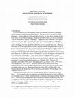
Download

