Article
Serine residues in the LAT adaptor are
essential for TCR-dependent signal
transduction
Mario Martı́nez-Florensa,* Antonio Garcı́a-Blesa,† José Yélamos,‡ Alba Muñoz-Suano,†
Margarita Domı́nguez-Villar,† Rut Valdor,§ Antonio Alonso,ⱍⱍ Francisco Garcı́a-Cózar,†
Pedro Aparicio,* Bernard Malissen,¶ and Enrique Aguado†,#,1
*Departamento de Bioquimı́ca, Biologı́a Molecular B e Inmunologı́a, Facultad de Medicina, Universidad de Murcia, Murcia,
Spain; †Universidad de Cádiz and #Fundación para la Gestión de la Investigación Biomédica de Cádiz, Hospital Universitario de
Puerto Real, Unidad de Investigación, Puerto Real, Spain; ‡Departamento de Inmunologı́a, IMIM-Hospital del Mar, Barcelona,
Spain; §Unidad de Transplantes, Hospital Universitario Virgen de la Arrixaca, Murcia, Spain; ⱍⱍServicio de Inmunologı́a,
Hospital Carlos Haya, Universidad de Málaga, Spain; and ¶Centre d'Immunologie de Marseille-Luminy, Université de la
Méditerranée, INSERM U631, CNRS UMR6102, Marseille, France
RECEIVED MAY 21, 2009; REVISED SEPTEMBER 9, 2010; ACCEPTED SEPTEMBER 10, 2010. DOI: 10.1189/jlb.0509342
ABSTRACT
Introduction
The adaptor protein LAT has a prominent role in the
transduction of intracellular signals elicited by the TCR/
CD3 complex. Upon TCR engagement, LAT becomes
tyrosine-phosphorylated and thereby, recruits to the
membrane several proteins implicated in the activation
of downstream signaling pathways. However, little is
known about the role of other conserved motifs
present in the LAT sequence. Here, we report that the
adaptor LAT contains several conserved serine-based
motifs, which are essential for proper signal transduction through the TCR. Mutation of these serine motifs in
the human T cell line Jurkat prevents proper calcium
influx, MAPK activation, and IL-2 production in response
to TCR/CD3 stimulation. Moreover, this mutant form of
LAT has a reduced ability to bind to PLC-␥1 and SLP-76,
although phosphorylation of tyrosine residues 132, 171,
and 191 is not decreased, raising a possible role for the
serine-based motifs of LAT for the binding of important
partners. The functional role of LAT serine-based motifs
in signal transduction could be mediated by an effect
on tyrosine phosphorylation, as their mutation significantly diminishes the phosphorylation of tyrosine residue 226. In addition, these serine motifs seem to have
a regulatory role, given that upon their mutation,
ZAP-70 shows enhanced phosphorylation. Therefore,
the LAT serine-based motifs likely regulate signaling
pathways that are essential for T cell physiology. J.
Leukoc. Biol. 89: 63–73; 2011.
T lymphocytes recognize foreign peptides bound to MHC molecules presented by APCs. Antigen recognition by the TCR
triggers the activation of the Lck tyrosine kinase, which in
turn, phosphorylates the tyrosines found in the ITAMs present
in the CD3 chains associated with the TCR [1, 2]. Phosphorylated ITAMs recruit ZAP-70 to the membrane, leading to its
activation by Lck. One prominent substrate of ZAP-70 is the
adaptor molecule LAT [3, 4], which is an integral transmembrane adaptor protein of 36 –38 kDa, expressed by peripheral
T lymphocytes, thymocytes, NK cells, mast cells, platelets, and
pre-B cells [3, 5]. The LAT cytoplasmic sequence contains
nine conserved tyrosine residues, which upon phosphorylation,
constitute docking sites for adaptors (Grb2, Grb2-related adaptor protein, Gads, SLP-76), effectors (PLC-␥1, Vav, Cbl), and
for the regulatory subunit of PI-3K [3, 6, 7]. Moreover, LAT has
an essential role in thymic development, as mice deficient in LAT
show an early block in ␣ and ␥␦ T cell development [8, 9].
In vitro approaches have shown that each of the LAT tyrosines manifests some specialization in the signaling proteins
it recruits. For instance, mutation of tyrosine 132 (corresponding to tyrosine 136 in the mouse) selectively eliminates binding to PLC-␥1, whereas the simultaneous mutation of tyrosines
171, 191, and 226 (alternatively called tyrosines 7, 8, and 9,
according to their order of appearance in the LAT intracytoplasmic segment) results in loss of binding to Gads and Grb2
adaptors [10 –12]. These data obtained in Jurkat T cells have
been corroborated by the analysis of knock-in mice harboring
the same mutated tyrosine residues. Indeed, LatY136F mutant
mice show a partial block in ␣ T cell development, whereas
LatY7/8/9F mice show a total block in ␣ T cell development
Abbreviations: Ca2⫹⫽calcium ions, Grb2⫽growth factor receptor-bound 2,
LAT⫽linker for activation of T cells, Ni⫽nickel, p⫽phosphorylation, SIN⫽selfinactivating, SLP-76⫽Src homology 2 domain-containing leukocyte protein
of 76 kDa, SPI⫽small peptide inhibitors
0741-5400/11/0089-0063 © Society for Leukocyte Biology
1. Correspondence: Hospital Universitario de Puerto Real, Unidad de Investigación, Carretera Nacional IV Km. 665, 11510 Puerto Real, Cádiz, Spain.
E-mail: enrique.aguado@uca.es
Volume 89, January 2011
Journal of Leukocyte Biology 63
�and a partially impeded ␥␦ T cell maturation program [8, 13,
14]. Both mutant mice showed TH2 lymphoproliferative disease involving CD4⫹ ␣ (in LatY136F mice) or ␥␦ T cells (in
the LatY7/8/9F mice). These observations suggest that in addition to its positive regulatory role, LAT plays a negative regulatory role in the signaling cassette operated by the TCR.
It has been shown recently that a threonine residue present
in human LAT is phosphorylated by Erk and thereby, negatively regulates TCR signaling events such as Ca2⫹ influx and
Erk activation itself [15]. Although this threonine residue is
not conserved in mouse and rat, we noticed that LAT contains
six serine-based motifs that are conserved in mouse, rat, and
human. As depicted (see Fig. 1), these motifs share the consensus Ser-X-X-Ser, where X represents any amino acid. Moreover, by searching for potential phosphorylation sites in the
LAT amino acid sequence using the NetPhos 2.0 server
(http://www.cbs.dtu.dk/services/NetPhos/) [16], we found
that the majority of these serine residues might represent potential phosphorylation sites for casein kinases I and II, which
constitute two distinct families of Ser/Thr kinases that are
ubiquitously expressed and have been proposed to regulate
the stability of their substrates and cell death induction [17–
19]. Using the Scansite server (http://scansite.mit.edu/) [20],
we confirmed further that several serine residues in LAT constitute potential phosphorylation sites for casein kinases.
Therefore, given the interspecies conservation of serinebased motifs, we have analyzed their functions in TCR-induced
intracellular signaling. Using the LAT mutant JCaM2.5 cells,
we demonstrate that these serine-based motifs are essential for
transducing activation signals coming from the TCR/CD3
complex and allow for the activation of PLC-␥1 kinase and
MEKs, as well as for IL-2 production. Therefore, our results
show that although LAT tyrosine-based motifs are essential for
the transduction of intracellular signals, other nontyrosinebased motifs are also necessary for its adaptor functions.
AGCGGGGAGGCCGCAGAAG 3⬘ and 5⬘ CTTCTGCGGCCTCCCCGCT 3⬘
for Ser180 to Ala mutation. For the generation of stable cell clones, the
mutated and WT forms of LAT were cloned into pcDNA3. For the GFPLAT fusion proteins, the cDNAs cloned into pcDNA3 were amplified by PCR
with primers 5⬘ GCAGGATCCACCATGGAGGAGGCCATCCTGGTCC 3⬘ and
5⬘ GAGAATTCGTTCAGCTCCTGCAGATTC 3⬘, digested with BamHI and
EcoRI, and cloned into pENTR11 (Invitrogen, Carlsbad, CA, USA); then, to
put the sequence in-frame, the plasmids were opened with NotI and XhoI, refilled with Klenow polymerase, and religated. For the WT LAT-6His and
LAT5S/A-6His fusion proteins, the IRES sequence from pIRES2-EGFP (Clontech, Palo Alto, CA, USA) was amplified with primers 5⬘ CTTCGAATTCTGCAGTCGAC 3⬘ and 5⬘ GAGTCTAGATGTGGCCATATTATCATC 3⬘, digested
with BamHI and EcoRI, and cloned into the pENTR11 vectors containing the
WT and mutant LAT, and then the oligonucleotide 5⬘ GAATTCGCAGGTGGAGGCGGTTCAGGCGGAGGTGGCGCTGGCGGTGGCGGATCGCATCATCACCATCACCATTAGCC GCGG 3⬘ was digested and introduced into EcoRI
and SacII sites. DNAs encoding LAT-GFP or LAT-6His-IRES were subcloned
in-frame with GFP in the SIN lentiviral transfer plasmid pHR’SINcPPT-Blast by
means of site-specific recombination (Gateway LR Clonase, Invitrogen). The
coding sequences of all LAT expression constructs made were verified by sequencing.
Lentiviral production and transduction of JCaM2.5
cells
Human embryo kidney 293-FT packaging cells (Invitrogen) were plated in
12-well plates at a density of 2.5 ⫻ 105 cells/well the day before transfection. Cells were washed with OptiMEM (Invitrogen) prior to transfection
and transfected with the corresponding expression vectors, together with
gag/pol and vsv capside, using Lipofectamine 2000 (Invitrogen), according
to the manufacturer’s guidelines. At 48 h and 72 h, transfection efficiency
was evaluated by FACS analysis using a CyanADP-MLE flow cytometer
(DakoCytomation, Denmark). Lentiviral supernatants were collected 48 h
and 72 h after transfection. JCaM2.5 cells were plated at a density of 105
cells/well (5⫻105 cells/ml) in 24-well plates. Lentiviral supernatant was
added and cells cultured for 48 h in a 37°C, 5% CO2 incubator. After selection with 10 g/ml blasticidin, expression of LAT and GFP was analyzed by
means of FACS analysis.
Cell culture and stable transfections
MATERIALS AND METHODS
Antibodies, primers, and plasmids
Stimulations were performed with the anti-CD3- chain mAb UCHT1 (Calbiochem, La Jolla, CA, USA) or OKT3 mAb (eBioscience, San Diego, CA,
USA). The rabbit polyclonal anti-LAT, anti-pLAT-Tyr226, and anti-Fas
(IgM) were obtained from Upstate Biotechnology (Lake Placid, NY, USA);
anti-LAT (V-19), anti-SLP-76 (H-300), anti-PLC-␥1 mAb, and anti-Erk were
from Santa Cruz Biotechnology (Santa Cruz, CA, USA); the antibodies
binding pErk, pMEK1/2, MEK1/2, pPLC-␥1, pZAP-70 (Tyr 319), pLAT-Tyr
171, and pLAT-Tyr191 were from Cell Signaling Technology (Beverly, MA,
USA); antiphosphotyrosine RC20 and anti-ZAP-70 were from Transduction
Laboratories (San José, CA, USA); and HRP-conjugated anti-phosphoserine
rabbit polyclonal antibody and anti-pLAT-Tyr132 were obtained from Abcam (Cambridge, UK). Mutant and WT LAT were expressed using the
pcDNA3 expression plasmid, which also encodes a neomycin-resistance
gene for production of stable lines. Mutation of LAT was performed by a
classical approach performing consecutive PCR reactions with the following
primers: 5⬘ CCTACGACGCCACAGCCTCAG 3⬘ and 5⬘ CTGAGGCTGTGGCGTCGTAGG 3⬘ for Ser38 to Ala and Ser40 to Ala mutation; 5⬘ GGTGCCAACGCTGTGGCGAG 3⬘ and 5⬘ CTCGCCACAGCGTTGGCACC 3⬘ for
Ser106 to Ala mutation; 5⬘ AGTGCCTTCGCCATGGAGTCC 3⬘ and 5⬘
GGACTCCATGGCGAAGGCACT 3⬘ for Ser164 to Ala mutation; 5⬘
64 Journal of Leukocyte Biology
Volume 89, January 2011
The LAT-deficient Jurkat mutant JCaM2.5 was a gift of Dr. Arthur Weiss
(University of California, San Francisco, CA, USA), and subsequent stable
transfectant lines were maintained as described [21, 22]. For stable transfections, 1 ⫻ 107 JCaM2.5 cells were resuspended in PBS and electroporated at 280 V, 960 microfarads, using a Gene Pulser electroporator (BioRad, Hercules, CA, USA). For generation of stable lines, transfected cells
were plated 24 h after electroporation in media containing 1.1 mg/ml
G418.
Confocal microscopy analysis
JCaM2.5 cells lentivirally transfected with WT LAT or the LAT5S/A mutant
fused to GFP were mounted on poly-D lysine-coated glass slides, and the
paraformaldehyde-fixed and permeabilized cells were stained with anti-LAT
(V-19) antibody conjugated to Oregon Green 488 (Molecular Probes, Eugene, OR, USA). Imaging was done using a Leica spectral confocal microscope (Leica Microsystems GmbH, Wetzlar, Germany).
TCR simulation and preparation of lysates
Transfectant cells were starved in RPMI without FCS for 18 h previous to
be stimulated with the indicated anti-CD3 mAb in RPMI 1640 at 37°C.
Cells were lysed at 108 cells/ml for 30 min on ice in lysis buffer: 1% Nonidet P-40, 150 mM NaCl, 20 mM HEPES, pH 7.6, 100 mM NaF, 1 mM
www.jleukbio.org
�Martı́nez-Florensa et al. Role of LAT serine-based motifs
EGTA, 1 mM PMSF, 1 mM Na3VO4, and 2 g/ml each SPI, antipain, chymostatin, leupeptin, and pepstatin. Nuclei were then removed by centrifugation at 12,000 g for 15 min at 4°C.
washed, resuspended in 1 ml PBS, and kept at 25°C in the dark until measurements. Before calcium measurements, cells were incubated at 37°C for
5 min. Measurements were performed by flow cytometry in a CyanADPMLE (DakoCytomation) using a UV enterprise laser set at 30 mW. The
baseline ratio is acquired before the addition of the anti-CD3 antibody.
Immunoprecipitation and Western blotting
For LAT immunoprecipitation, whole cell lysates from 107 transfectant cells
expressing LAT-6His were obtained as described above, but the lysis buffer
contained 1% Brij97, 200 mM NaCl, 20 mM Tris-HCl, pH 7.5, 1 mM PMSF,
1 mM Na3VO4, and 2 g/ml each SPI. The lysates were then subjected to
magnetic bead separation (Histidine Adem kit, Ademtech, Pessac, France),
as recommended by the supplier. Briefly, whole cell lysates were incubated
with 60 l magnetic beads for 40 min on ice. The samples were then
washed four times with 200 l binding buffer: 20 mM Tris, pH 7.5, 500
mM NaCl, Na3VO4 1 mM, pH 7.5, and 2 g/ml SPI in the magnetic field
of the Adem-Mag MSV device (Ademtech). Finally, LAT-6His was released
from the beads by applying 50 l elution buffer: 20 mM Tris, pH 7.5, 500
mM NaCl, 500 mM imidazole, 1 mM EDTA. For SLP-76 immunoprecipitation, whole cell lysates from 107 transfectant cells were obtained as described above, and lysates were incubated with 2 g anti-SLP-76 antibody
for 45 min on ice and then incubated with 60 l protein G-coupled magnetic beads for 40 min on ice (Ademtech). The samples were then washed
four times with 200 l binding buffer in the magnetic field of the AdemMag MSV device. Finally, SLP-76 was released from the beads by applying
50 l elution buffer: 20 mM Tris, pH 7.5, 500 mM NaCl, 500 mM imidazole, 1 mM EDTA. For Western blotting, whole cell lysates or immunoprecipitates were separated by SDS-PAGE and transferred to PVDF membranes, which were incubated with the indicated primary antibodies
followed by the appropriate secondary antibody conjugated to HRP. Reactive proteins were visualized using the ECL system and exposition to Hyperfilm-ECL (Amersham, Buckinghamshire, UK) or acquired directly with the
VersaDoc 5000 imaging system (Bio-Rad). For reprobing, PVDF membranes
were incubated for 10 min at room temperature with NaOH 0.1 N, followed by thorough washes with blocking buffer.
Measurement of intracellular Ca2ⴙ mobilization
with Fura-2
Where indicated, the measurement for intracellular-free Ca2⫹ was done
using Fura-2 AM (1 M; Molecular Probes) as described [23]. Briefly, 2 ⫻
106 transfectant cells were preloaded with the dye for 45 min at 25°C,
washed, resuspended in 2 ml PBS, and kept at 25°C in the dark until measurements. Calcium measurements were performed in the optical field of a
fluorescence spectrometer (Aminco Bowman 2, Microbeam, Barcelona,
Spain), under continuous stirring and at 37°C. The cells were excited alternatively with light of 340 and 380 nm wavelength and the light emitted at
510 nm, collected once/s. The calibration of Ca2⫹ was based on the signal
ratio at 340/380 nm and an established protocol as stated below. The Ca2⫹
was calculated according to the formula: Ca2⫹ ⫽ [(R–Rmin)/(Rmax–R)] ⫻
(Sf/Sb) ⫻ Kd, where R is the ratio of the 340/380 nm fluorescence signal,
Rmin is the 340/380 ratio in calcium-free buffer (EGTA-Tris 5 mM), Rmax is
the 340/380 ratio in the presence of saturating calcium (0.5% Triton
X-100 containing 1 mM CaCl2), Sf/Sb is the ratio of the 380-nm fluorescence measured in calcium-free conditions to that in calcium-repleted conditions, and Kd is 224 nM. The calibration procedure was done in every
experiment to take into account possible differences in the number of
cells.
Measurement of intracellular Ca2ⴙ mobilization
by flow cytometry
Where indicated, the measurement for intracellular-free Ca2⫹ was done
using Indo-1 AM (1 M; Molecular Probes) as described [24]. Briefly, 4 ⫻
106 transfectant cells were preloaded with the dye for 45 min at 25°C,
www.jleukbio.org
IL-2 measurement
For the induction of IL-2 expression, transfectant cells were cultured for
5 h in the presence of plate-bound OKT3 mAb at a final concentration of
10 g/ml and PMA (Sigma Chemical Co., St. Louis, MO, USA) at a final
concentration of 50 ng/ml. Before intracellular cytokine staining, cells
(1.5⫻106) were cultured in the presence of monensin (GolgiStop, BD
PharMingen, San Diego, CA, USA) at a final concentration of 2 M. Cells
were then placed immediately on ice, washed, and resuspended in PBS 1⫻,
1% FCS, 0.90% sodium azide. For intracellular IL-2 staining, cells were first
fixed using the Cytofix/Cytoperm kit (BD PharMingen,). Each cell sample
was subsequently split into aliquots that were stained with PE-conjugated
anti-IL-2 antibodies or fluorochrome-conjugated and isotype-matched negative control Ig (BD PharMingen). After a final wash, cells were analyzed on
a FACSort flow cytometer after gating out dead cells using forward- and
side-scatters and analyzed using CellQuest (BD PharMingen).
RESULTS
Generation of stable transfectants of JCaM2.5 cells
The LAT-deficient Jurkat cell line JCaM2.5 has been used extensively to dissect the function of LAT tyrosine-based motifs
during TCR signaling. JCaM2.5 cells are defective in RasMAPK activation and Ca2⫹ influx generation in response to
A
LAT
EC TM
B
CY
S--S*-S*--S
S*--S
S--S*--S
S--S*
35 38 40 43
106 109
161 164 167
177 180
human
36 39 41 44
109 112
165 168 171
181 184
mouse
36 39 41 44
109 112
165 168 171
181 184
rat
1
2
3
4
LAT
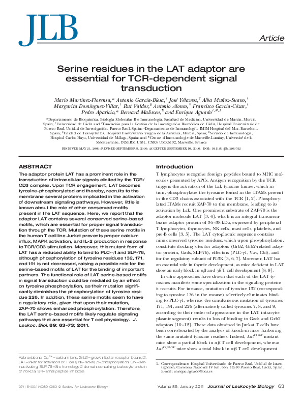
Figure 1. Production of stable LAT transfectants of JCaM2.5 cells.
(A) Diagram showing the different serine motifs found in the human,
mouse, and rat forms of LAT. The extracellular (EC), transmembrane
(TM), and cytoplasmic (CY) domains are indicated together with the
serine (S) residues found within the cytoplasmic region. *, Residues
mutated to alanine. The LAT mutant form, in which serine residues
38, 40, 106, 164, and 180 have been mutated to alanine, is denoted as
LAT5S/A. (B) Stable clones were generated expressing WT LAT or the
mutant LAT5S/A. LAT expression in JCaM2.5 cells (Lane 1) or transfected with empty vector (Lane 2) or with the cDNA coding for LAT
WT or LAT5S/A (Lanes 3 and 4, respectively) was analyzed by Western
blotting of 5 ⫻ 105 of each type of cells.
Volume 89, January 2011
Journal of Leukocyte Biology 65
�anti-CD3 antibody stimulation. Re-expression of a WT form of
LAT in JCaM2.5 cells restores all of the signaling events normally induced upon TCR engagement [10, 12, 22]. Therefore,
we used this cell line to test the role of the six serine-based
motifs (Ser-X-X-Ser), which we have identified to be conserved
in human, mouse, and rat forms of LAT (Fig. 1A). Mutation
of a single serine residue of serine-based motifs containing
proteins such as Bid, the transcription factor CREB, -catenin,
and E-cadherin suffices to impede or prevent their global
phosphorylation status or their biological function [25–28].
Accordingly, serine residues 38, 40, 106, 164, and 180 were
simultaneously mutated to alanines. Although this LAT mutant
still contains serine residues at positions 35, 43, 109, 161, 167,
and 177, the corresponding mutant will be subsequently denoted LAT5S/A. Once expressed in JCaM2.5 cells, this mutant
allows for the analysis of the functional consequence of serinebased motif disruption in LAT. It has to be pointed out that
by mutating Ser 164, two consecutive serine-based motifs (one
constituted by residues 161 and 164 and the other one by residues 164 and 167; see Fig. 1A) are eliminated simultaneously.
The cDNAs coding for WT LAT or LAT5S/A were introduced
into JCaM2.5 cells by electroporation. Stable clones were selected with G418 and screened for LAT expression using Western blot (Fig. 1B). Three clones expressing LAT5S/A mutant
molecules and three clones expressing LAT WT molecules displaying similar levels of LAT and TCR were selected for further studies. Therefore, disruption of the serine-based motif
was without noticeable effect on the stability of the LAT molecule. No significant biological and functional differences were
found among the three independent clones of each genotype.
It has been shown previously that subtle mutations disrupt
its function by preventing its recruitment to the plasma membrane [29]. To check whether LAT5S/A mutation affects its
membrane expression, we decided to analyze LAT subcellular
localization by confocal microscopy. For that, we generated a
lentiviral vector containing the WT LAT or the LAT5S/A mutant fused to GFP, and this vector was used to transduce
JCaM2.5 cells (Fig. 2A). We directly performed confocal microscopy with these cells, but the fluorescence level was not
high enough to visualize LAT subcellular localization (data not
shown). Therefore, we performed LAT staining over fixed and
permeabilized cells and confocal microscopy. As it can be seen
in Fig. 2B, the 5S/A mutation did not modify the LAT expression pattern, which is basically the same (i.e., mainly at the
cell membrane) in both types of cells.
Mutation of LAT serine residues negatively affects
activation of the pathway
To dissect the function of serine-based motifs of LAT in the
activation of the MAPK pathway, activation of Erk upon antiCD3 stimulation was analyzed. For that purpose, we used an
antibody specific for pErk at Thr 202 and Tyr 204, as phosphorylation of these two residues is indicative of Erk enzymatic
activation [30, 31]. As shown in Fig. 3A, JCaM2.5 cells expressing WT LAT showed a rapid phosphorylation of Erk. Conversely, JCaM2.5 cells expressing the LAT5S/A mutant were not
able to induce Erk phosphorylation at the same level observed
66 Journal of Leukocyte Biology
Volume 89, January 2011
A
wtLAT / LAT
5S/A
GFP
B
LAT wt
LAT 5S/A
Figure 2. Mutation of LAT serine-based motifs does not alter its membrane expression. (A) Diagram showing the LAT-GFP fusion proteins
(wt LAT/LAT5S/A) generated for their expression in JCaM2.5 cells by
means of the lentiviral system. (B) JCaM2.5 cells transfected with the
DNAs coding for the fusion proteins of WT LAT or the LAT5S/A mutant were fixed, permeabilized, and stained with anti-LAT antibody
conjugated to Oregon Green 488. Cells were examined with confocal
microscopy. Right panels show a magnification of cells in the frame in
left panels.
in WT LAT-expressing cells (Fig. 3A, right panel). The lower
panel in Fig. 3A shows the same membrane stripped and blotted with an anti-pan Erk mAb, demonstrating equal protein
loading.
Erk phosphorylation and activation proceed via the phosphorylation and activation of MEK-1, which in turn, phosphorylates regulatory threonine and tyrosine residues on Erk.
Therefore, to investigate MEK activation in response to antiCD3 stimulation, a mAb specific for pMEK-1/2 (Ser221) was
used to detect activated MEK in whole cell lysates obtained
from JCaM2.5 cells expressing WT LAT or LAT5S/A. As it can
be observed in Fig. 3B, mutation of serine residues of LAT
prevents MEK activation. The lower panel in Fig. 3B shows the
same membrane stripped and blotted with an anti-pan
MEK1/2 mAb, demonstrating equal protein loading. Consequently, mutation of LAT serine-based motifs greatly reduces
activation of the MAPK pathway upon anti-CD3 stimulation.
Altered calcium responses in JCaM2.5 cells
expressing LAT5S/A
The effect of the mutations introduced into LAT5S/A on TCRinduced PLC-␥1 activation was next analyzed. As shown in
Fig. 4A, anti-CD3 stimulation of JCaM2.5 cells expressing WT
LAT but not the LAT5S/A mutant induced phosphorylation of
PLC-␥1, a hallmark of its enzymatic activation [32]. The lower
www.jleukbio.org
�Martı́nez-Florensa et al. Role of LAT serine-based motifs
Figure 3. Mutation of serine motifs of
LAT impedes MAPK pathway activation.
JCaM2.5 cells stably expressing the vecAnti-CD3 (min):
0
3
10
30
0
3
10
30
0
3
10
30
tor alone, LAT WT, or the LAT5S/A mutant
were stimulated with the UCHT1
P-Erk
anti-CD3 mAb (1 g/106 cells) for the
indicated times. (A) Whole cell lysates
Erk
obtained from stimulated and nonstimulated cells were then probed by Western
blotting for the activation of Erk by usB
5S/A
pcDNA3
LAT wt
LAT
ing a mAb recognized as only dually,
Anti-CD3 (min):
doubly phosphorylated on specific thre0
3
10
30
0
3
10
30
0
3
10
30
onine and tyrosine residues on Erk (upP-MEK1/2 per panel). Stripped membranes were
also blotted with anti-pan Erk mAb to
show equal protein expression (lower
MEK 1/2
panel). (B) Whole cell lysates were assayed for MEK1/2 activation with a mAb
detecting specifically phosphorylation of Ser221. Stripped membranes were also blotted with anti-pan MEK1/2 mAb to show equal protein expression.
A
pcDNA3
LAT wt
panel in Fig. 4A shows the same membrane stripped and blotted with an anti-pan PLC-␥1 mAb, demonstrating equal protein loading. We investigated further whether these motifs are
necessary for calcium influx generation via the TCR. As can be
observed in Fig. 4B, re-expression of the WT form of LAT in
JCaM2.5 cells restored Ca2⫹ influx upon stimulation with antiCD3 mAb, but transfection of the LAT5S/A mutant did not restore normal Ca2⫹ influx generation upon anti-CD3 stimulation. The increase in Ca2⫹ triggered upon stimulation with
ionomycin excluded a general defect in the cellular calcium
machinery. To exclude that clonal variation could be responsible for the calcium defect observed in LAT5S/A-expressing
cells, we generated a lentiviral vector containing the WT LAT
or the LAT5S/A mutant fused to GFP, and this vector was used
to transduce JCaM2.5 cells. As shown in Fig. 4C (right panels),
most of the cells expressed the foreign gene upon infection
with the lentiviral vectors. These cells were stained with Indo-1,
and Ca2⫹ influx was determined by flow cytometry. As it can
be observed in Fig. 4C, mutation of serine motifs of LAT
greatly reduces Ca2⫹ influx upon anti-CD3 stimulation. Therefore, it can be concluded that the conserved serine motifs of
LAT are necessary for a proper PLC-␥1 activation and Ca2⫹
influx generation upon anti-CD3 stimulation.
Mutation of LAT serine residues negatively affects
phosphorylation of tyrosine 226
Given that the signaling activity of LAT is mainly funneled
through the four C-terminal tyrosine residues at positions 132,
171, 191, and 226, we next investigated whether mutation of
serine-based motifs in LAT had any influence on TCR-induced
phosphorylation of those tyrosine residues. For that purpose,
we used a panel of antibodies specific for the tyrosine-phosphorylated isoform of each of the last four C-terminal tyrosine
residues [33, 34]. Stably transfected JCaM2.5 cells were stimulated with anti-CD3 antibodies for up to 30 min to determine
the phosphorylation kinetics of the four C-terminal LAT tyrosines (Fig. 5). As shown in Fig. 5, cells expressing the WT
LAT or LAT5S/A mutant exhibited no major difference in intensity or kinetics of LAT tyrosines 132 or 191 phosphorylation.
www.jleukbio.org
LAT 5S/A
Interestingly, mutation of LAT serine motifs significantly
diminished the phosphorylation of tyrosine residue 226 upon
TCR/CD3 stimulation (Fig. 5, bottom pair of panels). With
regard to tyrosine 171, there was no major reduction of its activation-induced phosphorylation in LAT5S/A-expressing cells
compared with WT LAT, but its phosphorylation level was increased in resting cells expressing LAT5S/A compared with
those expressing the WT form. Therefore, conserved serine
motifs of LAT modify a basal level of phosphorylation of tyrosine 171 and are required specifically for proper TCR-induced phosphorylation of tyrosine residue 226.
Mutation of LAT serine motifs modifies pZAP-70
To analyze the impact of serine mutations on TCR/CD3 proximal signaling events, whole cell extracts from untreated cells
or cells stimulated with anti-CD3 mAb were analyzed by Western blot for ZAP-70 activation. For that purpose, an antibody
specific for the phosphorylated tyrosine residue found at position 319 of ZAP-70 was used, as this event constitutes a reporter of ZAP-70 enzymatic activation [35]. As reported previously [22], stimulation of LAT-deficient JCaM2.5 cells with
anti-CD3 mAb activated ZAP-70 kinase rapidly, demonstrating
that activation of this enzyme upon CD3 engagement does not
depend on LAT expression (Fig. 6A, upper panel). Also,
JCaM2.5 cells stably expressing the LAT5S/A mutant showed an
enhanced phosphorylation of tyrosine 319 of ZAP-70 upon anti-CD3 stimulation (Fig. 6A, upper panel). Unexpectedly, the
level of phosphorylation of tyrosine 319 of ZAP-70 was increased in resting cells deprived of LAT or expressing the
functional, defective LAT5S/A mutant (Fig. 6A).
To confirm the above finding using polyclonal populations
of LAT-expressing cells, we used a lentiviral expression system
to transduce JCaM2.5 cells with the LAT WT or LAT5S/A fused
to GFP (see Fig. 2A). Cells transduced with LAT5S/A-GFP
showed an increased level of phosphorylation of tyrosine 319
in ZAP-70 when compared with cells expressing WT LAT fused
to GFP (data not shown). Fig. 6B shows a diagram representing quantification of five experiments using cells expressing
WT and the LAT5S/A mutant. Therefore, it can be concluded
that LAT negatively regulates ZAP-70 activity, as the absence of
Volume 89, January 2011
Journal of Leukocyte Biology 67
�A
pcDNA3
Anti-CD3 (min):
0
2
5
10
LAT 5S/A
LAT wt
30
0
2
5
10
30
0
2
5
10
30
P-PLC-γ1
PLC-γ1
[Ca2+] nM
B
LAT-/-
LAT 5S/A
LAT wt
300
300
300
200
200
200
100
100
100
200
100
100
200
100
200
Time (seconds)
C
JCaM2.5
60
120
180
LAT wt-GFP
60
120
180
LAT 5S/A -GFP
60
120
180
Time (seconds)
GFP
Figure 4. Altered PLC-␥1 activation and calcium responses in JCaM2.5 cells expressing the mutant LAT5S/A. (A) Analysis of activation of PLC-␥1.
JCaM2.5 cells stably transfected with the vector alone, LAT WT, or the LAT5S/A mutant were stimulated with the UCHT1 anti-CD3 mAb (1 g/106 cells)
for the indicated times, and whole cell lysates were analyzed for the activation of PLC-␥1 by using a mAb recognizing phosphorylated tyrosine 783 (upper
panel). Stripped membranes were also blotted with anti-pan PLC-␥1 mAb to show equal protein expression. (B) JCaM2.5 cells stably expressing the vector alone, LAT WT, or the LAT5S/A mutant were loaded with Fura-2 and stimulated with UCHT1 mAb (1 g/ml) at the indicated time (white arrowheads). The intracellular Ca2⫹ concentration was determined at 37°C through the change in Fura-2 fluorescence. The charge of Fura-2 dye was assessed
by observing the response of the transfectants to stimulation with ionomycin (1 M, black arrowheads). (C) JCaM2.5 cells lentivirally transduced with
vectors for the expression of WT LAT-GFP or LAT5S/A -GFP fusion proteins were loaded with Indo-1 and stimulated with UCHT1 mAb (1 g/ml) at the
indicated time (white arrowheads). The change in intracellular Ca2⫹ concentration was determined by the change in Indo-1 fluorescence. The charge of
Indo-1 dye was assessed by observing the response to stimulation with ionomycin (1 M, black arrowheads). The level of expression of the LAT-GFP fusion proteins was assessed by flow cytometry (right panels). FL6Lin/FL7Lin, linear fluorescence 6/7.
68 Journal of Leukocyte Biology
Volume 89, January 2011
www.jleukbio.org
�Martı́nez-Florensa et al. Role of LAT serine-based motifs
LAT 5S/A
LAT wt
Anti-CD3 (min):
0
3
10
30
0
3
10
1.0
3.4
2.9
1.0
0.9
1.0
1.0
30
1.5
0.8
3.3
2.3
1.2
0.8
0.8
0.9
0.9
0.9
0.9
3.9
4.4
3.4
3.3
10.9
10.0
9.9
0.8
0.7
0.8
1.8
1.8
1.8
1.9
1.0
3.5
3.4
1.5
0.5
1.0
1.0
0.8
0.8
1.2
1.0
11.7
12.5
4.4
1.0
0.6
0.6
0.7
P-Y132-LAT
LAT
P-Y171-LAT
LAT
P-Y191-LAT
2.7
2.3
1.2
LAT
1.3
1.4
1.3
0.9
1.2
3.6
1.9
1.4
1.5
1.1
1.3
Figure 5. LAT tyrosine phosphorylation is specifically affected by mutation of conserved serine motifs. Transfectant JCaM2.5 cells stably expressing WT LAT or the
LAT5S/A mutant were stimulated with the anti-CD3 OKT-3
mAb (1 g/106 cells) for the indicated times, and whole
cell lysates were analyzed for the phosphorylation of specific LAT tyrosine residues with different antibodies. Each
membrane was stripped and blotted with anti-LAT antibody to show total protein expression. The numbers below
the panels indicate densitometry analysis of the relative
amount of each band. The data are representative of at
least three independent experiments.
P-Y226-LAT
LAT
this adaptor or the expression of a signaling-defective LAT5S/A
mutant induces the hyperphosphorylation of ZAP-70.
LAT5S/A, we decided to analyze if this mutant form of LAT
was still able to bind to PLC-␥1 upon TCR-dependent activation. Therefore, lentiviral vectors were generated containing
WT or the LAT5S/A mutant fused to a 6His tag and then an
IRES sequence preceding the coding sequence of GFP
(Fig. 7A). Upon transduction with the appropriate lentiviral
vectors, JCaM2.5 cells expressing the WT LAT-6His or the
Mutation of LAT serine-based motifs reduces binding
to PLC-␥1 and SLP-76
Given the reduced calcium influx generation and PLC-␥1
phosphorylation upon CD3 engagement in cells expressing
A
pcDNA3
Anti-CD3 (min):
LAT 5S/A
LAT wt
0
3
10
30
0
3
1.0
1.5
1.7
1.8
0.5
0.7
1.0
1.0
1.0
0.9
1.0
1.0
10
30
0
3
0.9
0.6
1.3
1.9
2.0
1.2
0.9
0.9
1.0
0.9
0.9
0.7
10
30
P-ZAP70
ZAP70
B
Relative ZAP-70 phosph
8,00
7,00
6,00
5,00
4,00
3,00
2,00
1,00
Figure 6. Activation of ZAP-70 in cells expressing WT LAT
or the LAT5S/A mutant. (A) JCaM2.5 cells stably transfected with the vector alone, LAT WT, or the LAT5S/A mutant were stimulated with the anti-CD3 OKT3 mAb (1 g/
106 cells) for the indicated times. Whole cell lysates were
prepared and analyzed for the activation of ZAP-70 by using a mAb recognizing phosphorylated tyrosine 319 (upper panel). Stripped membranes were also blotted with an
anti-pan ZAP-70 mAb to show total protein expression.
The numbers below the panels indicate densitometry analysis of the relative amount of each band. The data are representative of two independent experiments. (B) Diagram
representing quantification of five experiments using
JCaM2.5 cells expressing WT and the LAT5S/A mutant and
stimulated with anti-CD3 antibodies during the indicated
times. The bars represent relative densitometric data relative to nonstimulated cells expressing WT LAT.
0,00
0 min
10 min
Vector
www.jleukbio.org
0 min
LAT wt
10 min
0 min
10 min
LAT5S/A
Volume 89, January 2011
Journal of Leukocyte Biology 69
�A
B
wt LAT / LAT 5S/A
6-His
Anti-CD3 (min):
GFP
IRES
v
3
wt LAT
0
3
LAT 5S/A
0
3
PLC
C
v
wt LAT
LAT 5S/A
Anti-CD3 (min):
3
0
0
3
3
P-SLP-76
LAT
D
Anti-CD3 (min): 0
1
2
5
P-LAT
LAT 5S/A
wt LAT
10
0
1
2
SLP-76
5 10
CBL
Figure 7. Serine motifs of LAT are important for its binding to downstream effectors.
(A) Diagram showing the fusion protein generated for the expression by means of the lentiviral system. (B and D) JCaM2.5 cells (107) transduced with the vectors (v) containing LAT
LAT
WT
or the LAT5S/A fusion proteins were stimulated with 10 g OKT3 mAb for the indicated
1.0 1.3 1.3 1.4 1.3 1.0 1.1 1.0 1.0 1.3
times. Whole cell lysates were subjected to LAT purification with Ni-conjugated microbeads,
and the precipitates were analyzed by Western blot with anti-PLC-␥ or Cbl antibodies (upper panels). The membrane was then stripped and reblotted with anti-LAT antibody (lower panels). The data are representative of three independent experiments. (C) As in B and D, but the whole
lysates were subjected to SLP-76 immunoprecipitation with an anti-SLP-76 antibody, followed by incubation with protein G-coupled magnetic
beads. The precipitates were analyzed by Western blot with antiphosphotyrosine antibody (upper panel). The membrane was then stripped and
reblotted with anti-LAT antibody (not shown) or anti-SLP-76 (lower panel).
1.0 1.5 1.8 2.1 2.2 1.7 1.6 1.3 1.3 1.5
LAT5S/A-6His fusion proteins were stimulated with anti-CD3
mAb lysed and lysates used to purify LAT with Ni-coupled microbeads. As it can be observed in Fig. 7B, WT LAT coprecipitates with PLC-␥1 upon anti-CD3 stimulation, but in LAT5S/A6His-expressing cells, there is no coprecipitation of this enzyme. Given that SLP-76 and PLC-␥1 are known to form a
complex with LAT, which is absolutely required for PLC-␥ activation, we decided to check whether the mutations introduced
in LAT5S/A affected SLP-76 binding to phosphorylated LAT.
For that, we immunoprecipitated SLP-76 from cells expressing
WT LAT, the LAT5S/A mutant, or the empty expression vector,
and the precipitates were assayed by Western blotting. As it
can be observed in Fig. 7C, SLP-76 coprecipitated with tyrosine-phosphorylated LAT in TCR-stimulated cells expressing
WT LAT; by the contrary, SLP-76 did not bind to the LAT5S/A
mutant, although the SLP-76 levels were similar in all the cases
(Fig. 7C, lower panel).
It has been demonstrated previously that Cbl is able to bind
to LAT, and this binding is also required to stabilize PLC-␥1
recruitment [36 –39]. Therefore, it was of interest to analyze
the binding of LAT to Cbl. Accordingly, cells were stimulated
with anti-CD3 mAb, LAT was purified with Ni-coupled microbeads from cell lysates, and LAT-Cbl coprecipitation was
analyzed by Western blotting. As it can be observed in Fig. 7D,
Cbl binding to WT LAT augments in an activation-dependent
manner. However, this increase is not observed in cells expressing the LAT5S/A mutant. Also, it seems that basal binding
of Cbl is slightly increased in the LAT5S/A mutant with regard
to WT LAT, although this increase was not statistically significant (data not shown).
Defective IL-2 production in JCaM2.5 cells expressing
the LAT5S/A mutant
Given that mutation of serines 38, 40, 106, 164, and 180 prevents proper transduction of early intracellular signals triggered by the TCR/CD3 complex, we sought to evaluate
whether JCaM2.5 cells expressing the LAT5S/A mutant were
still capable of IL-2 production in response to stimulation
through the TCR/CD3 complex. As reported previously, to
70 Journal of Leukocyte Biology
Volume 89, January 2011
achieve detectable levels of IL-2 production in Jurkat cells,
cells had to be stimulated with plate-bound anti-CD3 antibody
(10 g/ml) plus PMA at a final concentration of 50 ng/ml
[40 – 42]. As shown in Fig. 8, JCaM2.5 cells expressing WT
LAT were capable of producing IL-2 upon stimulation with
PMA plus immobilized anti-CD3 antibody. In contrast, mutation of all of the conserved serine motifs of LAT totally prevented IL-2 production. However, stimulation with PMA plus
ionomycin, which bypasses LAT, induced identical IL-2 production in WT LAT- and LAT5S/A-expressing cells, demonstrating that the general IL-2 production machinery was intact in
LAT5S/A cells. Taken together, these data support an essential
role for the serine motifs of LAT in the transduction of activation signals coming from the TCR/CD3 complex.
DISCUSSION
It has been demonstrated previously that the ability of the
adaptor LAT to transduce intracellular signals is based on the
capacity of its phosphorylated tyrosines to recruit different signaling adaptors and effectors. To determine whether conserved, nontyrosine-based motifs could play a role in the LAT
signaling functions, we analyzed the consequences of mutating
the six conserved Ser-X-X-Ser motifs found in LAT. Our work
demonstrates that PLC-␥1 activation, Ca2⫹ influx generation,
and MEK/Erk activation in response to anti-CD3 stimulation
are strongly decreased in JCaM2.5 cells expressing mutant
LAT5S/A molecules. Consistent with these data, LAT5S/A-expressing cells are not able to produce IL-2 upon TCR/CD3mediated stimulation. Therefore, serine-based motifs are essential for the activation of signaling pathways triggered by the
TCR/CD3 complex.
Interestingly, we have also shown that mutation of LAT
serine-based motifs have a negative impact on the phosphorylation of Tyr 226, and it remains to be determined whether
this defect accounts for the signaling defects observed in cells
expressing LAT5S/A. In support of the view that the global defects associated with the LAT5S/A mutation are not fully accounted for by the reduced phosphorylation of tyrosine 226,
www.jleukbio.org
�Martı́nez-Florensa et al. Role of LAT serine-based motifs
Figure 8. JCaM2.5 cells expressing mutant LAT5S/A do not produce IL-2 upon
TCR/CD3 stimulation. Stable JCaM2.5
transfectants expressing LAT WT or the
mutant LAT5S/A were cultured at 37°C
for 5 h in the presence of plate-bound
OKT3 and PMA at 50 ng/ml in the presence of 2 M monensin. After stimulation, cells were washed and fixed, and
intracellular staining was performed
with PE-conjugated anti-IL-2 antibodies
or isotype-matched, negative control Ig.
As a positive control of IL-2 production,
cells were stimulated with 1 M ionomycin plus PMA at 20 ng/ml. Numbers
represent percentage of cells in each
quadrant. Dot-plots correspond to one
experiment representative of three.
FL1/2-H, Fluorescence 1/2-height.
Samelson and coworkers [12] showed that mutation of this
single tyrosine affects none of the intracellular signals triggered upon TCR engagement. Moreover, using a system in
which Rag-deficient mice were reconstituted with T cell precursors expressing the LATY235F mutant (in which tyrosine
235, the equivalent to tyrosine 226 in mice, was substituted by
phenylalanine), this mutant form of LAT was capable of reconstituting normal T cell development [43]. Therefore, it is
likely that the specific signaling phenotype observed in
JCaM2.5 cells expressing mutant LAT5S/A cannot be fully recapitulated by the expression of LAT molecules expressing a mutation of tyrosine 226, suggesting that the conserved serine
motifs may have a direct role in recruiting some effectors or
adaptors.
Although the phosphorylation of other tyrosine residues was
normal, the basal level of phosphorylation of tyrosine 171 in
resting cells seems to be augmented, and the significance of
this event remains to be determined. On the other hand, the
comparable level of induced phosphorylation of tyrosine 132
in the mutant LAT5S/A was in part unexpected, given that this
residue is primarily responsible for PLC-␥1 binding to LAT
and that activation of PLC-␥1 and Ca2⫹ influxes is impeded in
LAT5S/A cells. Therefore, although the activation of PLC-␥1
depends on the phosphorylation of tyrosine residue 132 and
the recruitment of SLP-76, it seems that the serine-based motifs of LAT might also play a role in the stabilization of the
LAT-SLP-76-PLC-␥1 complex. In agreement with this hypothesis, we have demonstrated that the mutant LAT5S/A has a reduced ability to bind to PLC-␥1 and SLP-76 upon CD3-dependent activation. Therefore, it is possible that the serine-based
motifs of LAT provide a proper alignment of the LAT tyrosines, allowing proper binding of the corresponding partners to allow PLC-␥1 activation. It remains to be determined
whether mutations introduced in LAT5S/A prevent serine phosphorylation of this adaptor molecule to explain the observed
LAT5S/A signaling defects. In contrast to results obtained by
Matsuda et al. [15], we have not been able to demonstrate significant serine phosphorylation of WT or mutant LAT upon
anti-CD3 stimulation (data not shown). As we have analyzed
www.jleukbio.org
serine phosphorylation by Western blotting of immunoprecipitated LAT or Ni-purified LAT-6His fusion molecules, an explanation for this controversial result could be a low proportion
of serine-phosphorylated LAT molecules or the low affinity of
the antiphosphoserine antibody used. Alternatively, it cannot
be discarded that nonphosphorylated serines could be involved in protein:protein interactions or have a regulatory role
over LAT phosphorylation by means of other post-translational
modifications such as O-linked N-acetylglucosamine additions
[44, 45]. Further work should help to unveil the underlying
mechanisms of the putative regulatory role of LAT serinebased motifs.
Remarkably, our work has shown that mutation of LAT
serine-based motifs increases ZAP-70 phosphorylation. This
finding is in agreement with the postulated negative regulatory
role of LAT in the intracellular signaling cassette operated by
the TCR/CD3 complex [46]. Normal ZAP-70 activation in cells
deficient in LAT expression has been reported previously [22,
47]. However, from the data presented from these groups, an
enhanced pZAP-70 in JCaM2 or ANJ3 LAT-deficient cells cannot be excluded, and it seems that this question should be analyzed more thoroughly. Our results demonstrate that in the
absence of any stimulation, LAT-deficient JCaM2.5 cells show a
hyperphosphorylation of ZAP-70 as compared with WT LATexpressing cells. Moreover, by using cells transduced with lentiviral vectors as well as stable cell clones, we have shown that
expression of the LAT5S/A mutant, which is incompetent to
transduce activation signals from the TCR, also induces constitutive hyperphosphorylation of ZAP-70. Thymocytes from LATdeficient mice transgenic for a gain-of-function mutant Lck
tyrosine kinase (LAT⫺/⫺ X LckY505F tg) also show an increase
in basal phosphorylation of tyrosine 319 of ZAP-70 compared
with Lat⫹/⫹ LckY505F thymocytes (B. Malissen et al., unpublished results). These results are in agreement with the negative regulatory function of the LAT adaptor revealed from the
analysis of LatY136F and LatY7/8/9F mutant mice [8, 13, 48]. It
remains to be determined whether hyperphosphorylation of
ZAP-70 in the absence of a completely functional LAT is dependent on Lck kinase or directly affects ZAP-70 itself.
Volume 89, January 2011
Journal of Leukocyte Biology 71
�In summary, LAT serine-based motifs seem to have a deep
impact on the functional capability of this adaptor molecule.
Further work aiming to analyze the significance of LAT serine
residues for its binding capacities would clarify the mechanisms of intracellular signaling modified in the LAT5S/A mutant.
10.
11.
12.
AUTHORSHIP
All authors contributed to discussions of experimental design
and data analysis. M.M-F. did all experimental studies unless
otherwise indicated; A.G-B. performed immunoprecipitations,
Western blots, calcium assays, and confocal imaging; A.M-S.,
M.D-V., and R.V. provided technical assistance; J.Y., A.A., F.GC., P.A., and B.M. provided suggestions; E.A. directed the
study and wrote the manuscript.
13.
14.
15.
16.
ACKNOWLEDGMENTS
This work was supported by Fundación Séneca (grants 03055/
PI/05 to P.A. and 00603/PI/04 to J.Y.), Consejerı́a de Salud
de Andalucı́a, Spain (grant PI-0007/2007 to F.G-C.), Instituto
de Salud Carlos III (grants CP06/00021 to E.A. and PI/020650
to P.A.), Spanish Ministerio de Ciencia e Innovación (grants
SAF2008-01572 to J.Y., SAF2009-09449 to F.G-C., and SAF2003310 to E.A.), Association pour la Recherche contre le Cancer
(to B.M.), and Fondation pour la Recherche Médicale (to
B.M.). We thank Dr. N. M. Atucha for helpful discussion
and technical advice in calcium analysis, A. Kissempfenig
for critical reading of the manuscript, and Dr. Weiss for the
JCaM2.5 cell line.
17.
18.
19.
20.
21.
22.
23.
DISCLOSURE
The authors have no conflicts of interest.
24.
REFERENCES
25.
1. Lin, J., Weiss, A. (2001) T cell receptor signaling. J. Cell Sci. 114, 243–244.
2. Koretzky, G. A., Abtahian, F., Derimanov, G. S., Dmowski, S. A., Guerriero, A., Jordan, M. S., Maltzman, J. S., Olenchock, B. A., Singer, A. L.,
Wu, J. N., Zhong, X. P. (2003) Regulation of hematopoietic cell development and activation by adapter proteins. Immunol. Res. 27, 357–366.
3. Zhang, W., Sloan-Lancaster, J., Kitchen, J., Trible, R. P., Samelson, L. E.
(1998) LAT: the ZAP-70 tyrosine kinase substrate that links T cell receptor to cellular activation. Cell 92, 83–92.
4. Weber, J. R., Orstavik, S., Torgersen, K. M., Danbolt, N. C., Berg, S. F.,
Ryan, J. C., Tasken, K., Imboden, J. B., Vaage, J. T. (1998) Molecular
cloning of the cDNA encoding pp36, a tyrosine-phosphorylated adaptor
protein selectively expressed by T cells and natural killer cells. J. Exp.
Med. 187, 1157–1161.
5. Oya, K., Wang, J., Watanabe, Y., Koga, R., Watanabe, T. (2003) Appearance of the LAT protein at an early stage of B-cell development and its
possible role. Immunology 109, 351–359.
6. Asada, H., Ishii, N., Sasaki, Y., Endo, K., Kasai, H., Tanaka, N., Takeshita,
T., Tsuchiya, S., Konno, T., Sugamura, K. (1999) Grf40, a novel Grb2
family member, is involved in T cell signaling through interaction with
SLP-76 and LAT. J. Exp. Med. 189, 1383–1390.
7. Liu, S. K., Fang, N., Koretzky, G. A., McGlade, C. J. (1999) The hematopoietic-specific adaptor protein Gads functions in T-cell signaling via interactions with the SLP-76 and LAT adaptors. Curr. Biol. 9, 67–75.
8. Nuñez-Cruz, S., Aguado, E., Richelme, S., Chetaille, B., Mura, A. M.,
Richelme, M., Pouyet, L., Jouvin-Marche, E., Xerri, L., Malissen, B., Malissen, M. (2003) LAT regulates ␥␦ T cell homeostasis and differentiation.
Nat. Immunol. 4, 999 –1008.
9. Zhang, W., Sommers, C. L., Burshtyn, D. N., Stebbins, C. C., DeJarnette,
J. B., Trible, R. P., Grinberg, A., Tsay, H. C., Jacobs, H. M., Kessler,
72 Journal of Leukocyte Biology
Volume 89, January 2011
26.
27.
28.
29.
30.
31.
32.
33.
C. M., Long, E. O., Love, P. E., Samelson, L. E. (1999) Essential role of
LAT in T cell development. Immunity 10, 323–332.
Lin, J., Weiss, A. (2001) Identification of the minimal tyrosine residues
required for linker for activation of T cell function. J. Biol. Chem. 276,
29588 –29595.
Paz, P. E., Wang, S., Clarke, H., Lu, X., Stokoe, D., Abo, A. (2001) Mapping the Zap-70 phosphorylation sites on LAT (linker for activation of T
cells) required for recruitment and activation of signaling proteins in T
cells. Biochem. J. 356, 461– 471.
Zhang, W., Trible, R. P., Zhu, M., Liu, S. K., McGlade, C. J., Samelson,
L. E. (2000) Association of Grb2, Gads, and phospholipase C-␥ 1 with
phosphorylated LAT tyrosine residues. Effect of LAT tyrosine mutations
on T cell antigen receptor-mediated signaling. J. Biol. Chem. 275, 23355–
23361.
Aguado, E., Richelme, S., Nunez-Cruz, S., Miazek, A., Mura, A. M., Richelme, M., Guo, X. J., Sainty, D., He, H. T., Malissen, B., Malissen, M.
(2002) Induction of T helper type 2 immunity by a point mutation in the
LAT adaptor. Science 296, 2036 –2040.
Sommers, C. L., Park, C. S., Lee, J., Feng, C., Fuller, C. L., Grinberg, A.,
Hildebrand, J. A., Lacana, E., Menon, R. K., Shores, E. W., Samelson,
L. E., Love, P. E. (2002) A LAT mutation that inhibits T cell development yet induces lymphoproliferation. Science 296, 2040 –2043.
Matsuda, S., Miwa, Y., Hirata, Y., Minowa, A., Tanaka, J., Nishida, E., Koyasu, S. (2004) Negative feedback loop in T-cell activation through
MAPK-catalyzed threonine phosphorylation of LAT. EMBO J. 23, 2577–
2585.
Blom, N., Gammeltoft, S., Brunak, S. (1999) Sequence and structurebased prediction of eukaryotic protein phosphorylation sites. J. Mol. Biol.
294, 1351–1362.
Gross, S. D., Anderson, R. A. (1998) Casein kinase I: spatial organization
and positioning of a multifunctional protein kinase family. Cell. Signal.
10, 699 –711.
Vielhaber, E., Virshup, D. M. (2001) Casein kinase I: from obscurity to
center stage. IUBMB Life 51, 73–78.
Zhu, J., Shibasaki, F., Price, R., Guillemot, J. C., Yano, T., Dotsch, V.,
Wagner, G., Ferrara, P., McKeon, F. (1998) Intramolecular masking of
nuclear import signal on NF-AT4 by casein kinase I and MEKK1. Cell 93,
851– 861.
Obenauer, J. C., Cantley, L. C., Yaffe, M. B. (2003) Scansite 2.0: proteome-wide prediction of cell signaling interactions using short sequence
motifs. Nucleic Acids Res. 31, 3635–3641.
Goldsmith, M. A., Dazin, P. F., Weiss, A. (1988) At least two non-antigenbinding molecules are required for signal transduction by the T-cell antigen receptor. Proc. Natl. Acad. Sci. USA 85, 8613– 8617.
Finco, T. S., Kadlecek, T., Zhang, W., Samelson, L. E., Weiss, A. (1998)
LAT is required for TCR-mediated activation of PLC␥1 and the Ras pathway. Immunity 9, 617– 626.
Atucha, N., Iyu, D., De Rycker, M., Soler, A., Garcia-Estan, J. (2003) Altered calcium regulation in freshly isolated aortic smooth muscle cells
from bile duct-ligated rats: role of nitric oxide. Cell Calcium 33, 129 –135.
Prinz, I., Gregoire, C., Mollenkopf, H., Aguado, E., Wang, Y., Malissen,
M., Kaufmann, S. H., Malissen, B. (2005) The type 1 cysteinyl leukotriene
receptor triggers calcium influx and chemotaxis in mouse ␣ - and ␥ ␦
effector T cells. J. Immunol. 175, 713–719.
Desagher, S., Osen-Sand, A., Montessuit, S., Magnenat, E., Vilbois, F.,
Hochmann, A., Journot, L., Antonsson, B., Martinou, J. C. (2001) Phosphorylation of bid by casein kinases I and II regulates its cleavage by
caspase 8. Mol. Cell 8, 601– 611.
Saeki, K., Yuo, A., Takaku, F. (1999) Cell-cycle-regulated phosphorylation
of cAMP response element-binding protein: identification of novel phosphorylation sites. Biochem. J. 338, 49 –54.
Marin, O., Bustos, V. H., Cesaro, L., Meggio, F., Pagano, M. A., Antonelli,
M., Allende, C. C., Pinna, L. A., Allende, J. E. (2003) A noncanonical sequence phosphorylated by casein kinase 1 in -catenin may play a role in
casein kinase 1 targeting of important signaling proteins. Proc. Natl. Acad.
Sci. USA 100, 10193–10200.
Lickert, H., Bauer, A., Kemler, R., Stappert, J. (2000) Casein kinase II
phosphorylation of E-cadherin increases E-cadherin/-catenin interaction
and strengthens cell– cell adhesion. J. Biol. Chem. 275, 5090 –5095.
Zhu, M., Shen, S., Liu, Y., Granillo, O., Zhang, W. (2005) Cutting edge:
localization of linker for activation of T cells to lipid rafts is not essential
in T cell activation and development. J. Immunol. 174, 31–35.
Payne, D. M., Rossomando, A. J., Martino, P., Erickson, A. K., Her, J. H.,
Shabanowitz, J., Hunt, D. F., Weber, M. J., Sturgill, T. W. (1991) Identification of the regulatory phosphorylation sites in pp42/mitogen-activated
protein kinase (MAP kinase). EMBO J. 10, 885– 892.
Sturgill, T. W., Ray, L. B., Erikson, E., Maller, J. L. (1988) Insulin-stimulated MAP-2 kinase phosphorylates and activates ribosomal protein S6
kinase II. Nature 334, 715–718.
Wang, Z., Gluck, S., Zhang, L., Moran, M. F. (1998) Requirement for
phospholipase C-␥1 enzymatic activity in growth factor-induced mitogenesis. Mol. Cell. Biol. 18, 590 –597.
Reynolds, L. F., de Bettignies, C., Norton, T., Beeser, A., Chernoff, J., Tybulewicz, V. L. J. (2004) Vav1 transduces T cell receptor signals to the
www.jleukbio.org
�Martı́nez-Florensa et al. Role of LAT serine-based motifs
34.
35.
36.
37.
38.
39.
40.
activation of the Ras/ERK pathway via LAT, Sos, and RasGRP1. J. Biol.
Chem. 279, 18239 –18246.
Houtman, J. C. D., Houghtling, R. A., Barda-Saad, M., Toda, Y., Samelson, L. E. (2005) Early phosphorylation kinetics of proteins involved in
proximal TCR-mediated signaling pathways. J. Immunol. 175, 2449 –2458.
Watts, J. D., Affolter, M., Krebs, D. L., Wange, R. L., Samelson, L. E., Aebersold, R. (1994) Identification by electrospray ionization mass spectrometry of the sites of tyrosine phosphorylation induced in activated Jurkat T cells on the protein tyrosine kinase ZAP-70. J. Biol. Chem. 269,
29520 –29529.
Rellahan, B. L., Graham, L. J., Tysgankov, A. Y., DeBell, K. E., Veri,
M. C., Noviello, C., Bonvini, E. (2003) A dynamic constitutive and inducible binding of c-Cbl by PLC␥1 SH3 and SH2 domains (negatively) regulates antigen receptor-induced PLC␥1 activation in lymphocytes. Exp. Cell
Res. 289, 184 –194.
Chiang, Y. J., Sommers, C. L., Jordan, M. S., Gu, H., Samelson, L. E., Koretzky, G. A., Hodes, R. J. (2004) Inactivation of c-Cbl reverses neonatal
lethality and T cell developmental arrest of SLP-76-deficient mice. J. Exp.
Med. 200, 25–34.
Braiman, A., Barda-Saad, M., Sommers, C. L., Samelson, L. E. (2006) Recruitment and activation of PLC␥1 in T cells: a new insight into old domains. EMBO J. 25, 774 –784.
Balagopalan, L., Barr, V. A., Sommers, C. L., Barda-Saad, M., Goyal, A.,
Isakowitz, M. S., Samelson, L. E. (2007) c-Cbl-mediated regulation of
LAT-nucleated signaling complexes. Mol. Cell. Biol. 27, 8622– 8636.
Jhun, B. S., Oh, Y. T., Lee, J. Y., Kong, Y., Yoon, K. S., Kim, S. S., Baik,
H. H., Ha, J., Kang, I. (2005) AICAR suppresses IL-2 expression through
inhibition of GSK-3 phosphorylation and NF-AT activation in Jurkat T
cells. Biochem. Biophys. Res. Commun. 332, 339 –346.
www.jleukbio.org
41. Yablonski, D., Kuhne, M. R., Kadlecek, T., Weiss, A. (1998) Uncoupling
of nonreceptor tyrosine kinases from PLC-␥1 in an SLP-76-deficient T
cell. Science 281, 413– 416.
42. Wang, Y., Johnson, P. (2005) Expression of CD45 lacking the catalytic
protein tyrosine phosphatase domain modulates Lck phosphorylation
and T cell activation. J. Biol. Chem. 280, 14318 –14324.
43. Ardouin, L., Rolink, A. G., Mura, A. M., Gommeaux, J., Melchers, F., Busslinger, M., Malissen, M., Malissen, B. (2005) Rapid in vivo analysis of
mutant forms of the LAT adaptor using Pax5-Lat double-deficient pro-B
cells. Eur. J. Immunol. 35, 977–986.
44. Ande, S. R., Moulik, S., Mishra, S. (2009) Interaction between O-GlcNAc
modification and tyrosine phosphorylation of prohibitin: implication for
a novel binary switch. PLoS ONE 4, e4586.
45. Golks, A., Guerini, D. (2008) The O-linked N-acetylglucosamine modification in cellular signaling and the immune system. “Protein modifications: beyond the usual suspects” review series. EMBO Rep. 9, 748 –753.
46. Malissen, B., Aguado, E., Malissen, M. (2005) Role of the LAT adaptor in
T cell development and Th2 differentiation. Adv. Immunol., 87, 1–25.
47. Zhang, W., Irvin, B. J., Trible, R. P., Abraham, R. T., Samelson, L. E.
(1999) Functional analysis of LAT in TCR-mediated signaling pathways
using a LAT-deficient Jurkat cell line. Int. Immunol. 11, 943–950.
48. Sommers, C. L., Menon, R. K., Grinberg, A., Zhang, W., Samelson, L. E.,
Love, P. E. (2001) Knock-in mutation of the distal four tyrosines of linker
for activation of T cells blocks murine T cell development. J. Exp. Med.
194, 135–142.
KEY WORDS:
CD3 䡠 signaling 䡠 PLC-␥1 䡠 MAPK kinases
Volume 89, January 2011
Journal of Leukocyte Biology 73
�
 Enrique Aguado