Glycobiology vol. 20 no. 2 pp. 235–247, 2010
doi:10.1093/glycob/cwp170
Advance Access publication on November 12, 2009
Two Arabidopsis thaliana Golgi α-mannosidase I enzymes are responsible for plant
N-glycan maturation
Hiroyuki Kajiura2 , Hisashi Koiwa3 , Yoshihisa Nakazawa4 ,
Atsushi Okazawa4 , Akio Kobayashi4 , Tatsuji Seki2 , and
Kazuhito Fujiyama1,2
2 International
Received on May 21, 2009; revised on October 13, 2009; accepted on October
22, 2009
N-Glycosylation is an important post-translational modification that occurs in many secreted and membrane proteins in eukaryotic cells. Golgi α-mannosidase I hydrolases
(MANI) are key enzymes that play a role in the early Nglycan modification pathway in the Golgi apparatus. In
Arabidopsis thaliana, two putative MANI genes, AtMANIa
(At3g21160) and AtMANIb (At1g51590), were identified.
Biochemical analysis using bacterially produced recombinant AtMANI isoforms revealed that both AtMANI isoforms
encode 1-deoxymannojirimycin-sensitive α-mannosidase I
and act on Man8 GlcNAc2 and Man9 GlcNAc2 structures
to yield Man5 GlcNAc2 . Structures of hydrolytic intermediates accumulated in the AtMANI reactions indicate that
AtMANIs employ hydrolytic pathways distinct from those
of mammalian MANIs. In planta, AtMANI-GFP/DsRed fusion proteins were detected in the Golgi stacks. Arabidopsis mutant lines manIa-1, manIa-2, manIb-1, and manIb-2
showed N-glycan profiles similar to that of wild type. On the
other hand, the manIa manIb double mutant lines produced
Man8 GlcNAc2 as the predominant N-glycan and lacked
plant-specific complex and hybrid N-glycans. These data
indicate that either AtMANIa or AtMANIb can function as
the Golgi α-mannosidase I that produces the Man5 GlcNAc2
N-glycan structure necessary for complex N-glycan
synthesis.
Keywords: Arabidopsis thaliana/N-glycan analysis/processing
of N-glycosylation/α1,/2-mannosidase
Introduction
In eukaryotic cells, many secreted and membrane proteins are
synthesized as glycoproteins through N-glycosylation, an important post-translational and ubiquitous protein modification
(Haltiwanger and Lowe 2004). In animals, N-glycans unique
1 To
whom correspondence should be addressed: Tel: +81-6-6879-7238; Fax:
+81-6-6879-7454; e-mail: fujiyama@icb.osaka-u.ac.jp
c The Author 2009. Published by Oxford University Press. All rights reserved. For permissions, please e-mail: journals.permissions@oxfordjournals.org
�
235
Downloaded from http://glycob.oxfordjournals.org/ by guest on December 1, 2015
Center for Biotechnology, Osaka University, 2-1 Yamada-oka,
Suita-shi, Osaka 565-0871, Japan; 3 Department of Horticultural Sciences,
Vegetable and Fruit Improvement Center, and Molecular and Environmental
Plant Science Program, Texas A&M University, College Station, TX
77843-2133, USA; and 4 Department of Biotechnology, Graduate School of
Engineering, Osaka University, 2-1 Yamada-oka, Suita, Osaka 565-0871,
Japan
to different cell stages and conditions not only regulate protein folding, assembly, and degradation rates, but are also involved in the cell–cell interaction and adhesion, protein targeting for specific organs, and immune response (Varki 1993).
The N-glycosylation of proteins in the endoplasmic reticulum
(ER) is conserved in eukaryotes and starts with an en bloc
transfer of preassembled core-N-glycan Glc3 Man9 GlcNAc2 to
Asn-X-Ser/Thr motif of a nascent peptide by the oligosaccharyltransferase (OST) complex (Helenius and Aebi 2001;
Kelleher and Gilmore 2006). The core-N-glycans are then
trimmed by α-glucosidases I and II and ER α-mannosidase
I (ER-MANI), which generate a high-mannose-type N-glycan
structure, Man8 GlcNAc2 (Tremblay and Herscovics 1999). Subsequently, the N-glycosylated proteins are transported into
the Golgi apparatus, and further modifications with glycosyltransferases and glycosylhydrolases in the Golgi apparatus produce mature complex N-glycans (Kornfeld and
Kornfeld 1985). In the Golgi apparatus, Golgi α-mannosidase Is
(MANI) and N-acetylglucosaminyltransferase I (GnTI) convert
Man8 GlcNAc2 to the common complex N-glycan intermediate, GlcNAcMan5 GlcNac2 (Kornfeld R and Kornfeld S 1985;
Steinkellner and Strasser 2003). The unique structures of plant
complex N-glycans are produced in the late N-glycan modification pathway, which employs the Golgi α-mannosidase II
(Strasser et al. 2006) and a series of plant-specific glycosyltransferases, such as β1,2-xylosyltransferase (XYLT) (Strasser
et al. 2000), two α1,3-fucosyltransferase (FUCT) (Wilson et al.
2001; Bondili et al. 2006), α1,4-FUCT (Léonard et al. 2002),
and β1,3-galactosyltransferase (GALT) (Strasser et al. 2007).
In animals, both the ER-localized and the Golgi-localized
components of the N-glycosylation pathway are essential for the
survival and normal development of individuals. Even relatively
mild defects in genes in the N-glycosylation pathway cause congenital disorder of glycosylation (Leroy 2006). In plants, the
ER-localized components of the N-glycosylation pathway are,
like in animals, essential. Severe mutations in OST subunits
and ER α-glucosidase I cause lethality (Boisson et al. 2001;
Gillmor et al. 2002; Koiwa et al. 2003). Partial loss of function in this component causes activation of unfolded protein
response, growth retardation, stress hypersensitivity, and cell
wall deficiencies (Burn et al. 2002; Martı́nez and Chrispeels
2003; Koiwa et al. 2003; Lerouxel et al. 2005). In contrast,
unlike in animal, defects in the plant Golgi N-glycan modification pathway do not cause visible phenotypes in plants growing
under normal conditions (von Schaewen et al. 1993; Strasser
et al. 2004, 2006, 2007). However, N-glycan modifications in
the Golgi apparatus are essential for plants to sustain root growth
under salt stress (Kang et al. 2008; von Schaewen et al. 2008).
The plant MANIs are homologs of the animal enzymes that
catalyze the first N-glycan modification in the Golgi apparatus
(Herscovics et al. 1994; Lal et al. 1994; Tremblay et al. 1998;
�H Kajiura et al.
Table I. Amino acid sequence similarities of Arabidopsis thaliana, Glycine max, and human α1,2-mannosidase
AtMANIa
AtMANIb
GmMANI
AtMANIa
AtMANIb
GmMANI
HsMANIA
HsMANIB
HsMANIC
–
81.3
71.9
81.3
–
73.4
71.9
73.4
–
33.0
34.6
33.2
34.3
36.4
34.6
32.5
34.3
33.0
The values (%) were obtained using CLUSTALW and are the mean amino acid similarities of each gene. AtMANIa, At3g21160, AAN41293; AtMANIb,
At1g51590, AAG52623; Glycine max (GmMANI), AAF16414; Homo sapiens (HsMANI), HsMANIA, CAA52831; HsMANIB, AAC26169; HsMANIC,
AAF97058.
Results
Two putative Golgi α-mannosidase I genes (AtMANIa and
AtMANIb) are identified in Arabidopsis thaliana
The carbohydrate-active enZYmes database (http://afmb.cnrsmrs.fr/CAZY/) has five entries for Arabidopsis glycosyl hydrolase family 47 proteins, which catalyze hydrolysis of the
terminal α1,2-linked D-mannose residues in the oligosaccharide. Two of them are homologous to human and soybean
Golgi α-mannosidase I (At3g21160, termed as AtMANIa, and
At1g51590, termed as AtMANIb), one for putative ER mannosidase (At1g30000), and two are without functional annotations (At5g43710 and At1g27520). The open reading frames
of AtMANIa and AtMANIb are 1719 and 1683 bp and encode
proteins of 573 (65.0 kDa) and 561 amino acids (63.5 kDa),
236
respectively. Amino acid sequence similarity is 81.3% between
AtMANIa and AtMANIb, and similarity to GmMANI is 72 and
73% for AtMANIa and AtMANIb, respectively (Table I). As
shown in Figure 1, similarity between AtMANIs and GmMANI
can be seen throughout the entire length of the peptides, including conserved class I α1,2-mannosidase motifs. Furthermore, a
hydrophobic transmembrane sequence found in the N-terminal
region of GmMANI is also conserved in AtMANIs, indicating
that AtMANIs have type II transmembrane topology like GmMANI (Figure 1). In contrast, their similarity to Homo sapiens
α-mannosidases (HsMANIA, HsMANIB, and HsMANIC) is
low (33–36%) outside of the class I α1,2-mannosidase motifs
(Table I and data not shown).
AtMANIs are Golgi-localized α-mannosidase homologs
cDNA fragments encoding an entire open reading frame of
each AtMANI isoform were isolated from Arabidopsis thaliana
rosette leaves. The cDNAs obtained had sequences identical to
the predicted sequences of AtMANIa and AtMANIb described
above. The expression profiles of the AtMANI isoforms were
determined by real-time RT-PCR analyses using total RNAs isolated from the rosette leaves, cauline leaves, stems, and roots of
Col-0 WT plants as well as from 4-, 8-, 12-, and 16 -day-old A.
thaliana suspension-cultured T87 cells. In the plant, the expression levels of AtMANIa and AtMANIb were highest in rosette
leaves (Figure 2A). Slightly lower expression levels were detected in other organs. The expression levels of AtMANIa and
AtMANIb in T87 cells decreased linearly during the cultivation,
but there was no difference between the ratios of the two AtMANIs in each culture (Figure 2B). This decline in the mRNA
expression levels of both AtMANIa and AtMANIb was associated
with the decline in protein production during the culture period
(data not shown). These results indicate that both AtMANIs are
expressed constitutively and there is no difference between the
expression levels of AtMANIa and AtMANIb and that both AtMANIs work synergistically and contribute to N-glycosylation
in the A. thaliana plant and cultured cells.
Importantly, the transmembrane regions of AtMANIs are homologous to that of GmMANI (Figure 1), which was sufficient
to target GmMANI to the ER-Golgi localization (Saint-JoreDupas et al. 2006). This suggests that AtMANIa and AtMANIb
encode specific α-mannosidase isoforms in the Golgi apparatus. To confirm their subcellular localization, the DsRed or
GFP-tagged cytosolic tail and transmembrane region (CT) of
AtMANIa or AtMANIb was transiently coexpressed in tobacco BY2-cultured cells (Figure 3A). AtMANIa-DsRed and
AtMANIb-DsRed were localized around the nucleus and at
the reticulate network throughout the cytoplasm (Figure 3B)
with some overlap with GFP-tagged CT of XYLT, an established marker for the medial-Golgi stacks (Pagny et al. 2003).
Downloaded from http://glycob.oxfordjournals.org/ by guest on December 1, 2015
Kawar et al. 2000; Tremblay and Herscovics 2000; Akao et al.
2006). Similar to the ER-MANI orthologs, the plant MANIs
belong to glycosyl hydrolase family 47 class I containing three
highly conserved amino acid sequences that are required for the
catalytic activity at their C-termini (Tremblay and Herscovics
1999; Mast and Moremen 2006). MANI orthologs are calciumdependent glycosylhydrolases, and their activity is specifically
inhibited by 1-deoxymannojirimycin (dMNj) (Fuhrmann et al.
1984). Localization studies of soybean MANI (GmMANI) have
shown that a transmembrane domain of GnMANI is sufficient to
target it to the ER and to the cis-Golgi (Saint-Jore-Dupas et al.
2006). Although the catalytic activity and in vivo function of
MANI have not been determined in plants, it is presumed that
they are essential for complex N-glycan biosynthesis because
mutations in the subsequent step catalyzed by GnTI causes a
total lack of complex N-glycans (cgl1 mutant plants described
by von Schaewen et al. (1993)).
In this study, two paralogous Arabidopsis MANI isoforms,
AtMANIa (At3g21160) and AtMANIb (At1g51590), were
characterized. Both AtMANI isoforms are ubiquitously expressed in plants and are localized in the Golgi apparatus.
Biochemical analysis of the AtMANI reaction using recombinant AtMANI isoforms and 2-aminoprydine (PA)-labeled sugar
chain substrates revealed isoform-specific modes of processing Man8 GlcNac2 -PA (M8A) and Man9 GlcNac2 -PA (M9) to
Man5 GlcNac2 -PA (M5). Detailed N-glycan profiles of AtMANI
T-DNA insertion lines show that the manIa manIb double mutants, but not single manI mutants, are defective in complex
N-glycan biosynthesis and accumulate high-mannose-type Nglycans. These results establish that AtMANIa and AtMANIb
function redundantly in the hydrolysis of terminal mannose
residues of N-glycans in the Golgi apparatus, which is a prerequisite for the production of plant complex N-glycans.
�AtMANIs are responsible for N-glycosylation in plant Golgi
Furthermore, both AtMANIs showed the same localization profiles in the cell (Figure 3B). The ER-Golgi localization profile
of AtMANIs is similar to the previously reported GmMANI
localization profile (Nebenführ et al. 1999; Saint-Jore-Dupas
et al. 2006). This supports that AtMANIa and AtMANIb are
indeed the Arabidopsis equivalent of GmMANI, the Golgi αmannosidase I.
Escherichia coli produces soluble and active AtMANIs
For the production of AtMANIa and AtMANIb in Escherichia
coli, AtMANIa and AtMANIb without the deduced transmembrane regions were generated by PCR using their full-length
cDNAs as templates. The truncated AtMANIs were expressed
as C-terminal (His)6 -tagged fusion proteins. The recombinant
AtMANIa and AtMANIb were purified by Co2+ -affinity chromatography. CBB stain and Western blot analysis of the purified
proteins using an anti-His-tag antibody revealed proteins with
a molecular mass of approximately 60 kDa, which is consistent
with the calculated molecular mass of AtMANIa and AtMANIb
with the histidine tag (Figure 4). AtMANIa preparation showed
additional bands (lower than 60 kDa), perhaps due to partial
degradation. We also tried to express glycosylated AtMANI
protein in Sf9 cells using the baculovirus expression system. Unfortunately, the recombinant proteins were produced in Sf9 cells
as insoluble forms (supplementary Figure S1). Therefore, bacterially produced AtMANI proteins were used for further studies.
AtMANIa and AtMANIb show the characteristics of class I
α-mannosidase
The enzymatic activities of the purified recombinant AtMANIa and AtMANIb (0.2 µg) were examined using PA-sugar
chains, Man8 GlcNAc2 (M8A) and M9, as substrates (Figure 5).
AtMANIa and AtMANIb partially digested both M8A and
M9 into M5. These activities toward M8A and M9 were also
observed in the well-characterized human Man9 -mannosidase
(Moran et al. 1998).
The activities and stabilities of MANI at various pHs and
temperatures and in the presence of ions and to known mannosidase inhibitors are summarized in Table II. AtMANIa showed
high activity at pH 7.0 and was stable at pH 5.0–10.0, whereas
AtMANIb showed high activity at pH 6.0 and was stable at
pH 4.5–6.5. The optimum temperature for both AtMANIs was
237
Downloaded from http://glycob.oxfordjournals.org/ by guest on December 1, 2015
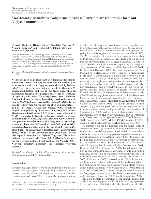
Fig. 1. Amino acid alignment of AtMANIa (At3g21160), AtMANIb (At1g51590), and G. max α-mannosidase (GmMANI) using CLUSTALW
(http://align.genome.jp/). Identical or similar sequences are shaded in black. The light gray box shows the putative transmembrane region. The potential
N-glycosylation sites, N-X-S/T, are labeled with asterisks. The conserved class I α1,2-mannosidase motifs are indicated by gray underlines.
�H Kajiura et al.
Fig. 2. Expression levels of AtMANIa and AtMANIb. The AtMANIa (dark
gray) and AtMANIb (light gray) expression levels (A) in A. thaliana rosette
leaves, cauline leaves, stems, and roots of Col-0 WT plants, and (B) in 4-, 8-,
12-, and 16-day-old A. thaliana T87 cultured cells were quantified by real-time
PCR. The relative transcript levels were calculated in comparison with those of
the rosette leaves of Col-0 WT and 4-day-old T87 cultured cells.
25◦ C. Furthermore, AtMANIa and AtMANIb were relatively
stable at lower temperature and still retained 50% of their original activities after incubation at 25◦ C for 1 h and showed a
significant decrease of activities at higher temperatures.
AtMANIs require Ca2+ for their optimum activities and were
inhibited by the addition of EDTA. AtMANIb activity in the
presence of EDTA was restored by the addition of Ca2+ . Other
metal ions such as Co2+ , Cu2+ , Mg2+ , Mn2+ , and Zn2+ had no
effect on their activities (data not shown). The MANI inhibitor
dMNj, but not the MANII inhibitor swainsonine, inhibited both
AtMANI activities (Table II). This establishes that AtMANIs
can hydrolyze the terminal α1,2-linked D-mannose residues
of M8A and M9 via the typical α-mannosidase I reaction
mechanism.
Distinct substrate specificity of AtMANIs suggests a synergistic
function of two isoforms
The structures of digestion intermediates of M8A or M9-PA
were analyzed by size fractionation (SF)- and reverse-phase
238
AtMANI double mutant plants accumulate only high-mannose
and oligomannosidic N-glycans
To determine the function of AtMANI isoforms in the production of complex N-glycans in planta, T-DNA insertion mutants
for AtMANIa and AtMANIb were identified in the SALK Institute Genome Analysis Laboratory (SIGnAL) T-DNA insertion
collection (Alonso et al. 2003). There are two independent alleles for each of the AtMANIa and AtMANIb genes, and they were
termed as manIa-1 (SALK_023251), manIa-2 (SALK_022849),
manIb-1 (SALK_076002), and manIb-2 (SALK_149737) (Figure 7A). Homozygous lines were identified by PCR using genomic DNA as templates (Figure 7B and data not shown), and
the T-DNA insertion position was confirmed by sequencing the
PCR products. The alleles manIa-1 and manIa-2 contain TDNAs in exon 4 and intron 5 of AtMANIa, whereas manIb1 and manIb-2 contain T-DNAs in intron 7 and exon 13 of
AtMANIb, respectively (Figure 7A).
The N-glycans of each line were PA-labeled and subjected
to RP-HPLC (Figure 8A) and SF-HPLC followed by matrixassisted laser desorption ionization time-of-flight (MALDITOF) mass spectrometry (MS) analysis. The relative amounts
of N-glycans detected in each line were calculated on the basis
of their SF-HPLC peak areas (Table III). All four manI single mutants showed N-glycan profiles similar to that of Col-0
WT and contained high-mannose-type and plant-specific hybrid or complex-type N-glycans. The potential redundancies
between AtMANI isoforms were tested using manIa manIb
double mutants. Homozygous double mutants were identified
in F2 generation after genetic crosses (Figure 7C and data
not shown). Double mutant plants were viable without obvious growth defects (data not shown). Interestingly, N-glycans
of all combinations of the double mutants tested show a
Downloaded from http://glycob.oxfordjournals.org/ by guest on December 1, 2015
(RP) high-performance liquid chromatography (HPLC) profiling using authentic PA-sugar chains as standards (supplementary Figure S2). The pathway for the trimming of M8A and
M9 was deduced as shown in Figure 6B and C, respectively.
AtMANIa and AtMANIb initially hydrolyzed mainly the α1,3linked terminal mannose residue (linkage III; Figure 6A) of
M8A, resulting in M7A. This contrasts with the reaction pathway of HsMANIs, which initiate the trimming at the branch of
the α1,6-linked terminal mannose residue (linkage I) and predominantly produce M7B. The ratios of M7A to M7B were 0.66
to 0.34 for AtMANIa and 0.71 to 0.29 for AtMANIb. Subsequent digestion of M7A and M7B yielded M6B, and the final
product was M5.
In the case of M9 trimming, however, the first removal
of the mannose residue was different between AtMANIa and
AtMANIb. AtMANIa tended to hydrolyze both α1,3- and α1,6linked terminal mannose residue. The ratios of M8A, M8B, and
M8C were 0.40, 0.48, and 0.12, respectively. On the other hand,
AtMANIb mainly hydrolyzed the α1,3-linked terminal mannose
residue (linkage III) and yielded M8B. The ratios of M8A, M8B,
and M8C were 0.18, 0.79, and 0.03, respectively. Although
the hydrolysis pathways were different between AtMANIa and
AtMANIb, all the intermediates were finally converted to the M5
structure, and no further trimmings into Man3 GlcNAc2 -PA or
ManGlcNAc2 -PA were detected. This indicates that AtMANIs
provide α-mannosidase activity in the maturation of N-glycans
in A. thaliana.
�AtMANIs are responsible for N-glycosylation in plant Golgi
Downloaded from http://glycob.oxfordjournals.org/ by guest on December 1, 2015
Fig. 3. Subcellular localization analysis of AtMANIa and AtMANIb in BY2 cultured cells. (A) Schematic of chimeric constructs used in this study. The putative
AtMANI cytosolic tail and transmembrane region were fused to GFP or to DsRed. The numbers indicate the length of the cytosolic tail and transmembrane region
of AtMANIa and AtMANIb. XylTCT -GFP was constructed for the control of the Golgi localization marker. (B) Dual-color imaging by confocal laser scanning
microscopy of transformed BY2 cultured cells. (Top) Cells coexpressing AtMANIaCT -DsRed and XylTCT -GFP. (Middle) Cells coexpressing AtMANIbCT -DsRed
and XylTCT -GFP. (Bottom) Cells coexpressing AtMANIaCT -DsRed and AtMANIbCT -GFP. Left, GFP fluorescence images; middle, DsRed fluorescence images;
right, merged images of GFP and DsRed. Bars = 10 µm.
predominant peak in RP-HPLC with the elution position corresponding to that of the authentic M8A (Figure 8C) as well
as small peaks corresponding to smaller oligomannosidic Nglycans. Other plant-specific hybrid and complex-type structures were not detected in their N-glycans. We noted that combinations that contain the manIb-2 allele produced a higher
level of smaller oligomannosidic N-glycans. This may indi-
cate that manIb-2 is a partial loss-of-function allele. Apparently, residual MANIb activities, if there are any, are not sufficient to drive the maturation of complex N-glycans in the
Golgi apparatus. Together, these results show that AtMANIa
and AtMANIb redundantly function in processing the M8A
form of N-glycans essential for the production of plant complex
N-glycans.
239
�H Kajiura et al.
Table II. Enzyme properties of recombinant AtMANIa and AtMANIb
expressed in E. coli. dMNj, 1-deoxymannojirimycin
Optimum pH
pH stability
Optimum temperature
AtMANIa
AtMANIb
6.0–8.0
5.0–10.0
5.0–6.0
4.5–6.5
20–30◦ C
Fig. 4. Purification of AtMANIa and AtMANIb expressed in E. coli. Purified
soluble AtMANI-(His)6 -fusion proteins were separated by SDS–PAGE and
detected by (i) CBB staining and (ii) using an anti-His-tag antibody. Two
active bands corresponding to the calculated size of AtMANIa and AtMANIb,
60 kDa, were detected in lanes 2 and 3, respectively. Lane 1 shows vector
control.
100%
600%
75%
35%
400%
10 µM dMNj
100 µM dMNj
50 µM swainsonine
500 µM swainsonine
70%
45%
100%
95%
Fig. 5. Result of SF-HPLC analysis of recombinant (i) AtMANIa and
(ii) AtMANIb reaction products. The reaction products of AtMANIa and
AtMANIb using M8A and M9 as substrates were subjected to SF-HPLC. The
peaks were identified by comparison with authentic PA-sugar chains, M5, M6,
M7, M8, and M9, as standards.
Discussion
α-Mannosidase I family proteins are type II membrane proteins localized in the early Golgi compartment and trim mannose residues from the M8 N-glycans exported from the ER.
In this study, we determined the in vitro and in vivo functions of two paralogous AtMANIs. The subcellular localization,
expression levels, and genetic redundancy of AtMANIs indicate that both AtMANIs participate in plant N-glycosylation
at the similar level. The biochemical characteristics of the
AtMANIs purified from E. coli are comparable with those of
human α1,2-mannosidases, which hydrolyze the high-mannosetype N-glycans (M6-M9 forms) to the M5 form. The activity of
AtMANIs, like with other α-mannosidases, is dependent on the
240
35%
20%
93%
90%
25◦ C (50%
relative activity)
30◦ C (10%
relative activity)
Ca2+ ion, and is inhibited not only by EDTA but also by a
specific MANI inhibitor, dMNj. Although the amino acid similarities between AtMANIs and HsMANIs are low, the amino
acids that coordinate a Ca2+ ion in animal MANIs (Vallee et al.
2000; Tempel et al. 2004) are highly conserved in AtMANIs.
Furthermore, when predicted three-dimensional structure models of AtMANIs are compared with a crystal structure model of
HsMANI, conformations of predicted AtMANIs’ catalytic sites
are similar to the dMNj-binding HsMANI catalytic site. The data
from biochemical analyses of AtMANIs were consistent with
these predictions. The N-glycan analysis data of manIa manIb
double mutant lines demonstrate that the AtMANI function is
required for the subsequent Golgi N-glycan modifications. However, processing the M8 form to smaller oligomannosidic forms
can occur at a reduced level even in the absence of AtMANIs.
Although the metal dependence and inhibitor sensitivity of
recombinant AtMANI isoforms are comparable, AtMANIa and
AtMANIb differ in their stability, hydrolytic pathway, and kinetics. The amino acid sequences of AtMANIa and AtMANIb
deduced from their cDNA sequences suggest that the each AtMANI has four potential N-glycosylation sites. In this study,
we used enzymatically active, unglycosylated AtMANIa and
AtMANIb produced in E. coli. Although it is not yet clear if
AtMANIa and AtMANIb are glycosylated in vivo, our data indicate that the non-glycosylated AtMANIa and AtMANIb are
catalytically active as has been shown for bacterially produced,
recombinant human MANI (Moran et al. 1998), GnTI that does
not have a potential N-glycosylation site (Fujiyama et al. 2001a),
GnTII (unpublished), β1,4-GalT (Shibatani et al. 2001), and
α2,6-sialyltransferase (Hidari et al. 2005). In these precedents,
the activities and properties of GnTI and β1,4-GalT were identical to those of the recombinant protein expressed in E. coli,
insects, fungal systems, or the purified native protein (Hollister
et al. 1998; Fujiyama et al. 2001a; Shibatani et al. 2001). Still,
at this point, no information is available for the enzymatic properties of glycosylated AtMANIs. Therefore, the interpretation
of our results should be limited to the unglycosylated forms
of AtMANIs. For example, we cannot exclude the possibility
Downloaded from http://glycob.oxfordjournals.org/ by guest on December 1, 2015
25◦ C (50%
relative activity)
30◦ C (10%
relative activity)
Metal-ion dependence (activity in the presence of)
No addition
100%
450%
15 mM CaCl2
95%
15 mM MgCl2
1 mM EDTA
20%
80%
1 mM EDTA + CaCl2
Temperature stability
�AtMANIs are responsible for N-glycosylation in plant Golgi
that N-glycan can affect the folding kinetics and/or stability of
AtMANIs. It should be noted, however, that protein stability
can vary substantially even between MANI isoforms produced
in a eukaryotic system (Lal et al. 1998). As for the AtMANIa
assay data, there was a lag period during the incubation before the hydrolytic activity was observed. At present, we cannot
delineate the cause of this observation. It may have been due
to partial unfolding during purification/dialysis, or due to its
slow binding kinetics to establish the initial substrate-enzyme
complex with M9 or M8. Further kinetic analyses are necessary to establish the isoform-specific characteristics of each
AtMANI.
In our N-glycan analysis of manIa manIb double mutant
lines, while all four double mutant lines lack complex-type
N-glycans and contain predominantly the M8 form of highmannose-type N-glycans, the amount of smaller oligomannosidic N-glycans varied among the lines. The double mutants
carrying the manIb-2 allele contain substantially smaller oligomannosidic N-glycans than plants with the manIb-1 allele. The
manIb-2 allele contains a T-DNA insertion in exon 13, which
is the most downstream T-DNA insertion site of all the manI
alleles used in this study. Therefore, it is possible that manIb-2
functions as a partial loss-of-function allele and produces a trace
amount of α-mannosidase activity that can be correctly targeted
to the Golgi apparatus; it is rather unlikely, however, that the
smaller oligomannosidic N-glycans detected are the direct products of AtMANIb-2 α-mannosidase activity. If AtMANIb-2 can
produce all the smaller oligomannosidic N-glycans observed,
the M5A structure produced in the double mutants, which is the
acceptor substrate for GnTI and the precursor of following Nglycan modifications, can be converted to complex N-glycans.
However, no plant-specific hybrid or complex-type N-glycans
were observed. This is indicative that the production of M5A
occurs in another subcellular compartment of the mutant.
241
Downloaded from http://glycob.oxfordjournals.org/ by guest on December 1, 2015
Fig. 6. Structure of Man9 GlcNAc2 oligosaccharide and deduced trimming pathways of AtMANIs. (A) Structure of Man9 GlcNAc2 . The linkage and labeling
designations for each of the mannose residues are shown. The four α1,2 linkages of Man9 GlcNAc2 are numbered. Reaction products of M8A (B) and M9
(C) substrates by recombinant AtMANIa (black) and AtMANIb (gray) were subjected to SF-HPLC, and each intermediate PA-sugar chain was collected and
subjected to subsequent RP-HPLC analysis. Their isoforms were determined by comparing with authentic PA-sugar chains. The relative ratios of the isoform
structures were calculated from the total concentration of processing intermediates. The nomenclatures used in this study were followed Yanagida et al. (1998).
�H Kajiura et al.
Which enzymes are responsible for the production of small
oligomannosidic N-glycans in the double mutants? Several
other genes encode α-mannosidase-like proteins in Arabidopsis.
These include three glycosyl hydrolase family 47 class I proteins and four glycosyl hydrolase family 38 proteins. Family 47
class I includes a putative ER-MANI (At1g30000) and unknown
proteins (At5g43710 and At1g27520) with limited similarity to
MANIs (20–26%). Family 38 proteins include α-mannosidase
II (AtGMII) (Strasser et al. 2006) and three α-mannosidaselike proteins (At3g26720, At5g13980, and At5g66150). Based
on the subcellular locations and substrate specificities, it is unlikely that currently known α-mannosidases in the Golgi and
242
Material and methods
Plant lines and DNA materials
Columbia T-DNA insertion mutants, SALK_023251
(AtMANIa-1), SALK_ 022849 (AtMANIa-2), SALK_076002
(AtMANIb-1), and SALK_149737 (AtMANIb-2) were identified
using the SIGnAL website at http://signal.salk.edu/ and
obtained from the Arabidopsis Biological Resource Center.
Mutant combinations of Columbia manIa/b genotypes were
prepared by genetic crossing, and their homozygous line was
identified by PCR methods as mentioned below.
Estimation of AtMANIa and AtMANIb mRNA expression levels
in A. thaliana T87 cultured cells and Col-0 WT plants by
real-time PCR
Total RNA was isolated from 4-, 8-, 12-, and 16-day-old A.
thaliana T87 cultured cells and from A. thaliana rosette leaves,
cauline leaves, stem, and root of Col-0 WT plants using the
Downloaded from http://glycob.oxfordjournals.org/ by guest on December 1, 2015
Fig. 7. Identification of homozygous manI mutants. (A) Schematic
representation of T-DNA insertion site in manI mutants. The DNA fragment
corresponding to AtMANIs was amplified by PCR using genomic DNA
extracted from each line as a template. The filled boxes indicate exons, and the
numbers and small arrows indicate the locations of the primers used for the
analysis. Primers 612 (AtMANIa-1-specific Fw primer) and 613
(AtMANIa-1-specific Re primer) were used to amplify wild-type AtMANIa-1
loci, and 188 (T-DNA-specific LBa1 primer) and 613 amplify the AtMANIa-1
mutant loci with the T-DNA insertion. Primers 608 (AtMANIb-1-specific Fw
primer) and 609 (AtMANIb-1-specific Re primer) amplify the wild-type
AtMANIb-1 loci, and 188 and 609 amplify the AtMANIb-1 mutant loci with the
T-DNA insertion. Other primers, 614, 615, and 608 to 611, were used to
amplify AtMANIa-2 loci and AtMANIb-2 loci and their mutant loci with the
T-DNA insertion. (B) PCR analysis of Col-0 WT and manI single mutants. The
DNA fragments corresponding to manIa-1, manIb-1, and T-DNA insertion loci
were PCR-amplified from genomic DNA extracted from Col-0 WT and
homozygous manI mutants. (C) PCR analysis of Col-0 WT and manI double
mutant. All the primer sets used for the detection of manIs loci are mentioned
above.
the ER, namely, AtGMII and the putative ER-MANI, produce
smaller oligomannosidic N-glycans (Herscovics 1999; Mast and
Moremen 2006). The other possible location of mannose trimming is in the vacuole (Boller and Kende 1979). Two family
38 mannosidases (At3g26720 and At5g13980) have been identified in a vacuolar proteome (Carter et al. 2004), and the αmannosidase activity of the At3g26720 gene product has been
demonstrated (Fujiyama et al. 2001b). Therefore, it is plausible
that some of the N-glycans in manIa manIb-2 plants were further processed in the vacuole. If this is the case, AtMANIb-2
may provide partial processing that may promote the vacuolar
mannose trimming, because N-glycans in manIa manIb-1 are
predominantly in the M8A form. This is also consistent with our
observation that AtMANIb can process the M8A form more efficiently than AtMANIa. At this point, vacuolar α-mannosidases
responsible for plant N-glycan trimming have not been identified, and therefore further studies are necessary to elucidate the
contribution of each α-mannosidase-like protein in the formation of mature N-glycan structures in plant cells.
Several studies have shown that N-glycosylation in the ER
is essential for the protein quality control process, and thus,
for the survival of plants. Blocking N-glycosylation/processing
in the ER by tunicamycin (Koizumi et al. 1999) or by pmm,
cyt1, rsw3, or knf/gcs1 mutation causes severe growth inhibition
and/or lethality (Nickle and Meinke 1998; Boisson et al. 2001;
Burn et al. 2002; Gillmor et al. 2002; Hoeberichts et al. 2008).
In contrast, studies on many N-glycan modification enzymes in
the Golgi apparatus did not reveal any phenotypic abnormality
in the loss-of-function mutant plants (von Schaewen et al. 1993;
Strasser et al. 2004, 2006, 2007). However, our recent study
demonstrated that N-glycan modifications in the Golgi apparatus
play important roles in plant osmotic stress tolerance and cell
wall biosynthesis through a function of the RSW2 protein (Kang
et al. 2008; von Schaewen et al. 2008). Under normal greenhouse
conditions, both manIa and manIb single mutants and manIa
manIb double mutants were viable and fertile with no obvious
growth phenotypes. This confirms the prediction that AtMANI
functions as the first enzyme in the Golgi pathway, which is
downstream of the essential protein quality control process in
the ER. Analysis of the growth response of manI mutant plants
under different environmental stress conditions is in progress.
�AtMANIs are responsible for N-glycosylation in plant Golgi
RNeasy Plant Mini Kit and treated with DNase (TaKaRa, Shiga,
Japan) to eliminate genomic DNA contamination. RNA aliquots
(0.5 µg) were reverse transcribed using the TaKaRa RNA PCR
Kit Ver.3.0 (TaKaRa).
R
�
Quantitative real-time PCR with the SYBR Green PCR
Master Mix (Applied Biosystems, Foster City, CA) was
performed with the prepared cDNA as a template using Gene
Amp 5700 (Applied Biosystems). In every real-time PCR run,
the A. thaliana ActII gene was used as a control to normalize the
amount of cDNA template. The specificity of the PCR amplification was confirmed by melt curve analysis. The amplification
data were processed using Gene Amp 5700 SDS software. The
reported values are the average of two independent trials with
similar results. The relative expression levels were calculated
as follows: on the basis of each tissue transcript level of the
ACTII gene, AtMANIa and AtMANIb transcript levels were
normalized and the tissue with the highest relative expression
was used as the standard for the comparison of every expression
level. The following gene-specific primer sets were used:
ActII gene (forward: 5′ -GACCTTTAACTCTCCCGCTATGTA3′ , reverse: 5′ -GTTGTGGTGAACATGTAACCTCTC-3′ ),
AtMANIa (forward: 5′ -GGGAGACAGTATTCTTGCAGATTC3′ , reverse: 5′ -GTAGACTGCGATGGATTAGCTGTA-3′ ),
AtMANIb (forward: 5′ -GGATATGTAGGCTTGAAGGATGTC3′ , reverse: 5′ -CTTCCTCTGGCGTAGTGCTATAGTT-3′ ).
Fluorescence reporter gene analysis
All fusion constructs were generated by PCR methods using
full-length AtMANIs, GFP, and DsRed cDNAs as templates,
and ligated with plant expression vectors pBI121 or pGPTVHPT. The resulting vectors, pBI121-XylTCT -GFP, pGPTV-HPTAtMANIaCT -DsRed, pGPTV-HPT-AtMANIbCT -DsRed, and
pGPTV-HPT-AtMANIaCT -DsRed-AtMANIbCT -GFP, were introduced into Agrobacterium tumefaciens LBA4404 via electroporation. Tobacco BY-2 cells were first transformed with
pBI121-XylTCT -GFP as described previously (Palacpac et al.
1999). Cells stably expressing XylTCT -GFP were selected and
supertransformed with DsRed fusion constructs. Fluorescence
signals were documented 3–4 days after the second transformation. Cells expressing GFP or DsRed fusion proteins were
analyzed with DIGITAL ECLIPSE C1si (Nikon, Tokyo, Japan)
equipped with CFI Plan Apo objectives, and EZ-C1 3.40 software (Nikon). Fluorescence was excited with the 488-nm line
of a solid laser (20 mW) and the 543-nm line of a G-HeNe laser
(10 mW) with laser power set to 1.0–5.0%. Image processing
243
Downloaded from http://glycob.oxfordjournals.org/ by guest on December 1, 2015
Fig. 8. N-Glycan analysis of A. thaliana Col-0 WT, manIa-1, manIa-2, manIb-1, manIb-2, and manIa manIb double mutant combinations. (A) Total N-glycans
prepared from glycoproteins and labeled with PA were analyzed by RP-HPLC with a C18 column. (i) Col-0 WT, (ii) manIa-1, (iii) manIa-2, (iv) manIb-1,
(v) manIb-2, (vi) manIa-1/b-1, (vii) manIa-1/b-2, (viii) manIa-2/b-1, and (ix) manIa-2/b-2. (B) MALDI-TOF MS analysis of the predominant PA-sugar chain from
the manIa-1/b-1 double mutant. The labeled peak represents the (M + Na)+ ion. Only one signal, (M8 + Na)+ , was observed. (C) An RP-HPLC profile of M8
structure derived from the manIa-1/b-1 double mutant: (i) authentic PA-sugar chain, M8A, M8B, and M8C, and (ii) the predominant PA-sugar chain from
manIa-1/b-1. The retention time of the manIa-1/b-1 PA-sugar chain corresponds to that of the M8A PA-sugar chain.
�H Kajiura et al.
Table III. Comparison and relative amount of N-glycan structures detected in Col-0 WT, manIa-1, manIa-2, manIb-1, or manIb-2 single mutant, and
manIa-1/manIb-1, manIa-1/manIb-2, manIa-2/manIb-1, or manIa-2/manIb-2 double mutant.
Ratio (%)
Structure
Col-0 WT
manIa-1
manIb-1
manIa-2
manIb-2
manIa-1/manIb-1
manIa-1/manIb-2
manIa-2/manIb-1
manIa-2/manIb-2
M5A
M6B
M7A
M8A
M9
M3FX
GnM3FX
M3X
Gn2M3X
Gn2M3FX
43.1
–
14.7
7.7
5.9
16.4
3.5
8.8
–
–
23.3
11.6
12.9
7.9
4.8
18.2
2.7
7.3
1.0
10.4
35.2
3.4
22.6
7.2
2.5
14.9
8.6
5.6
–
–
40.8
14.7
12.5
11.0
7.5
5.5
2.7
5.3
–
–
42.3
7.7
9.7
13.5
5.8
4.8
2.1
14.2
–
–
–
7.4
6.2
86.4
–
–
–
–
–
–
9.2
14.6
32.8
38.2
5.2
–
–
–
–
–
–
3.5
12.0
80.2
4.3
–
–
–
–
–
10.3
11.7
31.3
41.3
5.5
–
–
–
–
–
The relative ratio of the structures was calculated on the basis of the peak area as determined by HPLC.
Cloning and construction of E. coli expression vectors
containing each truncated form of two A. thaliana
α-mannosidases
Total RNA was isolated from A. thaliana rosette leaves using
the RNeasy Plant Mini kit (QIAGEN, Chatsworth, CA) and
was reverse transcribed into cDNA using the RNA PCR kit
Ver.2.1 (TaKaRa). The coding region of the two A. thaliana αmannosidases, AtMANIa and AtMANIb, was amplified from the
reverse transcription products using the KOD plus polymerase
(TOYOBO, Osaka, Japan) and primer sets (AtMANIa forward:
ATTGGATCCATGGCGAGGAATAAACTTGTA,
reverse:
ATTGAGCTCTTACTTCTTTGTTATCCGACC,
AtMANIb
forward:
ATTGGATCCATGGCGAGAAGTAGATCGATT,
reverse:
ATTGTCGACCTAAACGTTAATCTGATGACC).
The PCR products were subcloned into a pGEM T-Easy vector
(Promega, Tokyo, Japan), followed by sequencing using the
ABI PRISMTM Big DyeTM Terminator cycle sequencing kit
(Applied Biosystems).
cDNA fragments encoding soluble forms of AtMANIa and
AtMANIb were amplified from full-length AtMANI cDNAs
using the KOD plus polymerase and primer sets (AtMANIa
forward:
ATTCATATGGATCGTCAATCTCTTTCCCGA,
reverse:
ATTCTCGAGCTTCTTTGTTATCCGACCATA,
AtMANIb forward: ATTCATATGGATCGTATTAATCTTGCCCGA, reverse: ATTGTCGACAACGTTAATCTGATGACCAAA). The PCR products were subcloned into a pGEM
T-Easy vector, followed by sequencing, restriction enzyme
digestion, and ligation with a pET-23b vector (Novagen,
Darmstadt, Germany) to produce AtMANI-(His)6 fusion
proteins.
Expression of AtMANIa and AtMANIb in E. coli and their
purifications
Soluble AtMANI proteins were produced in E. coli Rosetta gami
B (DE3) (Novagen) harboring pET-AtMANIa or AtMANIb. The
cells were first cultivated in 2 mL of 2× YT medium (16 g L−1
tryptone, 10 g L−1 yeast extract, and 5 g L−1 NaCl) containing
50 µg mL−1 ampicillin for 12–14 h at 37◦ C. Two milliliters
of the preculture was transferred into 200 mL of the same
medium and further incubated until the O.D.600 reached 0.5. Af244
ter the incubation, IPTG induction was started by the addition of
1 mM IPTG, followed by incubation for 20 h at 15◦ C. The recombinant proteins were purified using a Co2+ column (Nexus
IMAC Resin, Valen, Atlanta, GA) following the manufacturer’s
protocols. The eluted fraction was dialyzed against a 1 mM
phosphate buffer (pH 6.0).
Western blot analysis of truncated and His-tag fusion
AtMANIa and AtMANIb
The purified protein samples were subjected to 12.5% SDS–
PAGE under reducing condition. The fractionated proteins
were electroblotted onto a PVDF membrane. The membrane
was incubated in phosphate-buffered saline (PBS) with 5%
skim milk at 4◦ C overnight and probed with a 1:1000-diluted
mouse anti-(His)5 -tag antibody (ZYMED, Invitrogen, Carlsbad,
CA). The membrane was then washed three times and
then placed in PBS with 5% skim milk containing 1:1000diluted peroxidase-conjugated anti-mouse IgG (Amersham,
Buckinghamshire, England). Specific bands were visualized using the POD immunostain set (Wako, Osaka, Japan) or chemiluminescent detection (ECL Plus Western Blotting Detection
System, Amersham).
Activity assays for AtMANIa and AtMANIb
AtMANIa and AtMANIb activities were assayed using PAlabeled sugar chains M8A and Man9 GlcNAc2 (M9) (TaKaRa)
as the substrates. The enzyme assays for purified AtMANIa
and AtMANIb were performed in 100 µL of total reaction volume containing a 20 mM cacodylic acid buffer (pH 7.0) for
AtMANIa activity or a 20 mM acetate buffer (pH 6.0) for AtMANIb activity, 15 mM CaCl2 , 10 pmol PA-labeled M8A or
M9, and 80 µL of dialyzed enzyme solution at 25◦ C for 24 h.
The enzymatic reactions were terminated by boiling for 5 min.
The samples were centrifuged at 4◦ C, 15,000 rpm, for 5 min, and
the supernatants were mixed with 4 volumes of acetonitrile and
subjected to SF-HPLC analysis as described previously (Misaki
et al. 2001).
Determination of optimal pH and temperature
The optimal pH for AtMANIa and AtMANIb activities was
determined by varying the pH between 4.0 and 10.0 at 0.5
interval. The reactions were carried out at 25◦ C for 24 h using 10 pmol PA-labeled Man6 GlcNAc2 (M6B). The enzymatic
Downloaded from http://glycob.oxfordjournals.org/ by guest on December 1, 2015
for both GFP and DsRed coloration was performed using Adobe
Photoshop CS3.
�AtMANIs are responsible for N-glycosylation in plant Golgi
activities were calculated as percent relative to the highest AtMANI activity sample observed in each assay.
To determine the optimal reaction temperature, the enzyme
reactions were performed at 0, 10, 20, 25, 30, 40, 50, 60, and
70◦ C using 10 pmol PA-labeled Man6 GlcNAc2 (M6B) as the
substrate. As in the pH analysis, the results are presented as%
relative activity against the highest sample.
Effect of metal ions and inhibitors on enzyme activity
To determine the effect of metal ions, the reaction was performed
as described previously (Moran et al. 1998).
Supplementary Data
Determination of the structure of digestion intermediates in the
AtMANIa and AtMANIb reaction
The enzyme assays for the trimming of the PA-sugar chains were
performed as described above. The reaction was monitored for
48 h. Samples were collected every 2 h in the first 12 h and every
12 h after 24 h from the start of the reaction and were analyzed
by SF-HPLC.
In the SF-HPLC analysis, the intermediates of the reaction
product resulting from the trimming of the substrate PA-sugar
chain were collected and subjected to RP-HPLC to determine
the isoform of each structure by comparing with the authentic
PA-sugar chains.
Acknowledgements
Identification of T-DNA insertion mutants
The screening of homozygous seedlings and the detection of
T-DNA localization were performed by PCR using the following primer sets: AtMANIa-1, forward primer, 612, (5′ -GTCACT
GAGGATGTGTCTGTCA-3′ ) and reverse primer, 613, (5′ GGGAATCGAACTTCAGTTAATGGA-3′ ) as well as a TDNA-specific primer, 188 (LBa1), (5′ -TGGTTCACGTAG
TGGGCCATCG-3′ ); AtMANIa-2, forward primer, 614, (5′ CAGGCTTGTCTCATCTTATTGAGA -3′ ) and reverse primer,
615, (5′ -TCGACAAGGAATATGACAGATAGGA -3′ ) and the
T-DNA-specific primer; AtMANb-1, forward primer, 608,
(5′ -GAGGACATGTGCTGTGTCAGT-3′ ) and reverse primer,
609, (5′ -ATGGTCACAAGACCCGACCA-3′ ) and the TDNA-specific primer; AtMANb-2, forward primer, 610, (5′ GAGTATAACTTTCTGCTGTGTCCT -3′ ) and reverse primer,
611, (5′ -CCACTCGAGGAGGGTATCTGA -3′ ) and the TDNA-specific primer.
Supplementary data for this article is available online at
http://glycob.oxfordjournals.org/.
We thank the ABRC for providing the sequence-indexed Arabidopsis T-DNA insertion mutants. We also thank Dr. Kanaya
(Osaka University) for his helpful discussions on the AtManI
activity assays.
Abbreviations
cgl1, complex glycan 1; CT, cytosolic tail and transmembrane region; ER, endoplasmic reticulum; FUCT, α1,3fucoyltransferase; GALT, β1,3-galactosyltransferase; GnTI,
N-acetylglucosaminlytransferase I; HPLC, high-performance
liquid chromatography; ManI, α1,2-mannosidase I; MALDITOF, matrix-assisted laser desorption ionization time-of-flight;
MS, mass spectrometry; OST, oligosaccharyltransferase; PA,
2-pyridylamino; RP, reverse phase; SIGnAL, SALK Institute
Genome Analysis Laboratory; SF, size fractionation; XYLT,
β1,2-xylosyltransferase.
References
Akao T, Yamaguchi M, Yahara A, Yoshiuchi K, Fujita H, Yamada O, Akita
O, Ohmachi T, Asada Y, Yoshida T. 2006. Cloning and expression of
1,2-a-mannosidase gene (fmanIB) from filamentous fungus Aspergillus
oryzae: In vivo visualization of the FmanIBp-GFP fusion protein. Biosci
Biotechnol Biochem. 70:471–479.
Alonso JM, Stepanova AN, Leisse TJ, Kim CJ, Chen H, Shinn P, Stevenson DK,
Zimmerman J, Barajas P, Cheuk R, et al. 2003. Genome-wide insertional
mutagenesis of Arabidopsis thaliana. Science. 301:653–657.
Boisson M, Gomord V, Audran C, Berger N, Dubreucq B, Granier F, Lerouge
P, Faye L, Caboche M, Lepiniec L. 2001. Arabidopsis glucosidase I mutants
245
Downloaded from http://glycob.oxfordjournals.org/ by guest on December 1, 2015
Effect of pH and temperature on enzyme activity and stability
For the determination of pH stability, the enzyme solution was
incubated with 10 µL of the following buffers at 200 mM: acetate buffer (pH 4.0–6.0), cacodylic acid (pH 6.0–8.0) or phosphate buffer (pH 6.0–8.0), Tris-HCl buffer (pH 8.0–9.0), and
boric acid buffer (pH 9.0–10.0) at 25◦ C for 1 h. Then, 10 µL of
1 M cacodylic acid buffer (pH 7.0) for AtMANIa or 1 M acetate
buffer (pH 6.0) for AtMANIb and 150 mM of CaCl2 were added,
followed by further incubation at 25◦ C for 24 h. The results are
presented as% relative activity against the highest sample.
For the determination of temperature stability, the enzyme
solution was incubated at various temperatures for 1 h and then
cooled on ice for 5 min. The activity assay was performed under
standard conditions.
Preparation of N-glycans and their structural analysis by
HPLC
Soil-grown A. thaliana Col-0 WT and T-DNA lines,
SALK_023251 (manIa-1), SALK_ 022849 (manIa-2),
SALK_076002 (manIb-1), and SALK_149737 (manIb-2), and
manIa manIb double mutants were disrupted by homogenization
and PA-labeled as described (Misaki et al. 2001). The reaction
products of the enzyme assays were resolved by SF-HPLC or
RP-HPLC as described previously (Misaki et al. 2001).
The PA-sugar chains eluted by SF-HPLC were collected
and lyophilized. The molecular mass of the PA-sugar chain
was estimated by MALDI-TOF MS on an Autoflex (BRUKER
DALTONICS) operated in the positive ion and reflector modes
using 2,5-dihydroxybenzoic acid (Sigma, St. Louis, MO) as a
matrix.
On the basis of the possible structure deduced from the
MALDI-TOF MS analysis, PA-sugar chains were digested with
suitable exogylcosidases, as described previously (Misaki et al.
2001). Their elution positions were compared with those of authentic PA-sugar chains purchased from TaKaRa and M3FX
prepared from horseradish peroxidase as described above. The
authentic PA-sugar chain isoforms are listed in Figure 5.
�H Kajiura et al.
246
involved in the processing of asparagine-linked oligosaccharides. J Biol
Chem. 269:9872–9881.
Léonard R, Costa G, Darrambide E, Lhernould S, Fleurat-Lessard P, Carlué
M, Gomord V, Faye L, Maftah A. 2002. The presence of Lewis a epitopes in Arabidopsis thaliana glycoconjugates depends on an active a4fucosyltransferase gene. Glycobiology. 12:299–306.
Lerouxel O, Mouille G, Andème-Onzighi C, Bruyant MP, Séveno M, LoutelierBourhis C, Driouich A, Höfte H, Lerouge P. 2005. Mutants in DEFECTIVE
GLYCOSYLATION, an Arabidopsis homolog of an oligosaccharyltransferase complex subunit, show protein underglycosylation and defects in cell
differentiation and growth. Plant J. 42:455–468
Leroy JG. 2006. Congenital disorders of N-glycosylation including diseases
associated with O- as well as N-glycosylation defects. Pediatr Res. 60:643–
656.
Martı́nez IM, Chrispeels MJ. 2003. Genomic analysis of the unfolded protein response in Arabidopsis shows its connection to important cellular processes.
Plant Cell. 15:561–576.
Mast SW, Moremen KW. 2006. Family 47 a-mannosidases in N-glycan processing. Methods Enzymol. 415:31–46.
Misaki R, Kimura Y, Fujiyama K, Seki T. 2001. Glycoproteins secreted
from suspension-cultured tobacco BY2 cells have distinct glycan structures from intracellular glycoproteins. Biosci Biotechnol Biochem. 65:2482–
2488.
Moran DG, Sakuradani S, Jun KH, Fujiyama K, Seki T, Yoshida T. 1998.
Characterization of recombinant human Man9 -mannosidase expressed in
Escherichia coli. J Ferment Bioeng. 86:277–283.
Nebenführ A, Gallagher LA, Dunahay TG, Frohlick JA, Mazurkiewicz AM,
Meehl JB, Staehelin LA. 1999. Stop-and-go movements of plant Golgi
stacks are mediated by the acto-myosin system. Plant Physiol. 121:1127–
1142.
Nickle TC, Meinke DW. 1998. A cytokinesis-defective mutant of Arabidopsis (cyt1) characterized by embryonic lethality, incomplete cell walls, and
excessive callose accumulation. Plant J. 15:321–332.
Pagny S, Bouissonnie F, Sarkar M, Follet-Gueye ML, Driouich A, Schachter H,
Faye L, Gomord V. 2003. Structural requirements for Arabidopsis b1,2xylosyltransferase activity and targeting to the Golgi. Plant J. 33:189–
203.
Palacpac NQ, Yoshida S, Sakai H, Kimura Y, Fujiyama K, Yoshida T, Seki
T. 1999. Stable expression of human β1,4-galactosyltransferase in plant
cells modifies N-linked glycosylation patterns. Proc Natl Acad Sci USA.
96:4692–4697.
Saint-Jore-Dupas C, Nebenführ A, Boulaflous A, Follet-Gueye M-L, Plasson
C, Hawes C, Driouich A, Faye L, Gomord V. 2006. Plant N-glycan processing enzymes employ different targeting mechanisms for their spatial
arrangement along the secretory pathway. Plant Cell. 18:3182–3200.
Shibatani S, Fujiyama K, Nishiguchi S, Seki T, Maekawa Y. 2001. Production
and characterization of active soluble human b1,4-galactosyltransferase in
Escherichia coli as a useful catalyst in synthesis of the Gal b1→4 GlcNAc
linkage. J Biosci Bioeng. 91:85–87.
Steinkellner H, Strasser R. 2003. Glycosyltransferases in the plant Golgi. In
Robinson DG, editor. The Golgi Apparatus and the Plant Secretory Pathway.
Vol 9. Oxford: Blackwell Publishing. p. 181–192.
Strasser R, Altmann F, Mach L, Glössl J, Steinkellner H. 2004. Generation of
Arabidopsis thaliana plants with complex N-glycans lacking b1,2-linked
xylose and core a1,3-linked fucose. FEBS Lett. 12:132–136.
Strasser R, Bondili JS, Vavra U, Schoberer J, Svoboda B, Glössl J, Léonard
R, Stadlmann J, Altmann F, Steinkellner H et al. 2007. A unique b1,3galactosyltransferase is indispensable for the biosynthesis of N-glycans
containing Lewis a structures in Arabidopsis thaliana. Plant Cell. 19:2278–
2292.
Strasser R, Mucha J, Mach L, Altmann F, Wilson IBH, Glössl J,
Steinkellner H. 2000. Molecular cloning and functional expression of b1,2xylosyltransferase cDNA from Arabidopsis thaliana. FEBS Lett. 472:105–
108.
Strasser R, Schoberer J, Jin C, Glossl J, Mach L, Steinkellner H. 2006. Molecular
cloning and characterization of Arabidopsis thaliana Golgi a-mannosidase
II, a key enzyme in the formation of complex N-glycans in plants. Plant J.
45:789–803.
Tempel W, Karaveg K, Liu ZJ, Rose J, Wang BC, Moremen KW. 2004. Structure of mouse Golgi alpha-mannosidase IA reveals the molecular basis for
substrate specificity among class 1 (family 47 glycosylhydrolase) alpha1,2mannosidases. J Biol Chem. 279:29774–29786.
Tremblay LO, Campbell Dyke N, Herscovics A. 1998. Molecular cloning,
chromosomal mapping and tissue-specific expression of a novel human
Downloaded from http://glycob.oxfordjournals.org/ by guest on December 1, 2015
reveal a critical role of N-glycan trimming in seed development. EMBO J.
20:1010–1019.
Boller T, Kende H. 1979. Hydrolytic enzymes in the central vacuole of plant
cells. Plant Physiol. 63:1123–1132.
Bondili JS, Castilho A, Mach L, Glössl J, Steinkellner H, Altmann F,
Strasser R. 2006. Molecular cloning and heterologous expression of b1,2xylosyltransferase and core a1,3-fucosyltransferase from maize. Phytochemistry. 67:2215–2224.
Burn JE, Hurley UA, Birch RJ, Arioli T, Cork A, Williamson RE. 2002. The
cellulose-deficient Arabidopsis mutant rsw3 is defective in a gene encoding
a putative glucosidase II, an enzyme processing N-glycans during ER quality
control. Plant J. 32:949–960.
Carter C, Pan S, Zouhar J, Avila EL, Girke T, Raikhel NV. 2004. The vegetative
vacuole proteome of Arabidopsis thaliana reveals predicted and unexpected
proteins. Plant Cell. 16:3285–3303.
Fuhrmann U, Bause E, Legler G, Ploegh H. 1984. Novel mannosidase inhibitor
blocking conversion of high mannose to complex oligosaccharides. Nature.
307:755–758.
Fujiyama K, Ido Y, Misaki R, Moran DG, Yanagihara I, Honda T, Nishimura S,
Yoshida T, Seki T. 2001a. Human N-acetylglucosaminyltransferase I. Expression in Escherichia coli as a soluble enzyme, and application as an immobilized enzyme for the chemoenzymatic synthesis of N-linked oligosaccharides. J Biosci Bioeng. 92:569–574.
Fujiyama K, Kira Y, Iizuka M, Kimura Y, Seki T. 2001b. Identification of
putative gene encoded on ORF16 of the 81 kb contig of Arabidopsis
thaliana chromosome III as alpha-mannosidase. J Biosci Bioeng. 92:401–
404.
Gillmor CS, Poindexter P, Lorieau J, Palcic MM, Somerville C. 2002. Alphaglucosidase I is required for cellulose biosynthesis and morphogenesis in
Arabidopsis. J Cell Biol. 156:1003–1013.
Haltiwanger RS, Lowe JB. 2004. Role of glycosylation in development. Annu
Rev Biochem. 73:491–537.
Helenius A, Aebi M. 2001. Intracellular functions of N-linked glycans. Science.
291:2364–2369.
Herscovics A. 1999. Importance of glycosidases in mammalian glycoprotein
biosynthesis. Biochim Biophys Acta. 1473:96–107.
Herscovics A, Schneikert J, Athanassiadis A, Moremen KW. 1994. Isolation of
a mouse Golgi mannosidase cDNA, a member of a gene family conserved
from yeast to mammals. J Biol Chem. 269:9864–9871.
Hoeberichts FA, Vaeck E, Kiddle G, Coppens E, van de Cotte B,
Adamantidis A, Ormenese S, Foyer CH, Zabeau M, Inzé D, et al. 2008
A temperature-sensitive mutation in the Arabidopsis thaliana phosphomannomutase gene disrupts protein glycosylation and triggers cell death. J Biol
Chem. 283:5708–5718.
Hollister JR, Shaper JH, Jarvis DL. 1998. Stable expression of mammalian b1,4galactosyltransferase extends the N-glycosylation pathway in insect cells.
Glycobiology. 8:473–480.
Hidari KI, Horie N, Murata T, Miyamoto D, Suzuki T, Usui T, Suzuki Y. 2005.
Purification and characterization of a soluble recombinant human ST6Gal I
functionally expressed in Escherichia coli. Glycoconj J. 22:1–11.
Kang JS, Frank J, Kang CH, Kajiura H, Vikram M, Ueda A, Kim S, Bahk JD,
Triplett B, Fujiyama K, et al. 2008. Salt tolerance of Arabidopsis thaliana
requires maturation of N-glycosylated proteins in the Golgi apparatus. Proc
Natl Acad Sci USA. 105:5933–5938.
Kawar Z, Romero PA, Herscovics A, Jarvis DL. 2000. N-Glycan processing by
a lepidopteran insect a1,2-mannosidase. Glycobiology. 10:347–355.
Kelleher DJ, Gilmore R. 2006. An evolving view of the eukaryotic oligosaccharyltransferase. Glycobiology. 16:47R–62R.
Koiwa H, Li F, McCully MG, Mendoza I, Koizumi N, Manabe Y, Nakagawa
Y, Zhu J, Rus A, Pardo JM, et al. 2003. The STT3a subunit isoform of
the Arabidopsis oligosaccharyltransferase controls adaptive responses to
salt/osmotic stress. Plant Cell. 15:2273–2284.
Koizumi N, Ujino T, Sano H, Chrispeels MJ. 1999. Overexpression of a gene that
encodes the first enzyme in the biosynthesis of asparagine-linked glycans
makes plants resistant to tunicamycin and obviates the tunicamycin-induced
unfolded protein response. Plant Physiol. 121:353–361.
Kornfeld R, Kornfeld S. 1985. Assembly of asparagine-linked oligosaccharides.
Annu Rev Biochem. 54:631–664.
Lal A, Pang P, Kalelkar S, Romero, PA, Herscovics A, Moremen KW. 1998.
Substrate specificities of recombinant murine Golgi a1,2-mannosidases IA
and IB and comparison with endoplasmic reticulum and Golgi processing
a1,2-mannosidases. Glycobiology. 8:981–995.
Lal A, Schutzbach JS, Forsee WT, Neame PJ, Moremen KW. 1994. Isolation
and expression of murine and rabbit cDNAs encoding an a1,2-mannosidase
�AtMANIs are responsible for N-glycosylation in plant Golgi
a1,2-mannosidase gene involved in N-glycan maturation. Glycobiology.
8:585–595.
Tremblay LO, Herscovics A. 1999. Cloning and expression of a specific human
a1,2-mannosidase that trims Man9 GlcNAc2 to Man8 GlcNAc2 isomer B
during N-glycan biosynthesis. Glycobiology. 9:1073–1078.
Tremblay LO, Herscovics A. 2000. Characterization of a cDNA encoding a novel
human Golgi a 1, 2-mannosidase (IC) involved in N-glycan biosynthesis.
J Biol Chem. 275:31655–31660.
Vallee F, Karaveg K, Herscovics A, Moremen KW, Howell PL. 2000. Structural basis for catalysis and inhibition of N-glycan processing class I a1,2mannosidases. J Biol Chem. 275:41287–41298.
Varki A. 1993. Biological roles of oligosaccharides: All of the theories are
correct. Glycobiology. 3:97–130.
von Schaewen A, Frank J, Koiwa H. 2008. Role of complex N-glycans in plant
stress tolerance. Plant Signaling Behav. 3:871–873.
von Schaewen A, Sturm A, O’Neill J, Chrispeels MJ. 1993. Isolation of a
mutant Arabidopsis plant that lacks N-acetyl glucosaminyl transferase I and
is unable to synthesize Golgi-modified complex N-linked glycans. Plant
Physiol. 102:1109–1118.
Wilson IBH, Rendić D, Freilinger A, Dumić J, Altmann F, Mucha J, Müller
S, Hauser MT. 2001. Cloning and expression of cDNAs encoding a1,3fucosyltransferase homologues from Arabidopsis thaliana. Biochim Biophys
Acta. 1527:88–96.
Yanagida K, Ogawa H, Omichi K, Hase S. 1998. Introduction of a new scale into
reversed-phase high-performance liquid chromatography of pyridylamino
sugar chains for structural assignment. J Chromatogr A. 800:187–198.
Downloaded from http://glycob.oxfordjournals.org/ by guest on December 1, 2015
247
�
 A. Okazawa