Characteristics of hypoxemic episodes
in very low birth weight infants on
ventilatory support
Mary A n n V. T. Dimaguila, MD, Juliann M. Di Fiore, BSEE,
Richard J. Martin, MD, a n d M a r t h a J. Miller, PhD, MD
From the Department of Pediatrics, Rainbow Babies and Childrens Hospital, Case Western
Reserve University, Cleveland, Ohio
Objective: To characterize hypoxemic episodes in very low birth weight infants
with mechanically ventilated lungs and to describe their natural history and the
effect of body position.
Study design: Tidal volume, respiratory rate, oxygen saturation, heart rate, and
body movement were continuously recorded in 10 very low birth weight infants
who exhibited episodes of hypoxemia during mechanical ventilation (birth
weight, 810 ± 133 gm; postconceptional age at study, 30 ± 1.6 weeks). Frequency of hypoxemic episodes was compared in both prone and supine positions.
Results:Seventy-eight percent of hypoxemic episodes began in association with
body movement as well as heart rate acceleration. Thereafter the spontaneous
and delivered minute ventilation both decreased during the first 15 seconds of
hypoxemia. The former decrease was due to a significant decrease in frequency
of spontaneous respiration, whereas the latter was associated with a significant
decrease in delivered tidal volume. Minute ventilation returned to normal before
recovery of oxygenation. A change in body position from supine to prone significantly decreased the frequency of hypoxemic episodes.
Conclusion: Hypoxemic episodes in infants who are on ventilatory support are
characterized by (I) movement and cardioacceleration at initiation; (2) a
decrease in both spontaneous and delivered minute ventilation, and (3) a lower
incidence in the prone position. We speculate that spontaneous movement during sleep can trigger cardiopulmonary reflex responses that initiate and propagate these episodes. (J Pediatr 1997; 130:577-83)
Hypoxemic episodes are a common occurrence in very low
birth weight infants during mechanical ventilation. Neither
the cause of these episodes nor their relationship to apnea of
prematurity is completely understood, and no trials documenting effective therapeutic interventions have been reported. In VLBW infants who are not on ventilatory support,
Supported by a grant from Wyeth Research.
Submitted for publication April 25, 1996; accepted Sept. 27, 1996.
Reprint requests: Martha J. Miller, PhD, MD, Department of Pediatrics, Rainbow Babies and Childrens Hospital, 11100 Euclid Ave.,
Cleveland, OH 44106-6010.
Copyright © 1997 by Mosby-Year Book, Inc.
0022-3476/97/$5.00 + 0 9/21/78374
respiratory instability leading to hypoxemia may be caused
by central inhibition of the respiratory drive coupled with
inadequate function of upper airway dilator muscles, leading
to upper airway obstruction. 1~ In infants who are on ventilatory support, the upper airway is bypassed and the cause
of hypoxemia must be related to an alteration in respiraVLBW
Very low birth weight
tory drive, or alveolar ventilation with a resultant change in
the balance of ventilation-perfusion. As first described by
Bolivar et al. 5 in 1995, changes in the respiratory pattern
and pulmonary mechanics can occur in association with ep-
577
�578
Dimaguila et al.
The Journal of Pediatrics
April 1997
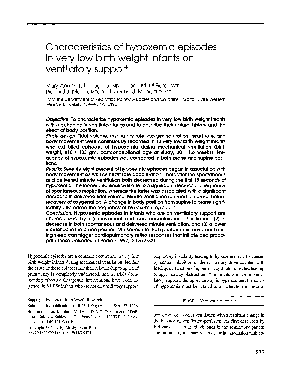
Pulse
Oximeter
Ouput
o~
Saturation
(~)
100 1
50
0
Heart Rate
(bpm)
0
Tidal Volume
(cc)
-25 ~
5 r
q
.
'~
Flow
(L/min)
10 see
Fig. l. Representative episode of hypoxemia in a 31-week infant on ventilatory support was characterized by (a) an initial
brief movement accompanied by an increase in baseline heart rate; (b) bradycardia with cardioacceleration accompanying
mechanically delivered breaths; (c) cessation of spontaneous breaths; and (d) decrease in delivered tidal volume. Pulse
oximeter output is the light phlethysmographic waveform.
isodes of crying and agitation in premature infants with an
endotracheal tube in place and can lead to prolonged hypoxemia.
In our nursery, we noted that hypoxemia in premature infants who were on ventilatory support could occur without
agitation and in some cases could be accompanied by central apnea. This study was therefore designed (1) to explore
changes in the spontaneous movement, respiratory output,
and cardiovascular reflex response that precede and accompany episodes of hypoxemia in VLBW infants on ventilatory
support and (2) to compare these characteristics with those
which we previously described as accompanying apnea of
prematurity.6 On the basis of our observations, we hypothesized that episodes of hypoxemia could involve both a decline in respiratory frequency and an alteration in pulmonary
mechanics, variably linked to periods of spontaneous movement and arousal. Furthermore, because change in body position may influence sleep state, ventilation, and apnea frequency in infants, v' 8 we compared the frequency of hypoxemic events in the prone and the supine positions to test
prone body position as a simple therapeutic modality.
METHODS
The study population consisted of 10 premature infants:
birth weight, 810 +- 133 gm; mean postconceptional age at
study, 30.0 -+ 1.6 weeks; postnatal age at study, 4.1 _+ 1.5
weeks. The size of the cohort for this study was estimated
to be adequate to detect a 50% difference between variables
or conditions with a p value of less than 0.05 and [3 equal to
0.10. All infants had exhibited at least five intermittent episodes of hypoxemia and bradycardia during a 24-hour pe-
riod during mechanical ventilation before entry into the
study. All infants were receiving ventilatory support (intermittent mechanical ventilation, <20; fractional inspired oxy g e n , <35%) (Infant Star, Infrasonics, San Diego, Calif.),
and none were sedated at the time of study. Five of the ten
infants were receiving aminophylline therapy (mean serum
theophylline level = 6 +_ 0.7 mg/dl). Subjects were excluded
from the study if any of the following were present: (1) congenital anomalies, including any major cardiac anomaly, (2)
grade IV intraventricular hemorrhage, (3) seizures, and (4)
sepsis documented by positive blood culture result. The investigation was approved by the institutional human research
coimnittee, and informed consent was obtained from the
parents.
Infants were studied for 3 hours while in their incubator
in the neonatal intensive care unit. At least two of the four
authors observed the infants continuously during the study.
For the purpose of this study, hypoxemia was defined as oxygen saturation measured by pulse oximeter of less than
90%, lasting for at least 20 seconds. If 02 saturation fell to
less than 75%, fractional inspired oxygen, intermittent mechanical ventilation, or both were increased. Inspiratory airflow and tidal volume were continuously measured via a Bicore pneumotachometer (Bicore Monitor System, Irvine,
Calif.) connected in line with the endotracheal tube. Flow
through the pneumotachometer was calibrated with a flow
meter and volume with an air-filled glass syringe. Oxygen
saturation was continuously recorded (N-1000 pulse oximeter, Nellcor, Inc., Hayward, Calif.). We observed spontaneous body or facial muscle movements at the initiation of
many hypoxemic episodes, ranging in amplitude from sub-
�The Journal of Pediatrics
Volume 130, Number 4
de facial or extremity movements to more general body
movement. To quantitate the association of spontaneous
movements (including those of low amplitude) with hypoxemia, we adopted the technique first described by Poets et
al.9 and used the light plethysmographic waveform from the
pulse oximeter for detection of movement. Simultaneous
beat-to-beat heart rate was monitored with a heart rate monitor (Biotachometer, Gould, Inc., Cleveland, Ohio). When
movement produced an artifact on the heart rate measurement, these data points were not included. Data were continuously recorded at a paper speed of 2 mm/sec on an
eight-channel chart recorder (Gould). Specific epochs lasting 15 seconds were analyzed immediately before hypoxemia, at 15 seconds and at 30 seconds during the hypoxemia,
and after recovery from the hypoxemic episode. To determine whether a change in functional residual capacity might
occur at or near the initiation of each hypoxemic episode, we
evaluated inspiratory and expiratory volumes over 10 breaths,
counting backward from the onset of hypoxemia. In three
infants an esophageal Catheter was positioned in the upper
third of the esophagus to determine whether a change in
baseline esophageal pressure, an increased frequency of
swallowing, or esophageal peristalsis occurred during hypoxemic episodes. The fidelity of pressure transmission was
tested as previously described in work from this laboratory.6
The frequency of hypoxemic episodes was evaluated in six
of these infants during sequential 60-minute epochs in both
prone and supine positions. In addition, baseline minute
ventilation and oxygen saturation were evaluated in prone
and supine control periods for three 50-second epochs per
period.
The results were expressed as mean _+ SD. Analysis of
variance with repeated measures and post-hoc comparison
by the Newman-Keuls test were used for statistical analysis
of the results.
RESULTS
In total, 60 episodes of hypoxemia with an oxygen saturation of less than 90% were observed in these infants (duration, 127 _+ 58 seconds; range, 39 to 259 seconds). Seventy-eight percent of the hypoxemic episodes were initiated by
movement and were associated with a brief initial heart rate
acceleration (from 154 -+ 7.9 to 158 _+ 8.7 beats/rain; p
<0.03) lasting for 9.4 _+ 3.0 seconds (Fig. 1). The types of
movement observed may be qualitatively described as ranging from slight facial muscle or extremity movements to
more generalized body movements resembling an arousal.
Forty percent of the episodes were associated with cessation of spontaneous breaths of at least 15 seconds' duration.
Total minute ventilation and oxygen saturation were significantly decreased (p <0.01 ) by 15 seconds into the episodes
(Figs. 1 and 2). Recovery of 02 saturation to greater than
Dimaguila et al.
579
E 17o
I:)..
°
160
,T,
Ij
A
m° 150
-10
4OO
100
*
"~
200
:~
100
90
85
0
15SEC
•
30SEC RECOVERY
OF
OXYGENATION
Fig. 2. Minute ventilation and 0 2 saturation significantly decreased by 15 seconds from initiation of the hypoxemia. In addition,
heart rate increased significantly by 15 seconds into the episode.
Recovery of minute ventilation preceded recovery of oxygen saturation.
90% occurred at 120 _+ 38 seconds from the start of hypoxemia. The spontaneous respiratory rate decreased significantly by 15 and 30 seconds during the hypoxemic period
(Fig. 3, A).
During the hypoxemic episodes, there was a decrease in
delivered tidal volume, from 9.4 _+ 3.1 ml to 7.2 -+ 3.3 ml,
by 15 seconds into the hypoxemia (p <0.05), with recovery
to 8.8 _+ 2.8 ml by 30 seconds (Fig. 3, B). This resulted in
a decrease in delivered minute ventilation from 167 -+ 69 to
134 _+ 57 ml/min (p <0.05) by 15 seconds into the hypoxemia. As a result, total minute ventilation (spontaneous plus
delivered tidal volume) decreased from 305 + 103 to
175 _+ 80 ml/min (p <0.0001) at 15 seconds into the hypoxemic episodes. Recovery of ventilation (defined as return to
minute ventilation before the hypoxemic episode) was noted
to occur before recovery of oxygenation (Fig. 2).
To determine whether a change in lung volume might occur at the onset of hypoxemic episodes, as has been described
by Bolivar et al.,5 inspiratory and expiratory tidal volumes
were compared over 10 breaths before hypoxemia. Of 60
episodes, nine (15%) resulted in a large expiratory breath in
�580
Dimaguila et al.
The Journal of Pediatrics
April 1997
35
12
3O
10
~Spontaneous
[ ~ Ventilafor
E
o
v
T
8
25
E
-i
o
>
o 20
6
"I0
°_
F-
"5_
15
4
10
2
0
15
30
Time (sec)
RECOVERY
OF
OXYGENATION
0
I~
15
,.30
Time (sec)
RECOVERY
OF
OXYGENATION
Fig. 3. A, Spontaneous respiratory rate decreased by 15 seconds into the hypoxemic episodes. B, Tidal volume, both that
delivered by the ventilator and that generated spontaneously by the infant, also decreased significantly by 15 seconds into
the hypoxemia.
which expiratory volume exceeded inspiratory volume by 6
cc or more during this interval.
The frequency of swallows during hypoxemic episodes
was evaluated in three of these infants who had indwelling
pressure catheters placed in the esophageal lumen. No difference in the swallow frequency per minute was noted when
control periods and hypoxemic episodes were compared
(1.3 -+ 1.2 vs 1.0 + 1.0 swallows per minute, respectively).
However, an increase in esophageal pressure greater than 2
cm H20 was observed at the initiation of 14 of 20 hypoxemic
episodes (70%) in these three infants.
During hypoxemia in these infants, we observed a rapid
fluctuation in heart rate, which was correlated with the onset
of each ventilator breath. Within approximately 0.6 second
of a delivered mechanical breath, heart rate increased and
then rapidly decreased during the ensuing brief interval before the next mechanical breath [Fig. 4]. This ventilator-dependent oscillation in heart rate during hypoxemia was observed in all infants studied.
To determine whether body position during sleep altered
the characteristic pattern of these hypoxemic episodes, we
studied six infants in both the prone and the supine positions.
We found that the hypoxemia occurred less often in the prone
position, 0.3 _+ 0.5 versus 1.7 + 1.5 episodes per hour
(prone vs supine, p <0.001). We also observed that both
baseline oxygen saturation and minute ventilation were
higher in the prone than in the supine position in this group
of infants (O2 saturation 99.2% -+ 1% vs 96.5% _+ 1.4%
[p <0.001] and minute ventilation 478 +_ 101 vs 319 ± 78
ml/min [p <0.005], prone vs supine, respectively).
DISCUSSION
The hypoxemic episodes observed in premature infants
during mechanical ventilation exhibit certain common characteristics. These include an initial, often subtle, episode of
body movement associated with a brief, small increase in
heart rate. The cardioacceleration-movement complex appears to be an intrinsic precursor of many hypoxemic
episodes. These episodes may be a manifestation of a startle
or other change in arousal status and may lead to a series of
exaggerated cardiorespiratory reflex responses in these immature infants.
After the cardioacceleration-movement complex, a decrease in spontaneous respiratory rate, accompanied by a
decrease in the delivered volume of ventilator-assisted
breaths, was noted. These results suggest that two physiologic changes are occurring: a decrease in central respiratory
drive and an alteration in pulmonary mechanic s . These data
are consistent with the work of Bolivar et al.,5 who described
a significant increase in resistance, a decrease in compliance,
and a decrease in lung volume during spontaneous hypoxemia in infants with an endotracheal tube in place. In this
study, as well as the Bolivar study, infants' lungs were ventilated with an uncuffed endotracheal tube. The measurement of delivered tidal volume may have been overestimated
in these infants because of an air leak around the endotracheal tube; this is a limitation of this aspect of the study.
Nevertheless, the consistent decrease in delivered tidal volume, during hypoxemia, does suggest that alteration in mechanical properties of the respiratory system is an intrinsic
characteristic of these episodes.
�The Journal of Pediatrics
Volume 130, Number 4
Dimaguila et al.
581
100-O~
Satu r ati o n
(%)
500
200-
Heart Rate
(bpm}
100-
ttttttttttttttt
2s°_
T i d a l Volume
O-
(cc)
-25
5-
Flow
(L/rain)
O-5.
I
I
10 s e c
~ =Ventilator B r e a t h
Fig. 4. In this example of a hypoxemic episode in a 28-week postconceptional age infant (weight 854 gin), heart rate was
modulated by reflex input elicited by pulmonary inflation. At each mechanical breath, acceleration of heart rate occurred
(q').
As had been noted previously by Bolivar et al., 5 we also
observed an increase in esophageal pressure at the onset of
hypoxemia, consistent with active recruitment of abdominal
muscles during expiration, although single episodes of prolonged expiration were rare in the immediate period preceding hypoxemia. Such active expiration could result in a further decrease in lung volume during the hypoxemia.
Modulation of heart rate by mechanical lung inflation, as
observed during hypoxemia in these infants, may reflect
cardiostimulatory afferent input from pulmonary receptors.
Support for this concept is derived from a number of observations in animals, showing that vagal parasympathetic input derived from stretch receptors in the airway is necessary
for cardiac acceleration caused by lung inflation. 1°, 11 During hypoxemia, opposing cardioinhibitory reflex input may
be derived from stimulation of carotid chemoreceptors.U' 12
The net changes in heart rate during hypoxemia could result
from the relative opposing contributions of these reflex inputs.
The degree of bradycardia accompanying hypoxemia was
greater in this study than previously reported by Bolivar et
al.5 This difference could have been due to the strategy of
ventilatory management of hypoxemia that was adopted. In
both studies the route of ventilation and positive end-expiratory pressure were similar. The same threshold for an increase in ventilator rate during hypoxemia (75% O2 saturation) was also used in both studies; however, in our study this
threshold was seldom reached (lowest mean saturation,
83%). An increase in the rate of mechanical ventilation
would produce a reflex increase in heart rate and may have
blunted the extent of bradycardia accompanying hypoxemia
observed by Bolivar et al. 5
The recovery phase of each hypoxemic episode exhibited
a delay in the return of oxygenation (as reflected in 02 saturation) to the baseline level when compared with minute
ventilation. Mismatching of ventilation and perfusion during
recovery from hypoxemia could be due to alveolar hypoventilation, pulmonary vasoconstriction, and/or accompanying
bronchoconstriction. The recovery of oxygenation would, in
turn, depend on the relative rapidity of reversal of these two
physiologic changes.
These hypoxemic episodes in infants on ventilatory support exhibit some elements in common with the well-characterized apneas observed in premature infants. 1-3 We previously observed that apnea was associated with an increase
in total pulmonary resistance before an episode and with an
increase in total and upper airway resistance after resolution
of the apnea. 6 In infants on ventiliatory support, in the current study, we found that hypoxemia is also associated with
a decrease in central respiratory drive. Furthermore, Bolivar
et al.5 clearly documented an increase in pulmonary resistance and a decrease in compliance during hypoxemic episodes. However, several differences between apnea of prematurity and the hypoxemias in infants with an endotracheal
tube in place are apparent, First, the cardioaccelerationmovement complex has not been described before apnea in
premature infants who are not on ventilatory support. Second, the increased frequency of spontaneous swallows that
�S82
Dimaguila et al.
occurs during idiopathic apnea (initially documented by
Menon et al. 14 and confirmed in our laboratory 15) was not
seen during these hypoxemic episodes in infants on ventiIatory support. We speculate that hypoxemia in these infants
represents an exaggerated reflex response, either to a spontaneous startle or to another, as yet uncharacterized reflex
input, such as gastroesophageal reflux or irritation of the
tracheal wall by the endotracheal tube. The resemblance of
these hypoxemic episodes to apnea of prematurity may be
attributed to the infant's limited repertoire of responses to
reflex input, which inhibits respiration. Specifically, many
reflexes may elicit a similar decrease in central drive and alteration in pulmonary mechanics. This hypothesis is supported by the observation that a wide diversity of metabolic,
infectious, and physiologic conditions commonly found in
premature infants may precipitate apnea)
In all infants the frequency of hypoxemic episodes
decreased in the prone position. The cause of this beneficial
effect of body position is not fully understood. There is evidence that the prone position has a number of effects on
ventilation in human infants 16, 17: specifically, placing the
infant prone results in increased compliance, 17 decreased
asynchrony of chest wall movement, is increased regularity
of breathing, 19 and increased arterial oxygen tension.17, is In
support of these findings, we also found that oxygenation and
minute ventilation were greater in the prone position in this
group of VLBW infants who were on ventilatory support.
Furthermore, improved central control of respiratory output,
reflected in lower apnea density, has been reported in infants
placed in the prone position, s The important contribution of
sleep state to control of respiration has been difficult to assess in very immature infants born at less than 32 weeks of
postconceptional age, such as those evaluated in this study.
As noted by Parmalee 2° and by Watanabe et al.,21 a clear
definition of active and quiet sleep is extremely difficult before 32 to 34 weeks of postconceptional age. Indeed, few
such infants spend any time in quiet sleep. 22 For this reason,
we did not attempt to characterize sleep state during the observation periods and cannot comment on the relationship of
state to the events.
One drawback of this study design was that the body position was not alternated to start in either the prone or the supine position. Both time limitation and standard nursing
practice in the intensive care unit limited the total duration
of observation. It is possible that these factors may have introduced a bias related to order of position change. Nevertheless, we speculate that the favorable balance of reflex inputs and improved oxygenation in the prone position may
contribute to the striking decrease in hypoxemic episodes
obgerved.
Current strategies to cope with recurrent hypoxemia in
infants who are on ventilatory support include acutely
The Journal of Pediatrics
April 1997
increasing the fractional inspired oxygen and, when hyPoventilation occurs, increasing the inspiratory pressure, the
ventilatory rate, or both. These strategies may acutely alleviate the hypoxemia and hypoventilation but, once the episode is resolved, may result in hyperoxia and hypocapnia if
prompt weaning from support is not initiated. At the present
time, no ventilatory technology is available that can independently detect and respond to the changes in pulmonary
mechanics and respiratory rate during both the hypoxemic
episodes and the ensuing recovery period. If such infants are
to be protected from inadvertent aggravation of the toxic effects of oxygen on the developing retina and lung, careful
attention must be paid to levels of oxygenation once the hypoxemic episode is over. Prone positioning of the infant was
effective in alleviating most, but not all, of the hypoxemic
episodes. Unfortunately, this practice cannot be continuously maintained and thus is only a temporizing measure.
Further exploration of the reflex responses that alter respi,
ration in VLBW infants may improve our understanding of
the cause of these hypoxemic episodes and may permit development of more specific pharmacologic and ventilatory
therapy.
REFERENCES
1. Martin ILl, Miller MJ, Carlo WA. Pathogenesis of apnea in
preterm infants. J Pediatr 1986;109:733-41.
2. Milner AD, Boon AW, Sannders RA, Hopkins IE. Upper airway obstruction and apnea in preterm babies. Arch Dis Child
1980;55:22-5.
3. Matthew OP, Roberts JL, Thach BT. Pharyngeal airway
obstruction in preterm infants during mixed and obstructive
apnea. J Pediatr 1982;100:964-8.
4. Dransfield DA, Spitzer AR, Fox WW. Episodic airway
obstruction in premature infants. Am J Dis Child 1983;
137:441-3.
5. Bolivar JM, Gerhardt T, Gonzalez A, et al. Mechanisms for
episodes of hypoxemia in preterm infants undergoing mechanical ventilation. J Pediatr 1995;127:767-73.
6. Miller MJ, Petrie TG, DiFiore JM. Changes in resistance and
ventilatory timing that accompany apnea in premature infants.
J Appl Physiol 1993;75:720-3.
7. Thach BT, Stark AR. Spontaneous neck flexion and airway
obstruction during apneic spells in preterm infants. J Pediatr
1979;94:275-81.
8. Heimler R, Langlois J, Hodel DJ, Nelin LD, Sasidharan P. Effect of positioning on the breathing pattern of preterm infants.
Arch Dis Child 1992;67:312-4.
9. Poets CF, Martin ILl. Noninvasive determination of blood
gases. In: Stocks J, Sly PD, Tepper RS, Morgan WJ, editors.
Infant respiratory pulmonary function testing. New York:
Wiley-Liss, 1996.
10. Hering E. (Jber den Einfluss der Athnung Alef den Krieslauf.
Zweite Mittheilnng: tiber die reflectorische Beziehnng zwischen lunge andherz. Sitzungsber Akad Wiss Wien 1871;64:33353.
11. Angell-James JE, de B Daley M. Cardiovascular responses in
apnoeic asphyxia, role of arterial chemoreceptors and the mod-
�The Journal of Pediatrics
Volume 130, Number 4
12.
13.
14.
15.
16.
Dimaguila et aL
ification of their effects by a pulmonary vagal inflation reflex.
J Physiol London 1969;201:87-104.
Angell-James JE, de B Daly M. The effects of artificial lung
inflation on reflexly induced bradycardia associated with
apnoea in the dog. J Physiol Lond 1978;274:349-60.
Martin RJ, DiFiore JM, Korenke CB, Randal H, Miller MJ,
Brooks LJ. Vulnerability of respiratory control in healthy preterm infants placed supine. J Pediatr 1995;127:609-14.
Menon AP, Scheffi GL, Thach BT. Frequency and significance
of swallowing during prolonged apnea in infants. Am Rev
Respir Dis 1984;130:969-73.
Miller MJ, DiFiore JM. A comparison of swallowing during
apnea and periodic breathing in premature infants. Pediatr Res
37:796-9.
Baird T, Paton JB, Fischer DE. Improved oxygenation with
prone positioning in neonates: stability of increased transcutaneous PO2. J Perinatol 1991;11:315-8.
O
583
17. Wagaman MJ, Shutack JG, Moomijan AS, Schwartz JG, Shaffer TH, Fox WW. Improved oxygenationand lung compliance
with prone positioning of neonates. J Pediatr 1979;94:787-91.
18. Martin RJ, Herrell N, Rubin D, Fanaroff A. Effect of supine and
prone positions on arterialoxygen tension in the preterm infant.
Pediatrics 1979;63:528-31.
19. Kravitz H, Elegant L, Block B, Babakitis M, Lundeen E. The
effect of position on the respiratory rate of premature and mature newborn infants. Pediatrics 1958;22:432-5.
20. Parmalee AH Jr. Ontogeny of sleep patterns and associatedperiodicities in infants. Mod Probl Paediatr 1974;13:299-311.
21. Watanabe K, Iwase K, Hara K. Development of slow-wave
sleep in low-birthweight infants. Dev Med Child Neurol
1974;16:23-31.
22. Holmes GL, Logan WJ, Kirkpatrick BU, Meyer ED. Central
nervous system maturation in the stressed premature. Ann
Neurol 1979;6:518-22.
N THE MOVE?
~end u s y o u r n e w a d d r e s s at least six w e e k s aheac
D o n ' t m i s s a s i n g l e i s s u e of t h e j o u r n a l ! To e n s u r e p r o m p t s e r v i c e w h e n y o u c h a n g e y o u r a d d r e s s ,
please photocopy and complete the form below.
Please send your change of address notification at least six weeks before your move to ensure continued service.
We regret we cannot guarantee replacement of issues missed due to late notification.
JOURNAL TITLE:
Fill in the rifle of the journal here.
OLD ADDRESS:
NEW ADDRESS:
Affix the address label from a recent issue of the journal here.
Clearly print your new address here.
Name
Address
City/State/ZIP
COPY A N D MAIL THIS FORM TO:
OR FAX TO:
OR PHONE:
J o u r n a l S u b s c r i p t i o n Services
M o s b y - Y e a r Book, Inc.
11830 W e s t l i n e I n d u s t r i a l Dr.
St. Louis, M O 63146-3318
314-432-1158
1-800-453-4351
O u t s i d e the U.S., call
314-453-4351
Mosby
�
 Juliann Di Fiore